
Filters
Clonality
Type
Reactivity
Gene Name
Isotype
Host
Application
Clone
10000 results for "Recombinant Proteins" - showing 8250-8300
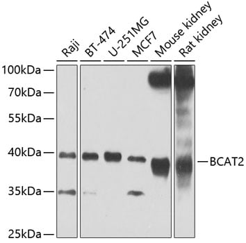 WB (Western Blot)
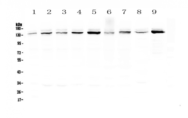
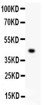
(Western blot analysis of extracts of various cell lines, using BCAT2 at 1:1000 dilution._Secondary antibody: HRP Goat Anti-Rabbit IgG (H+L) at 1:10000 dilution._Lysates/proteins: 25ug per lane._Blocking buffer: 3% nonfat dry milk in TBST._Detection: ECL Enhanced Kit (RM00021)._Exposure time: 60s.)
WB (Western Blot)
(Western blot analysis of extracts of various cell lines, using BCAT2 at 1:1000 dilution._Secondary antibody: HRP Goat Anti-Rabbit IgG (H+L) at 1:10000 dilution._Lysates/proteins: 25ug per lane._Blocking buffer: 3% nonfat dry milk in TBST._Detection: ECL Enhanced Kit (RM00021)._Exposure time: 60s.)
BCAT2, Polyclonal Antibody (Cat# AAA314041)
Full Name
BCAT2 Rabbit Polyclonal Antibody
Gene Names
BCAT2; BCAM; BCT2; PP18; BCATM
Reactivity
Human, Mouse, Rat
Applications
Immunofluorescence, Immunohistochemistry, Western Blot
Purity
Affinity purification
Pricing
 Application Data
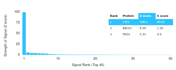
(Analysis of Protein Array containing more than 19,000 full-length human proteins using GM-CSF Mouse Monoclonal Antibody (CSF2/3402). Z- and S- Score: The Z-score represents the strength of a signal that a monoclonal antibody (MAb) (in combination with a fluorescently-tagged anti-IgG secondary antibody) produces when binding to a particular protein on the HuProtTM array. Z-scores are described in units of standard deviations (SD's) above the mean value of all signals generated on that array. If targets on HuProtTM are arranged in descending order of the Z-score, the S-score is the difference (also in units of SD's) between the Z-score. S-score therefore represents the relative target specificity of a MAb to its intended target. A MAb is considered to specific to its intended target, if the MAb has an S-score of at least 2.5. For example, if a MAb binds to protein X with a Z-score of 43 and to protein Y with a Z-score of 14, then the S-score for the binding of that MAb to protein X is equal to 29.)
Application Data
(Analysis of Protein Array containing more than 19,000 full-length human proteins using GM-CSF Mouse Monoclonal Antibody (CSF2/3402). Z- and S- Score: The Z-score represents the strength of a signal that a monoclonal antibody (MAb) (in combination with a fluorescently-tagged anti-IgG secondary antibody) produces when binding to a particular protein on the HuProtTM array. Z-scores are described in units of standard deviations (SD's) above the mean value of all signals generated on that array. If targets on HuProtTM are arranged in descending order of the Z-score, the S-score is the difference (also in units of SD's) between the Z-score. S-score therefore represents the relative target specificity of a MAb to its intended target. A MAb is considered to specific to its intended target, if the MAb has an S-score of at least 2.5. For example, if a MAb binds to protein X with a Z-score of 43 and to protein Y with a Z-score of 14, then the S-score for the binding of that MAb to protein X is equal to 29.)
GM-CSF, Monoclonal Antibody (Cat# AAA215377)
Full Name
GM-CSF (Granulocyte/Macrophage - Colony Stimulating Factor)
Gene Names
CSF2; CSF; GMCSF
Reactivity
Human
Applications
Immunohistochemistry
Purity
Purified Ab with BSA and Azide at 200ug/ml or Purified Ab with BSA and Azide at 200ug/ml or Purified Ab WITHOUT BSA and Azide at 1.0mg/ml
Pricing
 FCM/FACS (Flow Cytometry)
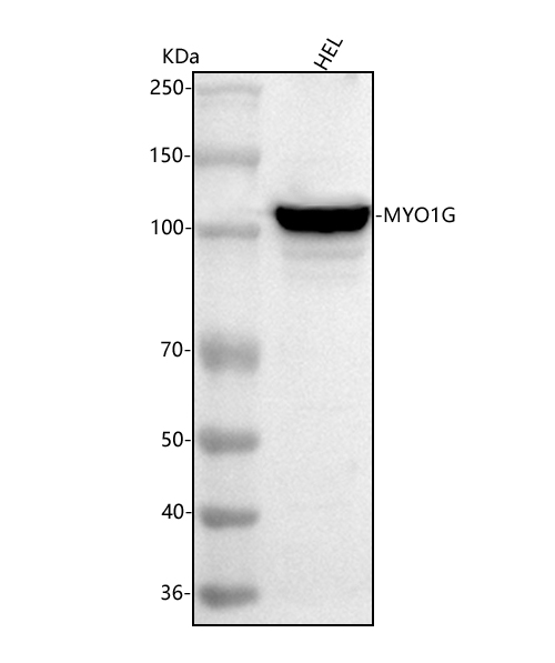
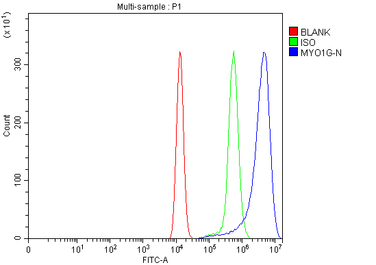
(Figure 2. Flow Cytometry analysis of HEL cells using anti-MYO1G antibody (AAA127526).Overlay histogram showing HEL cells stained with AAA127526 (Blue line). The cells were fixed with 4% paraformaldehyde and blocked with 10% normal goat serum. And then incubated with rabbit anti-MYO1G Antibody (AAA127526, 1ug/1x106 cells) for 30 min at 20 degree C. DyLight488 conjugated goat anti-rabbit IgG was used as secondary antibody for 30 minutes at 20 degree C. Isotype control antibody (Green line) was rabbit IgG (1ug/1x106) used under the same conditions. Unlabelled sample (Red line) was also used as a control.)
FCM/FACS (Flow Cytometry)
(Figure 2. Flow Cytometry analysis of HEL cells using anti-MYO1G antibody (AAA127526).Overlay histogram showing HEL cells stained with AAA127526 (Blue line). The cells were fixed with 4% paraformaldehyde and blocked with 10% normal goat serum. And then incubated with rabbit anti-MYO1G Antibody (AAA127526, 1ug/1x106 cells) for 30 min at 20 degree C. DyLight488 conjugated goat anti-rabbit IgG was used as secondary antibody for 30 minutes at 20 degree C. Isotype control antibody (Green line) was rabbit IgG (1ug/1x106) used under the same conditions. Unlabelled sample (Red line) was also used as a control.)
MYO1G, Polyclonal Antibody (Cat# AAA127526)
Full Name
Anti-MYO1G Antibody Picoband
Gene Names
MYO1G; HA2; MHAG
Reactivity
Human
Applications
Flow Cytometry, Western Blot
Purity
Immunogen affinity purified.
Pricing
 FCM/FACS (Flow Cytometry)
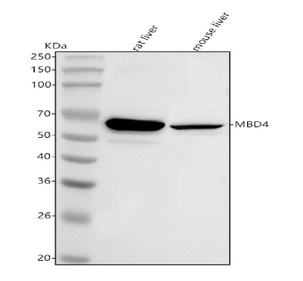
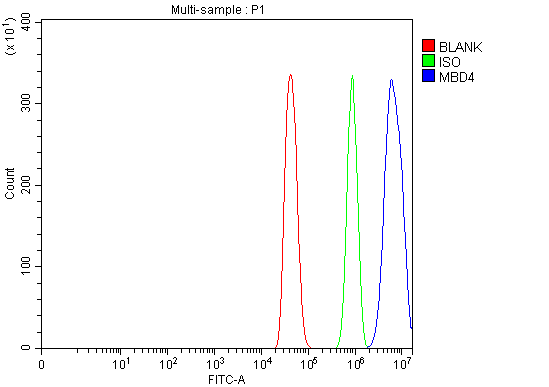
(Figure 2. Flow Cytometry analysis of C2C12 cells using anti-Mbd4 antibody (AAA127177).Overlay histogram showing C2C12 cells stained with AAA127177 (Blue line). To facilitate intracellular staining, cells were fixed with 4% paraformaldehyde and permeabilized with permeabilization buffer. The cells were blocked with 10% normal goat serum. And then incubated with rabbit anti-Mbd4 Antibody (AAA127177, 1ug/1x106 cells) for 30 min at 20 degree C. DyLight488 conjugated goat anti-rabbit IgG was used as secondary antibody for 30 minutes at 20 degree C. Isotype control antibody (Green line) was rabbit IgG (1ug/1x106) used under the same conditions. Unlabelled sample (Red line) was also used as a control.)
FCM/FACS (Flow Cytometry)
(Figure 2. Flow Cytometry analysis of C2C12 cells using anti-Mbd4 antibody (AAA127177).Overlay histogram showing C2C12 cells stained with AAA127177 (Blue line). To facilitate intracellular staining, cells were fixed with 4% paraformaldehyde and permeabilized with permeabilization buffer. The cells were blocked with 10% normal goat serum. And then incubated with rabbit anti-Mbd4 Antibody (AAA127177, 1ug/1x106 cells) for 30 min at 20 degree C. DyLight488 conjugated goat anti-rabbit IgG was used as secondary antibody for 30 minutes at 20 degree C. Isotype control antibody (Green line) was rabbit IgG (1ug/1x106) used under the same conditions. Unlabelled sample (Red line) was also used as a control.)
Mbd4, Polyclonal Antibody (Cat# AAA127177)
Full Name
Anti-Mbd4 Antibody Picoband
Gene Names
Mbd4; Med1
Reactivity
Mouse, Rat
Applications
Flow Cytometry, Western Blot
Purity
Immunogen affinity purified.
Pricing
 FCM/FACS (Flow Cytometry)
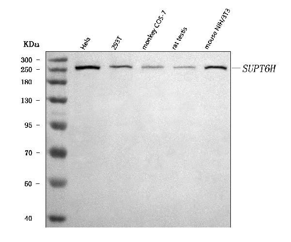
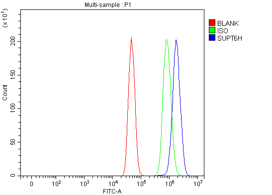
(Figure 2. Flow Cytometry analysis of U251 cells using anti-Spt6/SUPT6H antibody (AAA127377).Overlay histogram showing U251 cells stained with AAA127377 (Blue line). To facilitate intracellular staining, cells were fixed with 4% paraformaldehyde and permeabilized with permeabilization buffer. The cells were blocked with 10% normal goat serum. And then incubated with rabbit anti-Spt6/SUPT6H Antibody (AAA127377, 1ug/1x106 cells) for 30 min at 20 degree C. DyLight488 conjugated goat anti-rabbit IgG was used as secondary antibody for 30 minutes at 20 degree C. Isotype control antibody (Green line) was rabbit IgG (1ug/1x106) used under the same conditions. Unlabelled sample without incubation with primary antibody and secondary antibody (Red line) was used as a blank control.)
FCM/FACS (Flow Cytometry)
(Figure 2. Flow Cytometry analysis of U251 cells using anti-Spt6/SUPT6H antibody (AAA127377).Overlay histogram showing U251 cells stained with AAA127377 (Blue line). To facilitate intracellular staining, cells were fixed with 4% paraformaldehyde and permeabilized with permeabilization buffer. The cells were blocked with 10% normal goat serum. And then incubated with rabbit anti-Spt6/SUPT6H Antibody (AAA127377, 1ug/1x106 cells) for 30 min at 20 degree C. DyLight488 conjugated goat anti-rabbit IgG was used as secondary antibody for 30 minutes at 20 degree C. Isotype control antibody (Green line) was rabbit IgG (1ug/1x106) used under the same conditions. Unlabelled sample without incubation with primary antibody and secondary antibody (Red line) was used as a blank control.)
Spt6/SUPT6H, Polyclonal Antibody (Cat# AAA127377)
Full Name
Anti-Spt6/SUPT6H Antibody Picoband
Reactivity
Human, Monkey, Mouse, Rat
Applications
Flow Cytometry, Western Blot
Purity
Immunogen affinity purified.
Pricing
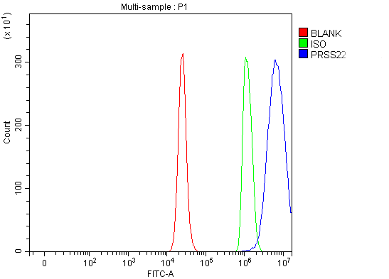 FCM/FACS (Flow Cytometry)
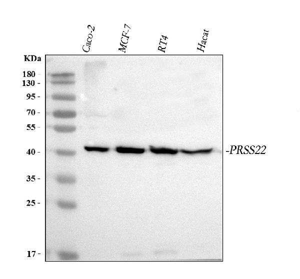
(Figure 2. Flow Cytometry analysis of RT4 cells using anti-PRSS22 antibody (AAA127799).Overlay histogram showing RT4 cells stained with AAA127799 (Blue line). The cells were fixed with 4% paraformaldehyde and blocked with 10% normal goat serum. And then incubated with rabbit anti-PRSS22 Antibody (AAA127799, 1ug/1x106 cells) for 30 min at 20 degree C. DyLight488 conjugated goat anti-rabbit IgG was used as secondary antibody for 30 minutes at 20 degree C. Isotype control antibody (Green line) was rabbit IgG (1ug/1x106) used under the same conditions. Unlabelled sample (Red line) was also used as a control.)
FCM/FACS (Flow Cytometry)
(Figure 2. Flow Cytometry analysis of RT4 cells using anti-PRSS22 antibody (AAA127799).Overlay histogram showing RT4 cells stained with AAA127799 (Blue line). The cells were fixed with 4% paraformaldehyde and blocked with 10% normal goat serum. And then incubated with rabbit anti-PRSS22 Antibody (AAA127799, 1ug/1x106 cells) for 30 min at 20 degree C. DyLight488 conjugated goat anti-rabbit IgG was used as secondary antibody for 30 minutes at 20 degree C. Isotype control antibody (Green line) was rabbit IgG (1ug/1x106) used under the same conditions. Unlabelled sample (Red line) was also used as a control.)
PRSS22, Polyclonal Antibody (Cat# AAA127799)
Full Name
Anti-PRSS22 Antibody Picoband
Gene Names
PRSS22; BSSP-4; SP001LA; hBSSP-4
Reactivity
Human
Applications
Western Blot, Flow Cytometry
Purity
Immunogen affinity purified.
Pricing
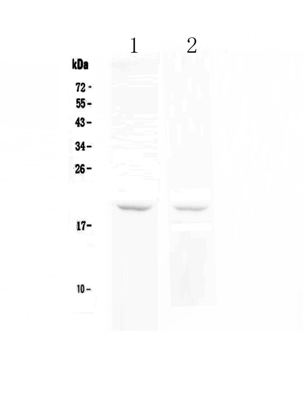
 IHC (Immunohiostchemistry)
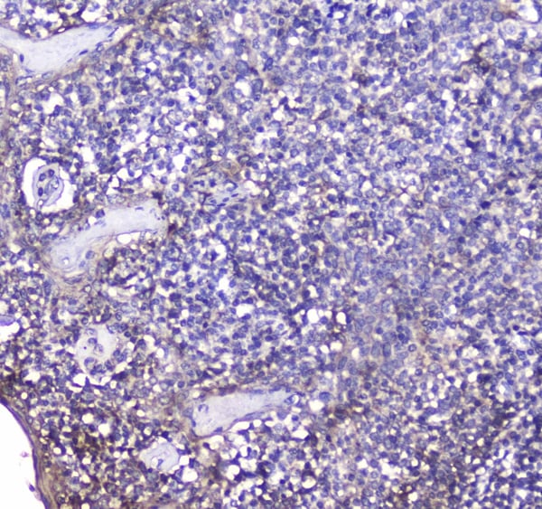
(Figure 2. IHC analysis of BCMA using anti-BCMA antibody (AAA124612).BCMA was detected in paraffin-embedded section of human tonsil tissue. Heat mediated antigen retrieval was performed in citrate buffer (pH6, epitope retrieval solution) for 20 mins. The tissue section was blocked with 10% goat serum. The tissue section was then incubated with 2ug/ml rabbit anti-BCMA Antibody (AAA124612) overnight at 4 degree C. Biotinylated goat anti-rabbit IgG was used as secondary antibody and incubated for 30 minutes at 37 degree C. The tissue section was developed using Strepavidin-Biotin-Complex (SABC) with DAB as the chromogen.)
IHC (Immunohiostchemistry)
(Figure 2. IHC analysis of BCMA using anti-BCMA antibody (AAA124612).BCMA was detected in paraffin-embedded section of human tonsil tissue. Heat mediated antigen retrieval was performed in citrate buffer (pH6, epitope retrieval solution) for 20 mins. The tissue section was blocked with 10% goat serum. The tissue section was then incubated with 2ug/ml rabbit anti-BCMA Antibody (AAA124612) overnight at 4 degree C. Biotinylated goat anti-rabbit IgG was used as secondary antibody and incubated for 30 minutes at 37 degree C. The tissue section was developed using Strepavidin-Biotin-Complex (SABC) with DAB as the chromogen.)
BCMA, Polyclonal Antibody (Cat# AAA124612)
Full Name
Anti-BCMA Picoband antibody
Gene Names
TNFRSF17; BCM; BCMA; CD269; TNFRSF13A
Reactivity
Human
No cross reactivity with other proteins.
No cross reactivity with other proteins.
Applications
Western Blot, Immunohistochemistry
Pricing

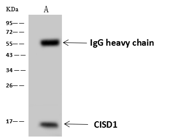 IP (Immunoprecipitation)
(CISD1 was immunoprecipitated using: Lane A:0.5 mg HepG2 Whole Cell Lysate 4 uL anti- CISD1 rabbit polyclonal antibody and 60 ug of Immunomagnetic beads Protein A/G. Primary antibody: Anti- CISD1 rabbit polyclonal antibody,at 1:100 dilution Secondary antibody: Goat Anti-Rabbit IgG (H+L)/HRP at 1/10000 dilution Developed using the ECL technique. Performed under reducing conditions. Predicted band size: 12kDa Observed band size :15 kDa)
IP (Immunoprecipitation)
(CISD1 was immunoprecipitated using: Lane A:0.5 mg HepG2 Whole Cell Lysate 4 uL anti- CISD1 rabbit polyclonal antibody and 60 ug of Immunomagnetic beads Protein A/G. Primary antibody: Anti- CISD1 rabbit polyclonal antibody,at 1:100 dilution Secondary antibody: Goat Anti-Rabbit IgG (H+L)/HRP at 1/10000 dilution Developed using the ECL technique. Performed under reducing conditions. Predicted band size: 12kDa Observed band size :15 kDa)
CISD1, Polyclonal Antibody (Cat# AAA258527)
Full Name
Anti-CISD1 Antibody, Rabbit Polyclonal
Gene Names
CISD1; ZCD1; MDS029; C10orf70; mitoNEET
Reactivity
Human
Applications
Immunoprecipitation, Western Blot
Purity
Protein A & Antigen Affinity
Pricing
 Application Data
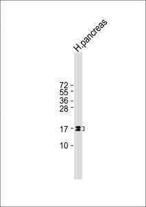
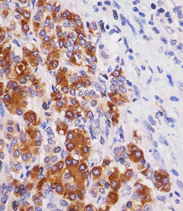
(AAA290840 staining REG3G in human pancreas tissue sections by Immunohistochemistry (IHC-P - paraformaldehyde-fixed, paraffin-embedded sections). Tissue was fixed with formaldehyde and blocked with 3% BSA for 0. 5 hour at room temperature; antigen retrieval was by heat mediation with a citrate buffer (pH6). Samples were incubated with primary antibody (1/25) for 1 hours at 37°C. A undiluted biotinylated goat polyvalent antibody was used as the secondary antibody.)
Application Data
(AAA290840 staining REG3G in human pancreas tissue sections by Immunohistochemistry (IHC-P - paraformaldehyde-fixed, paraffin-embedded sections). Tissue was fixed with formaldehyde and blocked with 3% BSA for 0. 5 hour at room temperature; antigen retrieval was by heat mediation with a citrate buffer (pH6). Samples were incubated with primary antibody (1/25) for 1 hours at 37°C. A undiluted biotinylated goat polyvalent antibody was used as the secondary antibody.)
REG3G, Monoclonal Antibody (Cat# AAA290840)
Full Name
REG3G Antibody
Gene Names
REG3G; PAP1B; PAPIB; PAP IB; PAP-1B; UNQ429; LPPM429; REG III; REG-III
Reactivity
Human
Applications
Western Blot
Pricing
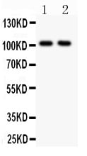
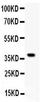
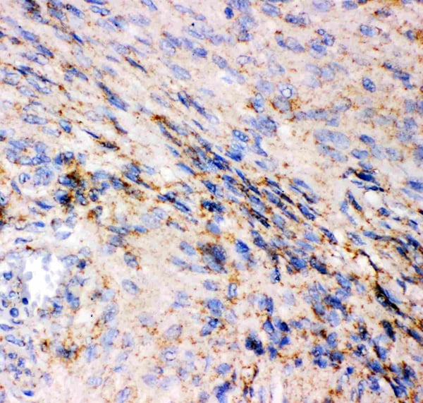 IHC (Immunohistochemistry)
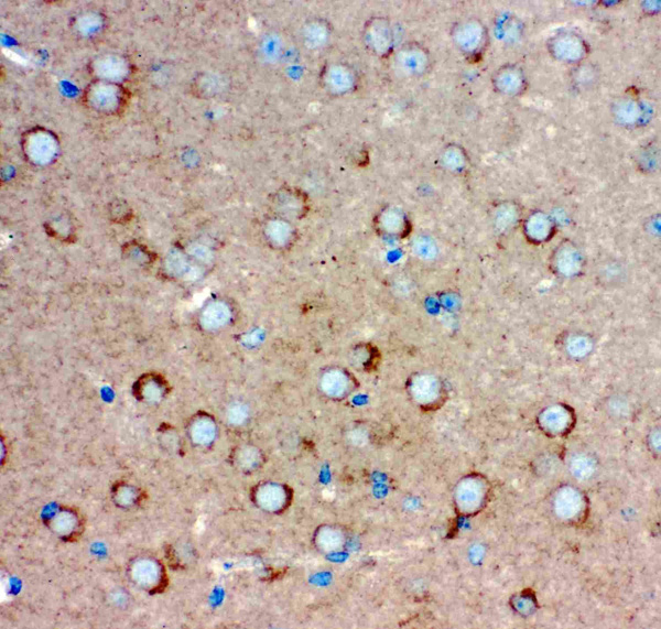
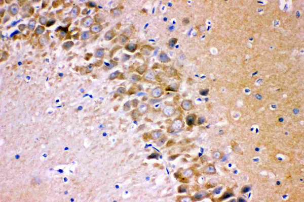
(Figure 5. IHC analysis of GRIA1 using anti-GRIA1 antibody (AAA46057).GRIA1 was detected in paraffin-embedded section of Human Meningeoma Tissue. Heat mediated antigen retrieval was performed in citrate buffer (pH6, epitope retrieval solution) for 20 mins. The tissue section was blocked with 10% goat serum. The tissue section was then incubated with 1ug/ml rabbit anti-GRIA1 Antibody (AAA46057) overnight at 4 degree C. Biotinylated goat anti-rabbit IgG was used as secondary antibody and incubated for 30 minutes at 37 degree C. The tissue section was developed using Strepavidin-Biotin-Complex (SABC) with DAB as the chromogen.)
IHC (Immunohistochemistry)
(Figure 5. IHC analysis of GRIA1 using anti-GRIA1 antibody (AAA46057).GRIA1 was detected in paraffin-embedded section of Human Meningeoma Tissue. Heat mediated antigen retrieval was performed in citrate buffer (pH6, epitope retrieval solution) for 20 mins. The tissue section was blocked with 10% goat serum. The tissue section was then incubated with 1ug/ml rabbit anti-GRIA1 Antibody (AAA46057) overnight at 4 degree C. Biotinylated goat anti-rabbit IgG was used as secondary antibody and incubated for 30 minutes at 37 degree C. The tissue section was developed using Strepavidin-Biotin-Complex (SABC) with DAB as the chromogen.)
Glutamate receptor 1, Polyclonal Antibody (Cat# AAA46057)
Full Name
Anti-GRIA1 Antibody
Gene Names
GRIA1; GLUH1; GLUR1; GLURA; GluA1; HBGR1
Reactivity
Human, Mouse, Rat. No cross reactivity with other proteins.
Applications
Immunohistochemistry, Western Blot
Purity
Immunogen affinity purified.
Pricing
Ephrin-A5, Active Protein (Cat# AAA48998)
Full Name
Recombinant human Ephrin-A5 protein
Gene Names
EFNA5; AF1; EFL5; RAGS; EPLG7; GLC1M; LERK7
Applications
Bioactivity, SDS-PAGE
Purity
> 95% by SDS-PAGE
Pricing
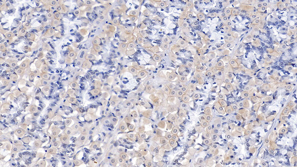
 IHC (Immunohiostchemistry)
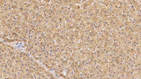
(DAB staining on IHCP;Sample: Human Liver Tissue; Primary Ab: 10ug/ml Mouse AntiHuman LSR AntibodySecond Ab: 2ug/mL HRPLinked Caprine AntiMouse IgG Polyclonal Antibody(Catalog: SAA544Mu19))
IHC (Immunohiostchemistry)
(DAB staining on IHCP;Sample: Human Liver Tissue; Primary Ab: 10ug/ml Mouse AntiHuman LSR AntibodySecond Ab: 2ug/mL HRPLinked Caprine AntiMouse IgG Polyclonal Antibody(Catalog: SAA544Mu19))
Lipolysis Stimulated Lipoprotein Receptor (LSR), Monoclonal Antibody (Cat# AAA151839)
Full Name
Monoclonal Antibody to Lipolysis Stimulated Lipoprotein Receptor (LSR)
Gene Names
LSR; ILDR3; LISCH7
Reactivity
Human, Pig
Applications
Immunoprecipitation, Immunocytochemistry, Immunohistochemistry, Western Blot
Purity
Protein A + Protein G affinity chromatography
Pricing
 FCM/FACS (Flow Cytometry)
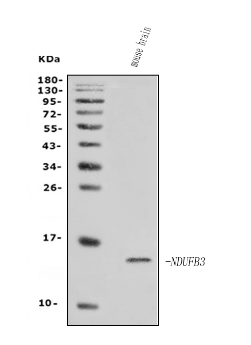
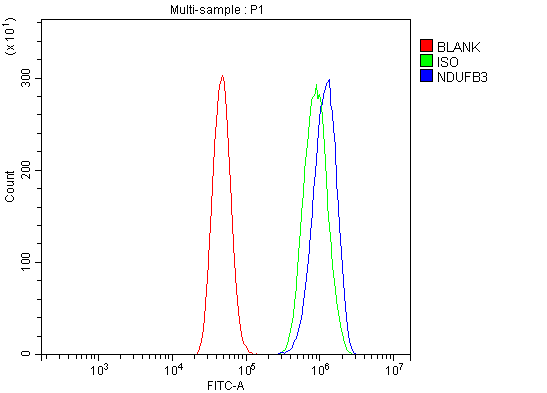
(Figure 2. Flow Cytometry analysis of A431 cells using anti-NDUFB3 antibody (AAA125817).Overlay histogram showing A431 cells stained with AAA125817 (Blue line). The cells were blocked with 10% normal goat serum. And then incubated with rabbit anti-NDUFB3 Antibody (AAA125817, 1μg/1x106 cells) for 30 min at 20 degree C. DyLight®488 conjugated goat anti-rabbit IgG (5-10μg/1x106 cells) was used as secondary antibody for 30 minutes at 20 degree C. Isotype control antibody (Green line) was rabbit IgG (1μg/1x106) used under the same conditions. Unlabelled sample (Red line) was also used as a control.)
FCM/FACS (Flow Cytometry)
(Figure 2. Flow Cytometry analysis of A431 cells using anti-NDUFB3 antibody (AAA125817).Overlay histogram showing A431 cells stained with AAA125817 (Blue line). The cells were blocked with 10% normal goat serum. And then incubated with rabbit anti-NDUFB3 Antibody (AAA125817, 1μg/1x106 cells) for 30 min at 20 degree C. DyLight®488 conjugated goat anti-rabbit IgG (5-10μg/1x106 cells) was used as secondary antibody for 30 minutes at 20 degree C. Isotype control antibody (Green line) was rabbit IgG (1μg/1x106) used under the same conditions. Unlabelled sample (Red line) was also used as a control.)
NDUFB3, Polyclonal Antibody (Cat# AAA125817)
Full Name
Anti-NDUFB3 Antibody
Gene Names
NDUFB3; B12; CI-B12
Reactivity
Human, Mouse
Applications
Direct ELISA, Flow Cytometry, Western Blot
Purity
Immunogen affinity purified.
Pricing
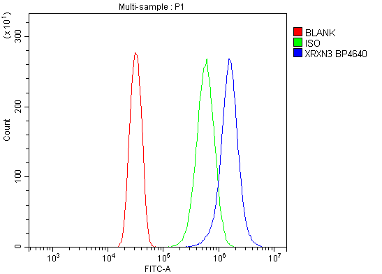 FCM/FACS (Flow Cytometry)
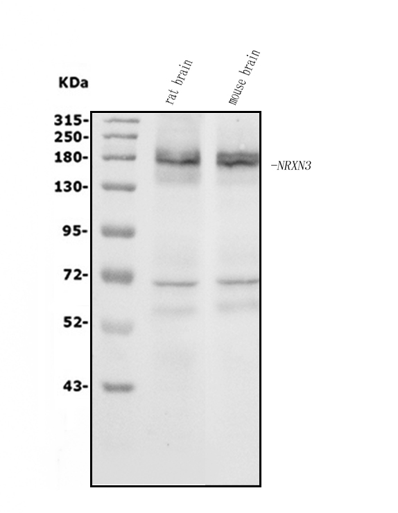
(Figure 2. Flow Cytometry analysis of U20S cells using anti-NRXN3 antibody (AAA125758).Overlay histogram showing U20S cells stained with AAA125758 (Blue line). The cells were blocked with 10% normal goat serum. And then incubated with rabbit anti-NRXN3 Antibody (AAA125758, 1μg/1x106 cells) for 30 min at 20 degree C. DyLight®488 conjugated goat anti-rabbit IgG (5-10μg/1x106 cells) was used as secondary antibody for 30 minutes at 20 degree C. Isotype control antibody (Green line) was rabbit IgG (1μg/1x106) used under the same conditions. Unlabelled sample (Red line) was also used as a control.)
FCM/FACS (Flow Cytometry)
(Figure 2. Flow Cytometry analysis of U20S cells using anti-NRXN3 antibody (AAA125758).Overlay histogram showing U20S cells stained with AAA125758 (Blue line). The cells were blocked with 10% normal goat serum. And then incubated with rabbit anti-NRXN3 Antibody (AAA125758, 1μg/1x106 cells) for 30 min at 20 degree C. DyLight®488 conjugated goat anti-rabbit IgG (5-10μg/1x106 cells) was used as secondary antibody for 30 minutes at 20 degree C. Isotype control antibody (Green line) was rabbit IgG (1μg/1x106) used under the same conditions. Unlabelled sample (Red line) was also used as a control.)
NRXN3, Polyclonal Antibody (Cat# AAA125758)
Full Name
Anti-NRXN3 Antibody
Gene Names
NRXN3; C14orf60
Reactivity
Human, Mouse, Rat
Applications
Direct ELISA, Flow Cytometry, Western Blot
Purity
Immunogen affinity purified.
Pricing
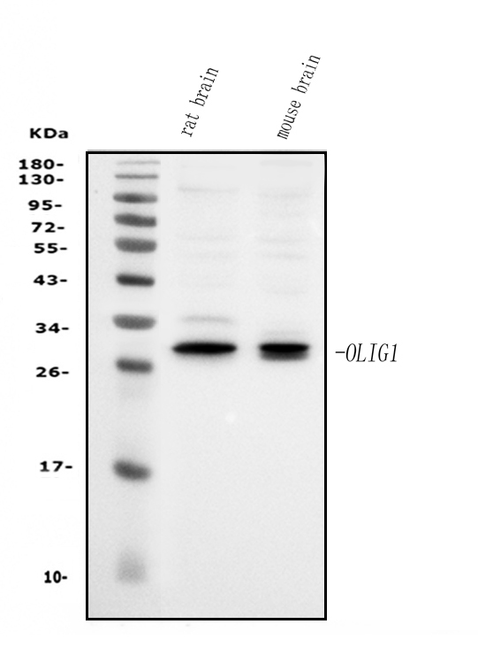
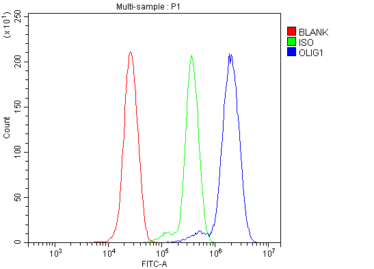 FCM/FACS (Flow Cytometry)
(Figure 2. Flow Cytometry analysis of THP-1 cells using anti-Olig1 antibody (AAA125768).Overlay histogram showing THP-1 cells stained with AAA125768 (Blue line). The cells were blocked with 10% normal goat serum. And then incubated with rabbit anti-Olig1 Antibody (AAA125768, 1μg/1x106 cells) for 30 min at 20 degree C. DyLight®488 conjugated goat anti-rabbit IgG (5-10μg/1x106 cells) was used as secondary antibody for 30 minutes at 20 degree C. Isotype control antibody (Green line) was rabbit IgG (1μg/1x106) used under the same conditions. Unlabelled sample (Red line) was also used as a control.)
FCM/FACS (Flow Cytometry)
(Figure 2. Flow Cytometry analysis of THP-1 cells using anti-Olig1 antibody (AAA125768).Overlay histogram showing THP-1 cells stained with AAA125768 (Blue line). The cells were blocked with 10% normal goat serum. And then incubated with rabbit anti-Olig1 Antibody (AAA125768, 1μg/1x106 cells) for 30 min at 20 degree C. DyLight®488 conjugated goat anti-rabbit IgG (5-10μg/1x106 cells) was used as secondary antibody for 30 minutes at 20 degree C. Isotype control antibody (Green line) was rabbit IgG (1μg/1x106) used under the same conditions. Unlabelled sample (Red line) was also used as a control.)
Olig1, Polyclonal Antibody (Cat# AAA125768)
Full Name
Anti-Olig1 Antibody
Gene Names
OLIG1; BHLHB6; BHLHE21
Reactivity
Human, Mouse, Rat
Applications
Direct ELISA, Flow Cytometry, Western Blot
Purity
Immunogen affinity purified.
Pricing
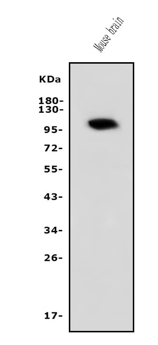
 FCM/FACS (Flow Cytometry)
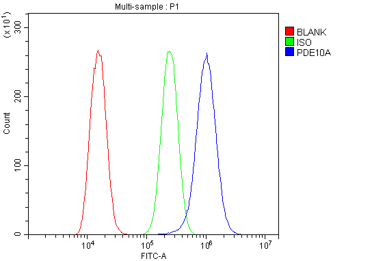
(Figure 2. Flow Cytometry analysis of A549 cells using anti-PDE10A antibody (AAA125667).Overlay histogram showing A549 cells stained with AAA125667 (Blue line). The cells were blocked with 10% normal goat serum. And then incubated with rabbit anti-PDE10A Antibody (AAA125667, 1μg/1x106 cells) for 30 min at 20 degree C. DyLight®488 conjugated goat anti-rabbit IgG (5-10μg/1x106 cells) was used as secondary antibody for 30 minutes at 20 degree C. Isotype control antibody (Green line) was rabbit IgG (1μg/1x106) used under the same conditions. Unlabelled sample (Red line) was also used as a control.)
FCM/FACS (Flow Cytometry)
(Figure 2. Flow Cytometry analysis of A549 cells using anti-PDE10A antibody (AAA125667).Overlay histogram showing A549 cells stained with AAA125667 (Blue line). The cells were blocked with 10% normal goat serum. And then incubated with rabbit anti-PDE10A Antibody (AAA125667, 1μg/1x106 cells) for 30 min at 20 degree C. DyLight®488 conjugated goat anti-rabbit IgG (5-10μg/1x106 cells) was used as secondary antibody for 30 minutes at 20 degree C. Isotype control antibody (Green line) was rabbit IgG (1μg/1x106) used under the same conditions. Unlabelled sample (Red line) was also used as a control.)
PDE10A, Polyclonal Antibody (Cat# AAA125667)
Full Name
Anti-PDE10A Antibody
Gene Names
PDE10A; HSPDE10A
Reactivity
Human, Mouse
Applications
Direct ELISA, Flow Cytometry, Western Blot
Purity
Immunogen affinity purified.
Pricing
 IP (Immunoprecipitation)
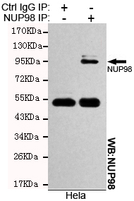
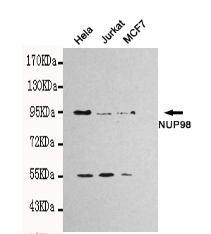
(Immunoprecipitation analysis of Hela cell lysates using NUP98 mouse mAb.)
IP (Immunoprecipitation)
(Immunoprecipitation analysis of Hela cell lysates using NUP98 mouse mAb.)
NUP98, Monoclonal Antibody (Cat# AAA320347)
Full Name
NUP98 Mouse mAb
Gene Names
NUP98; ADIR2; NUP96; NUP196
Reactivity
Human
Applications
Immunoprecipitation, Western Blot
Purity
The antibody was affinity-purified from mouse ascites by affinity-chromatography using epitope-specific immunogen.
Pricing
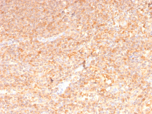
 IHC (Immunohistochemistry)
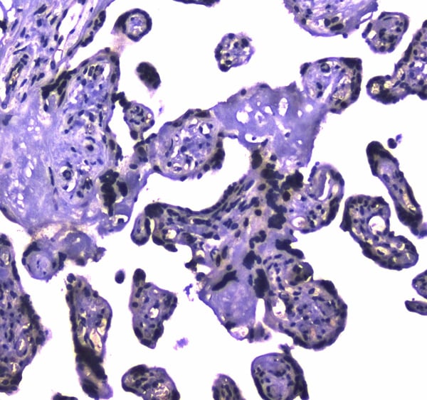
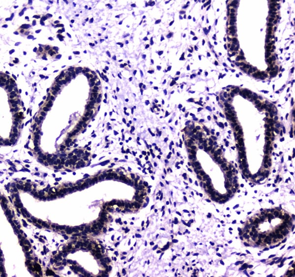
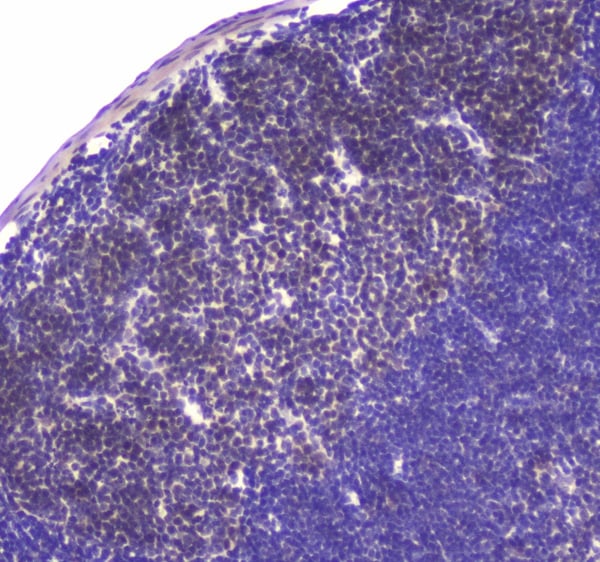
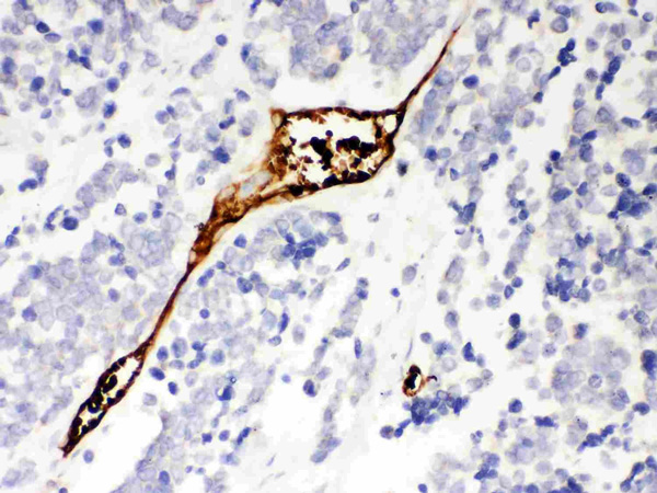
(Figure 4. IHC analysis of HAUSP/USP7 using anti-HAUSP/USP7 antibody (AAA124632).HAUSP/USP7 was detected in paraffin-embedded section of mouse Lymph nodes tissue. Heat mediated antigen retrieval was performed in citrate buffer (pH6, epitope retrieval solution) for 20 mins. The tissue section was blocked with 10% goat serum. The tissue section was then incubated with 1ug/ml rabbit anti-HAUSP/USP7 Antibody (AAA124632) overnight at 4 degree C. Biotinylated goat anti-rabbit IgG was used as secondary antibody and incubated for 30 minutes at 37 degree C. The tissue section was developed using Strepavidin-Biotin-Complex (SABC) with DAB as the chromogen.)
IHC (Immunohistochemistry)
(Figure 4. IHC analysis of HAUSP/USP7 using anti-HAUSP/USP7 antibody (AAA124632).HAUSP/USP7 was detected in paraffin-embedded section of mouse Lymph nodes tissue. Heat mediated antigen retrieval was performed in citrate buffer (pH6, epitope retrieval solution) for 20 mins. The tissue section was blocked with 10% goat serum. The tissue section was then incubated with 1ug/ml rabbit anti-HAUSP/USP7 Antibody (AAA124632) overnight at 4 degree C. Biotinylated goat anti-rabbit IgG was used as secondary antibody and incubated for 30 minutes at 37 degree C. The tissue section was developed using Strepavidin-Biotin-Complex (SABC) with DAB as the chromogen.)
HAUSP/USP7, Polyclonal Antibody (Cat# AAA124632)
Full Name
Anti-HAUSP/USP7 Picoband antibody
Gene Names
USP7; TEF1; HAUSP
Reactivity
Human, Mouse, Rat
No cross reactivity with other proteins.
No cross reactivity with other proteins.
Applications
Western Blot, Immunohistochemistry
Pricing
 Application Data
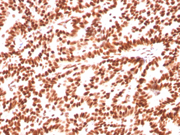
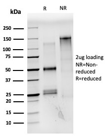
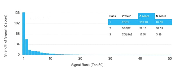
(Analysis of Protein Array containing more than 19,000 full-length human proteins using Estrogen Receptor alpha Mouse Monoclonal Antibody (ESR1/3559) Z- and S- Score: The Z-score represents the strength of a signal that a monoclonal antibody (MAb) (in combination with a fluorescently-tagged anti-IgG secondary antibody) produces when binding to a particular protein on the HuProtTM array. Z-scores are described in units of standard deviations (SD’s) above the mean value of all signals generated on that array. If targets on HuProtTM are arranged in descending order of the Z-score, the S-score is the difference (also in units of SD’s) between the Z-score. S-score therefore represents the relative target specificity of a MAb to its intended target. A MAb is considered to specific to its intended target, if the MAb has an S-score of at least 2.5. For example, if a MAb binds to protein X with a Z-score of 43 and to protein Y with a Z-score of 14, then the S-score for the binding of that MAb to protein X is equal to 29.)
Application Data
(Analysis of Protein Array containing more than 19,000 full-length human proteins using Estrogen Receptor alpha Mouse Monoclonal Antibody (ESR1/3559) Z- and S- Score: The Z-score represents the strength of a signal that a monoclonal antibody (MAb) (in combination with a fluorescently-tagged anti-IgG secondary antibody) produces when binding to a particular protein on the HuProtTM array. Z-scores are described in units of standard deviations (SD’s) above the mean value of all signals generated on that array. If targets on HuProtTM are arranged in descending order of the Z-score, the S-score is the difference (also in units of SD’s) between the Z-score. S-score therefore represents the relative target specificity of a MAb to its intended target. A MAb is considered to specific to its intended target, if the MAb has an S-score of at least 2.5. For example, if a MAb binds to protein X with a Z-score of 43 and to protein Y with a Z-score of 14, then the S-score for the binding of that MAb to protein X is equal to 29.)
Estrogen Receptor, alpha, Monoclonal Antibody (Cat# AAA214957)
Full Name
Estrogen Receptor, alpha (Marker of Estrogen Dependence)
Gene Names
ESR1; ER; ESR; Era; ESRA; ESTRR; NR3A1
Reactivity
Human
Applications
Immunohistochemistry, Flow Cytometry
Purity
Purified Ab with BSA and Azide at 200ug/ml OR Purified Ab WITHOUT BSA and Azide at 1.0mg/ml
Pricing
 IHC (Immunohiostchemistry)
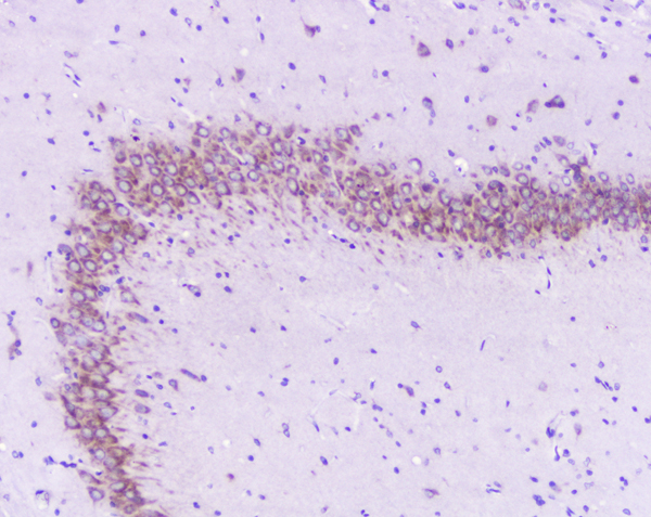
(Figure 3. IHC analysis of GM130 using anti-GM130 antibody.GM130 was detected in paraffin-embedded section of human oesophagus squama cancer tissue. Heat mediated antigen retrieval was performed in citrate buffer (pH6, epitope retrieval solution) for 20 mins. The tissue section was blocked with 10% goat serum. The tissue section was then incubated with 1ug/ml rabbit anti-GM130 antibody.GM130 was detected in paraffin-embedded section of human oesophagus squama cancer tissue. Heat mediated antigen retrieval was performed in citrate buffer (pH6, epitope retrieval solution) for 20 mins. The tissue section was blocked with 10% goat serum. The tissue section was then incubated with 1ug/ml rabbit anti-GM130 antibody.GM130 was detected in paraffin-embedded section of rat spleen tissue. Heat mediated antigen retrieval was performed in citrate buffer (pH6, epitope retrieval solution) for 20 mins. The tissue section was blocked with 10% goat serum. The tissue section was then incubated with 1ug/ml rabbit anti-GM130 antibody.GM130 was detected in paraffin-embedded section of human placenta tissue. Heat mediated antigen retrieval was performed in citrate buffer (pH6, epitope retrieval solution) for 20 mins. The tissue section was blocked with 10% goat serum. The tissue section was then incubated with 1ug/ml rabbit anti-GM130 antibody.GM130 was detected in paraffin-embedded section of human tonsil tissue. Heat mediated antigen retrieval was performed in citrate buffer (pH6, epitope retrieval solution) for 20 mins. The tissue section was blocked with 10% goat serum. The tissue section was then incubated with 1ug/ml rabbit anti-GM130 antibody.GM130 was detected in paraffin-embedded section of mouse intestine tissue. Heat mediated antigen retrieval was performed in citrate buffer (pH6, epitope retrieval solution) for 20 mins. The tissue section was blocked with 10% goat serum. The tissue section was then incubated with 1ug/ml rabbit anti-GM130 antibody.GM130 was detected in paraffin-embedded section of mouse spleen tissue. Heat mediated antigen retrieval was performed in citrate buffer (pH6, epitope retrieval solution) for 20 mins. The tissue section was blocked with 10% goat serum. The tissue section was then incubated with 1ug/ml rabbit anti-GM130 antibody.GM130 was detected in paraffin-embedded section of rat brain tissue. Heat mediated antigen retrieval was performed in citrate buffer (pH6, epitope retrieval solution) for 20 mins. The tissue section was blocked with 10% goat serum. The tissue section was then incubated with 1ug/ml rabbit anti-GM130 Antibody overnight at 4 degree C. Biotinylated goat anti-rabbit IgG was used as secondary antibody and incubated for 30 minutes at 37 degree C. The tissue section was developed using Strepavidin-Biotin-Complex (SABC) with DAB as the chromogen.)
IHC (Immunohiostchemistry)
(Figure 3. IHC analysis of GM130 using anti-GM130 antibody.GM130 was detected in paraffin-embedded section of human oesophagus squama cancer tissue. Heat mediated antigen retrieval was performed in citrate buffer (pH6, epitope retrieval solution) for 20 mins. The tissue section was blocked with 10% goat serum. The tissue section was then incubated with 1ug/ml rabbit anti-GM130 antibody.GM130 was detected in paraffin-embedded section of human oesophagus squama cancer tissue. Heat mediated antigen retrieval was performed in citrate buffer (pH6, epitope retrieval solution) for 20 mins. The tissue section was blocked with 10% goat serum. The tissue section was then incubated with 1ug/ml rabbit anti-GM130 antibody.GM130 was detected in paraffin-embedded section of rat spleen tissue. Heat mediated antigen retrieval was performed in citrate buffer (pH6, epitope retrieval solution) for 20 mins. The tissue section was blocked with 10% goat serum. The tissue section was then incubated with 1ug/ml rabbit anti-GM130 antibody.GM130 was detected in paraffin-embedded section of human placenta tissue. Heat mediated antigen retrieval was performed in citrate buffer (pH6, epitope retrieval solution) for 20 mins. The tissue section was blocked with 10% goat serum. The tissue section was then incubated with 1ug/ml rabbit anti-GM130 antibody.GM130 was detected in paraffin-embedded section of human tonsil tissue. Heat mediated antigen retrieval was performed in citrate buffer (pH6, epitope retrieval solution) for 20 mins. The tissue section was blocked with 10% goat serum. The tissue section was then incubated with 1ug/ml rabbit anti-GM130 antibody.GM130 was detected in paraffin-embedded section of mouse intestine tissue. Heat mediated antigen retrieval was performed in citrate buffer (pH6, epitope retrieval solution) for 20 mins. The tissue section was blocked with 10% goat serum. The tissue section was then incubated with 1ug/ml rabbit anti-GM130 antibody.GM130 was detected in paraffin-embedded section of mouse spleen tissue. Heat mediated antigen retrieval was performed in citrate buffer (pH6, epitope retrieval solution) for 20 mins. The tissue section was blocked with 10% goat serum. The tissue section was then incubated with 1ug/ml rabbit anti-GM130 antibody.GM130 was detected in paraffin-embedded section of rat brain tissue. Heat mediated antigen retrieval was performed in citrate buffer (pH6, epitope retrieval solution) for 20 mins. The tissue section was blocked with 10% goat serum. The tissue section was then incubated with 1ug/ml rabbit anti-GM130 Antibody overnight at 4 degree C. Biotinylated goat anti-rabbit IgG was used as secondary antibody and incubated for 30 minutes at 37 degree C. The tissue section was developed using Strepavidin-Biotin-Complex (SABC) with DAB as the chromogen.)
GM130/GOLGA2, Polyclonal Antibody (Cat# AAA124963)
Full Name
Anti-GM130/GOLGA2 Antibody
Gene Names
GOLGA2; GM130
Reactivity
Human, Mouse, Rat
Applications
Direct ELISA, Immunofluorescence, Immunocytochemistry, Immunohistochemistry, Western Blot
Purity
Immunogen Affinity Purified
Pricing
 IHC (Immunohistochemisry)
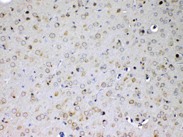
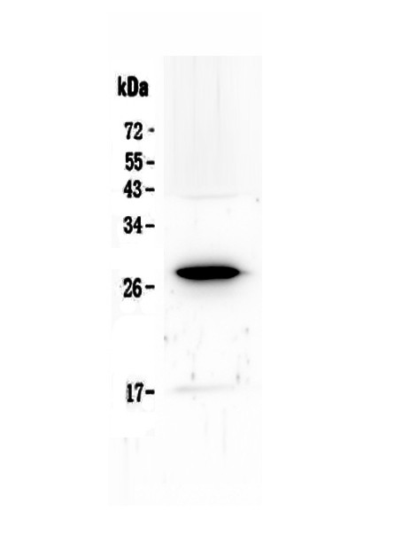
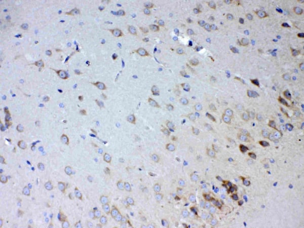
(Figure 3. IHC analysis of IL15RA using anti-IL15RA antibody (AAA124701).IL15RA was detected in paraffin-embedded section of rat brain tissue. Heat mediated antigen retrieval was performed in citrate buffer (pH6, epitope retrieval solution) for 20 mins. The tissue section was blocked with 10% goat serum. The tissue section was then incubated with 1ug/ml rabbit anti-IL15RA Antibody (AAA124701) overnight at 4 degree C. Biotinylated goat anti-rabbit IgG was used as secondary antibody and incubated for 30 minutes at 37 degree C. The tissue section was developed using Strepavidin-Biotin-Complex (SABC) with DAB as the chromogen.)
IHC (Immunohistochemisry)
(Figure 3. IHC analysis of IL15RA using anti-IL15RA antibody (AAA124701).IL15RA was detected in paraffin-embedded section of rat brain tissue. Heat mediated antigen retrieval was performed in citrate buffer (pH6, epitope retrieval solution) for 20 mins. The tissue section was blocked with 10% goat serum. The tissue section was then incubated with 1ug/ml rabbit anti-IL15RA Antibody (AAA124701) overnight at 4 degree C. Biotinylated goat anti-rabbit IgG was used as secondary antibody and incubated for 30 minutes at 37 degree C. The tissue section was developed using Strepavidin-Biotin-Complex (SABC) with DAB as the chromogen.)
IL15RA, Polyclonal Antibody (Cat# AAA124701)
Full Name
Anti-IL15RA Picoband Antibody
Gene Names
IL15RA; CD215
Reactivity
Human, Mouse, Rat
No cross reactivity with other proteins.
No cross reactivity with other proteins.
Applications
Western Blot, Immunohistochemistry
Purity
Immunogen affinity purified
Pricing

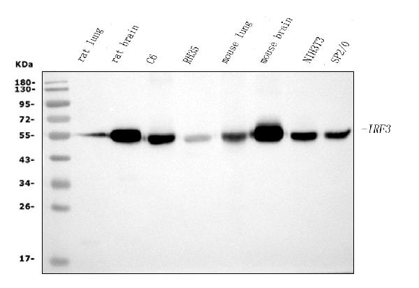
 FCM/FACS (Flow Cytometry)
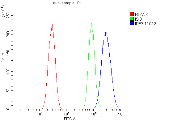
(Figure 3. Flow Cytometry analysis of HEPA1-6 cells using anti-IRF3 antibody (AAA125900).Overlay histogram showing HEPA1-6 cells stained with AAA125900 (Blue line). The cells were blocked with 10% normal goat serum. And then incubated with mouse anti- IRF3 Antibody (AAA125900, 1μg/1x106 cells) for 30 min at 20 degree C. DyLight®488 conjugated goat anti-mouse IgG (BA1126, 5-10μg/1x106 cells) was used as secondary antibody for 30 minutes at 20 degree C. Isotype control antibody (Green line) was mouse IgG (1μg/1x106) used under the same conditions. Unlabelled sample (Red line) was also used as a control.)
FCM/FACS (Flow Cytometry)
(Figure 3. Flow Cytometry analysis of HEPA1-6 cells using anti-IRF3 antibody (AAA125900).Overlay histogram showing HEPA1-6 cells stained with AAA125900 (Blue line). The cells were blocked with 10% normal goat serum. And then incubated with mouse anti- IRF3 Antibody (AAA125900, 1μg/1x106 cells) for 30 min at 20 degree C. DyLight®488 conjugated goat anti-mouse IgG (BA1126, 5-10μg/1x106 cells) was used as secondary antibody for 30 minutes at 20 degree C. Isotype control antibody (Green line) was mouse IgG (1μg/1x106) used under the same conditions. Unlabelled sample (Red line) was also used as a control.)
IRF3, Monoclonal Antibody (Cat# AAA125900)
Full Name
Anti-IRF3 Antibody (monoclonal, 11H2)
Gene Names
Irf3; IRF-3; C920001K05Rik
Reactivity
Mouse, Rat
Applications
Flow Cytometry, Immunofluorescence, Immunocytochemistry, Western Blot
Purity
Immunogen affinity purified.
Pricing
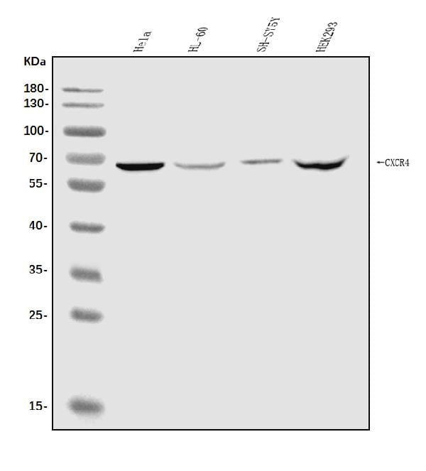
 FCM/FACS (Flow Cytometry)
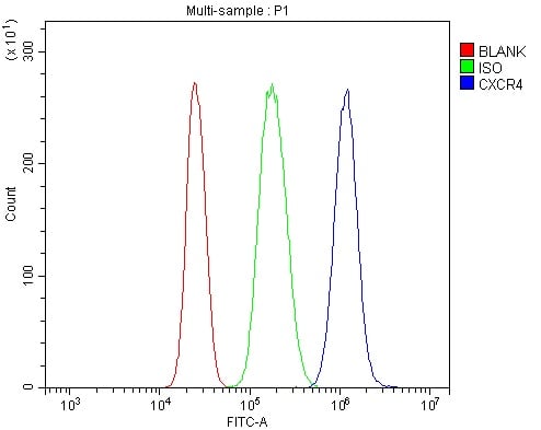
(Figure 2. Flow Cytometry analysis of U937 cells using anti-CXCR4 antibody (AAA125511).Overlay histogram showing U937 cells stained with AAA125511 (Blue line). The cells were blocked with 10% normal goat serum. And then incubated with rabbit anti-CXCR4 Antibody (AAA125511, 1μg/1x106 cells) for 30 min at 20 degree C. DyLight®488 conjugated goat anti-rabbit IgG (5-10μg/1x106 cells) was used as secondary antibody for 30 minutes at 20 degree C. Isotype control antibody (Green line) was rabbit IgG (1μg/1x106) used under the same conditions. Unlabelled sample (Red line) was also used as a control.)
FCM/FACS (Flow Cytometry)
(Figure 2. Flow Cytometry analysis of U937 cells using anti-CXCR4 antibody (AAA125511).Overlay histogram showing U937 cells stained with AAA125511 (Blue line). The cells were blocked with 10% normal goat serum. And then incubated with rabbit anti-CXCR4 Antibody (AAA125511, 1μg/1x106 cells) for 30 min at 20 degree C. DyLight®488 conjugated goat anti-rabbit IgG (5-10μg/1x106 cells) was used as secondary antibody for 30 minutes at 20 degree C. Isotype control antibody (Green line) was rabbit IgG (1μg/1x106) used under the same conditions. Unlabelled sample (Red line) was also used as a control.)
CXCR4, Polyclonal Antibody (Cat# AAA125511)
Full Name
Anti-CXCR4 Antibody
Gene Names
CXCR4; FB22; HM89; LAP3; LCR1; NPYR; WHIM; CD184; LESTR; NPY3R; NPYRL; HSY3RR; NPYY3R; D2S201E
Reactivity
Human
Applications
Direct ELISA, Flow Cytometry, Western Blot
Purity
Immunogen affinity purified.
Pricing
 IHC (Immunohistochemistry)
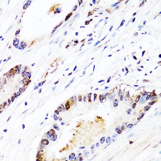
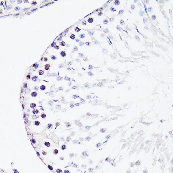
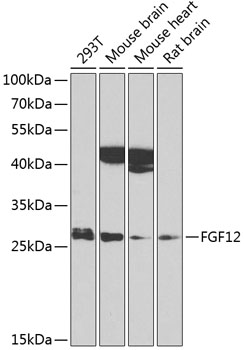
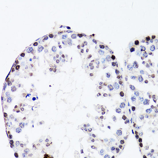
(Immunohistochemistry of paraffin-embedded mouse testis using FGF12 at dilution of 1:150 (40x lens).)
IHC (Immunohistochemistry)
(Immunohistochemistry of paraffin-embedded mouse testis using FGF12 at dilution of 1:150 (40x lens).)
FGF12, Polyclonal Antibody (Cat# AAA313812)
Full Name
FGF12 Rabbit Polyclonal Antibody
Gene Names
FGF12; FHF1; FGF12B
Reactivity
Human, Mouse, Rat
Applications
Immunofluorescence, Immunohistochemistry, Western Blot
Purity
Affinity purification
Pricing
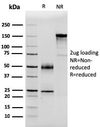
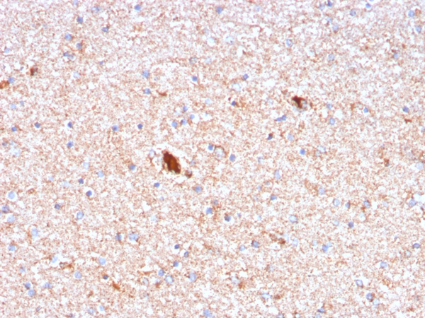
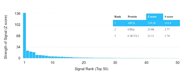 Application Data
(Analysis of Protein Array containing more than 19,000 full-length human proteins using Serum Amyloid P Mouse Monoclonal Antibody (APCS/3240). Z- and S- Score: The Z-score represents the strength of a signal that a monoclonal antibody (Monoclonal Antibody) (in combination with a fluorescently-tagged anti-IgG secondary antibody) produces when binding to a particular protein on the HuProtTM array. Z-scores are described in units of standard deviations (SD’s) above the mean value of all signals generated on that array. If targets on HuProtTM are arranged in descending order of the Z-score, the S-score is the difference (also in units of SD’s) between the Z-score. S-score therefore represents the relative target specificity of a Monoclonal Antibody to its intended target. A Monoclonal Antibody is considered to specific to its intended target, if the Monoclonal Antibody has an S-score of at least 2.5. For example, if a Monoclonal Antibody binds to protein X with a Z-score of 43 and to protein Y with a Z-score of 14, then the S-score for the binding of that Monoclonal Antibody to protein X is equal to 29.)
Application Data
(Analysis of Protein Array containing more than 19,000 full-length human proteins using Serum Amyloid P Mouse Monoclonal Antibody (APCS/3240). Z- and S- Score: The Z-score represents the strength of a signal that a monoclonal antibody (Monoclonal Antibody) (in combination with a fluorescently-tagged anti-IgG secondary antibody) produces when binding to a particular protein on the HuProtTM array. Z-scores are described in units of standard deviations (SD’s) above the mean value of all signals generated on that array. If targets on HuProtTM are arranged in descending order of the Z-score, the S-score is the difference (also in units of SD’s) between the Z-score. S-score therefore represents the relative target specificity of a Monoclonal Antibody to its intended target. A Monoclonal Antibody is considered to specific to its intended target, if the Monoclonal Antibody has an S-score of at least 2.5. For example, if a Monoclonal Antibody binds to protein X with a Z-score of 43 and to protein Y with a Z-score of 14, then the S-score for the binding of that Monoclonal Antibody to protein X is equal to 29.)
Serum Amyloid P/APCS, Monoclonal Antibody (Cat# AAA215016)
Full Name
Serum Amyloid P/APCS
Gene Names
APCS; SAP; PTX2; HEL-S-92n
Reactivity
Human
Applications
Immunohistochemistry
Purity
Purified Ab with BSA and Azide at 200ug/ml OR Purified Ab WITHOUT BSA and Azide at 1.0mg/ml
Pricing
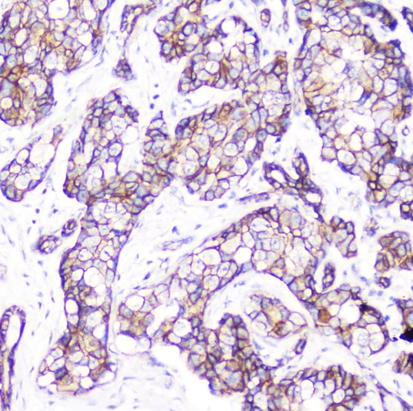
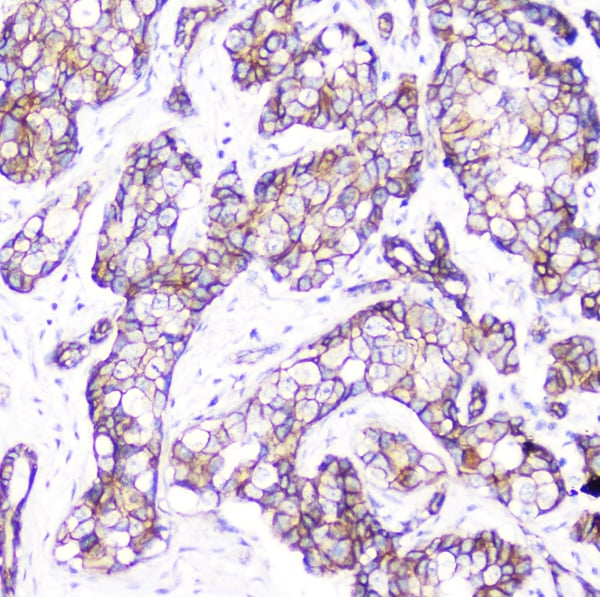
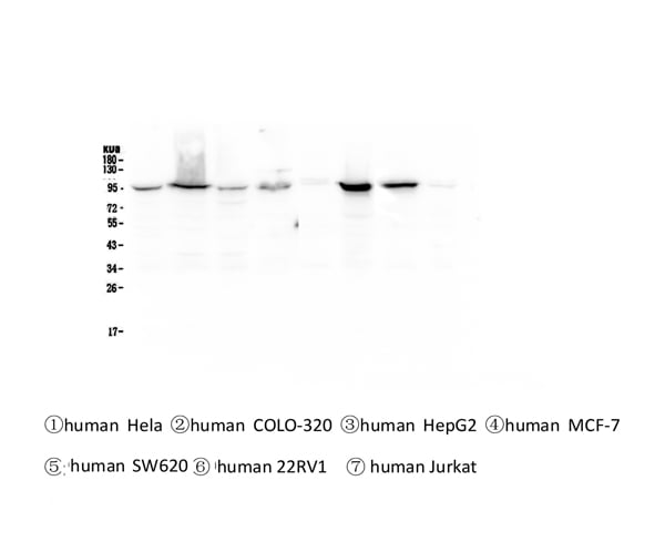
 FCM/FACS (Flow Cytometry)
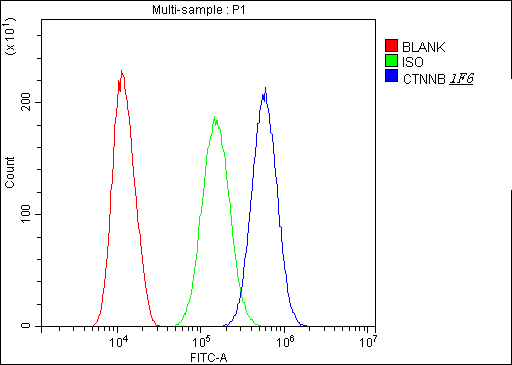
(Figure 5. Flow Cytometry analysis of SiHa cells using anti-beta Catenin antibody (M00004-2).Overlay histogram showing SiHa cells stained with M00004-2 (Blue line).The cells were blocked with 10% normal goat serum. And then incubated with mouse anti-beta Catenin Antibody (M00004-2, 1ug/1x106 cells) for 30 min at 20 degree C. DyLight® 488 conjugated goat anti-mouse IgG (BA1126, 5-10ug/1x106 cells) was used as secondary antibody for 30 minutes at 20 degree C. Isotype control antibody (Green line) was mouse IgG (1ug/1x106) used under the same conditions. Unlabelled sample (Red line) was also used as a control.)
FCM/FACS (Flow Cytometry)
(Figure 5. Flow Cytometry analysis of SiHa cells using anti-beta Catenin antibody (M00004-2).Overlay histogram showing SiHa cells stained with M00004-2 (Blue line).The cells were blocked with 10% normal goat serum. And then incubated with mouse anti-beta Catenin Antibody (M00004-2, 1ug/1x106 cells) for 30 min at 20 degree C. DyLight® 488 conjugated goat anti-mouse IgG (BA1126, 5-10ug/1x106 cells) was used as secondary antibody for 30 minutes at 20 degree C. Isotype control antibody (Green line) was mouse IgG (1ug/1x106) used under the same conditions. Unlabelled sample (Red line) was also used as a control.)
beta Catenin, Monoclonal Antibody (Cat# AAA125123)
Full Name
Anti-beta Catenin Antibody (monoclonal, 1F6)
Gene Names
CTNNB1; EVR7; CTNNB; MRD19; armadillo
Reactivity
Human, Mouse, Rat
Applications
Flow Cytometry, Immunocytochemistry, Immunohistochemistry, Western Blot
Purity
Immunogen Affinity Purified
Pricing
 FCM/FACS (Flow Cytometry)
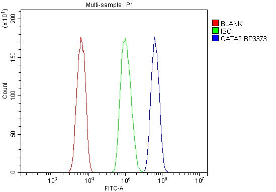
(Figure 3. Flow Cytometry analysis of HL-60 cells using anti- GATA2 antibody (AAA125607).Overlay histogram showing HL-60 cells stained withAAA125607 (Blue line). The cells were blocked with 10% normal goat serum. And then incubated with rabbit anti- GATA2 Antibody (AAA125607, 1μg/1x106 cells) for 30 min at 20 degree C. DyLight®488 conjugated goat anti-rabbit IgG (5-10μg/1x106 cells) was used as secondary antibody for 30 minutes at 20 degree C. Isotype control antibody (Green line) was rabbit IgG (1μg/1x106) used under the same conditions. Unlabelled sample (Red line) was also used as a control.)
FCM/FACS (Flow Cytometry)
(Figure 3. Flow Cytometry analysis of HL-60 cells using anti- GATA2 antibody (AAA125607).Overlay histogram showing HL-60 cells stained withAAA125607 (Blue line). The cells were blocked with 10% normal goat serum. And then incubated with rabbit anti- GATA2 Antibody (AAA125607, 1μg/1x106 cells) for 30 min at 20 degree C. DyLight®488 conjugated goat anti-rabbit IgG (5-10μg/1x106 cells) was used as secondary antibody for 30 minutes at 20 degree C. Isotype control antibody (Green line) was rabbit IgG (1μg/1x106) used under the same conditions. Unlabelled sample (Red line) was also used as a control.)
GATA2, Polyclonal Antibody (Cat# AAA125607)
Full Name
Anti-GATA2 Antibody
Gene Names
GATA2; DCML; NFE1B; MONOMAC
Reactivity
Human
Applications
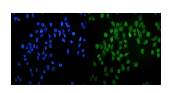
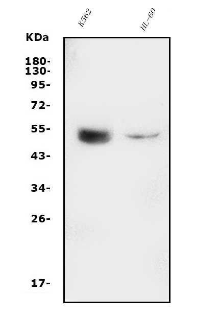
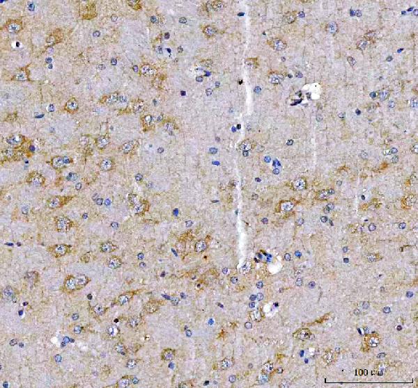
Direct ELISA, Flow Cytometry, Immunofluorescence, Immunocytochemistry, Western Blot
Purity
Immunogen affinity purified.
Pricing
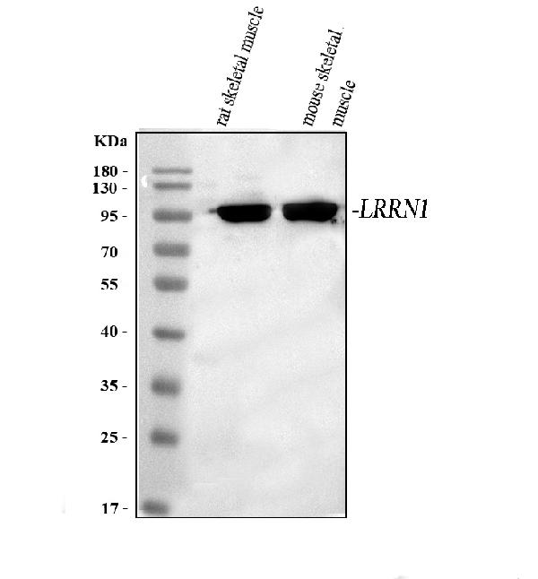
 FCM/FACS (Flow Cytometry)
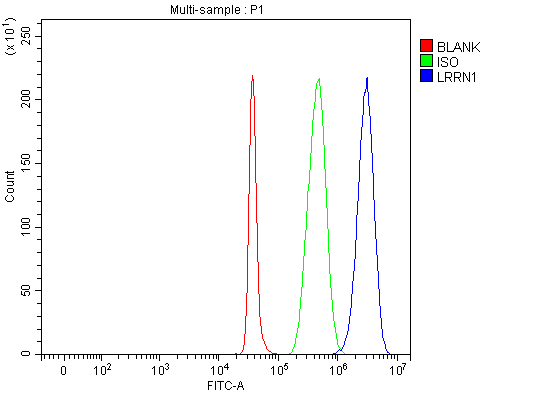
(Figure 3. Flow Cytometry analysis of SH-SY5Y cells using anti-LRRN1 antibody (AAA127745).Overlay histogram showing SH-SY5Y cells stained with AAA127745 (Blue line). The cells were fixed with 4% paraformaldehyde and blocked with 10% normal goat serum. And then incubated with rabbit anti-LRRN1 Antibody (AAA127745, 1ug/1x106 cells) for 30 min at 20 degree C. DyLight488 conjugated goat anti-rabbit IgG was used as secondary antibody for 30 minutes at 20 degree C. Isotype control antibody (Green line) was rabbit IgG (1ug/1x106) used under the same conditions. Unlabelled sample (Red line) was also used as a control.)
FCM/FACS (Flow Cytometry)
(Figure 3. Flow Cytometry analysis of SH-SY5Y cells using anti-LRRN1 antibody (AAA127745).Overlay histogram showing SH-SY5Y cells stained with AAA127745 (Blue line). The cells were fixed with 4% paraformaldehyde and blocked with 10% normal goat serum. And then incubated with rabbit anti-LRRN1 Antibody (AAA127745, 1ug/1x106 cells) for 30 min at 20 degree C. DyLight488 conjugated goat anti-rabbit IgG was used as secondary antibody for 30 minutes at 20 degree C. Isotype control antibody (Green line) was rabbit IgG (1ug/1x106) used under the same conditions. Unlabelled sample (Red line) was also used as a control.)
LRRN1, Polyclonal Antibody (Cat# AAA127745)
Full Name
Anti-LRRN1 Antibody Picoband
Gene Names
LRRN1; NLRR-1; FIGLER3
Reactivity
Human, Mouse, Rat
Applications
Flow Cytometry, Immunohistochemistry, Western Blot
Purity
Immunogen affinity purified.
Pricing
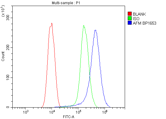
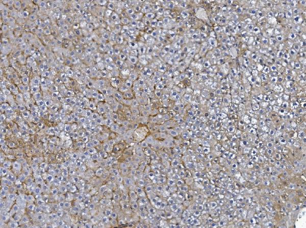
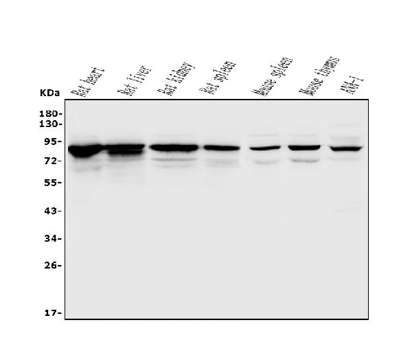
 FCM/FACS (Flow Cytometry)
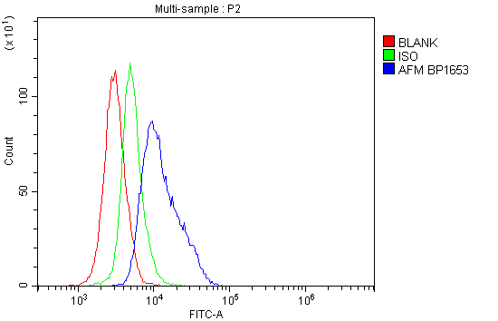
(Figure 4. Flow Cytometry analysis of mouse spleen tissues using anti-Afm antibody (AAA125534).Overlay histogram showing mouse spleen tissues stained with AAA125534 (Blue line). The tissues were blocked with 10% normal goat serum. And then incubated with rabbit anti-Afm Antibody (AAA125534, 1μg/1x106 cells) for 30 min at 20 degree C. DyLight®488 conjugated goat anti-rabbit IgG (5-10μg/1x106 cells) was used as secondary antibody for 30 minutes at 20 degree C. Isotype control antibody (Green line) was rabbit IgG (1μg/1x106) used under the same conditions. Unlabelled sample (Red line) was also used as a control.)
FCM/FACS (Flow Cytometry)
(Figure 4. Flow Cytometry analysis of mouse spleen tissues using anti-Afm antibody (AAA125534).Overlay histogram showing mouse spleen tissues stained with AAA125534 (Blue line). The tissues were blocked with 10% normal goat serum. And then incubated with rabbit anti-Afm Antibody (AAA125534, 1μg/1x106 cells) for 30 min at 20 degree C. DyLight®488 conjugated goat anti-rabbit IgG (5-10μg/1x106 cells) was used as secondary antibody for 30 minutes at 20 degree C. Isotype control antibody (Green line) was rabbit IgG (1μg/1x106) used under the same conditions. Unlabelled sample (Red line) was also used as a control.)
Afm, Polyclonal Antibody (Cat# AAA125534)
Full Name
Anti-Afm Antibody
Gene Names
Afm; Alf; alpha-Alb
Reactivity
Mouse, Rat
Applications
Direct ELISA, Flow Cytometry, Immunohistochemistry, Western Blot
Purity
Immunogen affinity purified.
Pricing
 IP (Immunoprecipitation)
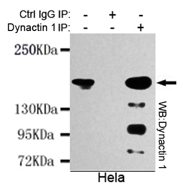
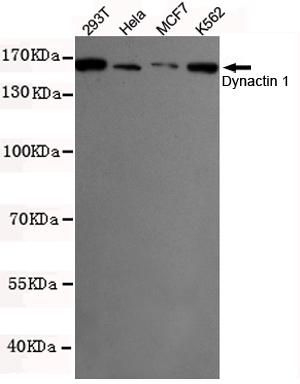
(Immunoprecipitation analysis of Hela cell lysates using Dynactin 1 mouse mAb.)
IP (Immunoprecipitation)
(Immunoprecipitation analysis of Hela cell lysates using Dynactin 1 mouse mAb.)
Dynactin 1, Monoclonal Antibody (Cat# AAA320293)
Full Name
Dynactin 1 (N-term) Mouse mAb
Gene Names
DCTN1; P135; DP-150; DAP-150
Reactivity
Human
Applications
Immunoprecipitation, Western Blot
Purity
The antibody was affinity-purified from mouse ascites by affinity-chromatography using epitope-specific immunogen.
Pricing
 FCM/FACS (Flow Cytometry)
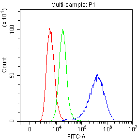
(Figure 3. Flow Cytometry analysis of SiHa cells using anti- Mesothelin antibody (AAA46561).Overlay histogram showing SiHa cells stained with AAA46561 (Blue line). Mesothelin cells were blocked with 10% normal goat serum. And then incubated with rabbit anti- Mesothelin Antibody (AAA46561,1ug/1x10^6 cells) for 30 min at 20 degree C. DyLight®488 conjugated goat anti-rabbit IgG (5-10ug/1x10^6 cells) was used as secondary antibody for 30 minutes at 20 degree C. Isotype control antibody (Green line) was rabbit IgG (1ug/1x106) used under the same conditions. Unlabelled sample (Red line) was also used as a control.)
FCM/FACS (Flow Cytometry)
(Figure 3. Flow Cytometry analysis of SiHa cells using anti- Mesothelin antibody (AAA46561).Overlay histogram showing SiHa cells stained with AAA46561 (Blue line). Mesothelin cells were blocked with 10% normal goat serum. And then incubated with rabbit anti- Mesothelin Antibody (AAA46561,1ug/1x10^6 cells) for 30 min at 20 degree C. DyLight®488 conjugated goat anti-rabbit IgG (5-10ug/1x10^6 cells) was used as secondary antibody for 30 minutes at 20 degree C. Isotype control antibody (Green line) was rabbit IgG (1ug/1x106) used under the same conditions. Unlabelled sample (Red line) was also used as a control.)
Mesothelin, Polyclonal Antibody (Cat# AAA46561)
Full Name
Anti-Mesothelin Antibody
Gene Names
MSLN; MPF; SMRP
Reactivity
Human
Applications
Immunocytochemistry, Flow Cytometry, Immunohistochemistry, Western Blot
Purity
Immunogen Affinity Purified
Pricing
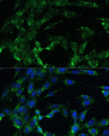
 IF (Immunofluorescence)
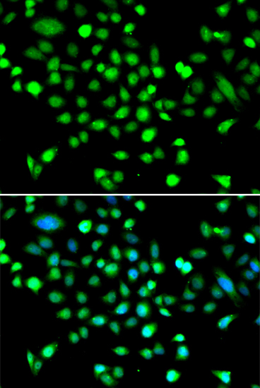
(Immunofluorescence analysis of MCF-7 cells using PRKAG1. Blue: DAPI for nuclear staining.)
IF (Immunofluorescence)
(Immunofluorescence analysis of MCF-7 cells using PRKAG1. Blue: DAPI for nuclear staining.)
PRKAG1, Polyclonal Antibody (Cat# AAA314021)
Full Name
PRKAG1 Rabbit Polyclonal Antibody
Gene Names
PRKAG1; AMPKG
Reactivity
Human, Mouse, Rat
Applications
Immunofluorescence, Immunohistochemistry, Western Blot
Purity
Affinity purification
Pricing
 Application Data
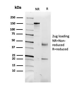
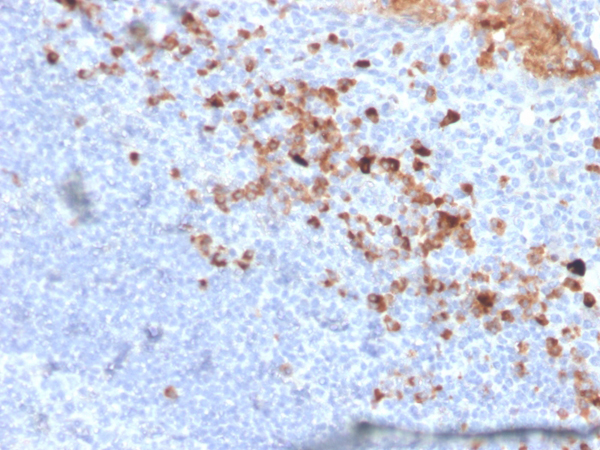
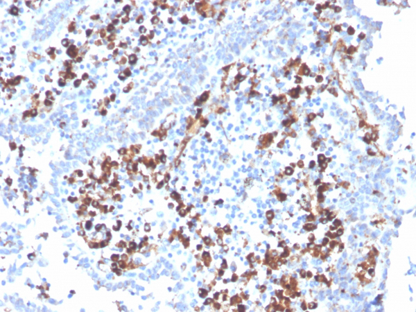
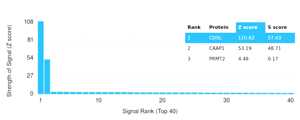
(Analysis of Protein Array containing more than 19,000 full-length human proteins using CD5L Mouse Monoclonal Antibody (CD5L/2991) Z- and S- Score: The Z-score represents the strength of a signal that a monoclonal antibody (MAb) (in combination with a fluorescently-tagged anti-IgG secondary antibody) produces when binding to a particular protein on the HuProtTM array. Z-scores are described in units of standard deviations (SD's) above the mean value of all signals generated on that array. If targets on HuProtTM are arranged in descending order of the Z-score, the S-score is the difference (also in units of SD's) between the Z-score. S-score therefore represents the relative target specificity of a MAb to its intended target. A MAb is considered to specific to its intended target, if the MAb has an S-score of at least 2.5. For example, if a MAb binds to protein X with a Z-score of 43 and to protein Y with a Z-score of 14, then the S-score for the binding of that MAb to protein X is equal to 29.)
Application Data
(Analysis of Protein Array containing more than 19,000 full-length human proteins using CD5L Mouse Monoclonal Antibody (CD5L/2991) Z- and S- Score: The Z-score represents the strength of a signal that a monoclonal antibody (MAb) (in combination with a fluorescently-tagged anti-IgG secondary antibody) produces when binding to a particular protein on the HuProtTM array. Z-scores are described in units of standard deviations (SD's) above the mean value of all signals generated on that array. If targets on HuProtTM are arranged in descending order of the Z-score, the S-score is the difference (also in units of SD's) between the Z-score. S-score therefore represents the relative target specificity of a MAb to its intended target. A MAb is considered to specific to its intended target, if the MAb has an S-score of at least 2.5. For example, if a MAb binds to protein X with a Z-score of 43 and to protein Y with a Z-score of 14, then the S-score for the binding of that MAb to protein X is equal to 29.)
CD5L/CD5LG/CD5, Monoclonal Antibody (Cat# AAA215361)
Full Name
CD5L/CD5LG/CD5 Ligand
Gene Names
CD5L; AIM; API6; CT-2; hAIM; PRO229; Spalpha; SP-ALPHA
Reactivity
Human
Applications
Western Blot, Immunohistochemistry, Immunofluorescence, Flow Cytometry
Purity
Purified Ab with BSA and Azide at 200ug/ml or Purified Ab with BSA and Azide at 200ug/ml or Purified Ab WITHOUT BSA and Azide at 1.0mg/ml
Pricing
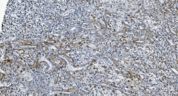
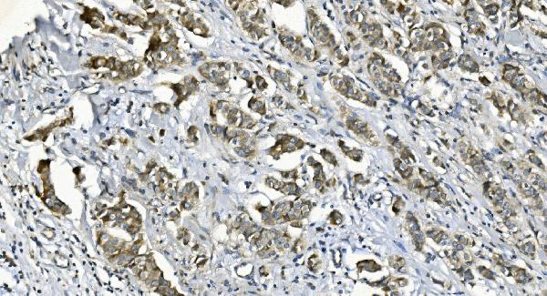
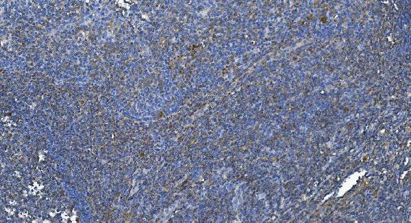
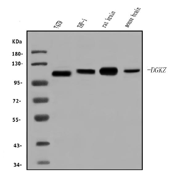
 FCM/FACS (Flow Cytometry)
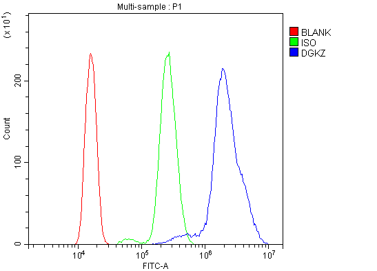
(Figure 5. Flow Cytometry analysis of HL-60 cells using anti-DGKZ/DGK-zeta antibody (AAA125777).Overlay histogram showing HL-60 cells stained with AAA125777 (Blue line). The cells were blocked with 10% normal goat serum. And then incubated with rabbit anti-DGKZ/DGK-zeta Antibody (AAA125777, 1μg/1x106 cells) for 30 min at 20 degree C. DyLight®488 conjugated goat anti-rabbit IgG (5-10μg/1x106 cells) was used as secondary antibody for 30 minutes at 20 degree C. Isotype control antibody (Green line) was rabbit IgG (1μg/1x106) used under the same conditions. Unlabelled sample (Red line) was also used as a control.)
FCM/FACS (Flow Cytometry)
(Figure 5. Flow Cytometry analysis of HL-60 cells using anti-DGKZ/DGK-zeta antibody (AAA125777).Overlay histogram showing HL-60 cells stained with AAA125777 (Blue line). The cells were blocked with 10% normal goat serum. And then incubated with rabbit anti-DGKZ/DGK-zeta Antibody (AAA125777, 1μg/1x106 cells) for 30 min at 20 degree C. DyLight®488 conjugated goat anti-rabbit IgG (5-10μg/1x106 cells) was used as secondary antibody for 30 minutes at 20 degree C. Isotype control antibody (Green line) was rabbit IgG (1μg/1x106) used under the same conditions. Unlabelled sample (Red line) was also used as a control.)
DGKZ/DGK-zeta, Polyclonal Antibody (Cat# AAA125777)
Full Name
Anti-DGKZ/DGK-zeta Antibody
Gene Names
DGKZ; DAGK5; DAGK6; DGK-ZETA; hDGKzeta
Reactivity
Human, Mouse, Rat
Applications
Direct ELISA, Flow Cytometry, Immunohistochemistry, Western Blot
Purity
Immunogen affinity purified.
Pricing
 FCM/FACS (Flow Cytometry)
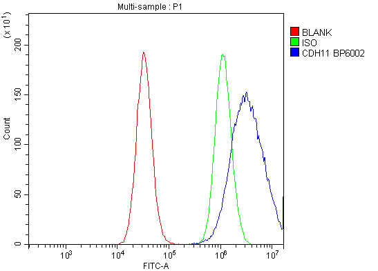
(Figure 4. Flow Cytometry analysis of PC-3 cells using anti-OB Cadherin/CDH11 antibody (AAA125724).Overlay histogram showing PC-3 cells stained with AAA125724 (Blue line). The cells were blocked with 10% normal goat serum. And then incubated with rabbit anti-OB Cadherin/CDH11 Antibody (AAA125724, 1μg/1x106 cells) for 30 min at 20 degree C. DyLight®488 conjugated goat anti-rabbit IgG (5-10μg/1x106 cells) was used as secondary antibody for 30 minutes at 20 degree C. Isotype control antibody (Green line) was rabbit IgG (1μg/1x106) used under the same conditions. Unlabelled sample (Red line) was also used as a control.)
FCM/FACS (Flow Cytometry)
(Figure 4. Flow Cytometry analysis of PC-3 cells using anti-OB Cadherin/CDH11 antibody (AAA125724).Overlay histogram showing PC-3 cells stained with AAA125724 (Blue line). The cells were blocked with 10% normal goat serum. And then incubated with rabbit anti-OB Cadherin/CDH11 Antibody (AAA125724, 1μg/1x106 cells) for 30 min at 20 degree C. DyLight®488 conjugated goat anti-rabbit IgG (5-10μg/1x106 cells) was used as secondary antibody for 30 minutes at 20 degree C. Isotype control antibody (Green line) was rabbit IgG (1μg/1x106) used under the same conditions. Unlabelled sample (Red line) was also used as a control.)
OB Cadherin/CDH11, Polyclonal Antibody (Cat# AAA125724)
Full Name
Anti-OB Cadherin/CDH11 Antibody
Gene Names
CDH11; OB; CAD11; CDHOB; OSF-4
Reactivity
Human, Mouse, Rat
Applications
Direct ELISA, Flow Cytometry, Immunohistochemistry, Western Blot
Purity
Immunogen affinity purified.
Pricing
 FCM/FACS (Flow Cytometry)
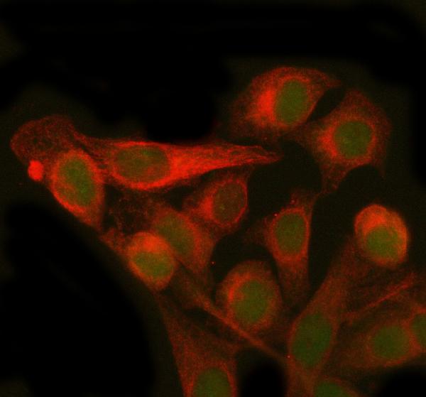
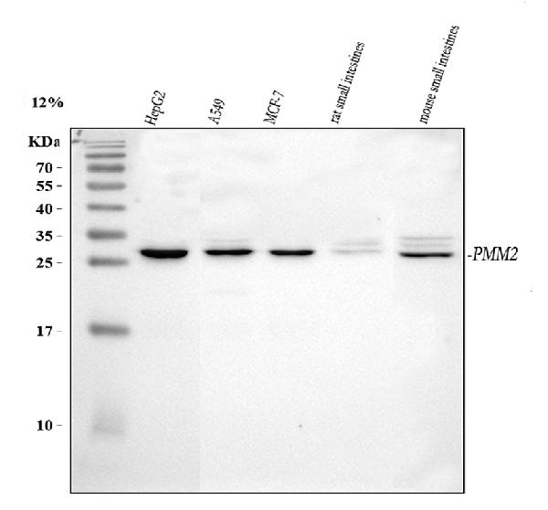
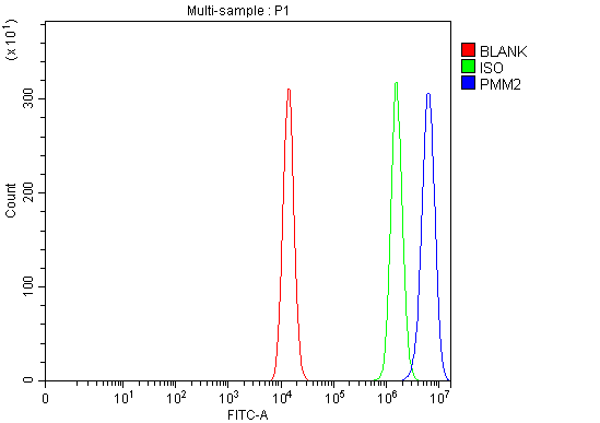
(Figure 3. Flow Cytometry analysis of HEL cells using anti-PMM2 antibody (AAA127163).Overlay histogram showing HEL cells stained with AAA127163 (Blue line). To facilitate intracellular staining, cells were fixed with 4% paraformaldehyde and permeabilized with permeabilization buffer. The cells were blocked with 10% normal goat serum. And then incubated with rabbit anti-PMM2 Antibody (AAA127163, 1ug/1x106 cells) for 30 min at 20 degree C. DyLight488 conjugated goat anti-rabbit IgG was used as secondary antibody for 30 minutes at 20 degree C. Isotype control antibody (Green line) was rabbit IgG (1ug/1x106) used under the same conditions. Unlabelled sample (Red line) was also used as a control.)
FCM/FACS (Flow Cytometry)
(Figure 3. Flow Cytometry analysis of HEL cells using anti-PMM2 antibody (AAA127163).Overlay histogram showing HEL cells stained with AAA127163 (Blue line). To facilitate intracellular staining, cells were fixed with 4% paraformaldehyde and permeabilized with permeabilization buffer. The cells were blocked with 10% normal goat serum. And then incubated with rabbit anti-PMM2 Antibody (AAA127163, 1ug/1x106 cells) for 30 min at 20 degree C. DyLight488 conjugated goat anti-rabbit IgG was used as secondary antibody for 30 minutes at 20 degree C. Isotype control antibody (Green line) was rabbit IgG (1ug/1x106) used under the same conditions. Unlabelled sample (Red line) was also used as a control.)
PMM2, Polyclonal Antibody (Cat# AAA127163)
Full Name
Anti-PMM2 Antibody Picoband
Gene Names
PMM2; PMI; CDG1; CDGS; PMI1; CDG1a; PMM 2
Reactivity
Human, Mouse, Rat
Applications
Flow Cytometry, Immunofluorescence, Immunocytochemistry, Western Blot
Purity
Immunogen affinity purified.
Pricing
 FCM/FACS (Flow Cytometry)
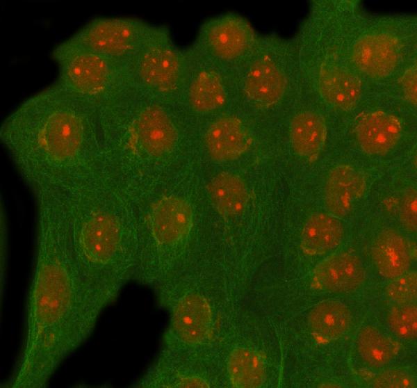
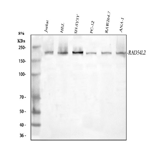
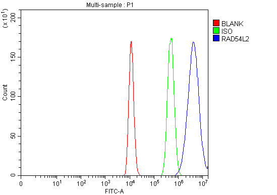
(Figure 3. Flow Cytometry analysis of HEL cells using anti-RAD54L2 antibody (AAA127729).Overlay histogram showing HEL cells stained with AAA127729 (Blue line). To facilitate intracellular staining, cells were fixed with 4% paraformaldehyde and permeabilized with permeabilization buffer. The cells were blocked with 10% normal goat serum. And then incubated with rabbit anti-RAD54L2 Antibody (AAA127729, 1ug/1x106 cells) for 30 min at 20 degree C. DyLight488 conjugated goat anti-rabbit IgG was used as secondary antibody for 30 minutes at 20 degree C. Isotype control antibody (Green line) was rabbit IgG (1ug/1x106) used under the same conditions. Unlabelled sample (Red line) was also used as a control.)
FCM/FACS (Flow Cytometry)
(Figure 3. Flow Cytometry analysis of HEL cells using anti-RAD54L2 antibody (AAA127729).Overlay histogram showing HEL cells stained with AAA127729 (Blue line). To facilitate intracellular staining, cells were fixed with 4% paraformaldehyde and permeabilized with permeabilization buffer. The cells were blocked with 10% normal goat serum. And then incubated with rabbit anti-RAD54L2 Antibody (AAA127729, 1ug/1x106 cells) for 30 min at 20 degree C. DyLight488 conjugated goat anti-rabbit IgG was used as secondary antibody for 30 minutes at 20 degree C. Isotype control antibody (Green line) was rabbit IgG (1ug/1x106) used under the same conditions. Unlabelled sample (Red line) was also used as a control.)
RAD54L2, Polyclonal Antibody (Cat# AAA127729)
Full Name
Anti-RAD54L2 Antibody Picoband
Gene Names
RAD54L2; ARIP4; HSPC325; SRISNF2L
Reactivity
Human, Mouse, Rat
Applications
Flow Cytometry, Immunofluorescence, Immunocytochemistry, Western Blot
Purity
Immunogen affinity purified.
Pricing
 Bioactivity
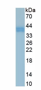
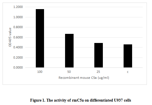
(Complement Component 5a (C5a) is a component of the complement system which plays a key role in promoting migration and adherence of neutrophils and monocytes to vessel walls. The activity of recombinant human C5a was also measured by its ability to induce N-acetyl-beta-D-glucosaminidase release from differentiated U937 human histiocytic lymphoma cells. 3.2*106 differentiated U937 cells were added to the 24-well plate and different concentrations of rhC5a was added to the 24-well plate and incubated at 37 degree C for 3min. The cells were centrifuged at 400 g for 3min, and the supernatant contained N-acetyl-beta-D-glucosaminidase. The enzyme activity of N-acetyl-beta-D-glucosaminidase was measured by the substrate of 4-Nitrophenyl 2-acetamido-2-deoxy-beta-D-glucopyranoside. The result was shown in figure 1, It was obvious that rhC5a can significantly induce N-acetyl-beta-D-glucosaminidase release.)
Bioactivity
(Complement Component 5a (C5a) is a component of the complement system which plays a key role in promoting migration and adherence of neutrophils and monocytes to vessel walls. The activity of recombinant human C5a was also measured by its ability to induce N-acetyl-beta-D-glucosaminidase release from differentiated U937 human histiocytic lymphoma cells. 3.2*106 differentiated U937 cells were added to the 24-well plate and different concentrations of rhC5a was added to the 24-well plate and incubated at 37 degree C for 3min. The cells were centrifuged at 400 g for 3min, and the supernatant contained N-acetyl-beta-D-glucosaminidase. The enzyme activity of N-acetyl-beta-D-glucosaminidase was measured by the substrate of 4-Nitrophenyl 2-acetamido-2-deoxy-beta-D-glucopyranoside. The result was shown in figure 1, It was obvious that rhC5a can significantly induce N-acetyl-beta-D-glucosaminidase release.)
Complement Component 5a (C5a), Active Protein (Cat# AAA161727)
Full Name
Active Complement Component 5a (C5a)
Gene Names
Hc; C5; He; C5a
Applications
Activity Assay, Cell Culture
Purity
Greater than 90% by SDS-PAGE
Pricing
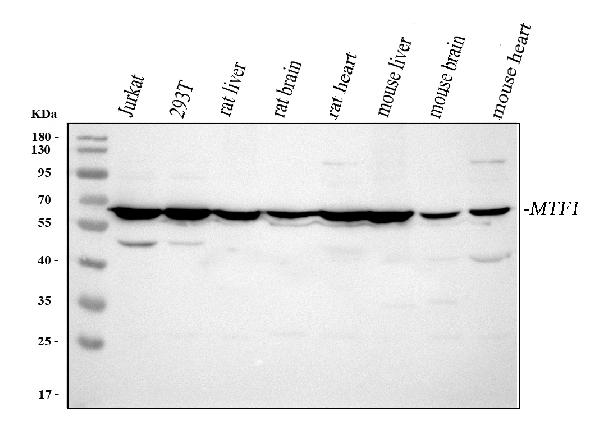
 FCM/FACS (Flow Cytometry)
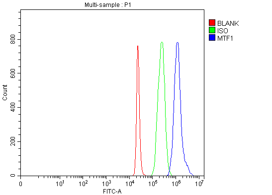
(Figure 5. Flow Cytometry analysis of 293T cells using anti-MTF1 antibody (AAA127249).Overlay histogram showing 293T cells stained with AAA127249 (Blue line). To facilitate intracellular staining, cells were fixed with 4% paraformaldehyde and permeabilized with permeabilization buffer. The cells were blocked with 10% normal goat serum. And then incubated with rabbit anti-MTF1 Antibody (AAA127249, 1ug/1x106 cells) for 30 min at 20 degree C. DyLight488 conjugated goat anti-rabbit IgG was used as secondary antibody for 30 minutes at 20 degree C. Isotype control antibody (Green line) was rabbit IgG (1ug/1x106) used under the same conditions. Unlabelled sample (Red line) was also used as a control.)
FCM/FACS (Flow Cytometry)
(Figure 5. Flow Cytometry analysis of 293T cells using anti-MTF1 antibody (AAA127249).Overlay histogram showing 293T cells stained with AAA127249 (Blue line). To facilitate intracellular staining, cells were fixed with 4% paraformaldehyde and permeabilized with permeabilization buffer. The cells were blocked with 10% normal goat serum. And then incubated with rabbit anti-MTF1 Antibody (AAA127249, 1ug/1x106 cells) for 30 min at 20 degree C. DyLight488 conjugated goat anti-rabbit IgG was used as secondary antibody for 30 minutes at 20 degree C. Isotype control antibody (Green line) was rabbit IgG (1ug/1x106) used under the same conditions. Unlabelled sample (Red line) was also used as a control.)
MTF1, Polyclonal Antibody (Cat# AAA127249)
Full Name
Anti-MTF1 Antibody Picoband
Gene Names
MTF1; ZRF; MTF-1
Reactivity
Human, Mouse, Rat
Applications
Flow Cytometry, Immunofluorescence, Immunocytochemistry, Immunohistochemistry, Western Blot
Purity
Immunogen affinity purified.
Pricing
 FCM/FACS (Flow Cytometry)
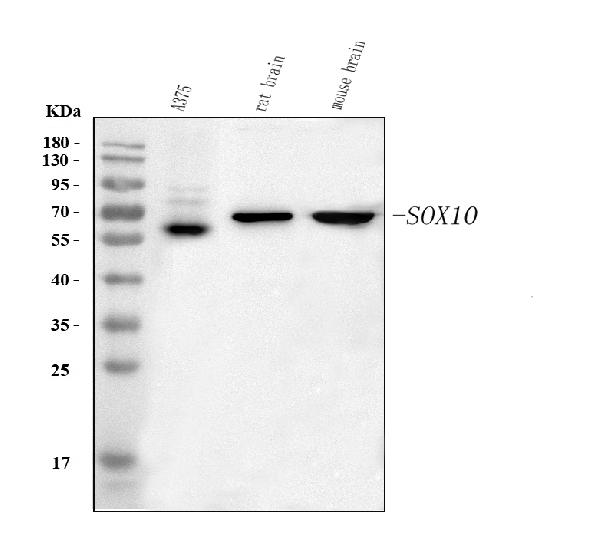
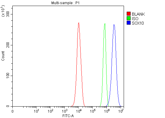
(Figure 3. Flow Cytometry analysis of PC-3 cells using anti-SOX10 antibody (AAA127013).Overlay histogram showing PC-3 cells stained with AAA127013 (Blue line). To facilitate intracellular staining, cells were fixed with 4% paraformaldehyde and permeabilized with permeabilization buffer. The cells were blocked with 10% normal goat serum. And then incubated with rabbit anti-SOX10 Antibody (AAA127013, 1ug/1x106 cells) for 30 min at 20 degree C. DyLight488 conjugated goat anti-rabbit IgG was used as secondary antibody for 30 minutes at 20 degree C. Isotype control antibody (Green line) was rabbit IgG (1ug/1x106) used under the same conditions. Unlabelled sample (Red line) was also used as a control.)
FCM/FACS (Flow Cytometry)
(Figure 3. Flow Cytometry analysis of PC-3 cells using anti-SOX10 antibody (AAA127013).Overlay histogram showing PC-3 cells stained with AAA127013 (Blue line). To facilitate intracellular staining, cells were fixed with 4% paraformaldehyde and permeabilized with permeabilization buffer. The cells were blocked with 10% normal goat serum. And then incubated with rabbit anti-SOX10 Antibody (AAA127013, 1ug/1x106 cells) for 30 min at 20 degree C. DyLight488 conjugated goat anti-rabbit IgG was used as secondary antibody for 30 minutes at 20 degree C. Isotype control antibody (Green line) was rabbit IgG (1ug/1x106) used under the same conditions. Unlabelled sample (Red line) was also used as a control.)
SOX10, Polyclonal Antibody (Cat# AAA127013)
Full Name
Anti-SOX10 Antibody Picoband
Gene Names
SOX10; DOM; WS4; PCWH; WS2E; WS4C
Reactivity
Human, Mouse, Rat
Applications
Flow Cytometry, Immunofluorescence, Immunocytochemistry, Western Blot
Purity
Immunogen affinity purified.
Pricing
 FCM/FACS (Flow Cytometry)
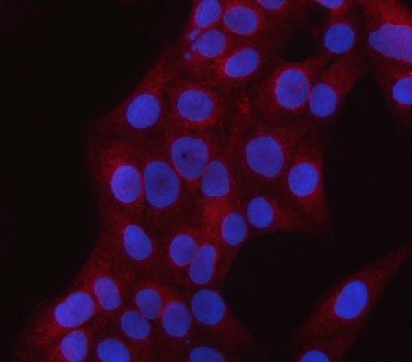
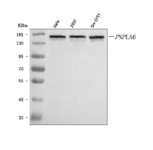
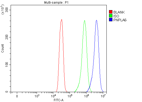
(Figure 3. Flow Cytometry analysis of SiHa cells using anti-PNPLA6 antibody (AAA127213).Overlay histogram showing SiHa cells stained with AAA127213 (Blue line). To facilitate intracellular staining, cells were fixed with 4% paraformaldehyde and permeabilized with permeabilization buffer. The cells were blocked with 10% normal goat serum. And then incubated with rabbit anti-PNPLA6 Antibody (AAA127213, 1ug/1x106 cells) for 30 min at 20 degree C. DyLight488 conjugated goat anti-rabbit IgG was used as secondary antibody for 30 minutes at 20 degree C. Isotype control antibody (Green line) was rabbit IgG (1ug/1x106) used under the same conditions. Unlabelled sample (Red line) was also used as a control.)
FCM/FACS (Flow Cytometry)
(Figure 3. Flow Cytometry analysis of SiHa cells using anti-PNPLA6 antibody (AAA127213).Overlay histogram showing SiHa cells stained with AAA127213 (Blue line). To facilitate intracellular staining, cells were fixed with 4% paraformaldehyde and permeabilized with permeabilization buffer. The cells were blocked with 10% normal goat serum. And then incubated with rabbit anti-PNPLA6 Antibody (AAA127213, 1ug/1x106 cells) for 30 min at 20 degree C. DyLight488 conjugated goat anti-rabbit IgG was used as secondary antibody for 30 minutes at 20 degree C. Isotype control antibody (Green line) was rabbit IgG (1ug/1x106) used under the same conditions. Unlabelled sample (Red line) was also used as a control.)
PNPLA6, Polyclonal Antibody (Cat# AAA127213)
Full Name
Anti-PNPLA6 Antibody Picoband
Gene Names
PNPLA6; NTE; sws; SPG39; NTEMND; iPLA2delta
Reactivity
Human
Applications
Flow Cytometry, Immunofluorescence, Immunocytochemistry, Western Blot
Purity
Immunogen affinity purified.
Pricing
 FCM/FACS (Flow Cytometry)
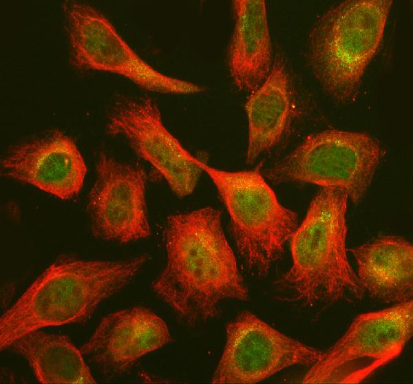
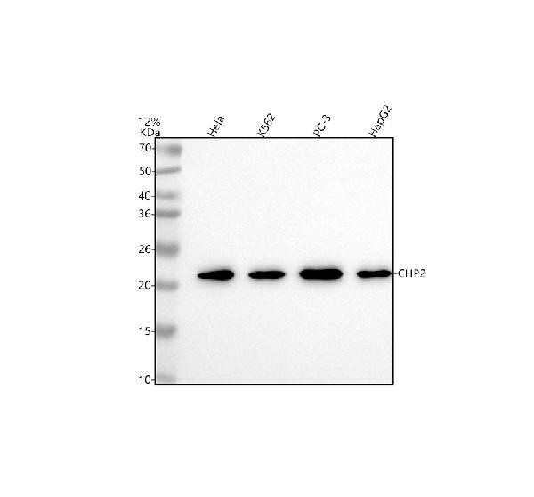
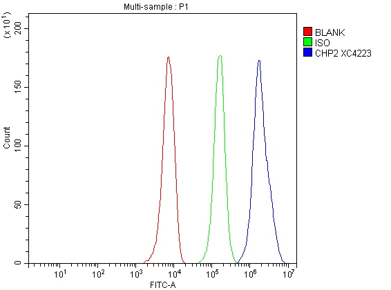
(Figure 3. Flow Cytometry analysis of HepG2 cells using anti-CHP2 antibody (AAA128233).Overlay histogram showing HepG2 cells stained with AAA128233 (Blue line). To facilitate intracellular staining, cells were fixed with 4% paraformaldehyde and permeabilized with permeabilization buffer. The cells were blocked with 10% normal goat serum. And then incubated with rabbit anti-CHP2 Antibody (AAA128233, 1ug/1x106 cells) for 30 min at 20 degree C. DyLight®488 conjugated goat anti-rabbit IgG was used as secondary antibody for 30 minutes at 20 degree C. Isotype control antibody (Green line) was rabbit IgG (1ug/1x106) used under the same conditions. Unlabelled sample without incubation with primary antibody and secondary antibody (Red line) was used as a blank control.)
FCM/FACS (Flow Cytometry)
(Figure 3. Flow Cytometry analysis of HepG2 cells using anti-CHP2 antibody (AAA128233).Overlay histogram showing HepG2 cells stained with AAA128233 (Blue line). To facilitate intracellular staining, cells were fixed with 4% paraformaldehyde and permeabilized with permeabilization buffer. The cells were blocked with 10% normal goat serum. And then incubated with rabbit anti-CHP2 Antibody (AAA128233, 1ug/1x106 cells) for 30 min at 20 degree C. DyLight®488 conjugated goat anti-rabbit IgG was used as secondary antibody for 30 minutes at 20 degree C. Isotype control antibody (Green line) was rabbit IgG (1ug/1x106) used under the same conditions. Unlabelled sample without incubation with primary antibody and secondary antibody (Red line) was used as a blank control.)
CHP2, Polyclonal Antibody (Cat# AAA128233)
Full Name
Anti-CHP2 Antibody Picoband
Reactivity
Human
Applications
Flow Cytometry, Immunofluorescence, Immunocytochemistry, Western Blot
Purity
Immunogen affinity purified.
Pricing
 FCM/FACS (Flow Cytometry)
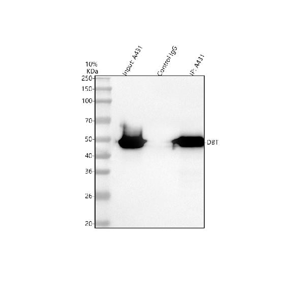
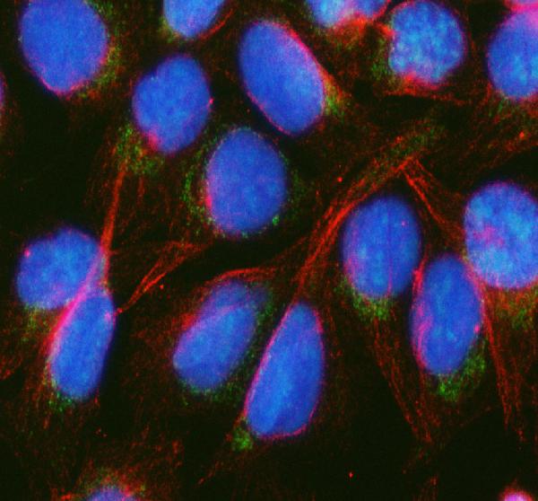
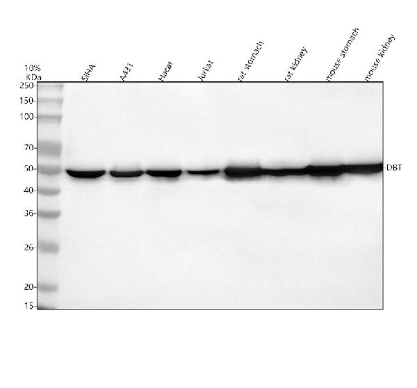
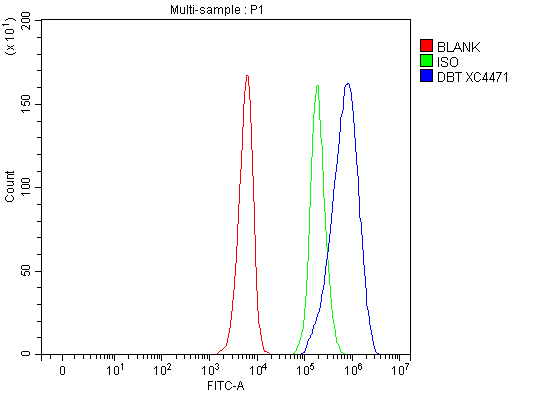
(Figure 4. Flow Cytometry analysis of SiHa cells using anti-DBT antibody (AAA128210).Overlay histogram showing SiHa cells stained with AAA128210 (Blue line). To facilitate intracellular staining, cells were fixed with 4% paraformaldehyde and permeabilized with permeabilization buffer. The cells were blocked with 10% normal goat serum. And then incubated with rabbit anti-DBT Antibody (AAA128210, 1ug/1x106 cells) for 30 min at 20 degree C. DyLight®488 conjugated goat anti-rabbit IgG was used as secondary antibody for 30 minutes at 20 degree C. Isotype control antibody (Green line) was rabbit IgG (1ug/1x106) used under the same conditions. Unlabelled sample without incubation with primary antibody and secondary antibody (Red line) was used as a blank control.)
FCM/FACS (Flow Cytometry)
(Figure 4. Flow Cytometry analysis of SiHa cells using anti-DBT antibody (AAA128210).Overlay histogram showing SiHa cells stained with AAA128210 (Blue line). To facilitate intracellular staining, cells were fixed with 4% paraformaldehyde and permeabilized with permeabilization buffer. The cells were blocked with 10% normal goat serum. And then incubated with rabbit anti-DBT Antibody (AAA128210, 1ug/1x106 cells) for 30 min at 20 degree C. DyLight®488 conjugated goat anti-rabbit IgG was used as secondary antibody for 30 minutes at 20 degree C. Isotype control antibody (Green line) was rabbit IgG (1ug/1x106) used under the same conditions. Unlabelled sample without incubation with primary antibody and secondary antibody (Red line) was used as a blank control.)
DBT, Polyclonal Antibody (Cat# AAA128210)
Full Name
Anti-DBT Antibody Picoband
Gene Names
DBT; E2; E2B; BCATE2; BCKADE2; BCKAD-E2
Reactivity
Human, Mouse, Rat
Applications
Flow Cytometry, Immunoprecipitation, Immunofluorescence, Immunocytochemistry, Western Blot
Purity
Immunogen affinity purified.
Pricing
 FCM/FACS (Flow Cytometry)
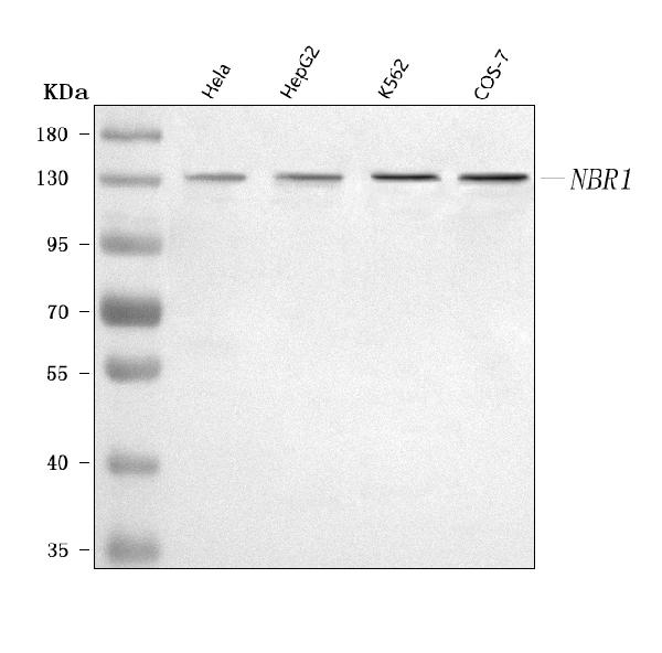
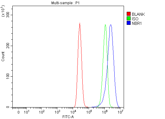
(Figure 3. Flow Cytometry analysis of MCF-7 cells using anti-NBR1 antibody (AAA127186).Overlay histogram showing MCF-7 cells stained with AAA127186 (Blue line). To facilitate intracellular staining, cells were fixed with 4% paraformaldehyde and permeabilized with permeabilization buffer. The cells were blocked with 10% normal goat serum. And then incubated with rabbit anti-NBR1 Antibody (AAA127186, 1ug/1x106 cells) for 30 min at 20 degree C. DyLight488 conjugated goat anti-rabbit IgG was used as secondary antibody for 30 minutes at 20 degree C. Isotype control antibody (Green line) was rabbit IgG (1ug/1x106) used under the same conditions. Unlabelled sample (Red line) was also used as a control.)
FCM/FACS (Flow Cytometry)
(Figure 3. Flow Cytometry analysis of MCF-7 cells using anti-NBR1 antibody (AAA127186).Overlay histogram showing MCF-7 cells stained with AAA127186 (Blue line). To facilitate intracellular staining, cells were fixed with 4% paraformaldehyde and permeabilized with permeabilization buffer. The cells were blocked with 10% normal goat serum. And then incubated with rabbit anti-NBR1 Antibody (AAA127186, 1ug/1x106 cells) for 30 min at 20 degree C. DyLight488 conjugated goat anti-rabbit IgG was used as secondary antibody for 30 minutes at 20 degree C. Isotype control antibody (Green line) was rabbit IgG (1ug/1x106) used under the same conditions. Unlabelled sample (Red line) was also used as a control.)
NBR1, Polyclonal Antibody (Cat# AAA127186)
Full Name
Anti-NBR1 Antibody Picoband
Gene Names
NBR1; IAI3B; M17S2; MIG19; 1A1-3B
Reactivity
Human, Monkey
Applications
Flow Cytometry, Immunofluorescence, Immunocytochemistry, Western Blot
Purity
Immunogen affinity purified.
Pricing
 FCM/FACS (Flow Cytometry)
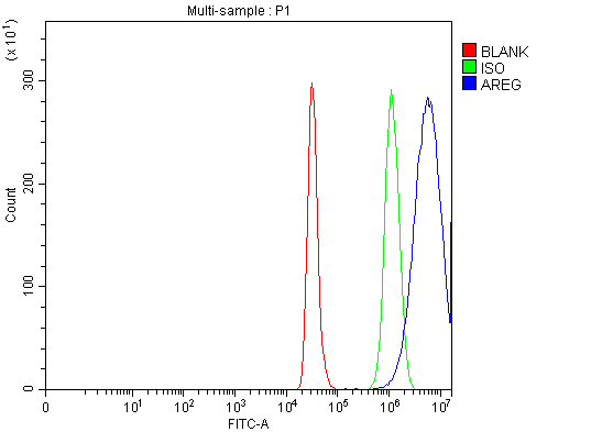
(Figure 2. Flow Cytometry analysis of CACO-2 cells using anti-Amphiregulin/AREG antibody (AAA126174).Overlay histogram showing CACO-2 cells stained with AAA126174 (Blue line). The cells were blocked with 10% normal goat serum. And then incubated with rabbit anti-Amphiregulin/AREG Antibody (AAA126174, 1 ug/1x10^6 cells) for 30 min at 20 degree C. DyLight488 conjugated goat anti-rabbit IgG was used as secondary antibody for 30 minutes at 20 degree C. Isotype control antibody (Green line) was rabbit IgG (1 ug/1x10^6) used under the same conditions. Unlabelled sample (Red line) was also used as a control.)
FCM/FACS (Flow Cytometry)
(Figure 2. Flow Cytometry analysis of CACO-2 cells using anti-Amphiregulin/AREG antibody (AAA126174).Overlay histogram showing CACO-2 cells stained with AAA126174 (Blue line). The cells were blocked with 10% normal goat serum. And then incubated with rabbit anti-Amphiregulin/AREG Antibody (AAA126174, 1 ug/1x10^6 cells) for 30 min at 20 degree C. DyLight488 conjugated goat anti-rabbit IgG was used as secondary antibody for 30 minutes at 20 degree C. Isotype control antibody (Green line) was rabbit IgG (1 ug/1x10^6) used under the same conditions. Unlabelled sample (Red line) was also used as a control.)
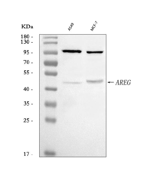
Amphiregulin/AREG, Polyclonal Antibody (Cat# AAA126174)
Full Name
Anti-Amphiregulin/AREG Antibody Picoband
Gene Names
AREG; AR; SDGF; CRDGF
Reactivity
Human
Applications
Western Blot, Flow Cytometry
Purity
Immunogen affinity purified.
Pricing
 FCM/FACS (Flow Cytometry)
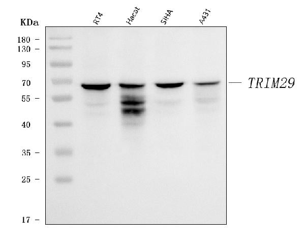
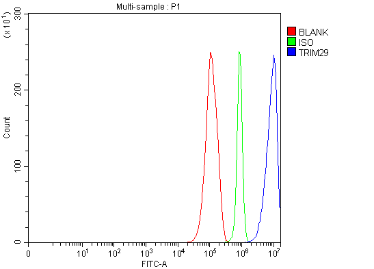
(Figure 2. Flow Cytometry analysis of RT4 cells using anti-TRIM29 antibody (AAA126384).Overlay histogram showing RT4 cells stained with AAA126384 (Blue line). The cells were blocked with 10% normal goat serum. And then incubated with rabbit anti-TRIM29 Antibody (AAA126384, 1 ug/1x10^6 cells) for 30 min at 20 degree C. DyLight488 conjugated goat anti-rabbit IgG was used as secondary antibody for 30 minutes at 20 degree C. Isotype control antibody (Green line) was rabbit IgG (1 ug/1x10^6) used under the same conditions. Unlabelled sample (Red line) was also used as a control.)
FCM/FACS (Flow Cytometry)
(Figure 2. Flow Cytometry analysis of RT4 cells using anti-TRIM29 antibody (AAA126384).Overlay histogram showing RT4 cells stained with AAA126384 (Blue line). The cells were blocked with 10% normal goat serum. And then incubated with rabbit anti-TRIM29 Antibody (AAA126384, 1 ug/1x10^6 cells) for 30 min at 20 degree C. DyLight488 conjugated goat anti-rabbit IgG was used as secondary antibody for 30 minutes at 20 degree C. Isotype control antibody (Green line) was rabbit IgG (1 ug/1x10^6) used under the same conditions. Unlabelled sample (Red line) was also used as a control.)
TRIM29, Polyclonal Antibody (Cat# AAA126384)
Full Name
Anti-TRIM29 Antibody Picoband
Gene Names
TRIM29; ATDC
Reactivity
Human
Applications
Flow Cytometry, Western Blot
Purity
Immunogen affinity purified.
Pricing
 IF (Immunofluorescence)
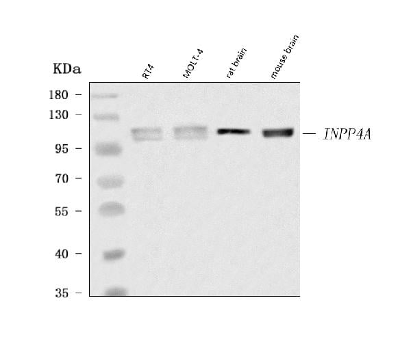
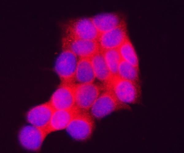
(Figure 2. IF analysis of INPP4A using anti-INPP4A antibody (AAA126485).INPP4A was detected in an immunocytochemical section of MCF-7 cells. Enzyme antigen retrieval was performed using IHC enzyme antigen retrieval reagent (AR0022) for 15 mins. The cells were blocked with 10% goat serum. And then incubated with 5 ug/mL rabbit anti-INPP4A Antibody (AAA126485) overnight at 4 degree C. Cy3 Conjugated Goat Anti-Rabbit IgG (BA1032) was used as secondary antibody at 1:100 dilution and incubated for 30 minutes at 37 degree C. The section was counterstained with DAPI. Visualize using a fluorescence microscope and filter sets appropriate for the label used.)
IF (Immunofluorescence)
(Figure 2. IF analysis of INPP4A using anti-INPP4A antibody (AAA126485).INPP4A was detected in an immunocytochemical section of MCF-7 cells. Enzyme antigen retrieval was performed using IHC enzyme antigen retrieval reagent (AR0022) for 15 mins. The cells were blocked with 10% goat serum. And then incubated with 5 ug/mL rabbit anti-INPP4A Antibody (AAA126485) overnight at 4 degree C. Cy3 Conjugated Goat Anti-Rabbit IgG (BA1032) was used as secondary antibody at 1:100 dilution and incubated for 30 minutes at 37 degree C. The section was counterstained with DAPI. Visualize using a fluorescence microscope and filter sets appropriate for the label used.)
INPP4A, Polyclonal Antibody (Cat# AAA126485)
Full Name
Anti-INPP4A Antibody Picoband
Gene Names
INPP4A; INPP4; TVAS1
Reactivity
Human, Mouse, Rat
Applications
Immunofluorescence, Immunocytochemistry, Western Blot
Purity
Immunogen affinity purified.
Pricing
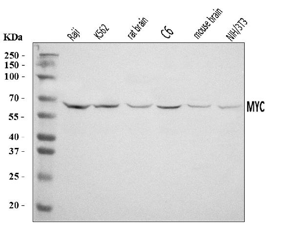
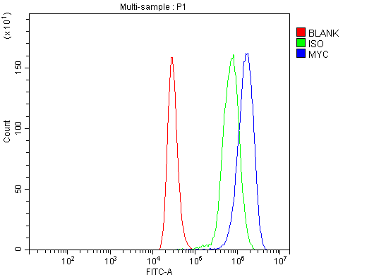 FCM/FACS (Flow Cytometry)
(Figure 2. Flow Cytometry analysis of THP-1 cells using anti-C-Myc/MYC antibody (AAA125945).Overlay histogram showing THP-1 cells stained with AAA125945 (Blue line). The cells were blocked with 10% normal goat serum. And then incubated with rabbit anti-C-Myc/MYC Antibody (AAA125945, 1 ug/1x10^6 cells) for 30 min at 20 degree C. DyLight488 conjugated goat anti-rabbit IgG was used as secondary antibody for 30 minutes at 20 degree C. Isotype control antibody (Green line) was rabbit IgG (1 ug/1x10^6) used under the same conditions. Unlabelled sample (Red line) was also used as a control.)
FCM/FACS (Flow Cytometry)
(Figure 2. Flow Cytometry analysis of THP-1 cells using anti-C-Myc/MYC antibody (AAA125945).Overlay histogram showing THP-1 cells stained with AAA125945 (Blue line). The cells were blocked with 10% normal goat serum. And then incubated with rabbit anti-C-Myc/MYC Antibody (AAA125945, 1 ug/1x10^6 cells) for 30 min at 20 degree C. DyLight488 conjugated goat anti-rabbit IgG was used as secondary antibody for 30 minutes at 20 degree C. Isotype control antibody (Green line) was rabbit IgG (1 ug/1x10^6) used under the same conditions. Unlabelled sample (Red line) was also used as a control.)
c-Myc/MYC, Polyclonal Antibody (Cat# AAA125945)
Full Name
Anti-c-Myc/MYC Antibody Picoband
Gene Names
MYC; MRTL; c-Myc; bHLHe39
Reactivity
Human, Mouse, Rat
Applications
Flow Cytometry, Western Blot
Purity
Immunogen affinity purified.
Pricing
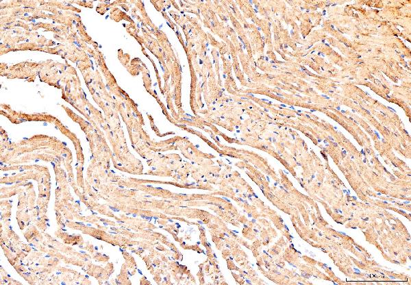
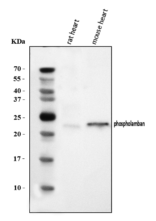
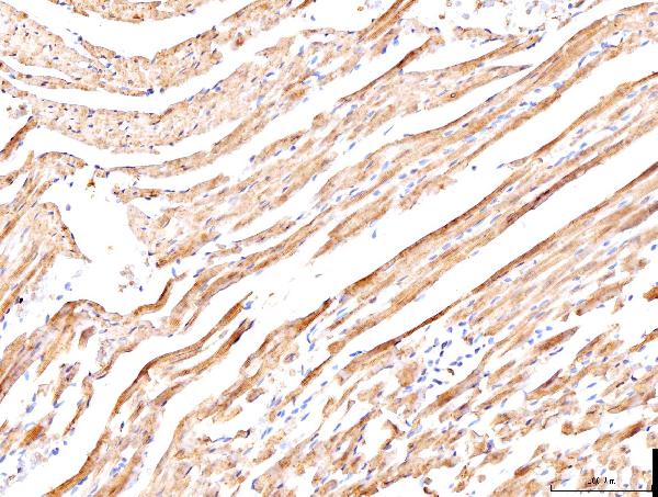 IHC (Immunohistochemisry)
(Figure 3. IHC analysis of Phospholamban/PLN using anti-Phospholamban/PLN antibody (AAA126140).Phospholamban/PLN was detected in a paraffin-embedded section of rat cardiac tissue. Heat mediated antigen retrieval was performed in EDTA buffer (pH 8.0, epitope retrieval solution). The tissue section was blocked with 10% goat serum. The tissue section was then incubated with 2 ug/ml rabbit anti-Phospholamban/PLN Antibody (AAA126140) overnight at 4 degree C. Biotinylated goat anti-rabbit IgG was used as secondary antibody and incubated for 30 minutes at 37 degree C. The tissue section was developed using Strepavidin-Biotin-Complex (SABC) with DAB as the chromogen.)
IHC (Immunohistochemisry)
(Figure 3. IHC analysis of Phospholamban/PLN using anti-Phospholamban/PLN antibody (AAA126140).Phospholamban/PLN was detected in a paraffin-embedded section of rat cardiac tissue. Heat mediated antigen retrieval was performed in EDTA buffer (pH 8.0, epitope retrieval solution). The tissue section was blocked with 10% goat serum. The tissue section was then incubated with 2 ug/ml rabbit anti-Phospholamban/PLN Antibody (AAA126140) overnight at 4 degree C. Biotinylated goat anti-rabbit IgG was used as secondary antibody and incubated for 30 minutes at 37 degree C. The tissue section was developed using Strepavidin-Biotin-Complex (SABC) with DAB as the chromogen.)
Phospholamban/PLN, Polyclonal Antibody (Cat# AAA126140)
Full Name
Anti-Phospholamban/PLN Antibody Picoband
Gene Names
PLN; PLB; CMD1P; CMH18
Reactivity
Human, Mouse, Rat
Applications
Immunohistochemistry, Western Blot
Purity
Immunogen affinity purified.
Pricing

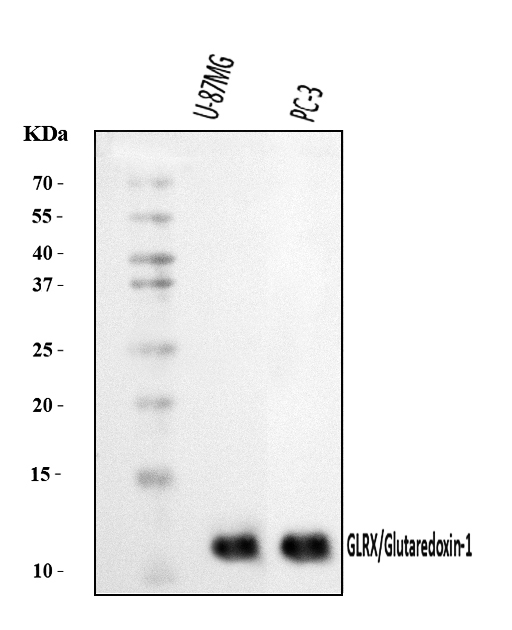
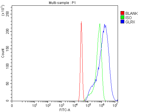 FCM/FACS (Flow Cytometry)
(Figure 3. Flow Cytometry analysis of HeLa cells using anti-Glutaredoxin 1/GLRX antibody (AAA126303).Overlay histogram showing HeLa cells stained with AAA126303 (Blue line). The cells were blocked with 10% normal goat serum. And then incubated with rabbit anti-Glutaredoxin 1/GLRX Antibody (AAA126303, 1 ug/1x10^6 cells) for 30 min at 20 degree C. DyLight488 conjugated goat anti-rabbit IgG was used as secondary antibody for 30 minutes at 20 degree C. Isotype control antibody (Green line) was rabbit IgG (1 ug/1x10^6) used under the same conditions. Unlabelled sample (Red line) was also used as a control.)
FCM/FACS (Flow Cytometry)
(Figure 3. Flow Cytometry analysis of HeLa cells using anti-Glutaredoxin 1/GLRX antibody (AAA126303).Overlay histogram showing HeLa cells stained with AAA126303 (Blue line). The cells were blocked with 10% normal goat serum. And then incubated with rabbit anti-Glutaredoxin 1/GLRX Antibody (AAA126303, 1 ug/1x10^6 cells) for 30 min at 20 degree C. DyLight488 conjugated goat anti-rabbit IgG was used as secondary antibody for 30 minutes at 20 degree C. Isotype control antibody (Green line) was rabbit IgG (1 ug/1x10^6) used under the same conditions. Unlabelled sample (Red line) was also used as a control.)
Glutaredoxin 1/GLRX, Polyclonal Antibody (Cat# AAA126303)
Full Name
Anti-Glutaredoxin 1/GLRX Antibody Picoband
Gene Names
GLRX; GRX; GRX1
Reactivity
Human
Applications
Western Blot, Immunocytochemistry, Immunofluorescence, Flow Cytometry
Purity
Immunogen affinity purified.
Pricing