
Filters
Clonality
Type
Reactivity
Gene Name
Isotype
Host
Application
Clone
10000 results for "Recombinant Proteins" - showing 9350-9400
FGF basic/FGF-2, Active Protein (Cat# AAA48985)
Full Name
Recombinant human FGF basic/FGF-2 protein
Gene Names
FGF2; BFGF; FGFB; FGF-2; HBGF-2
Applications
Bioactivity, SDS-PAGE
Purity
> 95% by SDS-PAGE
Pricing
 WB (Western Blot)
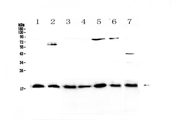
(Figure 1. Western blot analysis of IL23 using anti-IL23 antibody (AAA124620).Electrophoresis was performed on a 5-20% SDS-PAGE gel at 70V (Stacking gel) / 90V (Resolving gel) for 2-3 hours. The sample well of each lane was loaded with 50ug of sample under reducing conditions.Lane 1: human placenta tissue lysates,Lane 2: human PANC-1 cell lysates,Lane 3: rat lymph tissue lysates,Lane 4: rat small intestine tissue lysates,Lane 5: rat testis tissue lysates,Lane 6: rat ovary tissue lysates,Lane 7: mouse testis tissue lysates.After Electrophoresis, proteins were transferred to a Nitrocellulose membrane at 150mA for 50-90 minutes. Blocked the membrane with 5% Non-fat Milk/ TBS for 1.5 hour at RT. The membrane was incubated with rabbit anti-IL23 antigen affinity purified polyclonal antibody at 0.5ug/mL overnight at 4 degree C, then washed with TBS-0.1%Tween 3 times with 5 minutes each and probed with a goat anti-rabbit IgG-HRP secondary antibody at a dilution of 1:10000 for 1.5 hour at RT. The signal is developed using an Enhanced Chemiluminescent detection (ECL) kit with Tanon 5200 system. A specific band was detected for IL23 at approximately 19KD. The expected band size for IL23 is at 21KD.)
WB (Western Blot)
(Figure 1. Western blot analysis of IL23 using anti-IL23 antibody (AAA124620).Electrophoresis was performed on a 5-20% SDS-PAGE gel at 70V (Stacking gel) / 90V (Resolving gel) for 2-3 hours. The sample well of each lane was loaded with 50ug of sample under reducing conditions.Lane 1: human placenta tissue lysates,Lane 2: human PANC-1 cell lysates,Lane 3: rat lymph tissue lysates,Lane 4: rat small intestine tissue lysates,Lane 5: rat testis tissue lysates,Lane 6: rat ovary tissue lysates,Lane 7: mouse testis tissue lysates.After Electrophoresis, proteins were transferred to a Nitrocellulose membrane at 150mA for 50-90 minutes. Blocked the membrane with 5% Non-fat Milk/ TBS for 1.5 hour at RT. The membrane was incubated with rabbit anti-IL23 antigen affinity purified polyclonal antibody at 0.5ug/mL overnight at 4 degree C, then washed with TBS-0.1%Tween 3 times with 5 minutes each and probed with a goat anti-rabbit IgG-HRP secondary antibody at a dilution of 1:10000 for 1.5 hour at RT. The signal is developed using an Enhanced Chemiluminescent detection (ECL) kit with Tanon 5200 system. A specific band was detected for IL23 at approximately 19KD. The expected band size for IL23 is at 21KD.)
IL23, Polyclonal Antibody (Cat# AAA124620)
Full Name
Anti-IL23 Picoband antibody
Gene Names
IL23A; P19; SGRF; IL-23; IL-23A; IL23P19
Reactivity
Human, Mouse, Rat
No cross reactivity with other proteins.
No cross reactivity with other proteins.
Applications
Western Blot
Pricing

 WB (Western Blot)
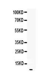
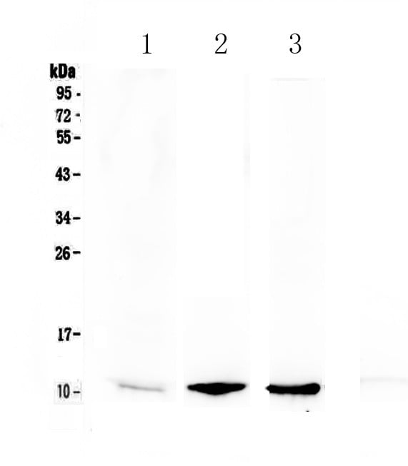
(Figure 1. Western blot analysis of EGF using anti-EGF antibody (AAA124567).Electrophoresis was performed on a 5-20% SDS-PAGE gel at 70V (Stacking gel) / 90V (Resolving gel) for 2-3 hours.Lane 1: recombinant rat EGF protein 1ng.After Electrophoresis, proteins were transferred to a Nitrocellulose membrane at 150mA for 50-90 minutes. Blocked the membrane with 5% Non-fat Milk/ TBS for 1.5 hour at RT. The membrane was incubated with rabbit anti-EGF antigen affinity purified polyclonal antibody at 0.5ug/mL overnight at 4 degree C, then washed with TBS-0.1%Tween 3 times with 5 minutes each and probed with a goat anti-rabbit IgG-HRP secondary antibody at a dilution of 1:10000 for 1.5 hour at RT. The signal is developed using an Enhanced Chemiluminescent detection (ECL) kit with Tanon 5200 system. A specific band was detected for EGF at approximately 6KD. The expected band size for EGF is at 6KD.)
WB (Western Blot)
(Figure 1. Western blot analysis of EGF using anti-EGF antibody (AAA124567).Electrophoresis was performed on a 5-20% SDS-PAGE gel at 70V (Stacking gel) / 90V (Resolving gel) for 2-3 hours.Lane 1: recombinant rat EGF protein 1ng.After Electrophoresis, proteins were transferred to a Nitrocellulose membrane at 150mA for 50-90 minutes. Blocked the membrane with 5% Non-fat Milk/ TBS for 1.5 hour at RT. The membrane was incubated with rabbit anti-EGF antigen affinity purified polyclonal antibody at 0.5ug/mL overnight at 4 degree C, then washed with TBS-0.1%Tween 3 times with 5 minutes each and probed with a goat anti-rabbit IgG-HRP secondary antibody at a dilution of 1:10000 for 1.5 hour at RT. The signal is developed using an Enhanced Chemiluminescent detection (ECL) kit with Tanon 5200 system. A specific band was detected for EGF at approximately 6KD. The expected band size for EGF is at 6KD.)
EGF, Polyclonal Antibody (Cat# AAA124567)
Full Name
Anti-EGF Picoband Antibody
Reactivity
Rat
No cross reactivity with other proteins.
No cross reactivity with other proteins.
Applications
Western Blot, Immunohistochemistry
Purity
Immunogen affinity purified
Pricing

 IHC (Immunohiostchemistry)
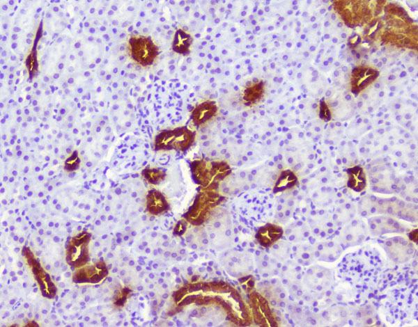
(Figure 3. IHC analysis of Calbindin using anti-Calbindin antibody.Calbindin was detected in paraffin-embedded section of mouse brain tissue. Heat mediated antigen retrieval was performed in citrate buffer (pH6, epitope retrieval solution) for 20 mins. The tissue section was blocked with 10% goat serum. The tissue section was then incubated with 1ug/ml rabbit anti-Calbindin antibody.Calbindin was detected in paraffin-embedded section of mouse brain tissue. Heat mediated antigen retrieval was performed in citrate buffer (pH6, epitope retrieval solution) for 20 mins. The tissue section was blocked with 10% goat serum. The tissue section was then incubated with 1ug/ml rabbit anti-Calbindin antibody.Calbindin was detected in paraffin-embedded section of mouse kidney tissue. Heat mediated antigen retrieval was performed in citrate buffer (pH6, epitope retrieval solution) for 20 mins. The tissue section was blocked with 10% goat serum. The tissue section was then incubated with 1ug/ml rabbit anti-Calbindin Antibody overnight at 4 degree C. Biotinylated goat anti-rabbit IgG was used as secondary antibody and incubated for 30 minutes at 37 degree C. The tissue section was developed using Strepavidin-Biotin-Complex (SABC) with DAB as the chromogen.)
IHC (Immunohiostchemistry)
(Figure 3. IHC analysis of Calbindin using anti-Calbindin antibody.Calbindin was detected in paraffin-embedded section of mouse brain tissue. Heat mediated antigen retrieval was performed in citrate buffer (pH6, epitope retrieval solution) for 20 mins. The tissue section was blocked with 10% goat serum. The tissue section was then incubated with 1ug/ml rabbit anti-Calbindin antibody.Calbindin was detected in paraffin-embedded section of mouse brain tissue. Heat mediated antigen retrieval was performed in citrate buffer (pH6, epitope retrieval solution) for 20 mins. The tissue section was blocked with 10% goat serum. The tissue section was then incubated with 1ug/ml rabbit anti-Calbindin antibody.Calbindin was detected in paraffin-embedded section of mouse kidney tissue. Heat mediated antigen retrieval was performed in citrate buffer (pH6, epitope retrieval solution) for 20 mins. The tissue section was blocked with 10% goat serum. The tissue section was then incubated with 1ug/ml rabbit anti-Calbindin Antibody overnight at 4 degree C. Biotinylated goat anti-rabbit IgG was used as secondary antibody and incubated for 30 minutes at 37 degree C. The tissue section was developed using Strepavidin-Biotin-Complex (SABC) with DAB as the chromogen.)
Calbindin/CALB1, Polyclonal Antibody (Cat# AAA124947)
Full Name
Anti-Calbindin/CALB1 Antibody
Gene Names
CALB1; CALB; D-28K
Reactivity
Human, Mouse, Rat
Applications
Direct ELISA, Immunohistochemistry, Western Blot
Purity
Immunogen Affinity Purified
Pricing
 IHC (Immunohiostchemistry)
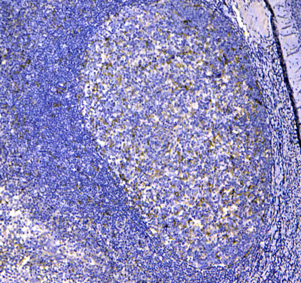
(Figure 2. IHC analysis of CD11c using anti-CD11c antibody (AAA124565).CD11c was detected in paraffin-embedded section of human tonsil tissue. Heat mediated antigen retrieval was performed in citrate buffer (pH6, epitope retrieval solution) for 20 mins. The tissue section was blocked with 10% goat serum. The tissue section was then incubated with 1ug/ml rabbit anti-CD11c Antibody (AAA124565) overnight at 4 degree C. Biotinylated goat anti-rabbit IgG was used as secondary antibody and incubated for 30 minutes at 37 degree C. The tissue section was developed using Strepavidin-Biotin-Complex (SABC) with DAB as the chromogen.)
IHC (Immunohiostchemistry)
(Figure 2. IHC analysis of CD11c using anti-CD11c antibody (AAA124565).CD11c was detected in paraffin-embedded section of human tonsil tissue. Heat mediated antigen retrieval was performed in citrate buffer (pH6, epitope retrieval solution) for 20 mins. The tissue section was blocked with 10% goat serum. The tissue section was then incubated with 1ug/ml rabbit anti-CD11c Antibody (AAA124565) overnight at 4 degree C. Biotinylated goat anti-rabbit IgG was used as secondary antibody and incubated for 30 minutes at 37 degree C. The tissue section was developed using Strepavidin-Biotin-Complex (SABC) with DAB as the chromogen.)
CD11c, Polyclonal Antibody (Cat# AAA124565)
Full Name
Anti-CD11c Picoband antibody
Gene Names
ITGAX; CD11C; SLEB6
Reactivity
Human, Mouse
No cross reactivity with other proteins.
No cross reactivity with other proteins.
Applications
Western Blot, Immunohistochemistry
Pricing
 IHC (Immunohiostchemistry)
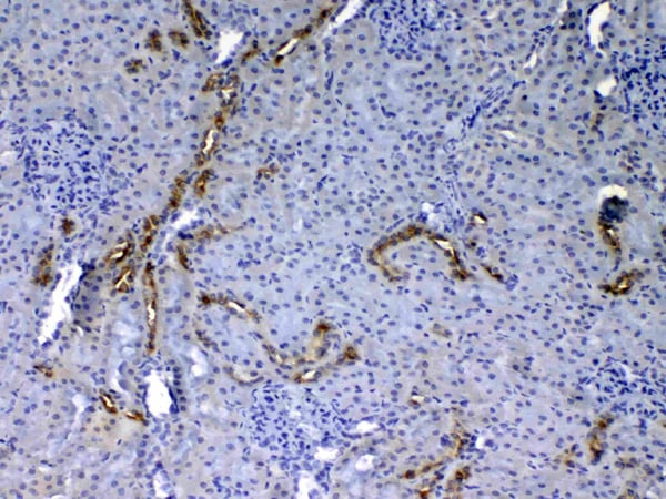
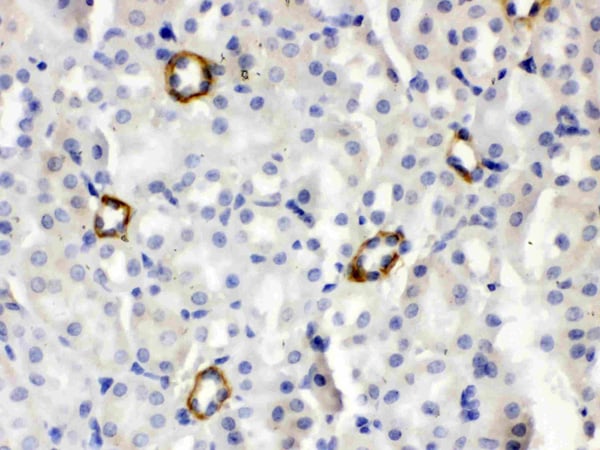
(Figure 2. IHC analysis of ISG15/Ucrp using anti- ISG15/Ucrp antibody (AAA46620).ISG15/Ucrp was detected in paraffin-embedded section of mouse kidney tissues. Heat mediated antigen retrieval was performed in citrate buffer (pH6, epitope retrieval solution) for 20 mins. The tissue section was blocked with 10% goat serum. The tissue section was then incubated with 1ug/ml rabbit anti- ISG15/Ucrp Antibody (AAA46620) overnight at 4 degree C. Biotinylated goat anti-rabbit IgG was used as secondary antibody and incubated for 30 minutes at 37 degree C. The tissue section was developed using Strepavidin-Biotin-Complex (SABC) with DAB as the chromogen.)
IHC (Immunohiostchemistry)
(Figure 2. IHC analysis of ISG15/Ucrp using anti- ISG15/Ucrp antibody (AAA46620).ISG15/Ucrp was detected in paraffin-embedded section of mouse kidney tissues. Heat mediated antigen retrieval was performed in citrate buffer (pH6, epitope retrieval solution) for 20 mins. The tissue section was blocked with 10% goat serum. The tissue section was then incubated with 1ug/ml rabbit anti- ISG15/Ucrp Antibody (AAA46620) overnight at 4 degree C. Biotinylated goat anti-rabbit IgG was used as secondary antibody and incubated for 30 minutes at 37 degree C. The tissue section was developed using Strepavidin-Biotin-Complex (SABC) with DAB as the chromogen.)
ISG15, Polyclonal Antibody (Cat# AAA46620)
Full Name
Anti-ISG15 Antibody
Gene Names
Isg15; G1p2; IP17; Irfp; UCRP; IGI15
Reactivity
Mouse
No cross reactivity with other proteins.
No cross reactivity with other proteins.
Applications
Immunohistochemistry, Western Blot
Purity
Immunogen affinity purified.
Pricing
 IHC (Immunohiostchemistry)
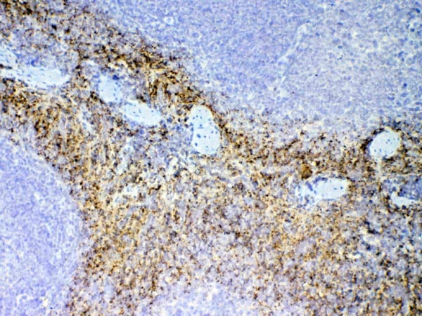
(Figure 2. IHC analysis of NAP2 using anti-NAP2 antibody (AAA124694).NAP2 was detected in paraffin-embedded section of mouse spleen tissue. Heat mediated antigen retrieval was performed in citrate buffer (pH6, epitope retrieval solution) for 20 mins. The tissue section was blocked with 10% goat serum. The tissue section was then incubated with 1ug/ml rabbit anti-NAP2 Antibody (AAA124694) overnight at 4 degree C. Biotinylated goat anti-rabbit IgG was used as secondary antibody and incubated for 30 minutes at 37 degree C. The tissue section was developed using Strepavidin-Biotin-Complex (SABC) with DAB as the chromogen.)
IHC (Immunohiostchemistry)
(Figure 2. IHC analysis of NAP2 using anti-NAP2 antibody (AAA124694).NAP2 was detected in paraffin-embedded section of mouse spleen tissue. Heat mediated antigen retrieval was performed in citrate buffer (pH6, epitope retrieval solution) for 20 mins. The tissue section was blocked with 10% goat serum. The tissue section was then incubated with 1ug/ml rabbit anti-NAP2 Antibody (AAA124694) overnight at 4 degree C. Biotinylated goat anti-rabbit IgG was used as secondary antibody and incubated for 30 minutes at 37 degree C. The tissue section was developed using Strepavidin-Biotin-Complex (SABC) with DAB as the chromogen.)
NAP2, Polyclonal Antibody (Cat# AAA124694)
Full Name
Anti-NAP2 Picoband antibody
Gene Names
Ppbp; TGB; LDGF; MDGF; TGB1; CTAP3; Cxcl7; NAP-2; Scyb7; b-TG1; LA-PF4; THBGB1; CTAPIII; beta-TG; AI854500; NAP-2-L1; 2400003M24Rik
Reactivity
Mouse
No cross reactivity with other proteins.
No cross reactivity with other proteins.
Applications
Western Blot, Immunohistochemistry
Pricing
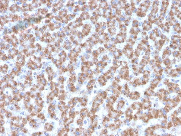
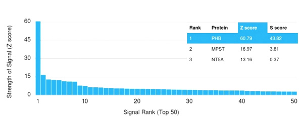 Application Data
(Analysis of Protein Array containing more than 19,000 full-length human proteins using Monospecific Mouse Monoclonal Antibody to Prohibitin (PHB/3193). Z- and S- Score: The Z-score represents the strength of a signal that a monoclonal antibody (MAb) (in combination with a fluorescently-tagged anti-IgG secondary antibody) produces when binding to a particular protein on the HuProtTM array. Z-scores are described in units of standard deviations (SD’s) above the mean value of all signals generated on that array. If targets on HuProtTM are arranged in descending order of the Z-score, the S-score is the difference (also in units of SD’s) between the Z-score. S-score therefore represents the relative target specificity of a MAb to its intended target. A MAb is considered to specific to its intended target, if the MAb has an S-score of at least 2.5. For example, if a MAb binds to protein X with a Z-score of 43 and to protein Y with a Z-score of 14, then the S-score for the binding of that MAb to protein X is equal to 29.)
Application Data
(Analysis of Protein Array containing more than 19,000 full-length human proteins using Monospecific Mouse Monoclonal Antibody to Prohibitin (PHB/3193). Z- and S- Score: The Z-score represents the strength of a signal that a monoclonal antibody (MAb) (in combination with a fluorescently-tagged anti-IgG secondary antibody) produces when binding to a particular protein on the HuProtTM array. Z-scores are described in units of standard deviations (SD’s) above the mean value of all signals generated on that array. If targets on HuProtTM are arranged in descending order of the Z-score, the S-score is the difference (also in units of SD’s) between the Z-score. S-score therefore represents the relative target specificity of a MAb to its intended target. A MAb is considered to specific to its intended target, if the MAb has an S-score of at least 2.5. For example, if a MAb binds to protein X with a Z-score of 43 and to protein Y with a Z-score of 14, then the S-score for the binding of that MAb to protein X is equal to 29.)
Prohibitin, Monoclonal Antibody (Cat# AAA215142)
Full Name
Prohibitin (Mitochondrial Marker)
Gene Names
PHB; PHB1; HEL-215; HEL-S-54e
Reactivity
Human
Applications
Immunohistochemistry, Western Blot
Purity
Purified Ab with BSA and Azide at 200ug/ml OR Purified Ab WITHOUT BSA and Azide at 1.0mg/ml
Pricing
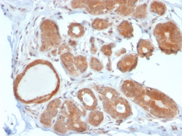
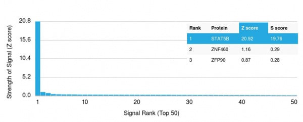 Application Data
(Analysis of Protein Array containing >19,000 full-length human proteins using STAT5B Mouse Monoclonal Antibody (STAT5B/2611) Z- and S- Score: The Z-score represents the strength of a signal that a monoclonal antibody (Monoclonal Antibody) (in combination with a fluorescently-tagged anti-IgG secondary antibody) produces when binding to a particular protein on the HuProtTM array. Z-scores are described in units of standard deviations (SD's) above the mean value of all signals generated on that array. If targets on HuProtTM are arranged in descending order of the Z-score, the S-score is the difference (also in units of SD's) between the Z-score. S-score therefore represents the relative target specificity of a Monoclonal Antibody to its intended target. A Monoclonal Antibody is considered to specific to its intended target, if the Monoclonal Antibody has an S-score of at least 2.5. For example, if a Monoclonal Antibody binds to protein X with a Z-score of 43 and to protein Y with a Z-score of 14, then the S-score for the binding of that Monoclonal Antibody to protein X is equal to 29.)
Application Data
(Analysis of Protein Array containing >19,000 full-length human proteins using STAT5B Mouse Monoclonal Antibody (STAT5B/2611) Z- and S- Score: The Z-score represents the strength of a signal that a monoclonal antibody (Monoclonal Antibody) (in combination with a fluorescently-tagged anti-IgG secondary antibody) produces when binding to a particular protein on the HuProtTM array. Z-scores are described in units of standard deviations (SD's) above the mean value of all signals generated on that array. If targets on HuProtTM are arranged in descending order of the Z-score, the S-score is the difference (also in units of SD's) between the Z-score. S-score therefore represents the relative target specificity of a Monoclonal Antibody to its intended target. A Monoclonal Antibody is considered to specific to its intended target, if the Monoclonal Antibody has an S-score of at least 2.5. For example, if a Monoclonal Antibody binds to protein X with a Z-score of 43 and to protein Y with a Z-score of 14, then the S-score for the binding of that Monoclonal Antibody to protein X is equal to 29.)
STAT5B, Monoclonal Antibody (Cat# AAA215210)
Full Name
STAT5B
Gene Names
STAT5B; STAT5
Reactivity
Human
Applications
Immunohistochemistry
Purity
Purified Ab with BSA and Azide at 200ug/ml OR Purified Ab WITHOUT BSA and Azide at 1.0mg/ml
Pricing
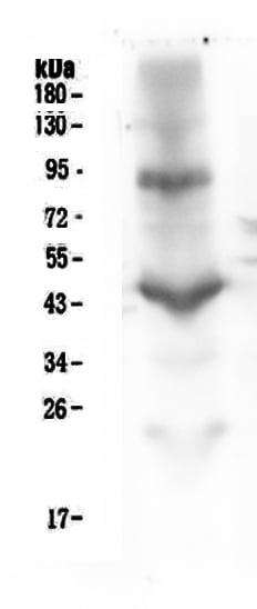
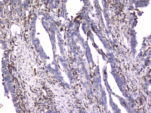
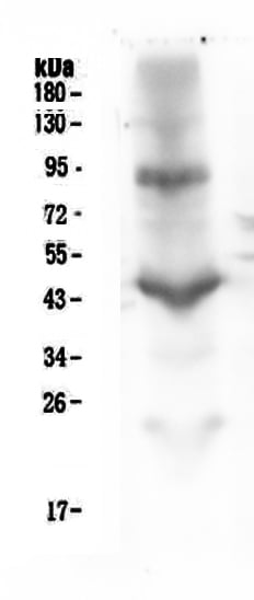 WB (Western Blot)
(Figure 3. Western blot analysis of CD18 using anti-CD18 antibody (M00458-1).Electrophoresis was performed on a 5-20% SDS-PAGE gel at 70V (Stacking gel)/90V (Resolving gel) for 2-3 hours. The sample well of each lane was loaded with 50ug of sample under reducing conditions.Lane 1: human placenta tissue lysate.After Electrophoresis, proteins were transferred to a Nitrocellulose membrane at 150mA for 50-90 minutes. Blocked the membrane with 5% Non-fat Milk/TBS for 1.5 hour at RT. The membrane was incubated with mouse anti-CD18 antigen affinity purified monoclonal antibody at 0.5 ug/ml overnight at 4 degree C, then washed with TBS-0.1%Tween 3 times with 5 minutes each and probed with a goat anti-mouse IgG-HRP secondary antibody at a dilution of 1:10000 for 1.5 hour at RT. The signal is developed using an Enhanced Chemiluminescent detection (ECL) kit with Tanon 5200 system.)
WB (Western Blot)
(Figure 3. Western blot analysis of CD18 using anti-CD18 antibody (M00458-1).Electrophoresis was performed on a 5-20% SDS-PAGE gel at 70V (Stacking gel)/90V (Resolving gel) for 2-3 hours. The sample well of each lane was loaded with 50ug of sample under reducing conditions.Lane 1: human placenta tissue lysate.After Electrophoresis, proteins were transferred to a Nitrocellulose membrane at 150mA for 50-90 minutes. Blocked the membrane with 5% Non-fat Milk/TBS for 1.5 hour at RT. The membrane was incubated with mouse anti-CD18 antigen affinity purified monoclonal antibody at 0.5 ug/ml overnight at 4 degree C, then washed with TBS-0.1%Tween 3 times with 5 minutes each and probed with a goat anti-mouse IgG-HRP secondary antibody at a dilution of 1:10000 for 1.5 hour at RT. The signal is developed using an Enhanced Chemiluminescent detection (ECL) kit with Tanon 5200 system.)
CD18, Monoclonal Antibody (Cat# AAA125132)
Full Name
Anti-CD18 Antibody (monoclonal, 1A3/2A10)
Gene Names
ITGB2; LAD; CD18; MF17; MFI7; LCAMB; LFA-1; MAC-1
Reactivity
Human
Applications
Flow Cytometry, Immunocytochemistry, Immunohistochemistry, Western Blot
Purity
Immunogen Affinity Purified
Pricing
RANK/TNFRSF11A, Active Protein (Cat# AAA49004)
Full Name
Recombinant mouse RANK/TNFRSF11A protein
Gene Names
Tnfrsf11a; OFE; ODFR; Rank; Ly109; mRANK; TRANCE-R
Applications
Bioactivity, SDS-PAGE
Purity
> 90% by SDS-PAGE
Pricing
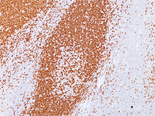
 Application Data
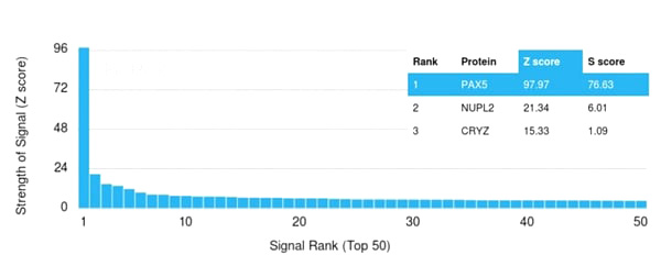
(Analysis of Protein Array containing more than 21,000 full-length human proteins using PAX5 Mouse Monoclonal Antibody (PAX5/3735) Z- and S- Score: The Z-score represents the strength of a signal that a monoclonal antibody (Monoclonal Antibody) (in combination with a fluorescently-tagged anti-IgG secondary antibody) produces when binding to a particular protein on the HuProtTM array. Z-scores are described in units of standard deviations (SD’s) above the mean value of all signals generated on that array. If targets on HuProtTM are arranged in descending order of the Z-score, the S-score is the difference (also in units of SD’s) between the Z-score. S-score therefore represents the relative target specificity of a Monoclonal Antibody to its intended target. A Monoclonal Antibody is considered to specific to its intended target, if the Monoclonal Antibody has an S-score of at least 2.5. For example, if a Monoclonal Antibody binds to protein X with a Z-score of 43 and to protein Y with a Z-score of 14, then the S-score for the binding of that Monoclonal Antibody to protein X is equal to 29.)
Application Data
(Analysis of Protein Array containing more than 21,000 full-length human proteins using PAX5 Mouse Monoclonal Antibody (PAX5/3735) Z- and S- Score: The Z-score represents the strength of a signal that a monoclonal antibody (Monoclonal Antibody) (in combination with a fluorescently-tagged anti-IgG secondary antibody) produces when binding to a particular protein on the HuProtTM array. Z-scores are described in units of standard deviations (SD’s) above the mean value of all signals generated on that array. If targets on HuProtTM are arranged in descending order of the Z-score, the S-score is the difference (also in units of SD’s) between the Z-score. S-score therefore represents the relative target specificity of a Monoclonal Antibody to its intended target. A Monoclonal Antibody is considered to specific to its intended target, if the Monoclonal Antibody has an S-score of at least 2.5. For example, if a Monoclonal Antibody binds to protein X with a Z-score of 43 and to protein Y with a Z-score of 14, then the S-score for the binding of that Monoclonal Antibody to protein X is equal to 29.)
PAX5/BSAP, Monoclonal Antibody (Cat# AAA215125)
Full Name
PAX5/BSAP (Early B-Cell Marker)
Gene Names
PAX5; ALL3; BSAP
Reactivity
Human
Applications
Immunohistochemistry
Purity
Purified Ab with BSA and Azide at 200ug/ml OR Purified Ab WITHOUT BSA and Azide at 1.0mg/ml
Pricing
 Application Data
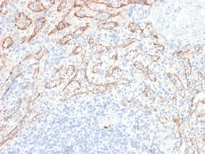
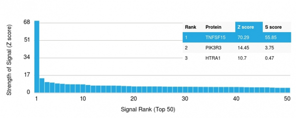
(Analysis of Protein Array containing more than 19, 000 full-length human proteins using VEGI Rabbit Recombinant Monoclonal Antibody (VEGI/2052R). Z- and S- Score: The Z-score represents the strength of a signal that a monoclonal antibody (MAb) (in combination with a fluorescently-tagged anti-IgG secondary antibody) produces when binding to a particular protein on the HuProtTM array. Z-scores are described in units of standard deviations (SD's) above the mean value of all signals generated on that array. If targets on HuProtTM are arranged in descending order of the Z-score, the S-score is the difference (also in units of SD's) between the Z-score. S-score therefore represents the relative target specificity of a MAb to its intended target. A MAb is considered to specific to its intended target, if the MAb has an S-score of at least 2.5. For example, if a MAb binds to protein X with a Z-score of 43 and to protein Y with a Z-score of 14, then the S-score for the binding of that MAb to protein X is equal to 29.)
Application Data
(Analysis of Protein Array containing more than 19, 000 full-length human proteins using VEGI Rabbit Recombinant Monoclonal Antibody (VEGI/2052R). Z- and S- Score: The Z-score represents the strength of a signal that a monoclonal antibody (MAb) (in combination with a fluorescently-tagged anti-IgG secondary antibody) produces when binding to a particular protein on the HuProtTM array. Z-scores are described in units of standard deviations (SD's) above the mean value of all signals generated on that array. If targets on HuProtTM are arranged in descending order of the Z-score, the S-score is the difference (also in units of SD's) between the Z-score. S-score therefore represents the relative target specificity of a MAb to its intended target. A MAb is considered to specific to its intended target, if the MAb has an S-score of at least 2.5. For example, if a MAb binds to protein X with a Z-score of 43 and to protein Y with a Z-score of 14, then the S-score for the binding of that MAb to protein X is equal to 29.)
TNFS15/VEGI (Vascular Endothelial Growth Inhibitor), Monoclonal Antibody (Cat# AAA214623)
Full Name
TNFS15/VEGI (Vascular Endothelial Growth Inhibitor)
Gene Names
TNFSF15; TL1; TL1A; VEGI; TNLG1B; VEGI192A
Reactivity
Human. Others not known.
Applications
Immunohistochemistry
Pricing
 FCM/FACS (Flow Cytometry)
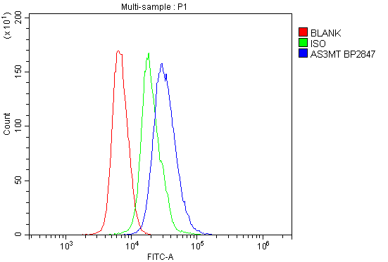
(Figure 2. Flow Cytometry analysis of mouse spleen tissues using anti-Cyt 19/As3mt antibody (AAA125740).Overlay histogram showing A431 cells stained with AAA125740 (Blue line). The tissues were blocked with 10% normal goat serum. And then incubated with rabbit anti-Cyt 19/As3mt Antibody (AAA125740,1μg/1x106 cells) for 30 min at 20 degree C. DyLight®488 conjugated goat anti-rabbit IgG (5-10μg/1x106 cells) was used as secondary antibody for 30 minutes at 20 degree C. Isotype control antibody (Green line) was rabbit IgG (1μg/1x106) used under the same conditions. Unlabelled sample (Red line) was also used as a control.)
FCM/FACS (Flow Cytometry)
(Figure 2. Flow Cytometry analysis of mouse spleen tissues using anti-Cyt 19/As3mt antibody (AAA125740).Overlay histogram showing A431 cells stained with AAA125740 (Blue line). The tissues were blocked with 10% normal goat serum. And then incubated with rabbit anti-Cyt 19/As3mt Antibody (AAA125740,1μg/1x106 cells) for 30 min at 20 degree C. DyLight®488 conjugated goat anti-rabbit IgG (5-10μg/1x106 cells) was used as secondary antibody for 30 minutes at 20 degree C. Isotype control antibody (Green line) was rabbit IgG (1μg/1x106) used under the same conditions. Unlabelled sample (Red line) was also used as a control.)
Cyt 19/As3mt, Polyclonal Antibody (Cat# AAA125740)
Full Name
Anti-Cyt 19/As3mt Antibody
Gene Names
As3mt; Cyt19; 2310045H08Rik
Reactivity
Mouse
Applications
Direct ELISA, Flow Cytometry, Western Blot
Purity
Immunogen affinity purified.
Pricing
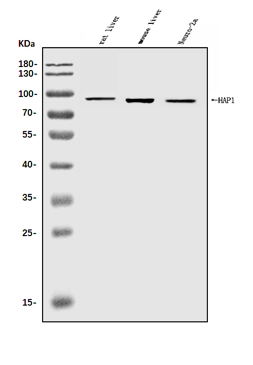
 FCM/FACS (Flow Cytometry)
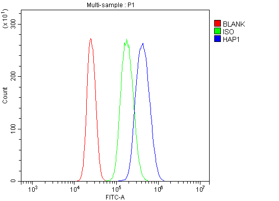
(Figure 3. Flow Cytometry analysis of U937 cells using anti-HAP1 antibody (AAA125615).Overlay histogram showing U937 cells stained with AAA125615 (Blue line). The cells were blocked with 10% normal goat serum. And then incubated with rabbit anti-HAP1 Antibody (AAA125615, 1μg/1x106 cells) for 30 min at 20 degree C. DyLight®488 conjugated goat anti-rabbit IgG (5-10μg/1x106 cells) was used as secondary antibody for 30 minutes at 20 degree C. Isotype control antibody (Green line) was rabbit IgG (1μg/1x106) used under the same conditions. Unlabelled sample (Red line) was also used as a control.)
FCM/FACS (Flow Cytometry)
(Figure 3. Flow Cytometry analysis of U937 cells using anti-HAP1 antibody (AAA125615).Overlay histogram showing U937 cells stained with AAA125615 (Blue line). The cells were blocked with 10% normal goat serum. And then incubated with rabbit anti-HAP1 Antibody (AAA125615, 1μg/1x106 cells) for 30 min at 20 degree C. DyLight®488 conjugated goat anti-rabbit IgG (5-10μg/1x106 cells) was used as secondary antibody for 30 minutes at 20 degree C. Isotype control antibody (Green line) was rabbit IgG (1μg/1x106) used under the same conditions. Unlabelled sample (Red line) was also used as a control.)
HAP1, Polyclonal Antibody (Cat# AAA125615)
Full Name
Anti-HAP1 Antibody
Gene Names
HAP1; HLP; HAP2; HIP5; hHLP1
Reactivity
Human, Mouse, Rat
Applications
Direct ELISA, Flow Cytometry, Immunofluorescence, Immunocytochemistry, Western Blot
Purity
Immunogen affinity purified.
Pricing
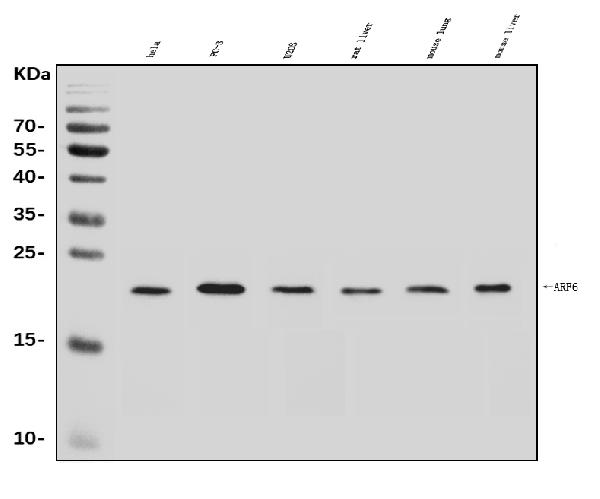
 IF (Immunofluorescence)
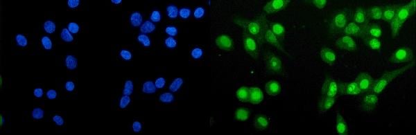
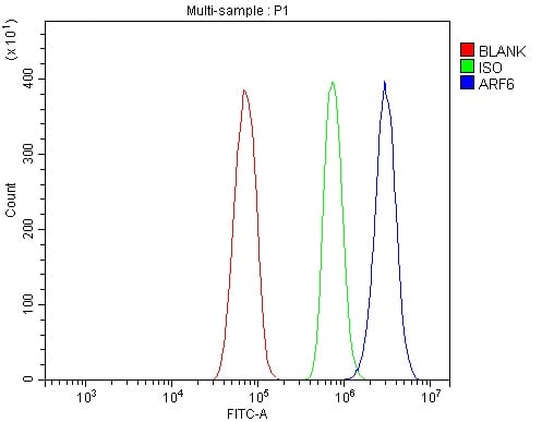
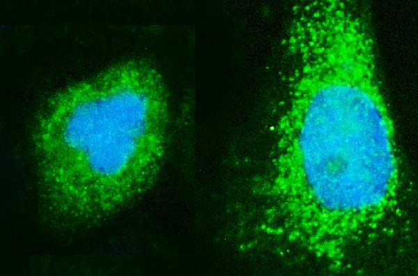
(Figure 4. IF analysis of ARF6 using anti- ARF6 antibody (AAA125580).ARF6 was detected in immunocytochemical section of A431 cells. Enzyme antigen retrieval was performed using IHC enzyme antigen retrieval reagent for 15 mins. The cells were blocked with 10% goat serum. And then incubated with 5μg/mL rabbit anti- ARF6 Antibody (AAA125580) overnight at 4 degree C. DyLight®488 Conjugated Goat Anti-Rabbit IgG was used as secondary antibody at 1:100 dilution and incubated for 30 minutes at 37 degree C. The section was counterstained with DAPI. Visualize using a fluorescence microscope and filter sets appropriate for the label used.)
IF (Immunofluorescence)
(Figure 4. IF analysis of ARF6 using anti- ARF6 antibody (AAA125580).ARF6 was detected in immunocytochemical section of A431 cells. Enzyme antigen retrieval was performed using IHC enzyme antigen retrieval reagent for 15 mins. The cells were blocked with 10% goat serum. And then incubated with 5μg/mL rabbit anti- ARF6 Antibody (AAA125580) overnight at 4 degree C. DyLight®488 Conjugated Goat Anti-Rabbit IgG was used as secondary antibody at 1:100 dilution and incubated for 30 minutes at 37 degree C. The section was counterstained with DAPI. Visualize using a fluorescence microscope and filter sets appropriate for the label used.)
ARF6, Polyclonal Antibody (Cat# AAA125580)
Full Name
Anti-ARF6 Antibody
Reactivity
Human, Mouse, Rat
Applications
Direct ELISA, Flow Cytometry, Immunofluorescence, Immunocytochemistry, Western Blot
Purity
Immunogen affinity purified.
Pricing
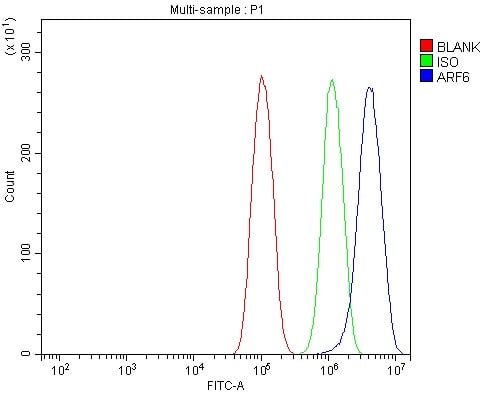
 FCM/FACS (Flow Cytometry)
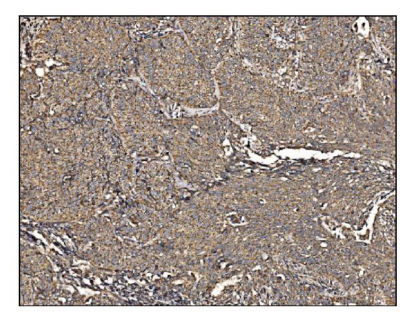
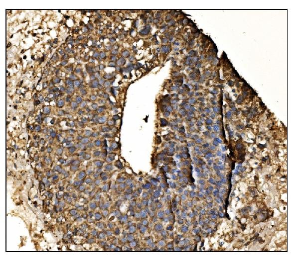
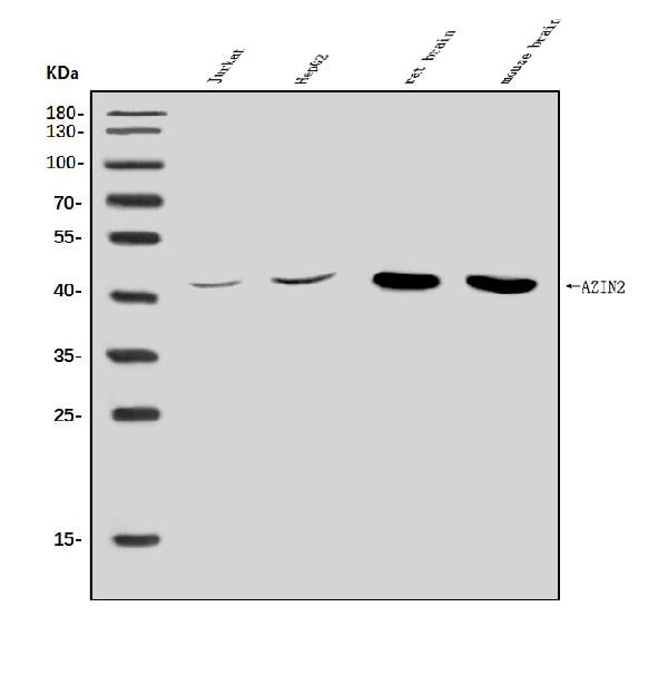
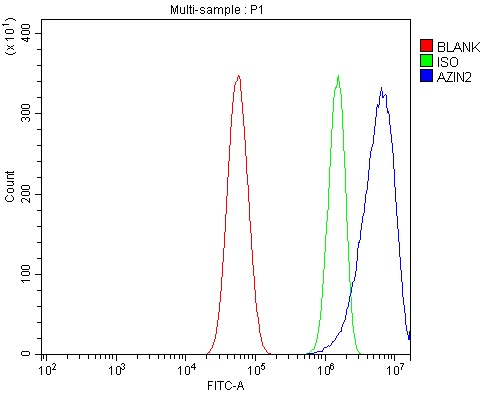
(Figure 5. Flow Cytometry analysis of U87 cells using anti-Arginine Decarboxylase/AZIN2 antibody (AAA125794).Overlay histogram showing U87 cells stained with AAA125794 (Blue line). The cells were blocked with 10% normal goat serum. And then incubated with rabbit anti-Arginine Decarboxylase/AZIN2 Antibody (AAA125794, 1 μg/1x106 cells) for 30 min at 20 degree C. DyLight®488 conjugated goat anti-rabbit IgG (5-10 μg/1 x106 cells) was used as secondary antibody for 30 minutes at 20 degree C. Isotype control antibody (Green line) was rabbit IgG (1 μg/1x106) used under the same conditions. Unlabelled sample (Red line) was also used as a control.)
FCM/FACS (Flow Cytometry)
(Figure 5. Flow Cytometry analysis of U87 cells using anti-Arginine Decarboxylase/AZIN2 antibody (AAA125794).Overlay histogram showing U87 cells stained with AAA125794 (Blue line). The cells were blocked with 10% normal goat serum. And then incubated with rabbit anti-Arginine Decarboxylase/AZIN2 Antibody (AAA125794, 1 μg/1x106 cells) for 30 min at 20 degree C. DyLight®488 conjugated goat anti-rabbit IgG (5-10 μg/1 x106 cells) was used as secondary antibody for 30 minutes at 20 degree C. Isotype control antibody (Green line) was rabbit IgG (1 μg/1x106) used under the same conditions. Unlabelled sample (Red line) was also used as a control.)
Arginine decarboxylase/AZIN2, Polyclonal Antibody (Cat# AAA125794)
Full Name
Anti-Arginine decarboxylase/AZIN2 Antibody
Gene Names
AZIN2; ADC; AZI2; ODCp; AZIB1; ODC-p; ODC1L
Reactivity
Human, Mouse, Rat
Applications
Direct ELISA, Flow Cytometry, Immunohistochemistry, Western Blot
Purity
Immunogen affinity purified.
Pricing
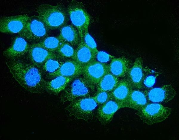
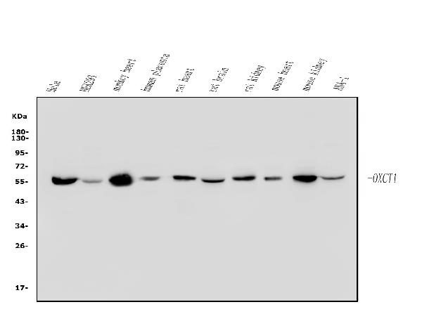
 FCM/FACS (Flow Cytometry)
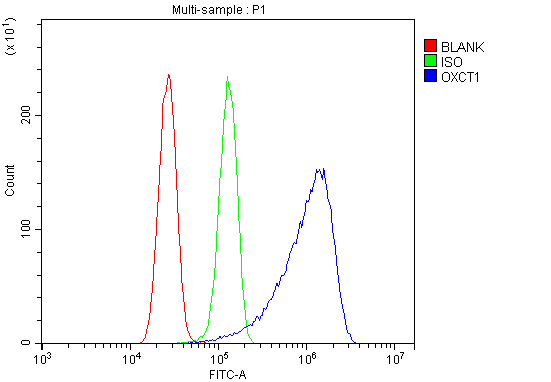
(Figure 3. Flow Cytometry analysis of U87 cells using anti-OXCT1/SCOT antibody (AAA125787).Overlay histogram showing U87 cells stained with AAA125787 (Blue line). The cells were blocked with 10% normal goat serum. And then incubated with rabbit anti-OXCT1/SCOT Antibody (AAA125787, 1μg/1x106 cells) for 30 min at 20 degree C. DyLight®488 conjugated goat anti-rabbit IgG (5-10μg/1x106 cells) was used as secondary antibody for 30 minutes at 20 degree C. Isotype control antibody (Green line) was rabbit IgG (1μg/1x106) used under the same conditions. Unlabelled sample (Red line) was also used as a control.)
FCM/FACS (Flow Cytometry)
(Figure 3. Flow Cytometry analysis of U87 cells using anti-OXCT1/SCOT antibody (AAA125787).Overlay histogram showing U87 cells stained with AAA125787 (Blue line). The cells were blocked with 10% normal goat serum. And then incubated with rabbit anti-OXCT1/SCOT Antibody (AAA125787, 1μg/1x106 cells) for 30 min at 20 degree C. DyLight®488 conjugated goat anti-rabbit IgG (5-10μg/1x106 cells) was used as secondary antibody for 30 minutes at 20 degree C. Isotype control antibody (Green line) was rabbit IgG (1μg/1x106) used under the same conditions. Unlabelled sample (Red line) was also used as a control.)
OXCT1/SCOT, Polyclonal Antibody (Cat# AAA125787)
Full Name
Anti-OXCT1/SCOT Antibody
Gene Names
OXCT1; OXCT; SCOT
Reactivity
Human, Mouse, Rat, Monkey
Applications
Direct ELISA, Flow Cytometry, Immunofluorescence, Immunocytochemistry, Western Blot
Purity
Immunogen affinity purified.
Pricing
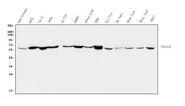 WB (Western Blot)
(Figure 2. Western blot analysis of CYP27A1 using anti-CYP27A1 antibody (AAA125635).Electrophoresis was performed on a 5-20% SDS-PAGE gel at 70V (Stacking gel) / 90V (Resolving gel) for 2-3 hours. The sample well of each lane was loaded with 50ug of sample under reducing conditions.Lane 1: human placenta tissue lysatesLane 2: human HEPG2 whole cell lysatesLane 3: human CACO-2 whole cell lysatesLane 4: human SW620 whole cell lysatesLane 5: human SGC-7901 whole cell lysatesLane 6: human HEK293 whole cell lysatesLane 7: monkey liver tissue lysatesLane 8: human U20S whole cell lysatesLane 9: rat liver tissue lysatesLane 10: rat heart tissue lysatesLane 11: mouse liver tissue lysatesLane 12: mouse heart tissue lysatesLane 13: mouse NIH/3T3 whole cell lysates.After Electrophoresis, proteins were transferred to a Nitrocellulose membrane at 150mA for 50-90 minutes. Blocked the membrane with 5% Non-fat Milk/ TBS for 1. 5 hour at RT. The membrane was incubated with rabbit anti-CYP27A1 antigen affinity purified polyclonal antibody (Catalog # AAA125635) at 0. 25 μg/mL overnight at 4 degree C, then washed with TBS-0. 1%Tween 3 times with 5 minutes each and probed with a goat anti-rabbit IgG-HRP secondary antibody at a dilution of 1:5000 for 1. 5 hour at RT. The signal is developed using an Enhanced Chemiluminescent detection (ECL) kit with Tanon 5200 system. A specific band was detected for CYP27A1 at approximately 60KD. The expected band size for CYP27A1 is at 60KD.)
WB (Western Blot)
(Figure 2. Western blot analysis of CYP27A1 using anti-CYP27A1 antibody (AAA125635).Electrophoresis was performed on a 5-20% SDS-PAGE gel at 70V (Stacking gel) / 90V (Resolving gel) for 2-3 hours. The sample well of each lane was loaded with 50ug of sample under reducing conditions.Lane 1: human placenta tissue lysatesLane 2: human HEPG2 whole cell lysatesLane 3: human CACO-2 whole cell lysatesLane 4: human SW620 whole cell lysatesLane 5: human SGC-7901 whole cell lysatesLane 6: human HEK293 whole cell lysatesLane 7: monkey liver tissue lysatesLane 8: human U20S whole cell lysatesLane 9: rat liver tissue lysatesLane 10: rat heart tissue lysatesLane 11: mouse liver tissue lysatesLane 12: mouse heart tissue lysatesLane 13: mouse NIH/3T3 whole cell lysates.After Electrophoresis, proteins were transferred to a Nitrocellulose membrane at 150mA for 50-90 minutes. Blocked the membrane with 5% Non-fat Milk/ TBS for 1. 5 hour at RT. The membrane was incubated with rabbit anti-CYP27A1 antigen affinity purified polyclonal antibody (Catalog # AAA125635) at 0. 25 μg/mL overnight at 4 degree C, then washed with TBS-0. 1%Tween 3 times with 5 minutes each and probed with a goat anti-rabbit IgG-HRP secondary antibody at a dilution of 1:5000 for 1. 5 hour at RT. The signal is developed using an Enhanced Chemiluminescent detection (ECL) kit with Tanon 5200 system. A specific band was detected for CYP27A1 at approximately 60KD. The expected band size for CYP27A1 is at 60KD.)
CYP27A1, Polyclonal Antibody (Cat# AAA125635)
Full Name
Anti-CYP27A1 Antibody
Gene Names
CYP27A1; CTX; CP27; CYP27
Reactivity
Human, Mouse, Rat, Monkey
Applications
Direct ELISA, Immunofluorescence, Immunocytochemistry, Western Blot
Purity
Immunogen affinity purified.
Pricing
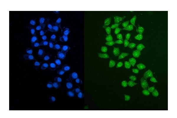
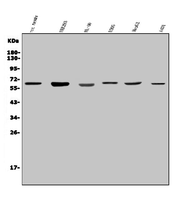
 FCM/FACS (Flow Cytometry)
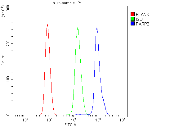
(Figure 3. Flow Cytometry analysis of HL-60 cells using anti-PARP2 antibody (AAA125690).Overlay histogram showing HL-60 cells stained with AAA125690 (Blue line). The cells were blocked with 10% normal goat serum. And then incubated with rabbit anti-PARP2 Antibody (AAA125690, 1μg/1x106 cells) for 30 min at 20 degree C. DyLight®488 conjugated goat anti-rabbit IgG (5-10μg/1x106 cells) was used as secondary antibody for 30 minutes at 20 degree C. Isotype control antibody (Green line) was rabbit IgG (1μg/1x106) used under the same conditions. Unlabelled sample (Red line) was also used as a control.)
FCM/FACS (Flow Cytometry)
(Figure 3. Flow Cytometry analysis of HL-60 cells using anti-PARP2 antibody (AAA125690).Overlay histogram showing HL-60 cells stained with AAA125690 (Blue line). The cells were blocked with 10% normal goat serum. And then incubated with rabbit anti-PARP2 Antibody (AAA125690, 1μg/1x106 cells) for 30 min at 20 degree C. DyLight®488 conjugated goat anti-rabbit IgG (5-10μg/1x106 cells) was used as secondary antibody for 30 minutes at 20 degree C. Isotype control antibody (Green line) was rabbit IgG (1μg/1x106) used under the same conditions. Unlabelled sample (Red line) was also used as a control.)
PARP2, Polyclonal Antibody (Cat# AAA125690)
Full Name
Anti-PARP2 Antibody
Gene Names
PARP2; ARTD2; ADPRT2; PARP-2; ADPRTL2; ADPRTL3; pADPRT-2
Reactivity
Human, Rat
Applications
Direct ELISA, Flow Cytometry, Immunofluorescence, Immunocytochemistry, Western Blot
Purity
Immunogen affinity purified.
Pricing
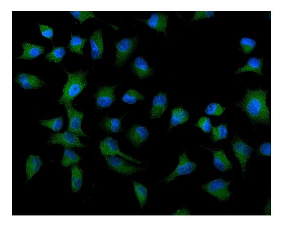
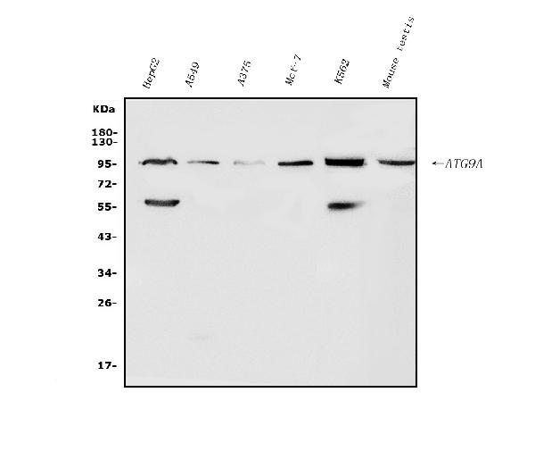
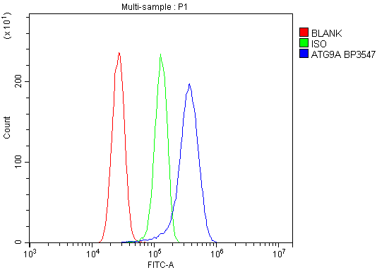 FCM/FACS (Flow Cytometry)
(Figure 3. Flow Cytometry analysis of U87 cells using anti-ATG9A antibody (AAA125702).Overlay histogram showing U87 cells stained with AAA125702 (Blue line). The cells were blocked with 10% normal goat serum. And then incubated with rabbit anti-ATG9A Antibody (AAA125702, 1μg/1x106 cells) for 30 min at 20 degree C. DyLight®488 conjugated goat anti-rabbit IgG (5-10μg/1x106 cells) was used as secondary antibody for 30 minutes at 20 degree C. Isotype control antibody (Green line) was rabbit IgG (1μg/1x106) used under the same conditions. Unlabelled sample (Red line) was also used as a control.)
FCM/FACS (Flow Cytometry)
(Figure 3. Flow Cytometry analysis of U87 cells using anti-ATG9A antibody (AAA125702).Overlay histogram showing U87 cells stained with AAA125702 (Blue line). The cells were blocked with 10% normal goat serum. And then incubated with rabbit anti-ATG9A Antibody (AAA125702, 1μg/1x106 cells) for 30 min at 20 degree C. DyLight®488 conjugated goat anti-rabbit IgG (5-10μg/1x106 cells) was used as secondary antibody for 30 minutes at 20 degree C. Isotype control antibody (Green line) was rabbit IgG (1μg/1x106) used under the same conditions. Unlabelled sample (Red line) was also used as a control.)
ATG9A, Polyclonal Antibody (Cat# AAA125702)
Full Name
Anti-ATG9A Antibody
Gene Names
ATG9A; mATG9; APG9L1; MGD3208
Reactivity
Human, Mouse
Applications
Direct ELISA, Flow Cytometry, Immunofluorescence, Immunocytochemistry, Western Blot
Purity
Immunogen affinity purified.
Pricing
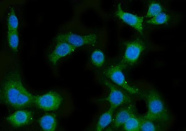
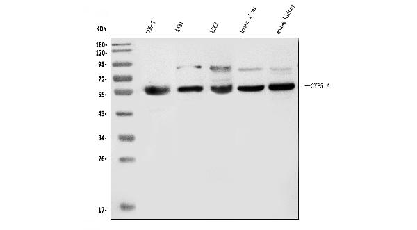
 FCM/FACS (Flow Cytometry)
(Figure 3. Flow Cytometry analysis of HepG2 cells using anti-CYP51A1/CYP51 antibody (AAA125712).Overlay histogram showing HepG2 cells stained with AAA125712 (Blue line). The cells were blocked with 10% normal goat serum. And then incubated with rabbit anti-CYP51A1/CYP51 Antibody (AAA125712, 1μg/1x106 cells) for 30 min at 20 degree C. DyLight®488 conjugated goat anti-rabbit IgG (5-10μg/1x106 cells) was used as secondary antibody for 30 minutes at 20 degree C. Isotype control antibody (Green line) was rabbit IgG (1μg/1x106) used under the same conditions. Unlabelled sample (Red line) was also used as a control.)
FCM/FACS (Flow Cytometry)
(Figure 3. Flow Cytometry analysis of HepG2 cells using anti-CYP51A1/CYP51 antibody (AAA125712).Overlay histogram showing HepG2 cells stained with AAA125712 (Blue line). The cells were blocked with 10% normal goat serum. And then incubated with rabbit anti-CYP51A1/CYP51 Antibody (AAA125712, 1μg/1x106 cells) for 30 min at 20 degree C. DyLight®488 conjugated goat anti-rabbit IgG (5-10μg/1x106 cells) was used as secondary antibody for 30 minutes at 20 degree C. Isotype control antibody (Green line) was rabbit IgG (1μg/1x106) used under the same conditions. Unlabelled sample (Red line) was also used as a control.)
CYP51A1/CYP51, Polyclonal Antibody (Cat# AAA125712)
Full Name
Anti-CYP51A1/CYP51 Antibody
Gene Names
CYP51A1; LDM; CP51; CYP51; CYPL1; P450L1; P450-14DM
Reactivity
Human, Mouse, Monkey
Applications
Direct ELISA, Flow Cytometry, Immunofluorescence, Immunocytochemistry, Western Blot
Purity
Immunogen affinity purified.
Pricing
 Bioactivity
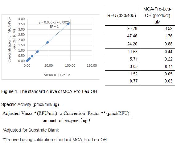
(MMP1 is a zinc-dependent enzymes capable of cleaving components of the extracellular matrix, which belongs to the matrix metalloproteinase (MMP) family. MMP-1 (interstitial collagenase), can degrade a broad range of substrates including types I, II, III, VII, VIII, and X collagens as well as casein, gelatin and so on. MMP-1 is expressed by fibroblasts, keratinocytes, endothelial cells, monocytes and macrophages. Structurally, MMP-1 may be divided into several distinct domains; a pro-domain which is cleaved upon activation; a catalytic domain containing the zinc binding site; a short hinge region and a carboxyl terminal (hemopexin-like) domain. The activity of recombinant pig MMP1 is measured by its ability to cleave a fluorogenic peptide substrate Mca-KPLGL-Dpa-AR-NH2 in the assay buffer 50 mM Tris, 10 mM CaCl2, 150 mM NaCl, 0.05% (w/v) Brij-35, pH 7.5. The rpMMP1 is diluted to 50 ug/ml in assay buffer, then activated by p-aminophenylmercuric acetate (APMA) in a final concentration of 1 mM incubated at 37 degree C for 2 hours. The activated rpMMP-1 is diluted to 1 ug/mL in assay buffer. Loading into a black well plate 50 uL of 1 ug/mL rpMMP-1 and start the reaction by adding 50 uL of 20 uM substrate, with a substrate blank containing 50 uL assay buffer, 50 uL substrate, and no rpMMP-1. Then read at excitiation and emission wavelengths of 320 nm and 405 nm, respectively, in kinetic mode for 5 minutes. The specific activity of recombinant pig MMP1 is > 10 pmol/min/ug.)
Bioactivity
(MMP1 is a zinc-dependent enzymes capable of cleaving components of the extracellular matrix, which belongs to the matrix metalloproteinase (MMP) family. MMP-1 (interstitial collagenase), can degrade a broad range of substrates including types I, II, III, VII, VIII, and X collagens as well as casein, gelatin and so on. MMP-1 is expressed by fibroblasts, keratinocytes, endothelial cells, monocytes and macrophages. Structurally, MMP-1 may be divided into several distinct domains; a pro-domain which is cleaved upon activation; a catalytic domain containing the zinc binding site; a short hinge region and a carboxyl terminal (hemopexin-like) domain. The activity of recombinant pig MMP1 is measured by its ability to cleave a fluorogenic peptide substrate Mca-KPLGL-Dpa-AR-NH2 in the assay buffer 50 mM Tris, 10 mM CaCl2, 150 mM NaCl, 0.05% (w/v) Brij-35, pH 7.5. The rpMMP1 is diluted to 50 ug/ml in assay buffer, then activated by p-aminophenylmercuric acetate (APMA) in a final concentration of 1 mM incubated at 37 degree C for 2 hours. The activated rpMMP-1 is diluted to 1 ug/mL in assay buffer. Loading into a black well plate 50 uL of 1 ug/mL rpMMP-1 and start the reaction by adding 50 uL of 20 uM substrate, with a substrate blank containing 50 uL assay buffer, 50 uL substrate, and no rpMMP-1. Then read at excitiation and emission wavelengths of 320 nm and 405 nm, respectively, in kinetic mode for 5 minutes. The specific activity of recombinant pig MMP1 is > 10 pmol/min/ug.)
Matrix Metalloproteinase 1 (MMP1), Active Protein (Cat# AAA161686)
Full Name
Active Matrix Metalloproteinase 1 (MMP1)
Gene Names
MMP1; Mmp1a
Applications
Activity Assay, Cell Culture
Purity
Greater than 90% by SDS-PAGE
Pricing
 IF (Immunofluorescence)
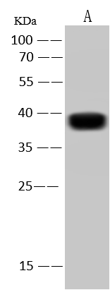
(EpCAM was immunoprecipitated using:Lane A:0.5 mg HCT116 Whole Cell LysateLane B:0.5 mg A431 Whole Cell LysateLane C:0.5 mg HT29 Whole Cell LysateLane D:0.5 mg HepG2 Whole Cell Lysate4 uL anti-EpCAM rabbit polyclonal antibody and 60 ug of Immunomagnetic beads Protein A/G.Primary antibody:Anti-EpCAM rabbit polyclonal antibody,at 1:100 dilutionSecondary antibody:Clean-Blot IP Detection Reagent (HRP) at 1:1000dilutionDeveloped using the ECL technique.Performed under reducing conditions.Predicted band size: 35 kDaObserved band size :35 kDa)
IF (Immunofluorescence)
(EpCAM was immunoprecipitated using:Lane A:0.5 mg HCT116 Whole Cell LysateLane B:0.5 mg A431 Whole Cell LysateLane C:0.5 mg HT29 Whole Cell LysateLane D:0.5 mg HepG2 Whole Cell Lysate4 uL anti-EpCAM rabbit polyclonal antibody and 60 ug of Immunomagnetic beads Protein A/G.Primary antibody:Anti-EpCAM rabbit polyclonal antibody,at 1:100 dilutionSecondary antibody:Clean-Blot IP Detection Reagent (HRP) at 1:1000dilutionDeveloped using the ECL technique.Performed under reducing conditions.Predicted band size: 35 kDaObserved band size :35 kDa)
EpCAM/TROP1, Polyclonal Antibody (Cat# AAA258828)
Full Name
Anti-EpCAM/TROP1 Antibody, Rabbit Polyclonal
Reactivity
Human
Applications
Immunofluorescence, Immunocytochemistry, Immunohistochemistry, Immunoprecipitation, Western Blot
Purity
Protein A & Antigen Affinity
Pricing
 FCM/FACS (Flow Cytometry)
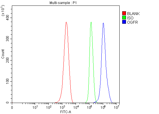
(Figure 2. Flow Cytometry analysis of ANA-1 cells using anti-Ogfr antibody (AAA127350).Overlay histogram showing ANA-1 cells stained with AAA127350 (Blue line). To facilitate intracellular staining, cells were fixed with 4% paraformaldehyde and permeabilized with permeabilization buffer. The cells were blocked with 10% normal goat serum. And then incubated with rabbit anti-Ogfr Antibody (AAA127350, 1ug/1x106 cells) for 30 min at 20 degree C. DyLight488 conjugated goat anti-rabbit IgG was used as secondary antibody for 30 minutes at 20 degree C. Isotype control antibody (Green line) was rabbit IgG (1ug/1x106) used under the same conditions. Unlabelled sample without incubation with primary antibody and secondary antibody (Red line) was used as a blank control.)
FCM/FACS (Flow Cytometry)
(Figure 2. Flow Cytometry analysis of ANA-1 cells using anti-Ogfr antibody (AAA127350).Overlay histogram showing ANA-1 cells stained with AAA127350 (Blue line). To facilitate intracellular staining, cells were fixed with 4% paraformaldehyde and permeabilized with permeabilization buffer. The cells were blocked with 10% normal goat serum. And then incubated with rabbit anti-Ogfr Antibody (AAA127350, 1ug/1x106 cells) for 30 min at 20 degree C. DyLight488 conjugated goat anti-rabbit IgG was used as secondary antibody for 30 minutes at 20 degree C. Isotype control antibody (Green line) was rabbit IgG (1ug/1x106) used under the same conditions. Unlabelled sample without incubation with primary antibody and secondary antibody (Red line) was used as a blank control.)
Ogfr, Polyclonal Antibody (Cat# AAA127350)
Full Name
Anti-Ogfr Antibody Picoband
Gene Names
Ogfr; 2010013E17Rik
Reactivity
Mouse
Applications
Flow Cytometry, Western Blot
Purity
Immunogen affinity purified.
Pricing
 WB (Western Blot)
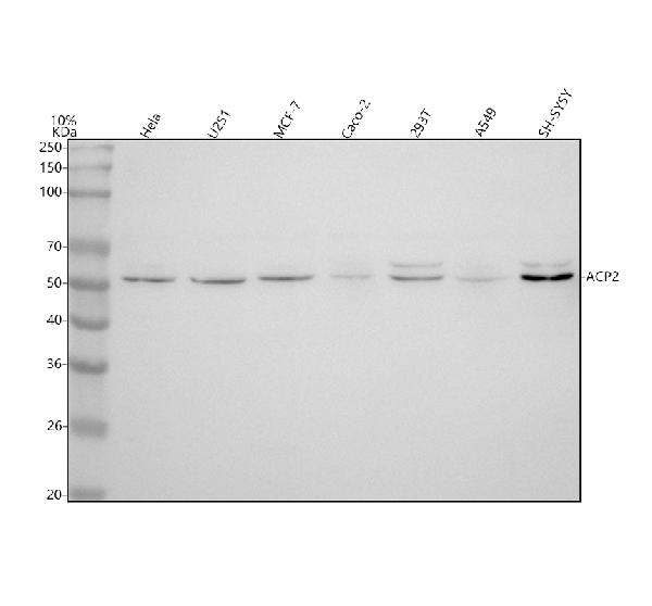
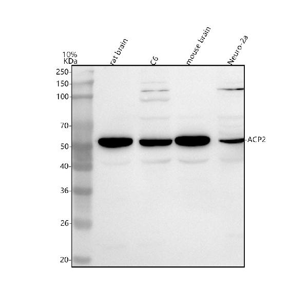
(Figure 2. Western blot analysis of ACP2 using anti-ACP2 antibody (AAA127385).Electrophoresis was performed on a 5-20% SDS-PAGE gel at 70V (Stacking gel)/90V (Resolving gel) for 2-3 hours. The sample well of each lane was loaded with 30 ug of sample under reducing conditions.Lane 1: rat brain tissue lysates,Lane 2: rat C6 whole cell lysates,Lane 3: mouse brain tissue lysates,Lane 4: mouse Neuro-2a whole cell lysates.After electrophoresis, proteins were transferred to a nitrocellulose membrane at 150 mA for 50-90 minutes. Blocked the membrane with 5% non-fat milk/TBS for 1.5 hour at RT. The membrane was incubated with rabbit anti-ACP2 antigen affinity purified polyclonal antibody (#AAA127385) at 0.5ug/mL overnight at 4 degree C, then washed with TBS-0.1%Tween 3 times with 5 minutes each and probed with a goat anti-rabbit IgG-HRP secondary antibody at a dilution of 1:5000 for 1.5 hour at RT. The signal is developed using an Enhanced Chemiluminescent detection (ECL) kit with Tanon 5200 system. A specific band was detected for ACP2 at approximately 52 kDa. The expected band size for ACP2 is at 48,18 kDa.)
WB (Western Blot)
(Figure 2. Western blot analysis of ACP2 using anti-ACP2 antibody (AAA127385).Electrophoresis was performed on a 5-20% SDS-PAGE gel at 70V (Stacking gel)/90V (Resolving gel) for 2-3 hours. The sample well of each lane was loaded with 30 ug of sample under reducing conditions.Lane 1: rat brain tissue lysates,Lane 2: rat C6 whole cell lysates,Lane 3: mouse brain tissue lysates,Lane 4: mouse Neuro-2a whole cell lysates.After electrophoresis, proteins were transferred to a nitrocellulose membrane at 150 mA for 50-90 minutes. Blocked the membrane with 5% non-fat milk/TBS for 1.5 hour at RT. The membrane was incubated with rabbit anti-ACP2 antigen affinity purified polyclonal antibody (#AAA127385) at 0.5ug/mL overnight at 4 degree C, then washed with TBS-0.1%Tween 3 times with 5 minutes each and probed with a goat anti-rabbit IgG-HRP secondary antibody at a dilution of 1:5000 for 1.5 hour at RT. The signal is developed using an Enhanced Chemiluminescent detection (ECL) kit with Tanon 5200 system. A specific band was detected for ACP2 at approximately 52 kDa. The expected band size for ACP2 is at 48,18 kDa.)
ACP2, Polyclonal Antibody (Cat# AAA127385)
Full Name
Anti-ACP2 Antibody Picoband
Gene Names
ACP2; LAP
Reactivity
Human, Mouse, Rat
Applications
Western Blot
Purity
Immunogen affinity purified.
Pricing
 IF (Immunofluorescence)
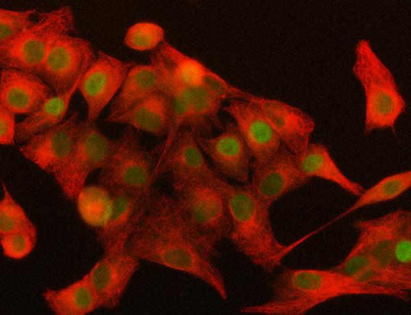
(Figure 2. IF analysis of DAX-1/NR0B1 using anti-DAX-1/NR0B1 antibody (AAA127063) and anti-Beta Tubulin antibody (M01857-3).DAX-1/NR0B1 was detected in immunocytochemical section of A549 cell. Enzyme antigen retrieval was performed using IHC enzyme antigen retrieval reagent for 15 mins. The cells were blocked with 10% goat serum. And then incubated with 5ug/mL rabbit anti-DAX-1/NR0B1 Antibody (AAA127063) and mouse anti-Beta Tubulin antibody (M01857-3) overnight at 4 degree C. DyLight488 Conjugated Goat Anti-Rabbit IgG and Cy3 Conjugated Goat Anti-Mouse IgG (BA1031) were used as secondary antibody at 1:500 dilution and incubated for 30 minutes at 37 degree C. Visualize using a fluorescence microscope and filter sets appropriate for the label used.)
IF (Immunofluorescence)
(Figure 2. IF analysis of DAX-1/NR0B1 using anti-DAX-1/NR0B1 antibody (AAA127063) and anti-Beta Tubulin antibody (M01857-3).DAX-1/NR0B1 was detected in immunocytochemical section of A549 cell. Enzyme antigen retrieval was performed using IHC enzyme antigen retrieval reagent for 15 mins. The cells were blocked with 10% goat serum. And then incubated with 5ug/mL rabbit anti-DAX-1/NR0B1 Antibody (AAA127063) and mouse anti-Beta Tubulin antibody (M01857-3) overnight at 4 degree C. DyLight488 Conjugated Goat Anti-Rabbit IgG and Cy3 Conjugated Goat Anti-Mouse IgG (BA1031) were used as secondary antibody at 1:500 dilution and incubated for 30 minutes at 37 degree C. Visualize using a fluorescence microscope and filter sets appropriate for the label used.)
DAX-1/NR0B1, Polyclonal Antibody (Cat# AAA127063)
Full Name
Anti-DAX-1/NR0B1 Antibody Picoband
Gene Names
NR0B1; AHC; AHX; DSS; GTD; HHG; AHCH; DAX1; DAX-1; NROB1; SRXY2
Reactivity
Human
Applications
Western Blot, Immunofluorescence, Immunocytochemistry
Purity
Immunogen affinity purified.
Pricing
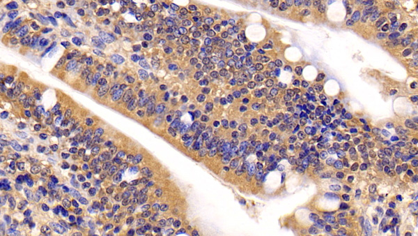
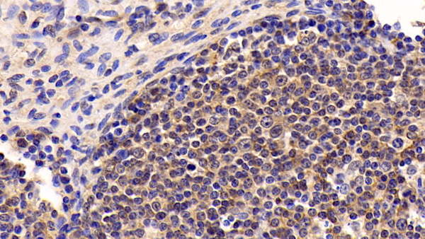 IHC (Immunohistochemistry)
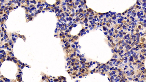
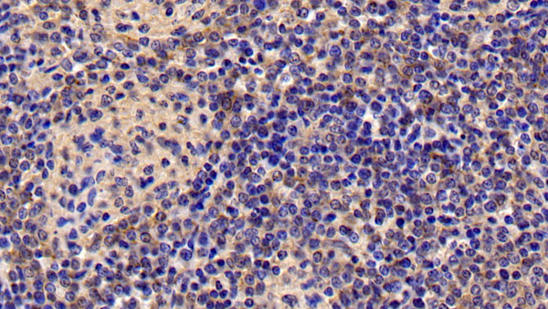
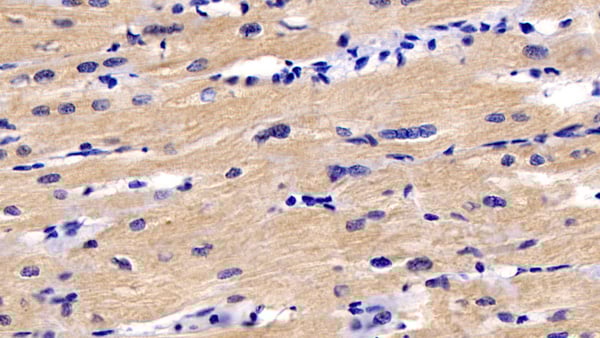
(DAB staining on IHCP;Sample: Human Lymph node Tissue; Primary Ab: 30ug/ml Mouse AntiHuman IL6 AntibodySecond Ab: 2ug/mL HRPLinked Caprine AntiMouse IgG Polyclonal Antibody(Catalog: SAA544Mu19))
IHC (Immunohistochemistry)
(DAB staining on IHCP;Sample: Human Lymph node Tissue; Primary Ab: 30ug/ml Mouse AntiHuman IL6 AntibodySecond Ab: 2ug/mL HRPLinked Caprine AntiMouse IgG Polyclonal Antibody(Catalog: SAA544Mu19))
Interleukin 6 (IL6), Monoclonal Antibody (Cat# AAA151463)
Full Name
Monoclonal Antibody to Interleukin 6 (IL6)
Gene Names
IL6; HGF; HSF; BSF2; IL-6; IFNB2
Reactivity
Human, Pig
Applications
Immunoprecipitation, Immunocytochemistry, Immunohistochemistry, Western Blot
Purity
Protein A + Protein G affinity chromatography
Pricing
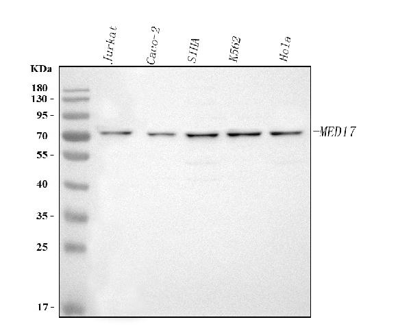
 FCM/FACS (Flow Cytometry)
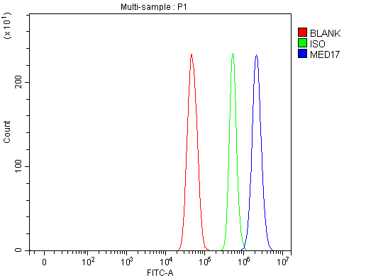
(Figure 2. Flow Cytometry analysis of U251 cells using anti-MED17 antibody (AAA127294).Overlay histogram showing U251 cells stained with AAA127294 (Blue line). To facilitate intracellular staining, cells were fixed with 4% paraformaldehyde and permeabilized with permeabilization buffer. The cells were blocked with 10% normal goat serum. And then incubated with rabbit anti-MED17 Antibody (AAA127294, 1ug/1x106 cells) for 30 min at 20 degree C. DyLight488 conjugated goat anti-rabbit IgG was used as secondary antibody for 30 minutes at 20 degree C. Isotype control antibody (Green line) was rabbit IgG (1ug/1x106) used under the same conditions. Unlabelled sample (Red line) was also used as a control.)
FCM/FACS (Flow Cytometry)
(Figure 2. Flow Cytometry analysis of U251 cells using anti-MED17 antibody (AAA127294).Overlay histogram showing U251 cells stained with AAA127294 (Blue line). To facilitate intracellular staining, cells were fixed with 4% paraformaldehyde and permeabilized with permeabilization buffer. The cells were blocked with 10% normal goat serum. And then incubated with rabbit anti-MED17 Antibody (AAA127294, 1ug/1x106 cells) for 30 min at 20 degree C. DyLight488 conjugated goat anti-rabbit IgG was used as secondary antibody for 30 minutes at 20 degree C. Isotype control antibody (Green line) was rabbit IgG (1ug/1x106) used under the same conditions. Unlabelled sample (Red line) was also used as a control.)
MED17, Polyclonal Antibody (Cat# AAA127294)
Full Name
Anti-MED17 Antibody Picoband
Gene Names
MED17; CRSP6; CRSP77; DRIP80; TRAP80
Reactivity
Human
Applications
Flow Cytometry, Western Blot
Purity
Immunogen affinity purified.
Pricing
 Bioactivity
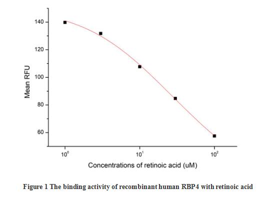
(Retinol-binding protein 4 (RBP4) is the specific carrier for retinol (also known as vitamin A), and is responsible for the conversion of unstable and insoluble retinol in aqueous solution into stable and soluble complex in plasma through their tight interaction. As a member of the lipocalin superfamily, RBP4 containing a beta-barrel structure with a well-defined cavity is secreted from the liver, and in turn delivers retinol from the liver stores to the peripheral tissues. In plasma, the RBP4-retinol complex interacts with transthyretin (TTR), and this binding is crucial for preventing RBP4 excretion through the kidney glomeruli. RBP4 expressed from an ectopic source efficiently delivers retinol to the eyes, and its deficiency affects night vision largely. Recently, RBP4 as an adipokine, is found to be expressed in adipose tissue and correlated with obesity, insulin resistance (IR) and type 2 diabetes (T2DM). The activity of recombinant human RBP4 was measured by its ability to bind all-trans retinoic acid. The binding of retinoic acid results in the quenching of Trp fluorescence in RBP4. RBP4 was diluted to 50 ug/ml in 50 mM Tris, 10 mM CaCl2, 150 mM NaCl, pH 7.5 (assay buffer) and the retinoic acid was diluted to 100, 30, 10, 3 and 1 uM in 95% ethanol. Mixing 112.5 uL of 50 ug/mL rhRBP4 and 12.5 uL of retinoic acid serial dilutions in microtubes and a blank containing 112.5 uL of 50 ug/mL rhRBP4 and 12.5 uL of 95% ethanol, then incubate at room temperature for 30 minutes. Loading 100 ul of the reaction mixtures and blank and read at excitation and emission wavelengths of 280 nm and 340 nm (top read), respectively, in endpoint mode. The result was shown in figure 1, the 50% binding concentration (BC50) is >30 uM.)
Bioactivity
(Retinol-binding protein 4 (RBP4) is the specific carrier for retinol (also known as vitamin A), and is responsible for the conversion of unstable and insoluble retinol in aqueous solution into stable and soluble complex in plasma through their tight interaction. As a member of the lipocalin superfamily, RBP4 containing a beta-barrel structure with a well-defined cavity is secreted from the liver, and in turn delivers retinol from the liver stores to the peripheral tissues. In plasma, the RBP4-retinol complex interacts with transthyretin (TTR), and this binding is crucial for preventing RBP4 excretion through the kidney glomeruli. RBP4 expressed from an ectopic source efficiently delivers retinol to the eyes, and its deficiency affects night vision largely. Recently, RBP4 as an adipokine, is found to be expressed in adipose tissue and correlated with obesity, insulin resistance (IR) and type 2 diabetes (T2DM). The activity of recombinant human RBP4 was measured by its ability to bind all-trans retinoic acid. The binding of retinoic acid results in the quenching of Trp fluorescence in RBP4. RBP4 was diluted to 50 ug/ml in 50 mM Tris, 10 mM CaCl2, 150 mM NaCl, pH 7.5 (assay buffer) and the retinoic acid was diluted to 100, 30, 10, 3 and 1 uM in 95% ethanol. Mixing 112.5 uL of 50 ug/mL rhRBP4 and 12.5 uL of retinoic acid serial dilutions in microtubes and a blank containing 112.5 uL of 50 ug/mL rhRBP4 and 12.5 uL of 95% ethanol, then incubate at room temperature for 30 minutes. Loading 100 ul of the reaction mixtures and blank and read at excitation and emission wavelengths of 280 nm and 340 nm (top read), respectively, in endpoint mode. The result was shown in figure 1, the 50% binding concentration (BC50) is >30 uM.)
Retinol Binding Protein 4 (RBP4), Active Protein (Cat# AAA161805)
Full Name
Active Retinol Binding Protein 4 (RBP4)
Gene Names
RBP4; RDCCAS
Applications
Activity Assay, Cell Culture
Purity
Greater than 90% by SDS-PAGE
Pricing
 FCM/FACS (Flow Cytometry)
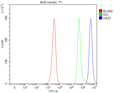
(Figure 2. Flow Cytometry analysis of JK cells using anti-GAS7 antibody (AAA127384).Overlay histogram showing JK cells stained with AAA127384 (Blue line). To facilitate intracellular staining, cells were fixed with 4% paraformaldehyde and permeabilized with permeabilization buffer. The cells were blocked with 10% normal goat serum. And then incubated with rabbit anti-GAS7 Antibody (AAA127384, 1ug/1x106 cells) for 30 min at 20 degree C. DyLight488 conjugated goat anti-rabbit IgG was used as secondary antibody for 30 minutes at 20 degree C. Isotype control antibody (Green line) was rabbit IgG (1ug/1x106) used under the same conditions. Unlabelled sample (Red line) was also used as a control.)
FCM/FACS (Flow Cytometry)
(Figure 2. Flow Cytometry analysis of JK cells using anti-GAS7 antibody (AAA127384).Overlay histogram showing JK cells stained with AAA127384 (Blue line). To facilitate intracellular staining, cells were fixed with 4% paraformaldehyde and permeabilized with permeabilization buffer. The cells were blocked with 10% normal goat serum. And then incubated with rabbit anti-GAS7 Antibody (AAA127384, 1ug/1x106 cells) for 30 min at 20 degree C. DyLight488 conjugated goat anti-rabbit IgG was used as secondary antibody for 30 minutes at 20 degree C. Isotype control antibody (Green line) was rabbit IgG (1ug/1x106) used under the same conditions. Unlabelled sample (Red line) was also used as a control.)
GAS7, Polyclonal Antibody (Cat# AAA127384)
Full Name
Anti-GAS7 Antibody Picoband
Gene Names
GAS7; MLL/GAS7
Reactivity
Human, Mouse, Rat
Applications
Flow Cytometry, Western Blot
Purity
Immunogen affinity purified.
Pricing
 FCM/FACS (Flow Cytometry)
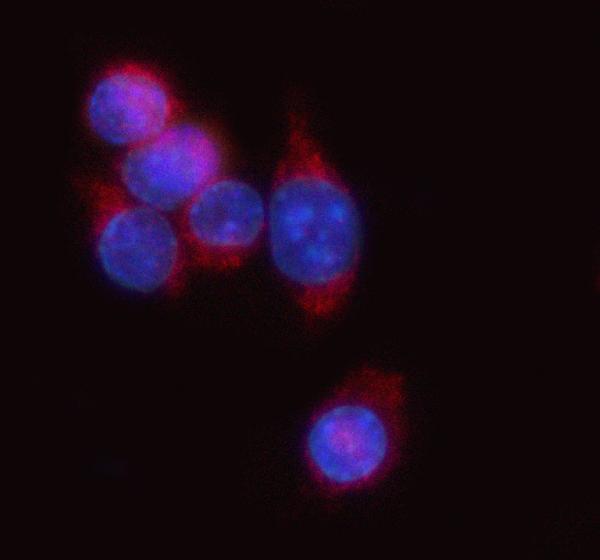
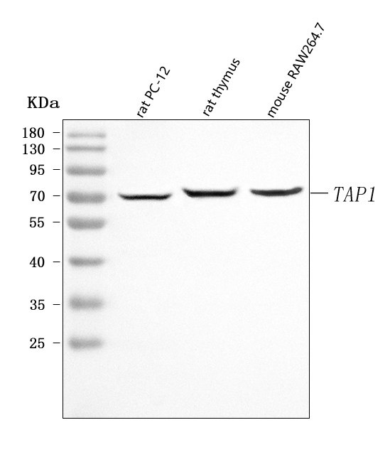
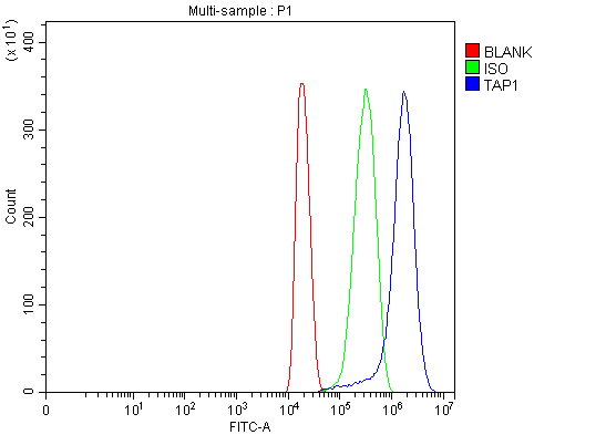
(Figure 3. Flow Cytometry analysis of HEPA1-6 cells using anti-Tap1 antibody (AAA127036).Overlay histogram showing HEPA1-6 cells stained with AAA127036 (Blue line). To facilitate intracellular staining, cells were fixed with 4% paraformaldehyde and permeabilized with permeabilization buffer. The cells were blocked with 10% normal goat serum. And then incubated with rabbit anti-Tap1 Antibody (AAA127036, 1ug/1x106 cells) for 30 min at 20 degree C. DyLight488 conjugated goat anti-rabbit IgG was used as secondary antibody for 30 minutes at 20 degree C. Isotype control antibody (Green line) was rabbit IgG (1ug/1x106) used under the same conditions. Unlabelled sample without incubation with primary antibody and secondary antibody (Red line) was used as a blank control.)
FCM/FACS (Flow Cytometry)
(Figure 3. Flow Cytometry analysis of HEPA1-6 cells using anti-Tap1 antibody (AAA127036).Overlay histogram showing HEPA1-6 cells stained with AAA127036 (Blue line). To facilitate intracellular staining, cells were fixed with 4% paraformaldehyde and permeabilized with permeabilization buffer. The cells were blocked with 10% normal goat serum. And then incubated with rabbit anti-Tap1 Antibody (AAA127036, 1ug/1x106 cells) for 30 min at 20 degree C. DyLight488 conjugated goat anti-rabbit IgG was used as secondary antibody for 30 minutes at 20 degree C. Isotype control antibody (Green line) was rabbit IgG (1ug/1x106) used under the same conditions. Unlabelled sample without incubation with primary antibody and secondary antibody (Red line) was used as a blank control.)
Tap1, Polyclonal Antibody (Cat# AAA127036)
Full Name
Anti-Tap1 Antibody Picoband
Gene Names
Tap1; Y3; TAP; APT1; Ham1; MTP1; PSF1; ABC17; Abcb2; Ham-1; RING4; Tap-1
Reactivity
Mouse, Rat
Applications
Flow Cytometry, Immunofluorescence, Immunocytochemistry, Western Blot
Purity
Immunogen affinity purified.
Pricing
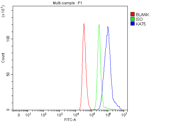 FCM/FACS (Flow Cytometry)
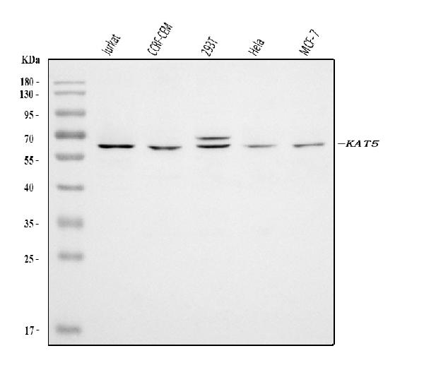
(Figure 2. Flow Cytometry analysis of THP-1 cells using anti-Tip60/KAT5 antibody (AAA126139).Overlay histogram showing THP-1 cells stained with AAA126139 (Blue line). The cells were blocked with 10% normal goat serum. And then incubated with rabbit anti-Tip60/KAT5 Antibody (AAA126139, 1 ug/1x10^6 cells) for 30 min at 20 degree C. DyLight488 conjugated goat anti-rabbit IgG was used as secondary antibody for 30 minutes at 20 degree C. Isotype control antibody (Green line) was rabbit IgG (1 ug/1x10^6) used under the same conditions. Unlabelled sample (Red line) was also used as a control.)
FCM/FACS (Flow Cytometry)
(Figure 2. Flow Cytometry analysis of THP-1 cells using anti-Tip60/KAT5 antibody (AAA126139).Overlay histogram showing THP-1 cells stained with AAA126139 (Blue line). The cells were blocked with 10% normal goat serum. And then incubated with rabbit anti-Tip60/KAT5 Antibody (AAA126139, 1 ug/1x10^6 cells) for 30 min at 20 degree C. DyLight488 conjugated goat anti-rabbit IgG was used as secondary antibody for 30 minutes at 20 degree C. Isotype control antibody (Green line) was rabbit IgG (1 ug/1x10^6) used under the same conditions. Unlabelled sample (Red line) was also used as a control.)
Tip60/KAT5, Polyclonal Antibody (Cat# AAA126139)
Full Name
Anti-Tip60/KAT5 Antibody Picoband
Gene Names
KAT5; TIP; ESA1; PLIP; TIP60; cPLA2; HTATIP; ZC2HC5; HTATIP1
Reactivity
Human
Applications
Western Blot, Flow Cytometry
Purity
Immunogen affinity purified.
Pricing
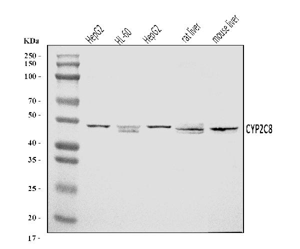
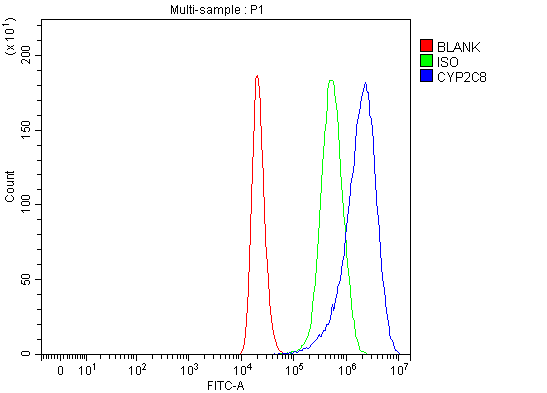 FCM/FACS (Flow Cytometry)
(Figure 2. Flow Cytometry analysis of 293T cells using anti-CYP2C8 antibody (AAA126118).Overlay histogram showing 293T cells stained with AAA126118 (Blue line). The cells were blocked with 10% normal goat serum. And then incubated with rabbit anti-CYP2C8 Antibody (AAA126118, 1 ug/1x10^6 cells) for 30 min at 20 degree C. DyLight488 conjugated goat anti-rabbit IgG was used as secondary antibody for 30 minutes at 20 degree C. Isotype control antibody (Green line) was rabbit IgG (1 ug/1x10^6) used under the same conditions. Unlabelled sample (Red line) was also used as a control.)
FCM/FACS (Flow Cytometry)
(Figure 2. Flow Cytometry analysis of 293T cells using anti-CYP2C8 antibody (AAA126118).Overlay histogram showing 293T cells stained with AAA126118 (Blue line). The cells were blocked with 10% normal goat serum. And then incubated with rabbit anti-CYP2C8 Antibody (AAA126118, 1 ug/1x10^6 cells) for 30 min at 20 degree C. DyLight488 conjugated goat anti-rabbit IgG was used as secondary antibody for 30 minutes at 20 degree C. Isotype control antibody (Green line) was rabbit IgG (1 ug/1x10^6) used under the same conditions. Unlabelled sample (Red line) was also used as a control.)
CYP2C8, Polyclonal Antibody (Cat# AAA126118)
Full Name
Anti-CYP2C8 Antibody Picoband
Gene Names
CYP2C8; CPC8; CYPIIC8; MP-12/MP-20
Reactivity
Human, Mouse, Rat
Applications
Flow Cytometry, Western Blot
Purity
Immunogen affinity purified.
Pricing
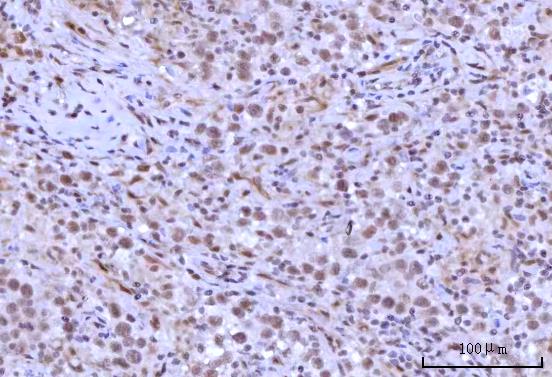
 IF (Immunofluorescence)
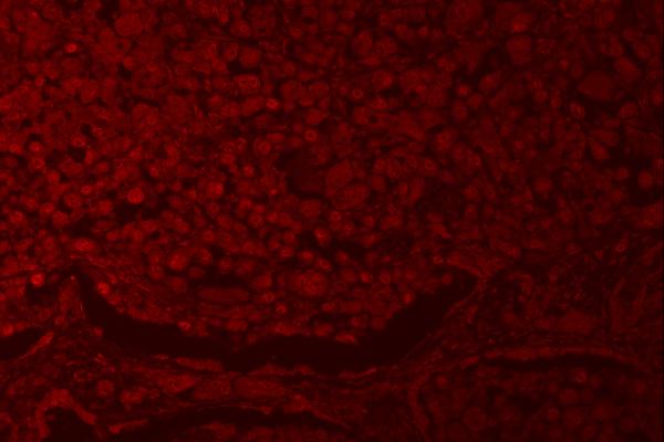
(Figure 5. IF analysis of RNF40 using anti-RNF401 antibody (AAA127414).RNF40 was detected in a paraffin-embedded section of human lung cancer tissue. Heat mediated antigen retrieval was performed in EDTA buffer (pH 8.0, epitope retrieval solution). The tissue section was blocked with 10% goat serum. The tissue section was then incubated with 5ug/mL rabbit anti-RNF40 Antibody (AAA127414) overnight at 4 degree C. DyLight550 Conjugated Goat Anti-Rabbit IgG (BA1135) was used as secondary antibody at 1:500 dilution and incubated for 30 minutes at 37 degree C. Visualize using a fluorescence microscope and filter sets appropriate for the label used.)
IF (Immunofluorescence)
(Figure 5. IF analysis of RNF40 using anti-RNF401 antibody (AAA127414).RNF40 was detected in a paraffin-embedded section of human lung cancer tissue. Heat mediated antigen retrieval was performed in EDTA buffer (pH 8.0, epitope retrieval solution). The tissue section was blocked with 10% goat serum. The tissue section was then incubated with 5ug/mL rabbit anti-RNF40 Antibody (AAA127414) overnight at 4 degree C. DyLight550 Conjugated Goat Anti-Rabbit IgG (BA1135) was used as secondary antibody at 1:500 dilution and incubated for 30 minutes at 37 degree C. Visualize using a fluorescence microscope and filter sets appropriate for the label used.)
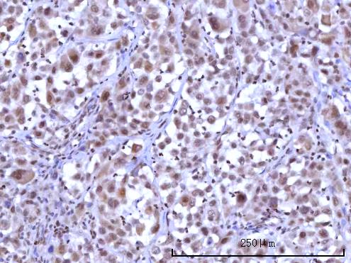
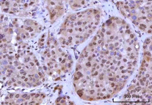
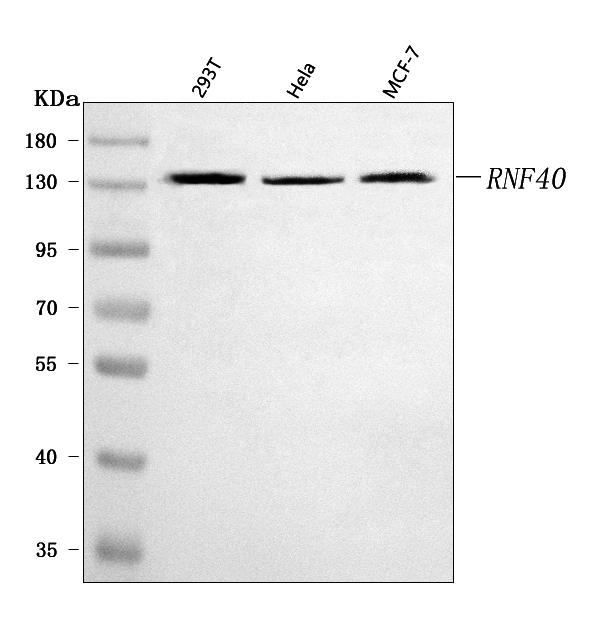
RNF40, Polyclonal Antibody (Cat# AAA127414)
Full Name
Anti-RNF40 Antibody Picoband
Gene Names
RNF40; BRE1B; RBP95; STARING
Reactivity
Human
Applications
Immunofluorescence, Immunohistochemistry, Western Blot
Purity
Immunogen affinity purified.
Pricing
 FCM/FACS (Flow Cytometry)
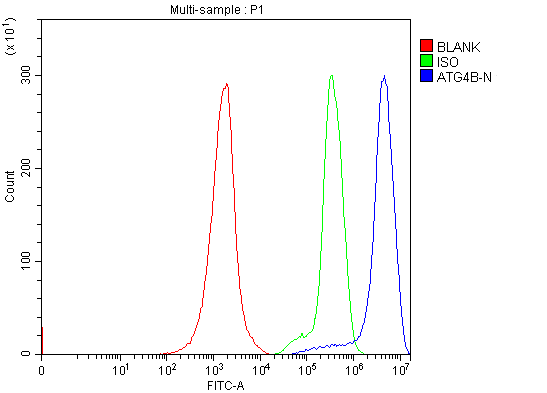
(Figure 2. Flow Cytometry analysis of Jurkat cells using anti-ATG4B antibody (AAA126267).Overlay histogram showing Jurkat cells stained with AAA126267 (Blue line). The cells were blocked with 10% normal goat serum. And then incubated with rabbit anti-ATG4B Antibody (AAA126267, 1 ug/1x10^6 cells) for 30 min at 20 degree C. DyLight488 conjugated goat anti-rabbit IgG was used as secondary antibody for 30 minutes at 20 degree C. Isotype control antibody (Green line) was rabbit IgG (1 ug/1x10^6) used under the same conditions. Unlabelled sample (Red line) was also used as a control.)
FCM/FACS (Flow Cytometry)
(Figure 2. Flow Cytometry analysis of Jurkat cells using anti-ATG4B antibody (AAA126267).Overlay histogram showing Jurkat cells stained with AAA126267 (Blue line). The cells were blocked with 10% normal goat serum. And then incubated with rabbit anti-ATG4B Antibody (AAA126267, 1 ug/1x10^6 cells) for 30 min at 20 degree C. DyLight488 conjugated goat anti-rabbit IgG was used as secondary antibody for 30 minutes at 20 degree C. Isotype control antibody (Green line) was rabbit IgG (1 ug/1x10^6) used under the same conditions. Unlabelled sample (Red line) was also used as a control.)
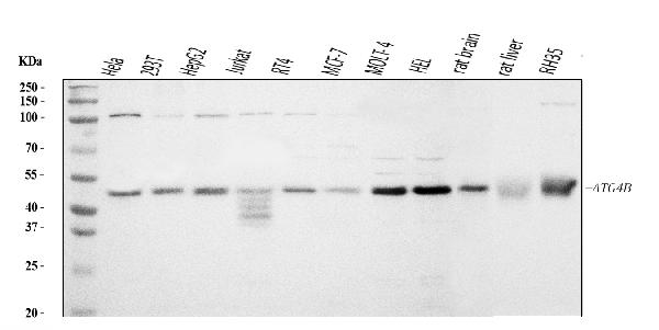
ATG4B, Polyclonal Antibody (Cat# AAA126267)
Full Name
Anti-ATG4B Antibody Picoband
Gene Names
ATG4B; APG4B; AUTL1
Reactivity
Human, Rat
Applications
Western Blot, Flow Cytometry
Purity
Immunogen affinity purified.
Pricing
 FCM/FACS (Flow Cytometry)
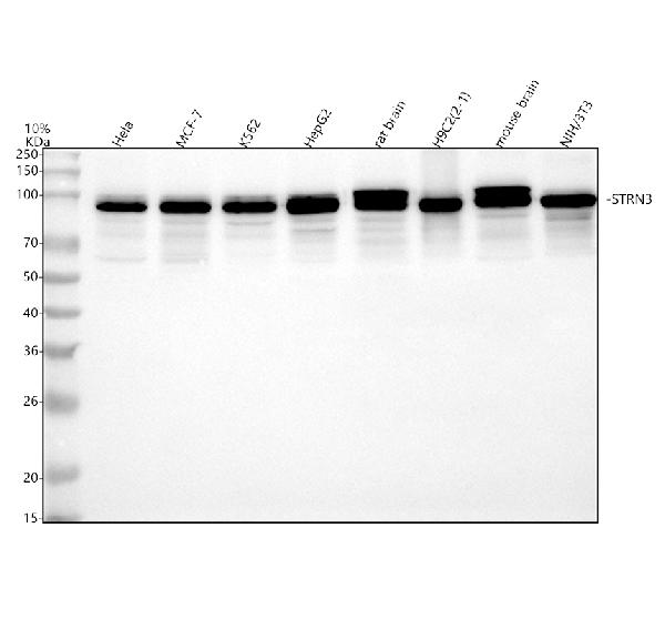
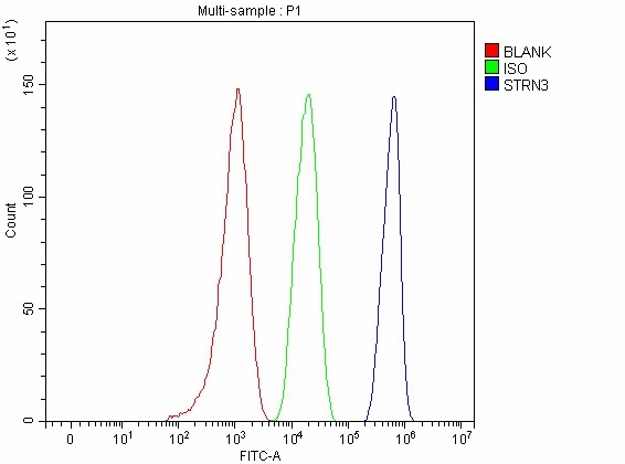
(Figure 3. Flow Cytometry analysis of HepG2 cells using anti-STRN3 antibody (AAA127395).Overlay histogram showing HepG2 cells stained with AAA127395 (Blue line). The cells were fixed with 4% paraformaldehyde and blocked with 10% normal goat serum. And then incubated with rabbit anti-STRN3 Antibody (AAA127395, 1ug/1x106 cells) for 30 min at 20 degree C. DyLight488 conjugated goat anti-rabbit IgG was used as secondary antibody for 30 minutes at 20 degree C. Isotype control antibody (Green line) was rabbit IgG (1ug/1x106) used under the same conditions. Unlabelled sample without incubation with primary antibody and secondary antibody (Red line) was used as a blank control.)
FCM/FACS (Flow Cytometry)
(Figure 3. Flow Cytometry analysis of HepG2 cells using anti-STRN3 antibody (AAA127395).Overlay histogram showing HepG2 cells stained with AAA127395 (Blue line). The cells were fixed with 4% paraformaldehyde and blocked with 10% normal goat serum. And then incubated with rabbit anti-STRN3 Antibody (AAA127395, 1ug/1x106 cells) for 30 min at 20 degree C. DyLight488 conjugated goat anti-rabbit IgG was used as secondary antibody for 30 minutes at 20 degree C. Isotype control antibody (Green line) was rabbit IgG (1ug/1x106) used under the same conditions. Unlabelled sample without incubation with primary antibody and secondary antibody (Red line) was used as a blank control.)
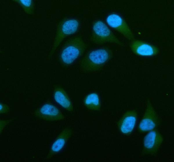
STRN3, Polyclonal Antibody (Cat# AAA127395)
Full Name
Anti-STRN3 Antibody Picoband
Gene Names
STRN3; SG2NA; S/G2NA; PPP2R6B
Reactivity
Human, Mouse, Rat
Applications
Flow Cytometry, Western Blot
Purity
Immunogen affinity purified.
Pricing
 FCM/FACS (Flow Cytometry)
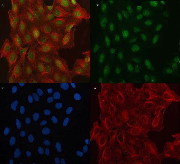
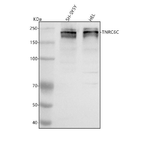
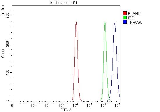
(Figure 3. Flow Cytometry analysis of HEL cells using anti-TNRC6C antibody (AAA127561).Overlay histogram showing HEL cells stained with AAA127561 (Blue line). To facilitate intracellular staining, cells were fixed with 4% paraformaldehyde and permeabilized with permeabilization buffer. The cells were blocked with 10% normal goat serum. And then incubated with rabbit anti-TNRC6C Antibody (AAA127561, 1ug/1x106 cells) for 30 min at 20 degree C. DyLight488 conjugated goat anti-rabbit IgG was used as secondary antibody for 30 minutes at 20 degree C. Isotype control antibody (Green line) was rabbit IgG (1ug/1x106) used under the same conditions. Unlabelled sample (Red line) was also used as a control.)
FCM/FACS (Flow Cytometry)
(Figure 3. Flow Cytometry analysis of HEL cells using anti-TNRC6C antibody (AAA127561).Overlay histogram showing HEL cells stained with AAA127561 (Blue line). To facilitate intracellular staining, cells were fixed with 4% paraformaldehyde and permeabilized with permeabilization buffer. The cells were blocked with 10% normal goat serum. And then incubated with rabbit anti-TNRC6C Antibody (AAA127561, 1ug/1x106 cells) for 30 min at 20 degree C. DyLight488 conjugated goat anti-rabbit IgG was used as secondary antibody for 30 minutes at 20 degree C. Isotype control antibody (Green line) was rabbit IgG (1ug/1x106) used under the same conditions. Unlabelled sample (Red line) was also used as a control.)
TNRC6C, Polyclonal Antibody (Cat# AAA127561)
Full Name
Anti-TNRC6C Antibody Picoband
Reactivity
Human
Applications
Flow Cytometry, Immunofluorescence, Immunocytochemistry, Western Blot
Purity
Immunogen affinity purified.
Pricing
 FCM/FACS (Flow Cytometry)
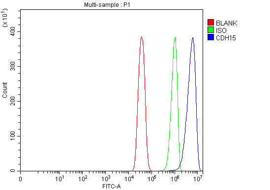
(Figure 4. Flow Cytometry analysis of C2C12 cells using anti-M Cadherin/CDH15 antibody (AAA126582).Overlay histogram showing C2C12 cells stained with AAA126582 (Blue line). The cells were blocked with 10% normal goat serum. And then incubated with rabbit anti-M Cadherin/CDH15 Antibody (AAA126582, 1 ug/1x10^6 cells) for 30 min at 20 degree C. DyLight488 conjugated goat anti-rabbit IgG was used as secondary antibody for 30 minutes at 20 degree C. Isotype control antibody (Green line) was rabbit IgG (1 ug/1x10^6) used under the same conditions. Unlabelled sample (Red line) was also used as a control.)
FCM/FACS (Flow Cytometry)
(Figure 4. Flow Cytometry analysis of C2C12 cells using anti-M Cadherin/CDH15 antibody (AAA126582).Overlay histogram showing C2C12 cells stained with AAA126582 (Blue line). The cells were blocked with 10% normal goat serum. And then incubated with rabbit anti-M Cadherin/CDH15 Antibody (AAA126582, 1 ug/1x10^6 cells) for 30 min at 20 degree C. DyLight488 conjugated goat anti-rabbit IgG was used as secondary antibody for 30 minutes at 20 degree C. Isotype control antibody (Green line) was rabbit IgG (1 ug/1x10^6) used under the same conditions. Unlabelled sample (Red line) was also used as a control.)
M Cadherin/CDH15, Polyclonal Antibody (Cat# AAA126582)
Full Name
Anti-M Cadherin/CDH15 Antibody Picoband
Gene Names
CDH15; CDH3; CDHM; MCAD; MRD3; CDH14
Reactivity
Human, Mouse, Rat
Applications
Western Blot, Immunohistochemistry, Flow Cytometry
Purity
Immunogen affinity purified.
Pricing
 FCM/FACS (Flow Cytometry)
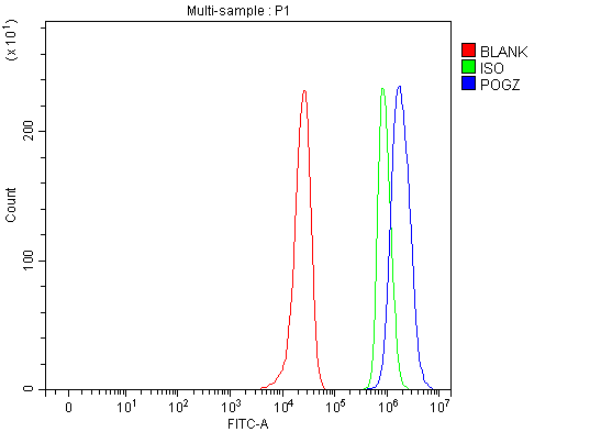
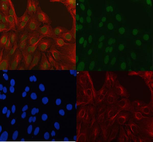
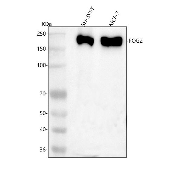
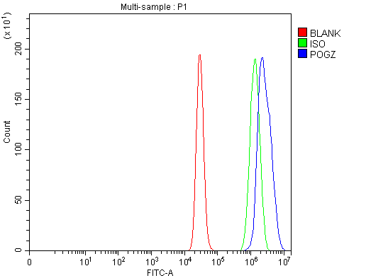
(Figure 4. Flow Cytometry analysis of MCF-7 cells using anti-POGZ antibody (AAA127348).Overlay histogram showing MCF-7 cells stained with AAA127348 (Blue line). To facilitate intracellular staining, cells were fixed with 4% paraformaldehyde and permeabilized with permeabilization buffer. The cells were blocked with 10% normal goat serum. And then incubated with rabbit anti-POGZ Antibody (AAA127348, 1ug/1x106 cells) for 30 min at 20 degree C. DyLight488 conjugated goat anti-rabbit IgG was used as secondary antibody for 30 minutes at 20 degree C. Isotype control antibody (Green line) was rabbit IgG (1ug/1x106) used under the same conditions. Unlabelled sample (Red line) was also used as a control.)
FCM/FACS (Flow Cytometry)
(Figure 4. Flow Cytometry analysis of MCF-7 cells using anti-POGZ antibody (AAA127348).Overlay histogram showing MCF-7 cells stained with AAA127348 (Blue line). To facilitate intracellular staining, cells were fixed with 4% paraformaldehyde and permeabilized with permeabilization buffer. The cells were blocked with 10% normal goat serum. And then incubated with rabbit anti-POGZ Antibody (AAA127348, 1ug/1x106 cells) for 30 min at 20 degree C. DyLight488 conjugated goat anti-rabbit IgG was used as secondary antibody for 30 minutes at 20 degree C. Isotype control antibody (Green line) was rabbit IgG (1ug/1x106) used under the same conditions. Unlabelled sample (Red line) was also used as a control.)
POGZ, Polyclonal Antibody (Cat# AAA127348)
Full Name
Anti-POGZ Antibody Picoband
Gene Names
POGZ; MRD37; WHSUS; ZNF635; ZNF280E; ZNF635m
Reactivity
Human
Applications
Flow Cytometry, Immunofluorescence, Immunocytochemistry, Western Blot
Purity
Immunogen affinity purified.
Pricing
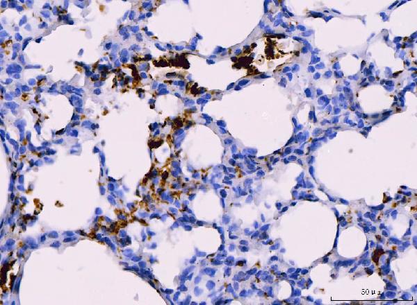
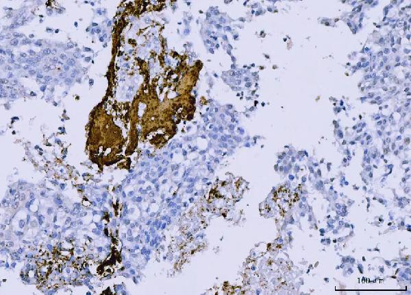
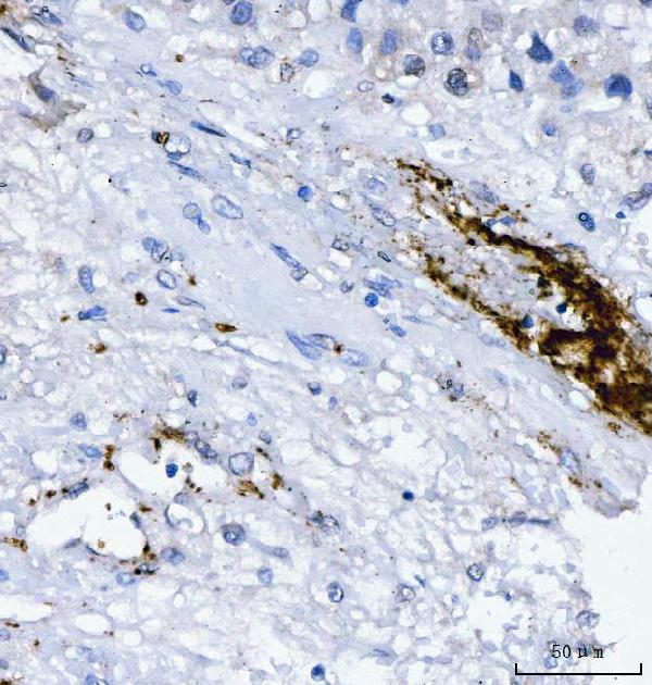
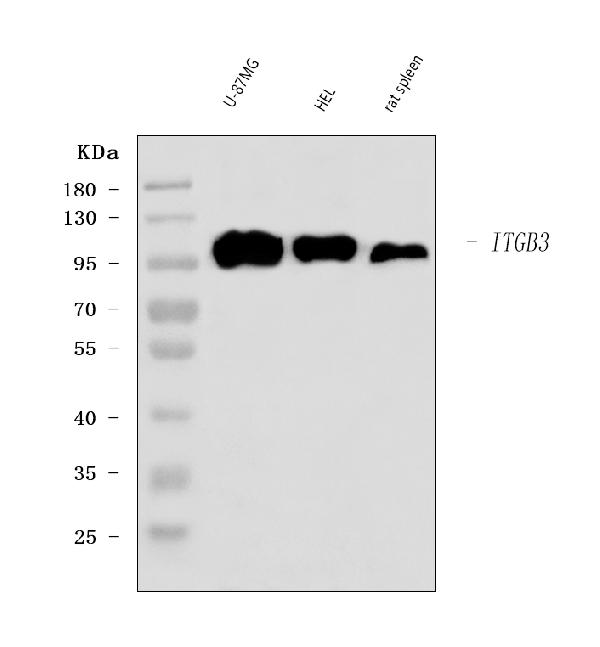
 FCM/FACS (Flow Cytometry)
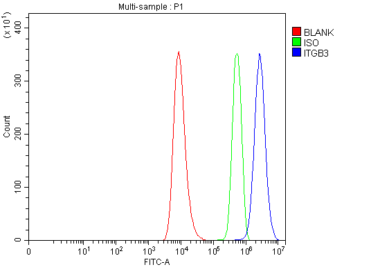
(Figure 5. Flow Cytometry analysis of HEL cells using anti-Integrin beta 3/ITGB3 antibody (AAA126045).Overlay histogram showing HEL cells stained with AAA126045 (Blue line). The cells were blocked with 10% normal goat serum. And then incubated with rabbit anti-Integrin beta 3/ITGB3 Antibody (AAA126045, 1 ug/1x10^6 cells) for 30 min at 20 degree C. DyLight488 conjugated goat anti-rabbit IgG was used as secondary antibody for 30 minutes at 20 degree C. Isotype control antibody (Green line) was rabbit IgG (1 ug/1x10^6) used under the same conditions. Unlabelled sample (Red line) was also used as a control.)
FCM/FACS (Flow Cytometry)
(Figure 5. Flow Cytometry analysis of HEL cells using anti-Integrin beta 3/ITGB3 antibody (AAA126045).Overlay histogram showing HEL cells stained with AAA126045 (Blue line). The cells were blocked with 10% normal goat serum. And then incubated with rabbit anti-Integrin beta 3/ITGB3 Antibody (AAA126045, 1 ug/1x10^6 cells) for 30 min at 20 degree C. DyLight488 conjugated goat anti-rabbit IgG was used as secondary antibody for 30 minutes at 20 degree C. Isotype control antibody (Green line) was rabbit IgG (1 ug/1x10^6) used under the same conditions. Unlabelled sample (Red line) was also used as a control.)
Integrin beta 3/ITGB3, Polyclonal Antibody (Cat# AAA126045)
Full Name
Anti-Integrin beta 3/ITGB3 Antibody Picoband
Gene Names
ITGB3; GT; CD61; GP3A; BDPLT2; GPIIIa; BDPLT16
Reactivity
Human, Rat
Applications
Western Blot, Immunohistochemistry, Flow Cytometry
Purity
Immunogen affinity purified.
Pricing
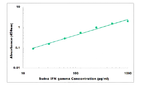 Standard Curve (Sample)
IFN gamma, ELISA Kit (Cat# AAA261812)
Full Name
Swine IFN gamma ELISA Kit
Gene Names
IFNG; IFG; IFI
Reactivity
Swine
Applications
Sandwich ELISA
Pricing
 SDS-PAGE
CDK8, Recombinant Protein (Cat# AAA72888)
Full Name
CDK8, Unactive recombinant protein
Gene Names
CDK8; K35
Applications
Western Blot
Purity
The purity was determined to be >80% by densitometry.
Pricing
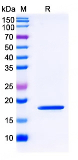 SDS-PAGE
(SDS-PAGE for Recombinant Mouse RNF43 protein)
SDS-PAGE
(SDS-PAGE for Recombinant Mouse RNF43 protein)
RNF43, Recombinant Protein (Cat# AAA120652)
Full Name
Recombinant Mouse RNF43 Protein, N-His
Gene Names
Rnf43; 4732452J19Rik
Applications
Western Blot
Purity
>90% as determined by SDS-PAGE
Pricing
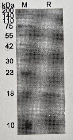 SDS-PAGE
(SDS PAGE for recombinant Human CLDN1 protein)
SDS-PAGE
(SDS PAGE for recombinant Human CLDN1 protein)
CLDN1, Recombinant Protein (Cat# AAA120421)
Full Name
Recombinant Human CLDN1, N-His
Gene Names
CLDN1; CLD1; SEMP1; ILVASC
Applications
Western Blot
Purity
>90% as determined by SDS-PAGE
Pricing

 SDS-PAGE
(Purity: Sample purity data. For lot-specific data, please refer to the respective datasheet.)
SDS-PAGE
(Purity: Sample purity data. For lot-specific data, please refer to the respective datasheet.)
COVID 19 Spike S1 (delta69-70 delta144 N501Y A570D D614G P681H) Coronavirus, Recombinant Protein (Cat# AAA73333)
Full Name
2019-nCoV Spike protein S1 (delta69-70 delta144 N501Y A570D D614G P681H)
Pricing
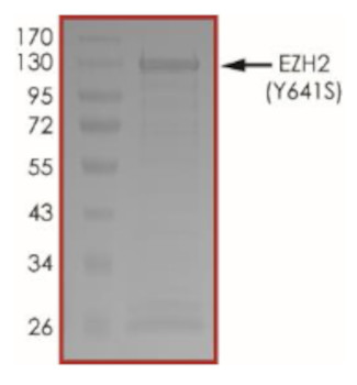 Application Data
(The purity of EZH2 (Y641S) was determined to be >80% by densitometry, approx. MW 130 kDa.)
Application Data
(The purity of EZH2 (Y641S) was determined to be >80% by densitometry, approx. MW 130 kDa.)
EZH2, Recombinant Protein (Cat# AAA72983)
Full Name
EZH2 (Y641S) Protein
Gene Names
EZH2; WVS; ENX1; KMT6; WVS2; ENX-1; EZH2b; KMT6A
Purity
>80%
Pricing
CD47, Recombinant Protein (Cat# AAA38019)
Full Name
Recombinant Human CD47
Gene Names
CD47; IAP; OA3; MER6
Purity
Greater than 95.0% as determined by SDS-PAGE.
Pricing
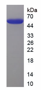
 Amino Acid Info
Interferon Alpha 11 (IFNa11), Recombinant Protein (Cat# AAA153748)
Full Name
Recombinant Interferon Alpha 11 (IFNa11)
Gene Names
Ifna11; IFN-[a]11
Applications
Western Blot
Purity
Greater than 90% by SDS-PAGE
Pricing
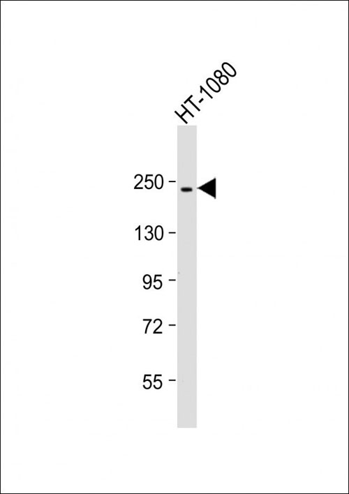
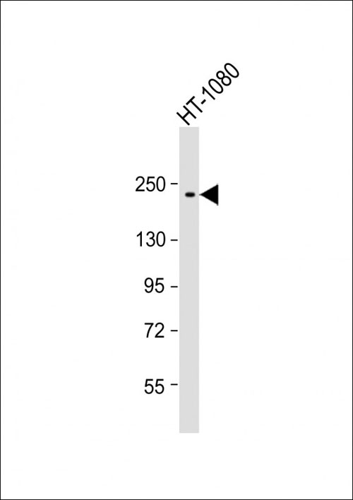 WB (Western Blot)
(Anti-PBRM1 Antibody at 1:500 dilution + HT-1080 whole cell lysateLysates/proteins at 20 ug per lane.SecondaryGoat Anti-mouse IgG, (H+L), Peroxidase conjugated at 1/10000 dilution.Predicted band size : 193 kDaBlocking/Dilution buffer: 5% NFDM/TBST.)
WB (Western Blot)
(Anti-PBRM1 Antibody at 1:500 dilution + HT-1080 whole cell lysateLysates/proteins at 20 ug per lane.SecondaryGoat Anti-mouse IgG, (H+L), Peroxidase conjugated at 1/10000 dilution.Predicted band size : 193 kDaBlocking/Dilution buffer: 5% NFDM/TBST.)
PBRM1, Monoclonal Antibody (Cat# AAA290611)
Full Name
PBRM1 Antibody
Gene Names
PBRM1; PB1; BAF180
Reactivity
Human
Applications
Western Blot
Pricing