
Filters
Clonality
Type
Reactivity
Gene Name
Isotype
Host
Application
Clone
10000 results for "Recombinant Proteins" - showing 9450-9500
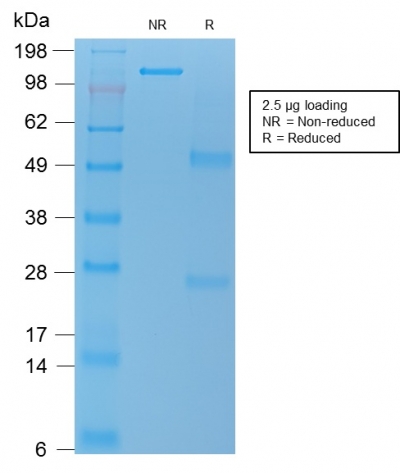
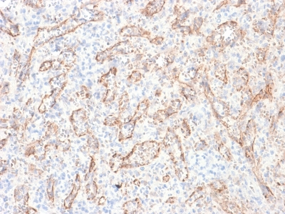
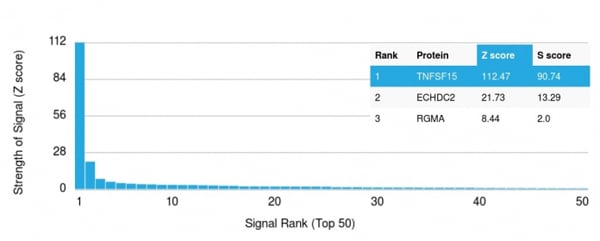 Application Data
(Analysis of Protein Array containing more than 19, 000 full-length human proteins using TNFS15 / VEGI Recombinant Mouse Monoclonal Antibody (rVEGI/1283).Z- and S- Score: The Z-score represents the strength of a signal that a monoclonal antibody (MAb) (in combination with a fluorescently-tagged anti-IgG secondary antibody) produces when binding to a particular protein on the HuProtTM array. Z-scores are described in units of standard deviations (SD's) above the mean value of all signals generated on that array. If targets on HuProtTM are arranged in descending order of the Z-score, the S-score is the difference (also in units of SD's) between the Z-score. S-score therefore represents the relative target specificity of a MAb to its intended target. A MAb is considered to specific to its intended target, if the MAb has an S-score of at least 2.5. For example, if a MAb binds to protein X with a Z-score of 43 and to protein Y with a Z-score of 14, then the S-score for the binding of that MAb to protein X is equal to 29.)
Application Data
(Analysis of Protein Array containing more than 19, 000 full-length human proteins using TNFS15 / VEGI Recombinant Mouse Monoclonal Antibody (rVEGI/1283).Z- and S- Score: The Z-score represents the strength of a signal that a monoclonal antibody (MAb) (in combination with a fluorescently-tagged anti-IgG secondary antibody) produces when binding to a particular protein on the HuProtTM array. Z-scores are described in units of standard deviations (SD's) above the mean value of all signals generated on that array. If targets on HuProtTM are arranged in descending order of the Z-score, the S-score is the difference (also in units of SD's) between the Z-score. S-score therefore represents the relative target specificity of a MAb to its intended target. A MAb is considered to specific to its intended target, if the MAb has an S-score of at least 2.5. For example, if a MAb binds to protein X with a Z-score of 43 and to protein Y with a Z-score of 14, then the S-score for the binding of that MAb to protein X is equal to 29.)
TNFS15/VEGI (Vascular Endothelial Growth Inhibitor), Monoclonal Antibody (Cat# AAA214622)
Full Name
TNFS15/VEGI (Vascular Endothelial Growth Inhibitor)
Gene Names
TNFSF15; TL1; TL1A; VEGI; TNLG1B; VEGI192A
Reactivity
Human. Others not known.
Applications
Immunohistochemistry
Pricing
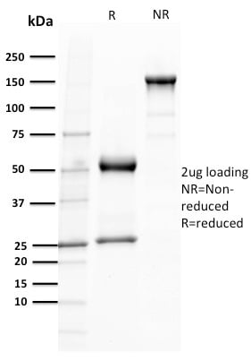
 Application Data
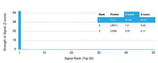
(Analysis of Protein Array containing more than 19,000 full-length human proteins using TCL1 Mouse Monoclonal Antibody (TCL1/2079). Z- and S- Score: The Z-score represents the strength of a signal that a monoclonal antibody (MAb) (in combination with a fluorescently-tagged anti-IgG secondary antibody) produces when binding to a particular protein on the HuProtTM array. Z-scores are described in units of standard deviations (SD's) above the mean value of all signals generated on that array. If targets on HuProtTM are arranged in descending order of the Z-score, the S-score is the difference (also in units of SD's) between the Z-score. S-score therefore represents the relative target specificity of a MAb to its intended target. A MAb is considered to specific to its intended target, if the MAb has an S-score of at least 2.5. For example, if a MAb binds to protein X with a Z-score of 43 and to protein Y with a Z-score of 14, then the S-score for the binding of that MAb to protein X is equal to 29.)
Application Data
(Analysis of Protein Array containing more than 19,000 full-length human proteins using TCL1 Mouse Monoclonal Antibody (TCL1/2079). Z- and S- Score: The Z-score represents the strength of a signal that a monoclonal antibody (MAb) (in combination with a fluorescently-tagged anti-IgG secondary antibody) produces when binding to a particular protein on the HuProtTM array. Z-scores are described in units of standard deviations (SD's) above the mean value of all signals generated on that array. If targets on HuProtTM are arranged in descending order of the Z-score, the S-score is the difference (also in units of SD's) between the Z-score. S-score therefore represents the relative target specificity of a MAb to its intended target. A MAb is considered to specific to its intended target, if the MAb has an S-score of at least 2.5. For example, if a MAb binds to protein X with a Z-score of 43 and to protein Y with a Z-score of 14, then the S-score for the binding of that MAb to protein X is equal to 29.)
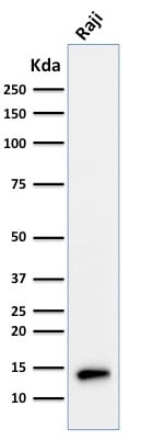
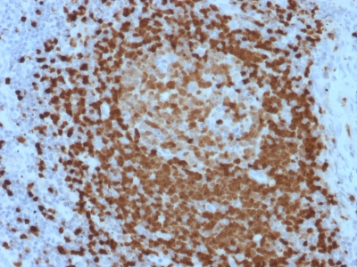
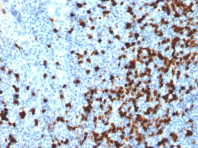
TCL1, Monoclonal Antibody (Cat# AAA214840)
Full Name
TCL1 (T-Cell Marker)
Gene Names
TCL1A; TCL1
Reactivity
Human. Others not known.
Applications
Immunohistochemistry, Western Blot
Purity
Purified Ab with BSA and Azide at 200ug/ml OR Purified Ab WITHOUT BSA and Azide at 1.0mg/ml
Pricing
 Application Data
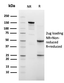
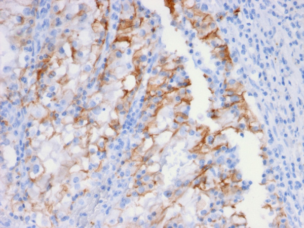
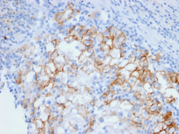
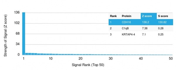
(Analysis of Protein Array containing more than 19,000 full-length human proteins using Cadherin 16 (CDH16) Mouse Monoclonal Antibody (CDH16/2448). Z- and S- Score: The Z-score represents the strength of a signal that a monoclonal antibody (MAb) (in combination with a fluorescently-tagged anti-IgG secondary antibody) produces when binding to a particular protein on the HuProtTM array. Z-scores are described in units of standard deviations (SD's) above the mean value of all signals generated on that array. If targets on HuProtTM are arranged in descending order of the Z-score, the S-score is the difference (also in units of SD's) between the Z-score. S-score therefore represents the relative target specificity of a MAb to its intended target. A MAb is considered to specific to its intended target, if the MAb has an S-score of at least 2.5. For example, if a MAb binds to protein X with a Z-score of 43 and to protein Y with a Z-score of 14, then the S-score for the binding of that MAb to protein X is equal to 29.)
Application Data
(Analysis of Protein Array containing more than 19,000 full-length human proteins using Cadherin 16 (CDH16) Mouse Monoclonal Antibody (CDH16/2448). Z- and S- Score: The Z-score represents the strength of a signal that a monoclonal antibody (MAb) (in combination with a fluorescently-tagged anti-IgG secondary antibody) produces when binding to a particular protein on the HuProtTM array. Z-scores are described in units of standard deviations (SD's) above the mean value of all signals generated on that array. If targets on HuProtTM are arranged in descending order of the Z-score, the S-score is the difference (also in units of SD's) between the Z-score. S-score therefore represents the relative target specificity of a MAb to its intended target. A MAb is considered to specific to its intended target, if the MAb has an S-score of at least 2.5. For example, if a MAb binds to protein X with a Z-score of 43 and to protein Y with a Z-score of 14, then the S-score for the binding of that MAb to protein X is equal to 29.)
Ksp-Cadherin/CDH16, Monoclonal Antibody (Cat# AAA214871)
Full Name
Ksp-Cadherin/CDH16 (Renal Cell Marker)
Reactivity
Human
Applications
Immunohistochemistry
Purity
Purified Ab with BSA and Azide at 200ug/ml OR Purified Ab WITHOUT BSA and Azide at 1.0mg/ml
Pricing
 Application Data
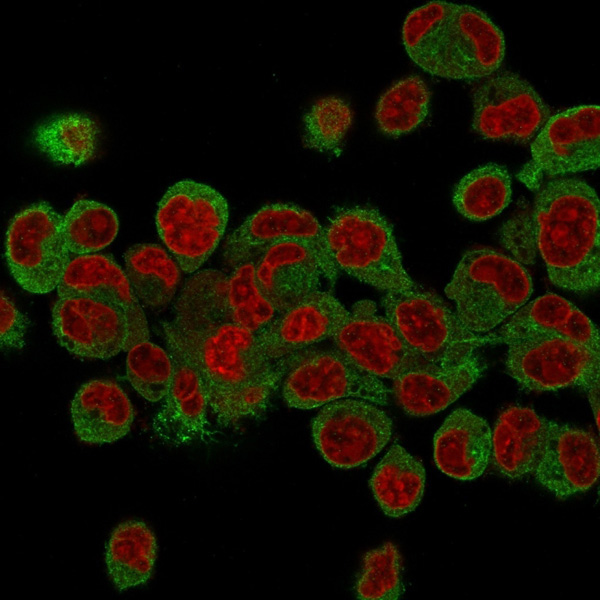
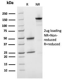
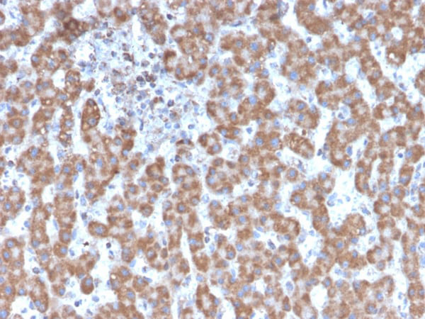
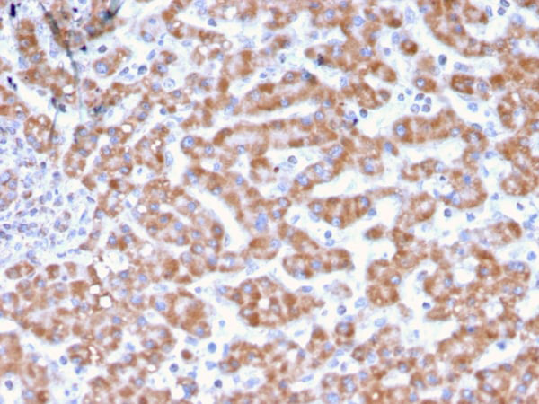
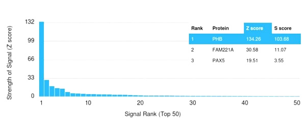
(Analysis of Protein Array containing more than 19,000 full-length human proteins using Monospecific Mouse Monoclonal Antibody to Prohibitin (PHB/3231). Z- and S- Score: The Z-score represents the strength of a signal that a monoclonal antibody (MAb) (in combination with a fluorescently-tagged anti-IgG secondary antibody) produces when binding to a particular protein on the HuProtTM array. Z-scores are described in units of standard deviations (SD's) above the mean value of all signals generated on that array. If targets on HuProtTM are arranged in descending order of the Z-score, the S-score is the difference (also in units of SD's) between the Z-score. S-score therefore represents the relative target specificity of a MAb to its intended target. A MAb is considered to specific to its intended target, if the MAb has an S-score of at least 2.5. For example, if a MAb binds to protein X with a Z-score of 43 and to protein Y with a Z-score of 14, then the S-score for the binding of that MAb to protein X is equal to 29.)
Application Data
(Analysis of Protein Array containing more than 19,000 full-length human proteins using Monospecific Mouse Monoclonal Antibody to Prohibitin (PHB/3231). Z- and S- Score: The Z-score represents the strength of a signal that a monoclonal antibody (MAb) (in combination with a fluorescently-tagged anti-IgG secondary antibody) produces when binding to a particular protein on the HuProtTM array. Z-scores are described in units of standard deviations (SD's) above the mean value of all signals generated on that array. If targets on HuProtTM are arranged in descending order of the Z-score, the S-score is the difference (also in units of SD's) between the Z-score. S-score therefore represents the relative target specificity of a MAb to its intended target. A MAb is considered to specific to its intended target, if the MAb has an S-score of at least 2.5. For example, if a MAb binds to protein X with a Z-score of 43 and to protein Y with a Z-score of 14, then the S-score for the binding of that MAb to protein X is equal to 29.)
Prohibitin, Monoclonal Antibody (Cat# AAA215138)
Full Name
Prohibitin (Mitochondrial Marker)
Gene Names
PHB; PHB1; HEL-215; HEL-S-54e
Reactivity
Human
Applications
Immunohistochemistry, Immunofluorescence, Western Blot
Purity
Purified Ab with BSA and Azide at 200ug/ml OR Purified Ab WITHOUT BSA and Azide at 1.0mg/ml
Pricing
 Application Data
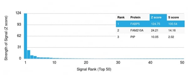
(Analysis of Protein Array containing more than 19,000 full-length human proteins using FABP5 Mouse Monoclonal Antibody (CPTC-FABP5-3). Z- and S- Score: The Z-score represents the strength of a signal that a monoclonal antibody (MAb) (in combination with a fluorescently-tagged anti-IgG secondary antibody) produces when binding to a particular protein on the HuProtTM array. Z-scores are described in units of standard deviations (SD's) above the mean value of all signals generated on that array. If targets on HuProtTM are arranged in descending order of the Z-score, the S-score is the difference (also in units of SD's) between the Z-score. S-score therefore represents the relative target specificity of a MAb to its intended target. A MAb is considered to specific to its intended target, if the MAb has an S-score of at least 2.5. For example, if a MAb binds to protein X with a Z-score of 43 and to protein Y with a Z-score of 14, then the S-score for the binding of that MAb to protein X is equal to 29.)
Application Data
(Analysis of Protein Array containing more than 19,000 full-length human proteins using FABP5 Mouse Monoclonal Antibody (CPTC-FABP5-3). Z- and S- Score: The Z-score represents the strength of a signal that a monoclonal antibody (MAb) (in combination with a fluorescently-tagged anti-IgG secondary antibody) produces when binding to a particular protein on the HuProtTM array. Z-scores are described in units of standard deviations (SD's) above the mean value of all signals generated on that array. If targets on HuProtTM are arranged in descending order of the Z-score, the S-score is the difference (also in units of SD's) between the Z-score. S-score therefore represents the relative target specificity of a MAb to its intended target. A MAb is considered to specific to its intended target, if the MAb has an S-score of at least 2.5. For example, if a MAb binds to protein X with a Z-score of 43 and to protein Y with a Z-score of 14, then the S-score for the binding of that MAb to protein X is equal to 29.)
FABP5, Monoclonal Antibody (Cat# AAA214737)
Full Name
FABP5 (Marker of Metastatic Potential in Colorectal Cancer)
Gene Names
FABP5; EFABP; KFABP; E-FABP; PAFABP; PA-FABP
Reactivity
Human. Others not tested.
Applications
Immunohistochemistry, Western Blot
Purity
Purified Ab with BSA and Azide at 200ug/ml OR Purified Ab WITHOUT BSA and Azide at 1.0mg/ml
Pricing
 IF (Immunofluorescence)
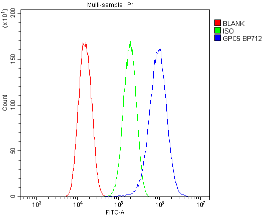
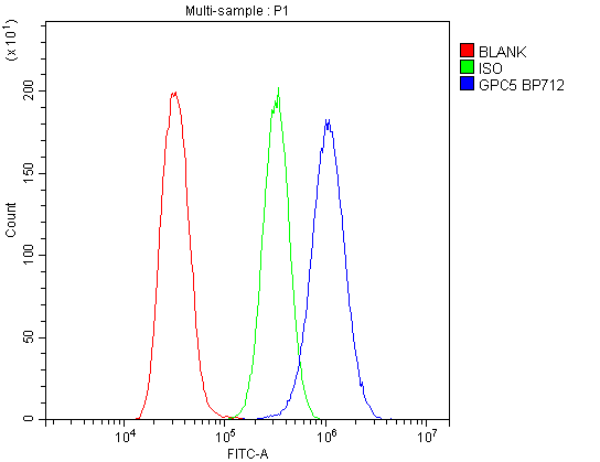
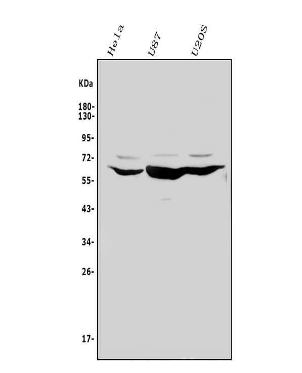
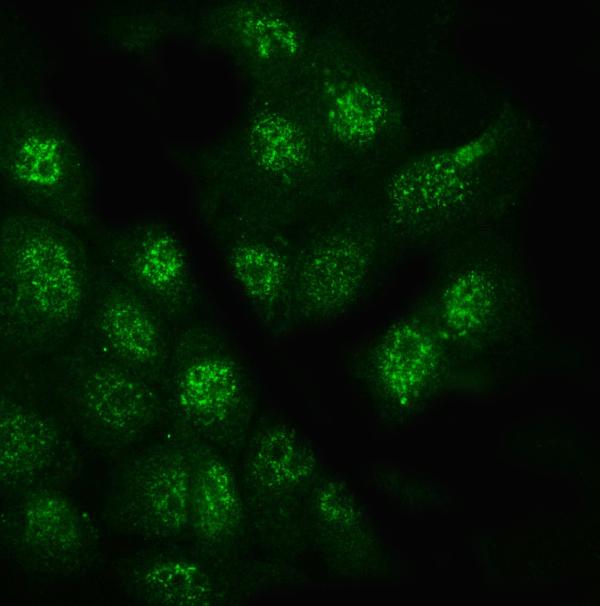
(Figure 4. IF analysis of Glypican 5/GPC5 using anti- Glypican 5/GPC5 antibody (AAA125791).Glypican 5/GPC5 was detected in immunocytochemical section of A431 cells. Enzyme antigen retrieval was performed using IHC enzyme antigen retrieval reagent for 15 mins. The cells were blocked with 10% goat serum. And then incubated with 4μg/mL rabbit anti- Glypican 5/GPC5 Antibody (AAA125791) overnight at 4 degree C. DyLight®488 Conjugated Goat Anti-Rabbit IgG was used as secondary antibody at 1:100 dilution and incubated for 30 minutes at 37 degree C. The section was counterstained with DAPI. Visualize using a fluorescence microscope and filter sets appropriate for the label used.)
IF (Immunofluorescence)
(Figure 4. IF analysis of Glypican 5/GPC5 using anti- Glypican 5/GPC5 antibody (AAA125791).Glypican 5/GPC5 was detected in immunocytochemical section of A431 cells. Enzyme antigen retrieval was performed using IHC enzyme antigen retrieval reagent for 15 mins. The cells were blocked with 10% goat serum. And then incubated with 4μg/mL rabbit anti- Glypican 5/GPC5 Antibody (AAA125791) overnight at 4 degree C. DyLight®488 Conjugated Goat Anti-Rabbit IgG was used as secondary antibody at 1:100 dilution and incubated for 30 minutes at 37 degree C. The section was counterstained with DAPI. Visualize using a fluorescence microscope and filter sets appropriate for the label used.)
Glypican 5/GPC5, Polyclonal Antibody (Cat# AAA125791)
Full Name
Anti-Glypican 5/GPC5 Antibody
Reactivity
Human
Applications
Direct ELISA, Flow Cytometry, Immunofluorescence, Immunocytochemistry, Western Blot
Purity
Immunogen affinity purified.
Pricing
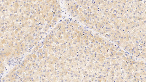
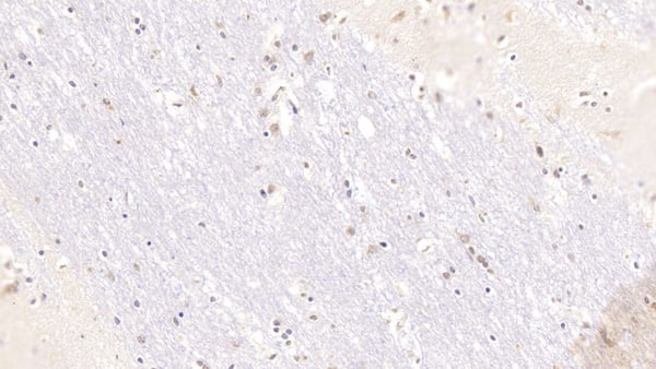 IHC (Immunohiostchemistry)
(DAB staining on IHCP;Samples: Human Cerebrum Tissue; Primary Ab: 10ug/ml Mouse AntiHuman ZPI AntibodySecond Ab: 2ug/mL HRPLinked Caprine AntiMouse IgG Polyclonal Antibody(Catalog: SAA544Mu19))
IHC (Immunohiostchemistry)
(DAB staining on IHCP;Samples: Human Cerebrum Tissue; Primary Ab: 10ug/ml Mouse AntiHuman ZPI AntibodySecond Ab: 2ug/mL HRPLinked Caprine AntiMouse IgG Polyclonal Antibody(Catalog: SAA544Mu19))
Serpin A10 (SERPINA10), Monoclonal Antibody (Cat# AAA151778)
Full Name
Monoclonal Antibody to Serpin A10 (SERPINA10)
Gene Names
SERPINA10; PZI; ZPI
Reactivity
Human, Pig
Applications
Immunoprecipitation, Immunocytochemistry, Immunohistochemistry, Western Blot
Purity
Protein A + Protein G affinity chromatography
Pricing
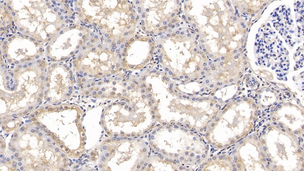
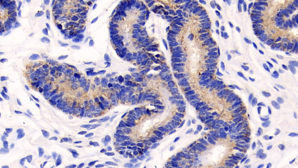
 IHC (Immunohistochemisry)
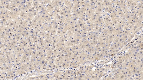
(DAB staining on IHCP;Sample: Human Liver Tissue; Primary Ab: 40ug/ml Mouse AntiHuman MMP13 AntibodySecond Ab: 2ug/mL HRPLinked Caprine AntiMouse IgG Polyclonal Antibody(Catalog: SAA544Mu19))
IHC (Immunohistochemisry)
(DAB staining on IHCP;Sample: Human Liver Tissue; Primary Ab: 40ug/ml Mouse AntiHuman MMP13 AntibodySecond Ab: 2ug/mL HRPLinked Caprine AntiMouse IgG Polyclonal Antibody(Catalog: SAA544Mu19))
Matrix Metalloproteinase 13 (MMP13), Monoclonal Antibody (Cat# AAA151476)
Full Name
Monoclonal Antibody to Matrix Metalloproteinase 13 (MMP13)
Gene Names
MMP13; CLG3; MANDP1
Reactivity
Human, Pig
Applications
Immunoprecipitation, Immunocytochemistry, Immunohistochemistry, Western Blot
Purity
Protein A + Protein G affinity chromatography
Pricing
 FCM/FACS (Flow Cytometry)
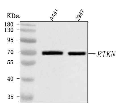
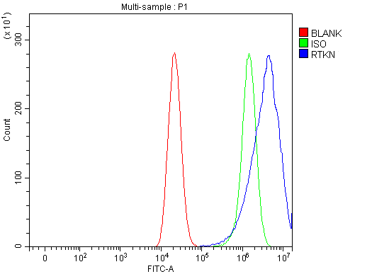
(Figure 2. Flow Cytometry analysis of CACO-2 cells using anti-RTKN antibody (AAA127459).Overlay histogram showing CACO-2 cells stained with AAA127459 (Blue line). To facilitate intracellular staining, cells were fixed with 4% paraformaldehyde and permeabilized with permeabilization buffer. The cells were blocked with 10% normal goat serum. And then incubated with rabbit anti-RTKN Antibody (AAA127459, 1ug/1x106 cells) for 30 min at 20 degree C. DyLight488 conjugated goat anti-rabbit IgG was used as secondary antibody for 30 minutes at 20 degree C. Isotype control antibody (Green line) was rabbit IgG (1ug/1x106) used under the same conditions. Unlabelled sample without incubation with primary antibody and secondary antibody (Red line) was used as a blank control.)
FCM/FACS (Flow Cytometry)
(Figure 2. Flow Cytometry analysis of CACO-2 cells using anti-RTKN antibody (AAA127459).Overlay histogram showing CACO-2 cells stained with AAA127459 (Blue line). To facilitate intracellular staining, cells were fixed with 4% paraformaldehyde and permeabilized with permeabilization buffer. The cells were blocked with 10% normal goat serum. And then incubated with rabbit anti-RTKN Antibody (AAA127459, 1ug/1x106 cells) for 30 min at 20 degree C. DyLight488 conjugated goat anti-rabbit IgG was used as secondary antibody for 30 minutes at 20 degree C. Isotype control antibody (Green line) was rabbit IgG (1ug/1x106) used under the same conditions. Unlabelled sample without incubation with primary antibody and secondary antibody (Red line) was used as a blank control.)
RTKN, Polyclonal Antibody (Cat# AAA127459)
Full Name
Anti-RTKN Antibody Picoband
Reactivity
Human
Applications
Flow Cytometry, Western Blot
Purity
Immunogen affinity purified.
Pricing
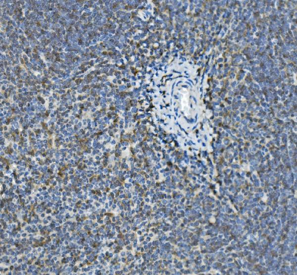
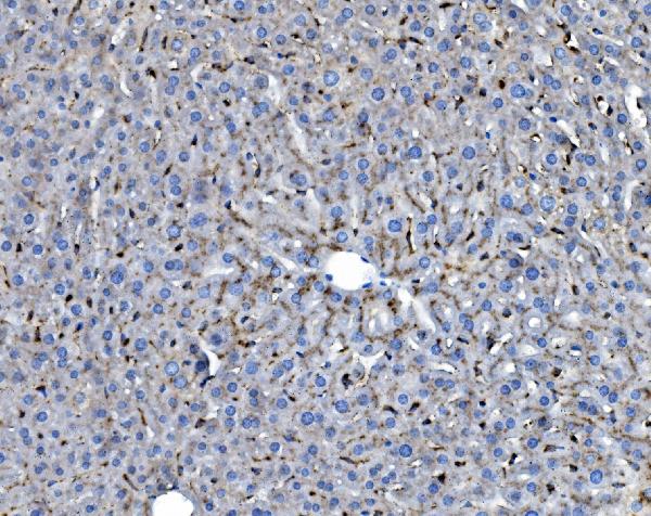

 FCM/FACS (Flow Cytometry)
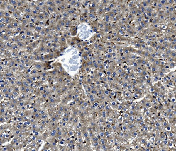
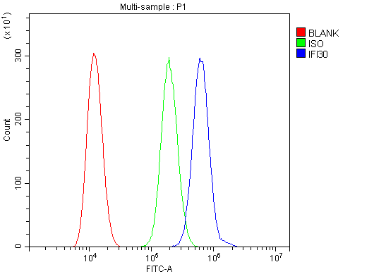
(Figure 5. Flow Cytometry analysis of HEPA1-6 cells using anti-GILT/IFI30 antibody (AAA125756).Overlay histogram showing HEPA1-6 cells stained with AAA125756 (Blue line). The cells were blocked with 10% normal goat serum. And then incubated with rabbit anti-GILT/IFI30 Antibody (AAA125756, 1μg/1x106 cells) for 30 min at 20 degree C. DyLight®488 conjugated goat anti-rabbit IgG (5-10μg/1x106 cells) was used as secondary antibody for 30 minutes at 20 degree C. Isotype control antibody (Green line) was rabbit IgG (1μg/1x106) used under the same conditions. Unlabelled sample (Red line) was also used as a control.)
FCM/FACS (Flow Cytometry)
(Figure 5. Flow Cytometry analysis of HEPA1-6 cells using anti-GILT/IFI30 antibody (AAA125756).Overlay histogram showing HEPA1-6 cells stained with AAA125756 (Blue line). The cells were blocked with 10% normal goat serum. And then incubated with rabbit anti-GILT/IFI30 Antibody (AAA125756, 1μg/1x106 cells) for 30 min at 20 degree C. DyLight®488 conjugated goat anti-rabbit IgG (5-10μg/1x106 cells) was used as secondary antibody for 30 minutes at 20 degree C. Isotype control antibody (Green line) was rabbit IgG (1μg/1x106) used under the same conditions. Unlabelled sample (Red line) was also used as a control.)
GILT/IFI30, Polyclonal Antibody (Cat# AAA125756)
Full Name
Anti-GILT/IFI30 Antibody
Gene Names
Ifi30; GILT; IP30
Reactivity
Mouse, Rat
Applications
Direct ELISA, Flow Cytometry, Immunohistochemistry, Western Blot
Purity
Immunogen affinity purified.
Pricing
 WB (Western Blot)
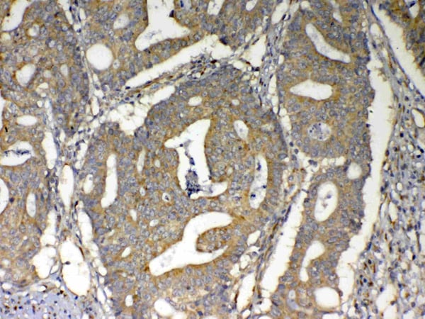
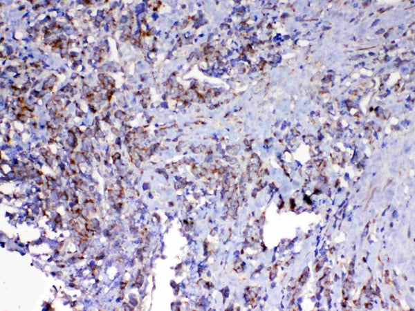
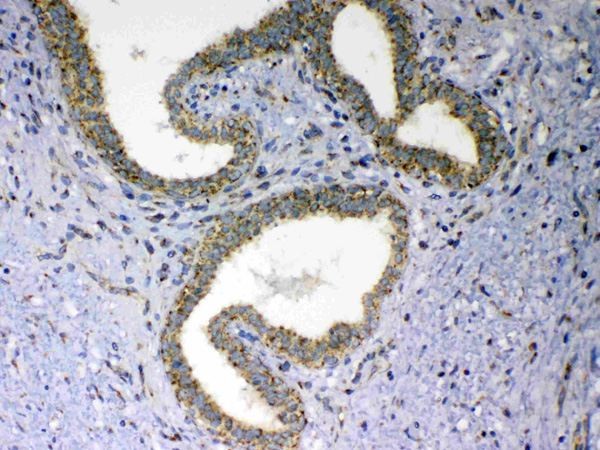
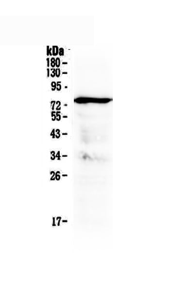
(Figure 1. Western blot analysis of MAVS using anti-MAVS antibody (AAA124553).Electrophoresis was performed on a 5-20% SDS-PAGE gel at 70V (Stacking gel) / 90V (Resolving gel) for 2-3 hours. The sample well of each lane was loaded with 50ug of sample under reducing conditions.Lane 1: human placenta tissue lysate.After Electrophoresis, proteins were transferred to a Nitrocellulose membrane at 150mA for 50-90 minutes. Blocked the membrane with 5% Non-fat Milk/ TBS for 1.5 hour at RT. The membrane was incubated with rabbit anti-MAVS antigen affinity purified polyclonal antibody at 0.5ug/mL overnight at 4 degree C, then washed with TBS-0.1%Tween 3 times with 5 minutes each and probed with a goat anti-rabbit IgG-HRP secondary antibody at a dilution of 1:10000 for 1.5 hour at RT. The signal is developed using an Enhanced Chemiluminescent detection (ECL) kit with Tanon 5200 system. A specific band was detected for MAVS at approximately 75KD. The expected band size for MAVS is at 57KD.)
WB (Western Blot)
(Figure 1. Western blot analysis of MAVS using anti-MAVS antibody (AAA124553).Electrophoresis was performed on a 5-20% SDS-PAGE gel at 70V (Stacking gel) / 90V (Resolving gel) for 2-3 hours. The sample well of each lane was loaded with 50ug of sample under reducing conditions.Lane 1: human placenta tissue lysate.After Electrophoresis, proteins were transferred to a Nitrocellulose membrane at 150mA for 50-90 minutes. Blocked the membrane with 5% Non-fat Milk/ TBS for 1.5 hour at RT. The membrane was incubated with rabbit anti-MAVS antigen affinity purified polyclonal antibody at 0.5ug/mL overnight at 4 degree C, then washed with TBS-0.1%Tween 3 times with 5 minutes each and probed with a goat anti-rabbit IgG-HRP secondary antibody at a dilution of 1:10000 for 1.5 hour at RT. The signal is developed using an Enhanced Chemiluminescent detection (ECL) kit with Tanon 5200 system. A specific band was detected for MAVS at approximately 75KD. The expected band size for MAVS is at 57KD.)
MAVS, Polyclonal Antibody (Cat# AAA124553)
Full Name
Anti-MAVS Picoband Antibody
Gene Names
MAVS; IPS1; VISA; IPS-1; CARDIF
Reactivity
Human
No cross reactivity with other proteins.
No cross reactivity with other proteins.
Applications
Western Blot, Immunohistochemistry
Purity
Immunogen affinity purified
Pricing
 FCM/FACS (Flow Cytometry)
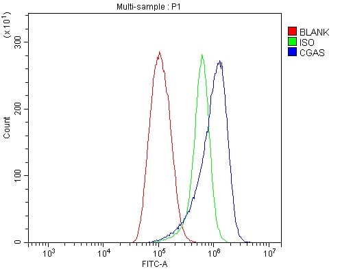
(Figure 4. Flow Cytometry analysis of U937 cells using anti-CGAS antibody (AAA125828).Overlay histogram showing U937 cells stained with AAA125828 (Blue line). The cells were blocked with 10% normal goat serum. And then incubated with rabbit anti-CGAS Antibody (AAA125828, 1μg/1x106 cells) for 30 min at 20 degree C. DyLight®488 conjugated goat anti-rabbit IgG (5-10μg/1x106 cells) was used as secondary antibody for 30 minutes at 20 degree C. Isotype control antibody (Green line) was rabbit IgG (1μg/1x106) used under the same conditions. Unlabelled sample (Red line) was also used as a control.)
FCM/FACS (Flow Cytometry)
(Figure 4. Flow Cytometry analysis of U937 cells using anti-CGAS antibody (AAA125828).Overlay histogram showing U937 cells stained with AAA125828 (Blue line). The cells were blocked with 10% normal goat serum. And then incubated with rabbit anti-CGAS Antibody (AAA125828, 1μg/1x106 cells) for 30 min at 20 degree C. DyLight®488 conjugated goat anti-rabbit IgG (5-10μg/1x106 cells) was used as secondary antibody for 30 minutes at 20 degree C. Isotype control antibody (Green line) was rabbit IgG (1μg/1x106) used under the same conditions. Unlabelled sample (Red line) was also used as a control.)
CGAS, Polyclonal Antibody (Cat# AAA125828)
Full Name
Anti-CGAS Antibody
Gene Names
MB21D1; cGAS; h-cGAS; C6orf150
Reactivity
Human
Applications
Direct ELISA, Flow Cytometry, Immunofluorescence, Immunocytochemistry, Western Blot
Purity
Immunogen affinity purified.
Pricing

 IF (Immunofluorescence)
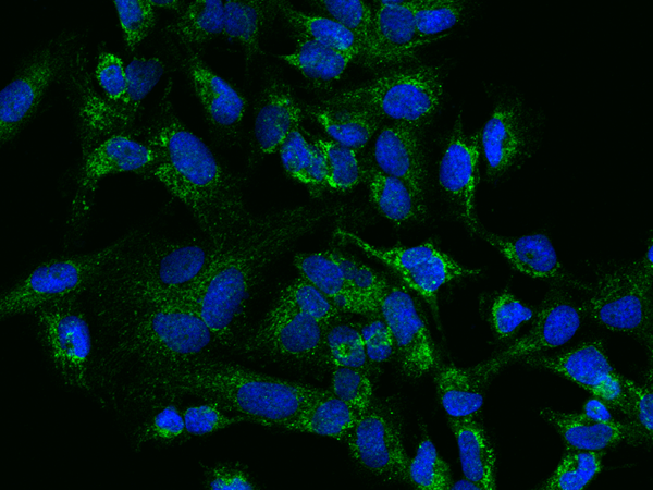
(Immunofluorescence staining of His-THEM2 in U251MG cells. Cells were fixed with 4% PFA, permeabilzed with 0.3% Triton X-100 in PBS,blocked with 10% serum, and incubated with rabbit anti-Human His-THEM2 polyclonal antibody (dilution ratio 1:100) at 4? overnight. Then cells were stained with the Alexa Fluor488-conjugated Goat Anti-rabbit IgG secondary antibody (green) and counterstained with DAPI (blue).Positive staining was localized to Nucleus and Cytoplasm.)
IF (Immunofluorescence)
(Immunofluorescence staining of His-THEM2 in U251MG cells. Cells were fixed with 4% PFA, permeabilzed with 0.3% Triton X-100 in PBS,blocked with 10% serum, and incubated with rabbit anti-Human His-THEM2 polyclonal antibody (dilution ratio 1:100) at 4? overnight. Then cells were stained with the Alexa Fluor488-conjugated Goat Anti-rabbit IgG secondary antibody (green) and counterstained with DAPI (blue).Positive staining was localized to Nucleus and Cytoplasm.)
ACOT13/THEM2, Polyclonal Antibody (Cat# AAA258924)
Full Name
Anti-ACOT13/THEM2 Antibody, Rabbit Polyclonal
Reactivity
Human
Applications
Immunofluorescence, Immunocytochemistry, Western Blot
Purity
Protein A & Antigen Affinity
ACOT13/THEM2 specific IgG was purified by Human ACOT13/THEM2 affinity chromatography.
ACOT13/THEM2 specific IgG was purified by Human ACOT13/THEM2 affinity chromatography.
Pricing
 FCM/FACS (Flow Cytometry)
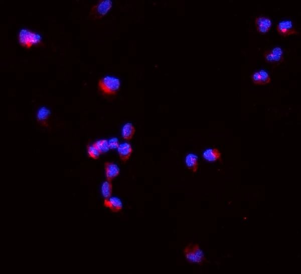
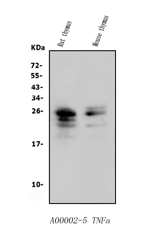
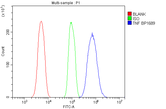
(Figure 3. Flow Cytometry analysis of Raw264. 7 cells using anti-TNF Alpha/Tnf antibody (AAA125508).Overlay histogram showing Raw264. 7 cells stained with AAA125508 (Blue line). The cells were blocked with 10% normal goat serum. And then incubated with rabbit anti-TNF Alpha/Tnf Antibody (AAA125508, 1μg/1x106 cells) for 30 min at 20 degree C. DyLight®488 conjugated goat anti-rabbit IgG (5-10μg/1x106 cells) was used as secondary antibody for 30 minutes at 20 degree C. Isotype control antibody (Green line) was rabbit IgG (1μg/1x106) used under the same conditions. Unlabelled sample (Red line) was also used as a control.)
FCM/FACS (Flow Cytometry)
(Figure 3. Flow Cytometry analysis of Raw264. 7 cells using anti-TNF Alpha/Tnf antibody (AAA125508).Overlay histogram showing Raw264. 7 cells stained with AAA125508 (Blue line). The cells were blocked with 10% normal goat serum. And then incubated with rabbit anti-TNF Alpha/Tnf Antibody (AAA125508, 1μg/1x106 cells) for 30 min at 20 degree C. DyLight®488 conjugated goat anti-rabbit IgG (5-10μg/1x106 cells) was used as secondary antibody for 30 minutes at 20 degree C. Isotype control antibody (Green line) was rabbit IgG (1μg/1x106) used under the same conditions. Unlabelled sample (Red line) was also used as a control.)
TNF alpha/Tnf, Polyclonal Antibody (Cat# AAA125508)
Full Name
Anti-TNF alpha/Tnf Antibody
Gene Names
Tnf; DIF; Tnfa; TNF-a; TNFSF2; Tnlg1f; Tnfsf1a; TNFalpha; TNF-alpha
Reactivity
Mouse, Rat
Applications
Direct ELISA, Flow Cytometry, Immunofluorescence, Immunocytochemistry, Western Blot
Purity
Immunogen affinity purified.
Pricing
 Bioactivity
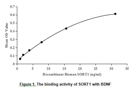
(Brain-derived neurotrophic factor, also known as BDNF, is a member of the neurotrophin family of growth factors, which are related to the canonical Nerve Growth Factor. BDNF acts on certain neurons of the central nervous system and the peripheral nervous system, helping to support the survival of existing neurons, and encourage the growth and differentiation of new neurons and synapses. Besides, Sortilin 1 (SORT1) has been identified as an interactor of BDNF, thus a binding ELISA assay was conducted to detect the interaction of recombinant human BDNF and recombinant human SORT1. Briefly, SORT1 were diluted serially in PBS with 0.01% BSA (pH 7.4). Duplicate samples of 100ul were then transferred to BDNF-coated microtiter wells and incubated for 2h at 37 degree C. Wells were washed with PBST and incubated for 1h with anti-SORT1 pAb, then aspirated and washed 3 times. After incubation with HRP labelled secondary antibody, wells were aspirated and washed 3 times. With the addition of substrate solution, wells were incubated 15-25 minutes at 37 degree C. Finally, add 50uL stop solution to the wells and read at 450/630nm immediately. The binding activity of of BDNF and SORT1 was shown in Figure 1, and this effect was in a dose dependent manner.)
Bioactivity
(Brain-derived neurotrophic factor, also known as BDNF, is a member of the neurotrophin family of growth factors, which are related to the canonical Nerve Growth Factor. BDNF acts on certain neurons of the central nervous system and the peripheral nervous system, helping to support the survival of existing neurons, and encourage the growth and differentiation of new neurons and synapses. Besides, Sortilin 1 (SORT1) has been identified as an interactor of BDNF, thus a binding ELISA assay was conducted to detect the interaction of recombinant human BDNF and recombinant human SORT1. Briefly, SORT1 were diluted serially in PBS with 0.01% BSA (pH 7.4). Duplicate samples of 100ul were then transferred to BDNF-coated microtiter wells and incubated for 2h at 37 degree C. Wells were washed with PBST and incubated for 1h with anti-SORT1 pAb, then aspirated and washed 3 times. After incubation with HRP labelled secondary antibody, wells were aspirated and washed 3 times. With the addition of substrate solution, wells were incubated 15-25 minutes at 37 degree C. Finally, add 50uL stop solution to the wells and read at 450/630nm immediately. The binding activity of of BDNF and SORT1 was shown in Figure 1, and this effect was in a dose dependent manner.)
Brain Derived Neurotrophic Factor (BDNF), Active Protein (Cat# AAA152273)
Full Name
Active Brain Derived Neurotrophic Factor (BDNF)
Gene Names
BDNF; ANON2; BULN2
Applications
Activity Assay, Cell Culture
Purity
Greater than 95% by SDS-PAGE
Pricing
 FCM/FACS (Flow Cytometry)
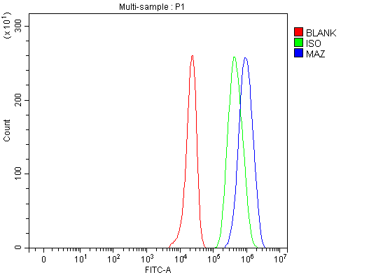
(Figure 3. Flow Cytometry analysis of HepG2 cells using anti-MAZ antibody (AAA127313).Overlay histogram showing HepG2 cells stained with AAA127313 (Blue line). To facilitate intracellular staining, cells were fixed with 4% paraformaldehyde and permeabilized with permeabilization buffer. The cells were blocked with 10% normal goat serum. And then incubated with rabbit anti-MAZ Antibody (AAA127313, 1ug/1x106 cells) for 30 min at 20 degree C. DyLight488 conjugated goat anti-rabbit IgG was used as secondary antibody for 30 minutes at 20 degree C. Isotype control antibody (Green line) was rabbit IgG (1ug/1x106) used under the same conditions. Unlabelled sample (Red line) was also used as a control.)
FCM/FACS (Flow Cytometry)
(Figure 3. Flow Cytometry analysis of HepG2 cells using anti-MAZ antibody (AAA127313).Overlay histogram showing HepG2 cells stained with AAA127313 (Blue line). To facilitate intracellular staining, cells were fixed with 4% paraformaldehyde and permeabilized with permeabilization buffer. The cells were blocked with 10% normal goat serum. And then incubated with rabbit anti-MAZ Antibody (AAA127313, 1ug/1x106 cells) for 30 min at 20 degree C. DyLight488 conjugated goat anti-rabbit IgG was used as secondary antibody for 30 minutes at 20 degree C. Isotype control antibody (Green line) was rabbit IgG (1ug/1x106) used under the same conditions. Unlabelled sample (Red line) was also used as a control.)
MAZ, Polyclonal Antibody (Cat# AAA127313)
Full Name
Anti-MAZ Antibody Picoband
Gene Names
MAZ; PUR1; ZF87; Pur-1; SAF-1; SAF-2; SAF-3; Zif87; ZNF801
Reactivity
Human
Applications
Flow Cytometry, Immunofluorescence, Immunocytochemistry, Western Blot
Purity
Immunogen affinity purified.
Pricing
 FCM/FACS (Flow Cytometry)
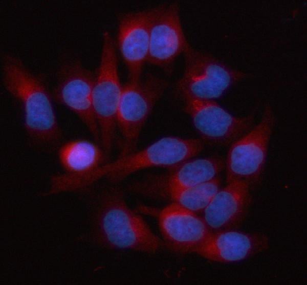
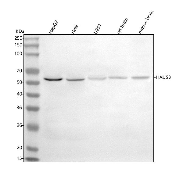
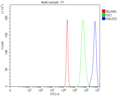
(Figure 3. Flow Cytometry analysis of HepG2 cells using anti-HAUS3 antibody (AAA127771).Overlay histogram showing HepG2 cells stained with AAA127771 (Blue line). To facilitate intracellular staining, cells were fixed with 4% paraformaldehyde and permeabilized with permeabilization buffer. The cells were blocked with 10% normal goat serum. And then incubated with rabbit anti-HAUS3 Antibody (AAA127771, 1ug/1x106 cells) for 30 min at 20 degree C. DyLight488 conjugated goat anti-rabbit IgG was used as secondary antibody for 30 minutes at 20 degree C. Isotype control antibody (Green line) was rabbit IgG (1ug/1x106) used under the same conditions. Unlabelled sample (Red line) was also used as a control.)
FCM/FACS (Flow Cytometry)
(Figure 3. Flow Cytometry analysis of HepG2 cells using anti-HAUS3 antibody (AAA127771).Overlay histogram showing HepG2 cells stained with AAA127771 (Blue line). To facilitate intracellular staining, cells were fixed with 4% paraformaldehyde and permeabilized with permeabilization buffer. The cells were blocked with 10% normal goat serum. And then incubated with rabbit anti-HAUS3 Antibody (AAA127771, 1ug/1x106 cells) for 30 min at 20 degree C. DyLight488 conjugated goat anti-rabbit IgG was used as secondary antibody for 30 minutes at 20 degree C. Isotype control antibody (Green line) was rabbit IgG (1ug/1x106) used under the same conditions. Unlabelled sample (Red line) was also used as a control.)
HAUS3, Polyclonal Antibody (Cat# AAA127771)
Full Name
Anti-HAUS3 Antibody Picoband
Gene Names
HAUS3; IT1; dgt3; C4orf15
Reactivity
Human, Mouse, Rat
Applications
Flow Cytometry, Immunofluorescence, Immunocytochemistry, Western Blot
Purity
Immunogen affinity purified.
Pricing
 FCM/FACS (Flow Cytometry)
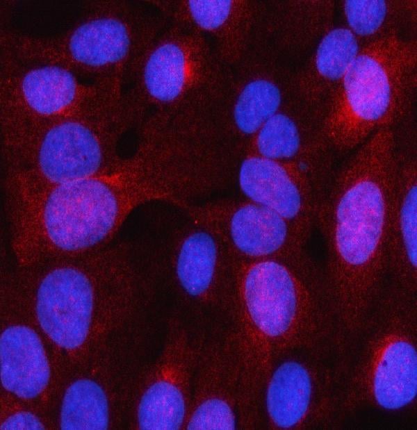
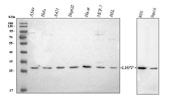
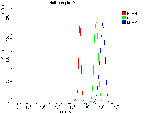
(Figure 3. Flow Cytometry analysis of JK cells using anti-LHPP antibody (AAA127669).Overlay histogram showing JK cells stained with AAA127669 (Blue line). To facilitate intracellular staining, cells were fixed with 4% paraformaldehyde and permeabilized with permeabilization buffer. The cells were blocked with 10% normal goat serum. And then incubated with rabbit anti-LHPP Antibody (AAA127669, 1ug/1x106 cells) for 30 min at 20 degree C. DyLight488 conjugated goat anti-rabbit IgG was used as secondary antibody for 30 minutes at 20 degree C. Isotype control antibody (Green line) was rabbit IgG (1ug/1x106) used under the same conditions. Unlabelled sample (Red line) was also used as a control.)
FCM/FACS (Flow Cytometry)
(Figure 3. Flow Cytometry analysis of JK cells using anti-LHPP antibody (AAA127669).Overlay histogram showing JK cells stained with AAA127669 (Blue line). To facilitate intracellular staining, cells were fixed with 4% paraformaldehyde and permeabilized with permeabilization buffer. The cells were blocked with 10% normal goat serum. And then incubated with rabbit anti-LHPP Antibody (AAA127669, 1ug/1x106 cells) for 30 min at 20 degree C. DyLight488 conjugated goat anti-rabbit IgG was used as secondary antibody for 30 minutes at 20 degree C. Isotype control antibody (Green line) was rabbit IgG (1ug/1x106) used under the same conditions. Unlabelled sample (Red line) was also used as a control.)
LHPP, Polyclonal Antibody (Cat# AAA127669)
Full Name
Anti-LHPP Antibody Picoband
Gene Names
LHPP; HDHD2B
Reactivity
Human, Mouse, Rat
Applications
Flow Cytometry, Immunofluorescence, Immunocytochemistry, Western Blot
Purity
Immunogen affinity purified.
Pricing
 FCM/FACS (Flow Cytometry)
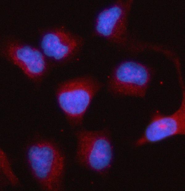
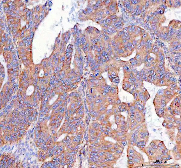
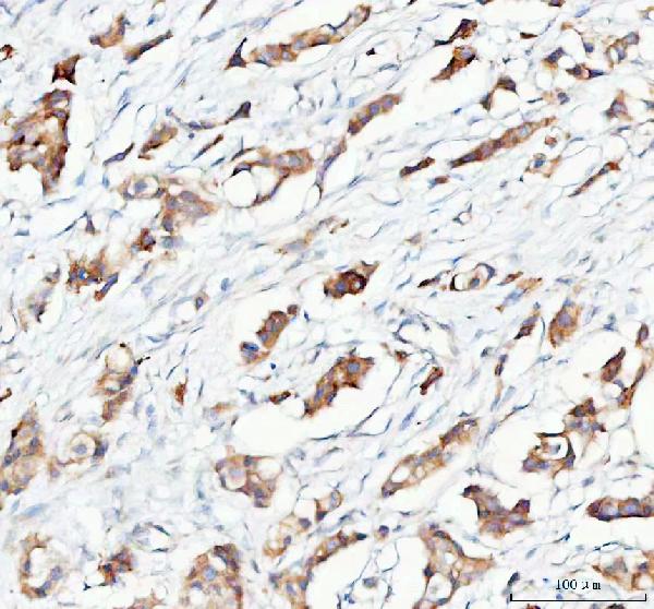
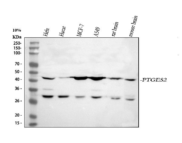
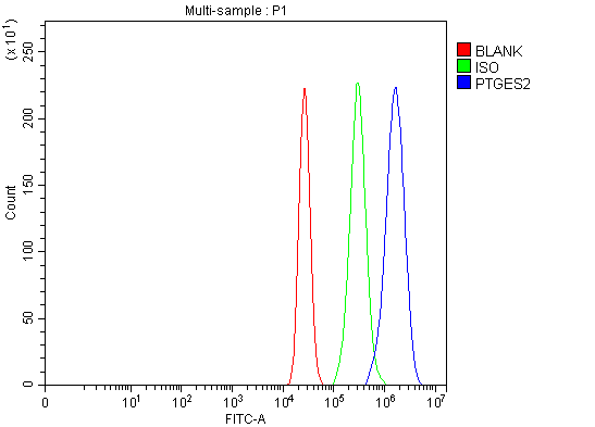
(Figure 5. Flow Cytometry analysis of MCF-7 cells using anti-PTGES2/Gbf1 antibody (AAA127397).Overlay histogram showing MCF-7 cells stained with AAA127397 (Blue line). To facilitate intracellular staining, cells were fixed with 4% paraformaldehyde and permeabilized with permeabilization buffer. The cells were blocked with 10% normal goat serum. And then incubated with rabbit anti-PTGES2/Gbf1 Antibody (AAA127397, 1ug/1x106 cells) for 30 min at 20 degree C. DyLight488 conjugated goat anti-rabbit IgG was used as secondary antibody for 30 minutes at 20 degree C. Isotype control antibody (Green line) was rabbit IgG (1ug/1x106) used under the same conditions. Unlabelled sample (Red line) was also used as a control.)
FCM/FACS (Flow Cytometry)
(Figure 5. Flow Cytometry analysis of MCF-7 cells using anti-PTGES2/Gbf1 antibody (AAA127397).Overlay histogram showing MCF-7 cells stained with AAA127397 (Blue line). To facilitate intracellular staining, cells were fixed with 4% paraformaldehyde and permeabilized with permeabilization buffer. The cells were blocked with 10% normal goat serum. And then incubated with rabbit anti-PTGES2/Gbf1 Antibody (AAA127397, 1ug/1x106 cells) for 30 min at 20 degree C. DyLight488 conjugated goat anti-rabbit IgG was used as secondary antibody for 30 minutes at 20 degree C. Isotype control antibody (Green line) was rabbit IgG (1ug/1x106) used under the same conditions. Unlabelled sample (Red line) was also used as a control.)
PTGES2/Gbf1, Polyclonal Antibody (Cat# AAA127397)
Full Name
Anti-PTGES2/Gbf1 Antibody Picoband
Gene Names
PTGES2; GBF1; GBF-1; PGES2; C9orf15; mPGES-2
Reactivity
Human, Mouse, Rat
Applications
Flow Cytometry, Western Blot, Immunocytochemistry, Immunohistochemistry, Immunofluorescence
Purity
Immunogen affinity purified.
Pricing
 FCM/FACS (Flow Cytometry)
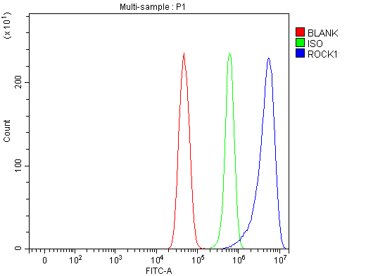
(Figure 2. Flow Cytometry analysis of U251 cells using anti-ROCK1 antibody (AAA127007).Overlay histogram showing U251 cells stained with AAA127007 (Blue line). To facilitate intracellular staining, cells were fixed with 4% paraformaldehyde and permeabilized with permeabilization buffer. The cells were blocked with 10% normal goat serum. And then incubated with rabbit anti-ROCK1 Antibody (AAA127007, 1ug/1x106 cells) for 30 min at 20 degree C. DyLight488 conjugated goat anti-rabbit IgG was used as secondary antibody for 30 minutes at 20 degree C. Isotype control antibody (Green line) was rabbit IgG (1ug/1x106) used under the same conditions. Unlabelled sample (Red line) was also used as a control.)
FCM/FACS (Flow Cytometry)
(Figure 2. Flow Cytometry analysis of U251 cells using anti-ROCK1 antibody (AAA127007).Overlay histogram showing U251 cells stained with AAA127007 (Blue line). To facilitate intracellular staining, cells were fixed with 4% paraformaldehyde and permeabilized with permeabilization buffer. The cells were blocked with 10% normal goat serum. And then incubated with rabbit anti-ROCK1 Antibody (AAA127007, 1ug/1x106 cells) for 30 min at 20 degree C. DyLight488 conjugated goat anti-rabbit IgG was used as secondary antibody for 30 minutes at 20 degree C. Isotype control antibody (Green line) was rabbit IgG (1ug/1x106) used under the same conditions. Unlabelled sample (Red line) was also used as a control.)
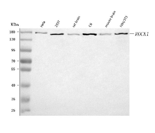
ROCK1, Polyclonal Antibody (Cat# AAA127007)
Full Name
Anti-ROCK1 Antibody Picoband
Gene Names
ROCK1; ROCK-I; P160ROCK
Reactivity
Human, Mouse, Rat
Applications
Flow Cytometry, Western Blot
Purity
Immunogen affinity purified.
Pricing
 IP (Immunoprecipitation)
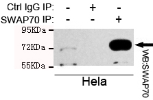
(Immunoprecipitation analysis of Hela cell lysate using SWAP70 mouse mAb.)
IP (Immunoprecipitation)
(Immunoprecipitation analysis of Hela cell lysate using SWAP70 mouse mAb.)
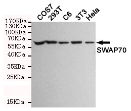
SWAP70, Monoclonal Antibody (Cat# AAA320353)
Full Name
SWAP70 Mouse mAb
Gene Names
SWAP70; HSPC321; SWAP-70
Reactivity
Human, Mouse, Rat, Monkey
Applications
Immunoprecipitation, Western Blot
Purity
The antibody was affinity-purified from mouse ascites by affinity-chromatography using epitope-specific immunogen.
Pricing
 IHC (Immunohistochemisry)
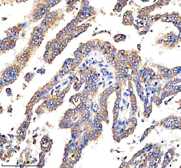
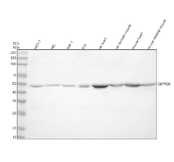
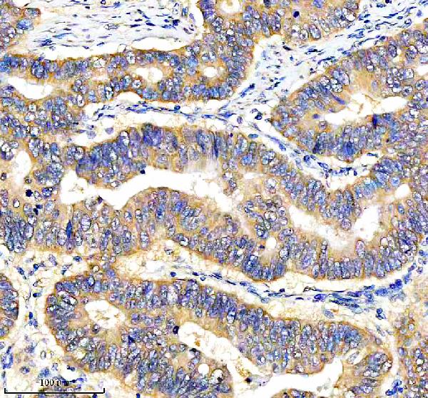
(Figure 3. IHC analysis of DEPDC6/DEPTOR using anti-DEPDC6/DEPTOR antibody (AAA128211).DEPDC6/DEPTOR was detected in a paraffin-embedded section of human stomach cancer tissue. Heat mediated antigen retrieval was performed in EDTA buffer (pH 8.0, epitope retrieval solution). The tissue section was blocked with 10% goat serum. The tissue section was then incubated with 2ug/ml rabbit anti-DEPDC6/DEPTOR Antibody (AAA128211) overnight at 4 degree C. Peroxidase Conjugated Goat Anti-rabbit IgG was used as secondary antibody and incubated for 30 minutes at 37 degree C. The tissue section was developed using HRP Conjugated Rabbit IgG Super Vision Assay Kit with DAB as the chromogen.)
IHC (Immunohistochemisry)
(Figure 3. IHC analysis of DEPDC6/DEPTOR using anti-DEPDC6/DEPTOR antibody (AAA128211).DEPDC6/DEPTOR was detected in a paraffin-embedded section of human stomach cancer tissue. Heat mediated antigen retrieval was performed in EDTA buffer (pH 8.0, epitope retrieval solution). The tissue section was blocked with 10% goat serum. The tissue section was then incubated with 2ug/ml rabbit anti-DEPDC6/DEPTOR Antibody (AAA128211) overnight at 4 degree C. Peroxidase Conjugated Goat Anti-rabbit IgG was used as secondary antibody and incubated for 30 minutes at 37 degree C. The tissue section was developed using HRP Conjugated Rabbit IgG Super Vision Assay Kit with DAB as the chromogen.)
DEPDC6/DEPTOR, Polyclonal Antibody (Cat# AAA128211)
Full Name
Anti-DEPDC6/DEPTOR Antibody Picoband
Gene Names
DEPTOR; DEP.6; DEPDC6
Reactivity
Human, Mouse, Rat
Applications
Flow Cytometry, Immunohistochemistry, Western Blot
Purity
Immunogen affinity purified.
Pricing
 FCM/FACS (Flow Cytometry)
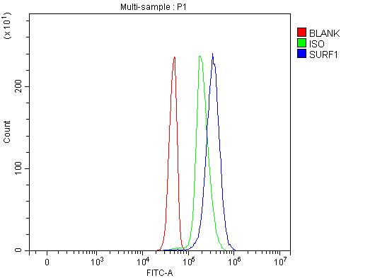
(Figure 3. Flow Cytometry analysis of Jurkat cells using anti-SURF1 antibody (AAA127193).Overlay histogram showing Jurkat cells stained with AAA127193 (Blue line). To facilitate intracellular staining, cells were fixed with 4% paraformaldehyde and permeabilized with permeabilization buffer. The cells were blocked with 10% normal goat serum. And then incubated with rabbit anti-SURF1 Antibody (AAA127193, 1ug/1x106 cells) for 30 min at 20 degree C. DyLight488 conjugated goat anti-rabbit IgG was used as secondary antibody for 30 minutes at 20 degree C. Isotype control antibody (Green line) was rabbit IgG (1ug/1x106) used under the same conditions. Unlabelled sample without incubation with primary antibody and secondary antibody (Red line) was used as a blank control.)
FCM/FACS (Flow Cytometry)
(Figure 3. Flow Cytometry analysis of Jurkat cells using anti-SURF1 antibody (AAA127193).Overlay histogram showing Jurkat cells stained with AAA127193 (Blue line). To facilitate intracellular staining, cells were fixed with 4% paraformaldehyde and permeabilized with permeabilization buffer. The cells were blocked with 10% normal goat serum. And then incubated with rabbit anti-SURF1 Antibody (AAA127193, 1ug/1x106 cells) for 30 min at 20 degree C. DyLight488 conjugated goat anti-rabbit IgG was used as secondary antibody for 30 minutes at 20 degree C. Isotype control antibody (Green line) was rabbit IgG (1ug/1x106) used under the same conditions. Unlabelled sample without incubation with primary antibody and secondary antibody (Red line) was used as a blank control.)
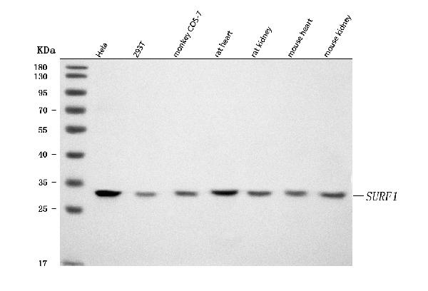
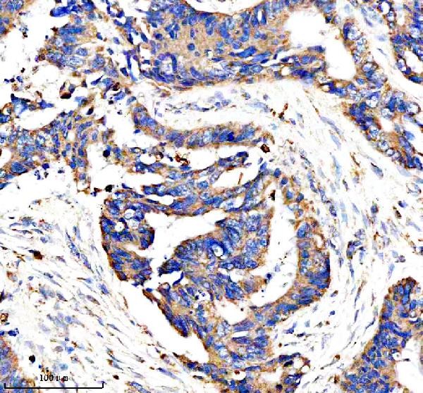
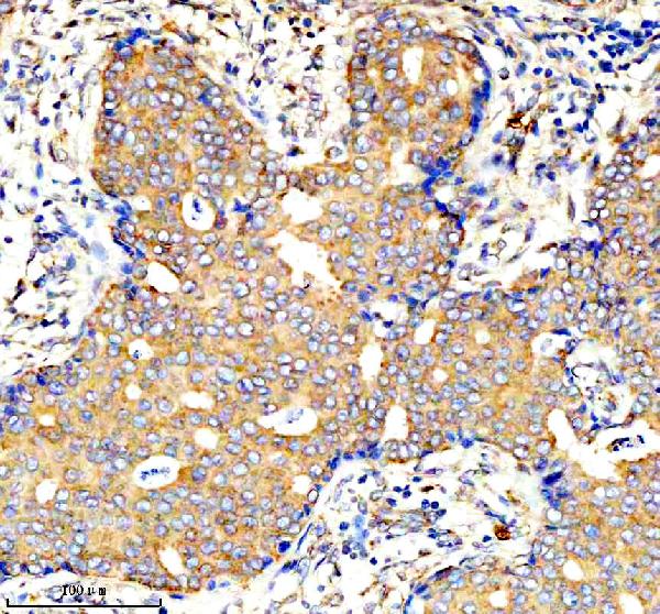
SURF1, Polyclonal Antibody (Cat# AAA127193)
Full Name
Anti-SURF1 Antibody Picoband
Reactivity
Human, Monkey, Mouse, Rat
Applications
Flow Cytometry, Immunofluorescence, Immunocytochemistry, Western Blot
Purity
Immunogen affinity purified.
Pricing
 FCM/FACS (Flow Cytometry)
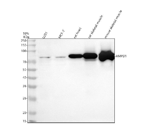
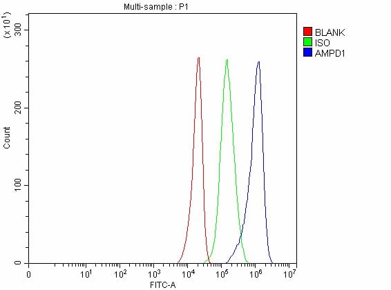
(Figure 4. Flow Cytometry analysis of U251 cells using anti-AMPD1 antibody (AAA127219).Overlay histogram showing U251 cells stained with AAA127219 (Blue line). To facilitate intracellular staining, cells were fixed with 4% paraformaldehyde and permeabilized with permeabilization buffer. The cells were blocked with 10% normal goat serum. And then incubated with rabbit anti-AMPD1 Antibody (AAA127219, 1ug/1x106 cells) for 30 min at 20 degree C. DyLight488 conjugated goat anti-rabbit IgG was used as secondary antibody for 30 minutes at 20 degree C. Isotype control antibody (Green line) was rabbit IgG (1ug/1x106) used under the same conditions. Unlabelled sample without incubation with primary antibody and secondary antibody (Red line) was used as a blank control.)
FCM/FACS (Flow Cytometry)
(Figure 4. Flow Cytometry analysis of U251 cells using anti-AMPD1 antibody (AAA127219).Overlay histogram showing U251 cells stained with AAA127219 (Blue line). To facilitate intracellular staining, cells were fixed with 4% paraformaldehyde and permeabilized with permeabilization buffer. The cells were blocked with 10% normal goat serum. And then incubated with rabbit anti-AMPD1 Antibody (AAA127219, 1ug/1x106 cells) for 30 min at 20 degree C. DyLight488 conjugated goat anti-rabbit IgG was used as secondary antibody for 30 minutes at 20 degree C. Isotype control antibody (Green line) was rabbit IgG (1ug/1x106) used under the same conditions. Unlabelled sample without incubation with primary antibody and secondary antibody (Red line) was used as a blank control.)
AMPD1, Polyclonal Antibody (Cat# AAA127219)
Full Name
Anti-AMPD1 Antibody Picoband
Gene Names
AMPD1; MAD; MADA
Reactivity
Human, Mouse, Rat
Applications
Flow Cytometry, Immunohistochemistry, Western Blot
Purity
Immunogen affinity purified.
Pricing
 FCM/FACS (Flow Cytometry)
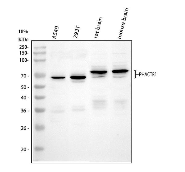
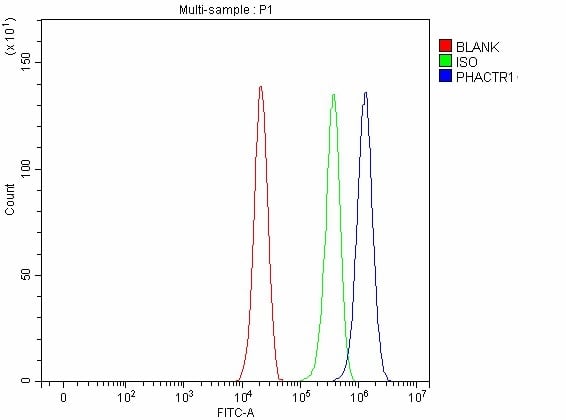
(Figure 2. Flow Cytometry analysis of A549 cells using anti-PHACTR1 antibody (AAA127359).Overlay histogram showing A549 cells stained with AAA127359 (Blue line). To facilitate intracellular staining, cells were fixed with 4% paraformaldehyde and permeabilized with permeabilization buffer. The cells were blocked with 10% normal goat serum. And then incubated with rabbit anti-PHACTR1 Antibody (AAA127359, 1ug/1x106 cells) for 30 min at 20 degree C. DyLight488 conjugated goat anti-rabbit IgG was used as secondary antibody for 30 minutes at 20 degree C. Isotype control antibody (Green line) was rabbit IgG (1ug/1x106) used under the same conditions. Unlabelled sample without incubation with primary antibody and secondary antibody (Red line) was used as a blank control.)
FCM/FACS (Flow Cytometry)
(Figure 2. Flow Cytometry analysis of A549 cells using anti-PHACTR1 antibody (AAA127359).Overlay histogram showing A549 cells stained with AAA127359 (Blue line). To facilitate intracellular staining, cells were fixed with 4% paraformaldehyde and permeabilized with permeabilization buffer. The cells were blocked with 10% normal goat serum. And then incubated with rabbit anti-PHACTR1 Antibody (AAA127359, 1ug/1x106 cells) for 30 min at 20 degree C. DyLight488 conjugated goat anti-rabbit IgG was used as secondary antibody for 30 minutes at 20 degree C. Isotype control antibody (Green line) was rabbit IgG (1ug/1x106) used under the same conditions. Unlabelled sample without incubation with primary antibody and secondary antibody (Red line) was used as a blank control.)
PHACTR1, Polyclonal Antibody (Cat# AAA127359)
Full Name
Anti-PHACTR1 Antibody Picoband
Gene Names
PHACTR1; RPEL; RPEL1; dJ257A7.2
Reactivity
Human, Mouse, Rat
Applications
Flow Cytometry, Western Blot
Purity
Immunogen affinity purified.
Pricing
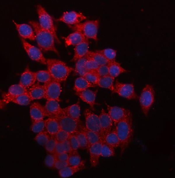
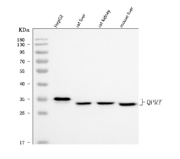
 FCM/FACS (Flow Cytometry)
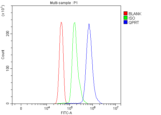
(Figure 3. Flow Cytometry analysis of Jurkat cells using anti-QPRT antibody (AAA127617).Overlay histogram showing Jurkat cells stained with AAA127617 (Blue line). To facilitate intracellular staining, cells were fixed with 4% paraformaldehyde and permeabilized with permeabilization buffer. The cells were blocked with 10% normal goat serum. And then incubated with rabbit anti-QPRT Antibody (AAA127617, 1ug/1x106 cells) for 30 min at 20 degree C. DyLight488 conjugated goat anti-rabbit IgG was used as secondary antibody for 30 minutes at 20 degree C. Isotype control antibody (Green line) was rabbit IgG (1ug/1x106) used under the same conditions. Unlabelled sample without incubation with primary antibody and secondary antibody (Red line) was used as a blank control.)
FCM/FACS (Flow Cytometry)
(Figure 3. Flow Cytometry analysis of Jurkat cells using anti-QPRT antibody (AAA127617).Overlay histogram showing Jurkat cells stained with AAA127617 (Blue line). To facilitate intracellular staining, cells were fixed with 4% paraformaldehyde and permeabilized with permeabilization buffer. The cells were blocked with 10% normal goat serum. And then incubated with rabbit anti-QPRT Antibody (AAA127617, 1ug/1x106 cells) for 30 min at 20 degree C. DyLight488 conjugated goat anti-rabbit IgG was used as secondary antibody for 30 minutes at 20 degree C. Isotype control antibody (Green line) was rabbit IgG (1ug/1x106) used under the same conditions. Unlabelled sample without incubation with primary antibody and secondary antibody (Red line) was used as a blank control.)
QPRT, Polyclonal Antibody (Cat# AAA127617)
Full Name
Anti-QPRT Antibody Picoband
Gene Names
QPRT; QPRTase; HEL-S-90n
Reactivity
Human, Mouse, Rat
Applications
Flow Cytometry, Immunofluorescence, Immunocytochemistry, Western Blot
Purity
Immunogen affinity purified.
Pricing
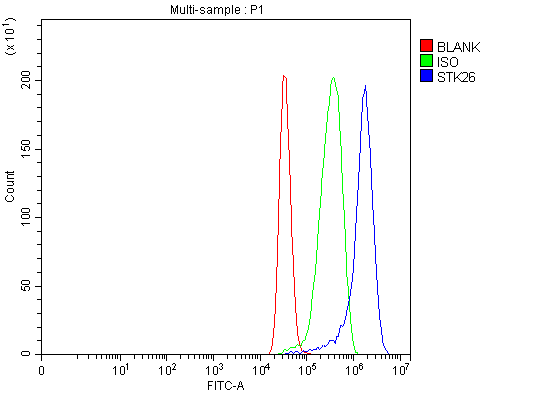 FCM/FACS (Flow Cytometry)
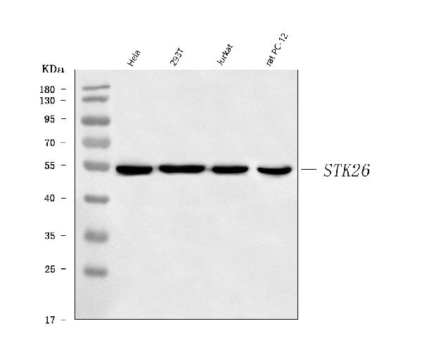
(Figure 2. Flow Cytometry analysis of MCF-7 cells using anti-MST4/STK26 antibody (AAA126554).Overlay histogram showing MCF-7 cells stained with AAA126554 (Blue line). The cells were blocked with 10% normal goat serum. And then incubated with rabbit anti-MST4/STK26 Antibody (AAA126554, 1 ug/1x10^6 cells) for 30 min at 20 degree C. DyLight488 conjugated goat anti-rabbit IgG was used as secondary antibody for 30 minutes at 20 degree C. Isotype control antibody (Green line) was rabbit IgG (1 ug/1x10^6) used under the same conditions. Unlabelled sample (Red line) was also used as a control.)
FCM/FACS (Flow Cytometry)
(Figure 2. Flow Cytometry analysis of MCF-7 cells using anti-MST4/STK26 antibody (AAA126554).Overlay histogram showing MCF-7 cells stained with AAA126554 (Blue line). The cells were blocked with 10% normal goat serum. And then incubated with rabbit anti-MST4/STK26 Antibody (AAA126554, 1 ug/1x10^6 cells) for 30 min at 20 degree C. DyLight488 conjugated goat anti-rabbit IgG was used as secondary antibody for 30 minutes at 20 degree C. Isotype control antibody (Green line) was rabbit IgG (1 ug/1x10^6) used under the same conditions. Unlabelled sample (Red line) was also used as a control.)
MST4/STK26, Polyclonal Antibody (Cat# AAA126554)
Full Name
Anti-MST4/STK26 Antibody Picoband
Gene Names
MST4; MASK
Reactivity
Human, Rat
Applications
Western Blot, Flow Cytometry
Purity
Immunogen affinity purified.
Pricing
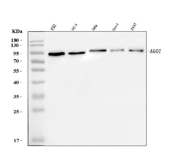
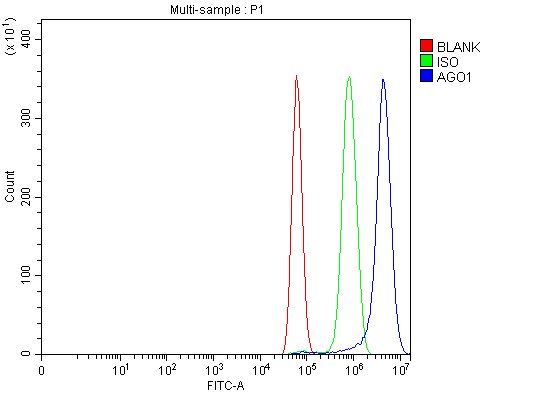 FCM/FACS (Flow Cytometry)
(Figure 2. Flow Cytometry analysis of U87 cells using anti-AGO1 antibody (AAA126085).Overlay histogram showing U87 cells stained with AAA126085 (Blue line). The cells were blocked with 10% normal goat serum. And then incubated with rabbit anti-AGO1 Antibody (AAA126085, 1 ug/1x10^6 cells) for 30 min at 20 degree C. DyLight488 conjugated goat anti-rabbit IgG was used as secondary antibody for 30 minutes at 20 degree C. Isotype control antibody (Green line) was rabbit IgG (1 ug/1x10^6) used under the same conditions. Unlabelled sample (Red line) was also used as a control.)
FCM/FACS (Flow Cytometry)
(Figure 2. Flow Cytometry analysis of U87 cells using anti-AGO1 antibody (AAA126085).Overlay histogram showing U87 cells stained with AAA126085 (Blue line). The cells were blocked with 10% normal goat serum. And then incubated with rabbit anti-AGO1 Antibody (AAA126085, 1 ug/1x10^6 cells) for 30 min at 20 degree C. DyLight488 conjugated goat anti-rabbit IgG was used as secondary antibody for 30 minutes at 20 degree C. Isotype control antibody (Green line) was rabbit IgG (1 ug/1x10^6) used under the same conditions. Unlabelled sample (Red line) was also used as a control.)
AGO1, Polyclonal Antibody (Cat# AAA126085)
Full Name
Anti-AGO1 Antibody Picoband
Gene Names
AGO1; Q99; EIF2C; EIF2C1; GERP95
Reactivity
Human
Applications
Western Blot, Flow Cytometry
Purity
Immunogen affinity purified.
Pricing
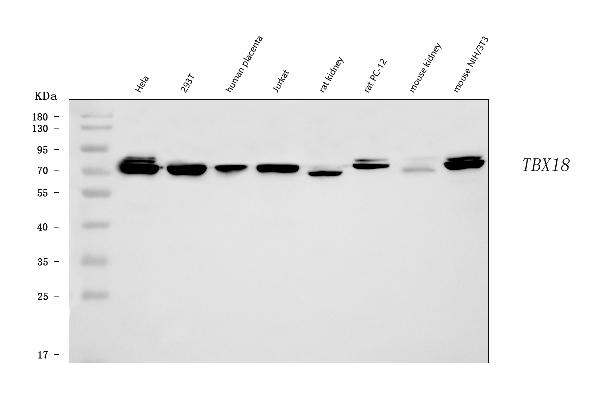
 FCM/FACS (Flow Cytometry)
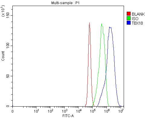
(Figure 2. Flow Cytometry analysis of U87 cells using anti-TBX18 antibody (AAA126450).Overlay histogram showing U87 cells stained with AAA126450 (Blue line). The cells were blocked with 10% normal goat serum. And then incubated with rabbit anti-TBX18 Antibody (AAA126450, 1 ug/1x10^6 cells) for 30 min at 20 degree C. DyLight488 conjugated goat anti-rabbit IgG was used as secondary antibody for 30 minutes at 20 degree C. Isotype control antibody (Green line) was rabbit IgG (1 ug/1x10^6) used under the same conditions. Unlabelled sample (Red line) was also used as a control.)
FCM/FACS (Flow Cytometry)
(Figure 2. Flow Cytometry analysis of U87 cells using anti-TBX18 antibody (AAA126450).Overlay histogram showing U87 cells stained with AAA126450 (Blue line). The cells were blocked with 10% normal goat serum. And then incubated with rabbit anti-TBX18 Antibody (AAA126450, 1 ug/1x10^6 cells) for 30 min at 20 degree C. DyLight488 conjugated goat anti-rabbit IgG was used as secondary antibody for 30 minutes at 20 degree C. Isotype control antibody (Green line) was rabbit IgG (1 ug/1x10^6) used under the same conditions. Unlabelled sample (Red line) was also used as a control.)
TBX18, Polyclonal Antibody (Cat# AAA126450)
Full Name
Anti-TBX18 Antibody Picoband
Reactivity
Human, Mouse, Rat
Applications
Flow Cytometry, Western Blot
Purity
Immunogen affinity purified.
Pricing
 IF (Immunofluorescence)
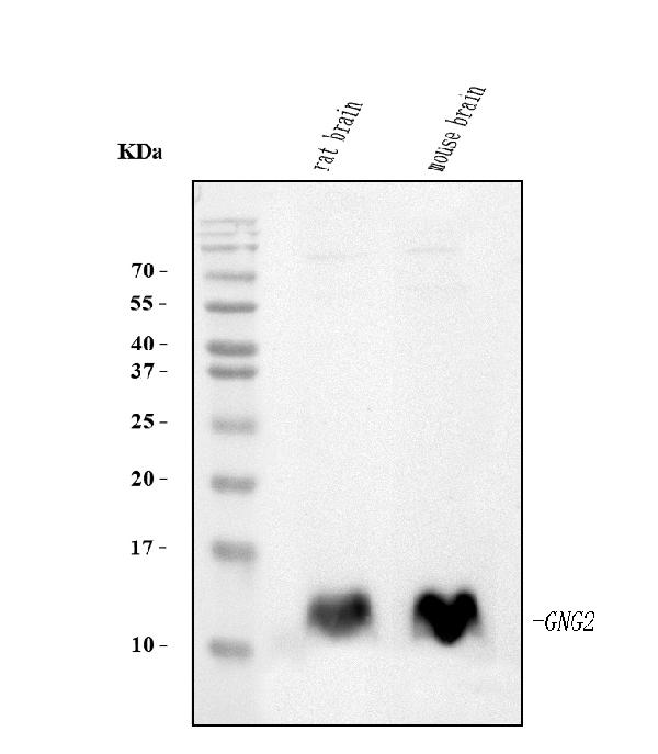
(Figure 2. IF analysis of GNG2 using anti-GNG2 antibody (AAA126953).GNG2 was detected in an immunocytochemical section of Caco-2 cells. Enzyme antigen retrieval was performed using IHC enzyme antigen retrieval reagent (AR0022) for 15 mins. The cells were blocked with 10% goat serum. And then incubated with 5 ug/mL mouse anti-GNG2 Antibody (AAA126953) overnight at 4 degree C. DyLight488 Conjugated Goat Anti-Mouse IgG was used as secondary antibody at 1:100 dilution and incubated for 30 minutes at 37 degree C. The section was counterstained with DAPI. Visualize using a fluorescence microscope and filter sets appropriate for the label used.)
IF (Immunofluorescence)
(Figure 2. IF analysis of GNG2 using anti-GNG2 antibody (AAA126953).GNG2 was detected in an immunocytochemical section of Caco-2 cells. Enzyme antigen retrieval was performed using IHC enzyme antigen retrieval reagent (AR0022) for 15 mins. The cells were blocked with 10% goat serum. And then incubated with 5 ug/mL mouse anti-GNG2 Antibody (AAA126953) overnight at 4 degree C. DyLight488 Conjugated Goat Anti-Mouse IgG was used as secondary antibody at 1:100 dilution and incubated for 30 minutes at 37 degree C. The section was counterstained with DAPI. Visualize using a fluorescence microscope and filter sets appropriate for the label used.)
GNG2, Monoclonal Antibody (Cat# AAA126953)
Full Name
Anti-GNG2 Antibody Picoband (monoclonal, 7C13)
Reactivity
Human, Mouse, Rat
Applications
Immunofluorescence, Immunocytochemistry, Western Blot
Purity
Immunogen affinity purified.
Pricing
 FCM/FACS (Flow Cytometry)
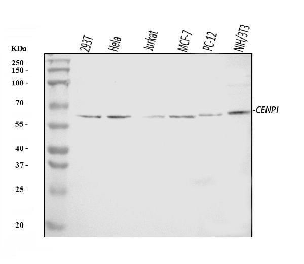
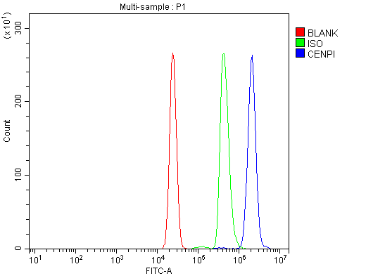
(Figure 2. Flow Cytometry analysis of U937 cells using anti-CENPI antibody (AAA126569).Overlay histogram showing U937 cells stained with AAA126569 (Blue line). The cells were blocked with 10% normal goat serum. And then incubated with rabbit anti-CENPI Antibody (AAA126569, 1 ug/1x10^6 cells) for 30 min at 20 degree C. DyLight488 conjugated goat anti-rabbit IgG was used as secondary antibody for 30 minutes at 20 degree C. Isotype control antibody (Green line) was rabbit IgG (1 ug/1x10^6) used under the same conditions. Unlabelled sample (Red line) was also used as a control.)
FCM/FACS (Flow Cytometry)
(Figure 2. Flow Cytometry analysis of U937 cells using anti-CENPI antibody (AAA126569).Overlay histogram showing U937 cells stained with AAA126569 (Blue line). The cells were blocked with 10% normal goat serum. And then incubated with rabbit anti-CENPI Antibody (AAA126569, 1 ug/1x10^6 cells) for 30 min at 20 degree C. DyLight488 conjugated goat anti-rabbit IgG was used as secondary antibody for 30 minutes at 20 degree C. Isotype control antibody (Green line) was rabbit IgG (1 ug/1x10^6) used under the same conditions. Unlabelled sample (Red line) was also used as a control.)
CENPI, Polyclonal Antibody (Cat# AAA126569)
Full Name
Anti-CENPI Antibody Picoband
Gene Names
CENPI; Mis6; LRPR1; CENP-I; FSHPRH1
Reactivity
Human, Mouse, Rat
Applications
Flow Cytometry, Western Blot
Purity
Immunogen affinity purified.
Pricing
 FCM/FACS (Flow Cytometry)
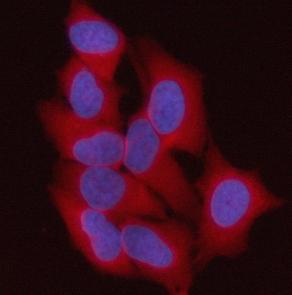
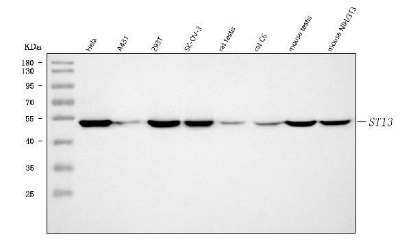
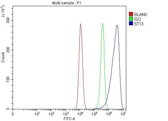
(Figure 3. Flow Cytometry analysis of HCT116 cells using anti-ST13 antibody (AAA127318).Overlay histogram showing HCT116 cells stained with AAA127318 (Blue line). To facilitate intracellular staining, cells were fixed with 4% paraformaldehyde and permeabilized with permeabilization buffer. The cells were blocked with 10% normal goat serum. And then incubated with rabbit anti-ST13 Antibody (AAA127318, 1ug/1x106 cells) for 30 min at 20 degree C. DyLight488 conjugated goat anti-rabbit IgG was used as secondary antibody for 30 minutes at 20 degree C. Isotype control antibody (Green line) was rabbit IgG (1ug/1x106) used under the same conditions. Unlabelled sample without incubation with primary antibody and secondary antibody (Red line) was used as a blank control.)
FCM/FACS (Flow Cytometry)
(Figure 3. Flow Cytometry analysis of HCT116 cells using anti-ST13 antibody (AAA127318).Overlay histogram showing HCT116 cells stained with AAA127318 (Blue line). To facilitate intracellular staining, cells were fixed with 4% paraformaldehyde and permeabilized with permeabilization buffer. The cells were blocked with 10% normal goat serum. And then incubated with rabbit anti-ST13 Antibody (AAA127318, 1ug/1x106 cells) for 30 min at 20 degree C. DyLight488 conjugated goat anti-rabbit IgG was used as secondary antibody for 30 minutes at 20 degree C. Isotype control antibody (Green line) was rabbit IgG (1ug/1x106) used under the same conditions. Unlabelled sample without incubation with primary antibody and secondary antibody (Red line) was used as a blank control.)
ST13, Polyclonal Antibody (Cat# AAA127318)
Full Name
Anti-ST13 Antibody Picoband
Gene Names
ST13; HIP; HOP; P48; AAG2; SNC6; HSPABP; FAM10A1; FAM10A4; HSPABP1; PRO0786
Reactivity
Human, Mouse, Rat
Applications
Flow Cytometry, Immunofluorescence, Immunocytochemistry, Western Blot
Purity
Immunogen affinity purified.
Pricing
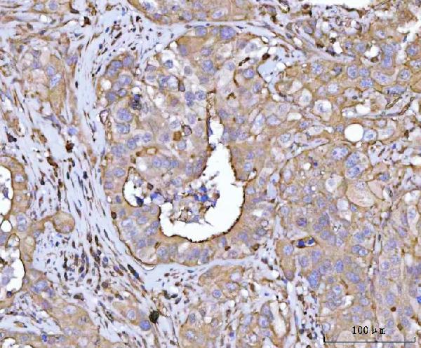
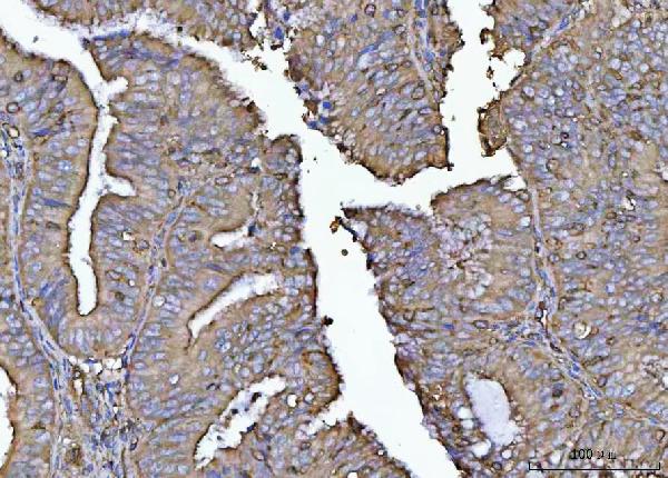
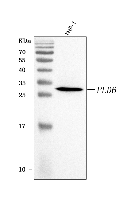
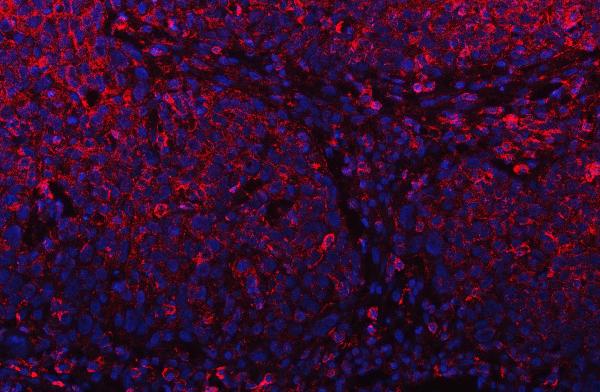 IF (Immunofluorescence)
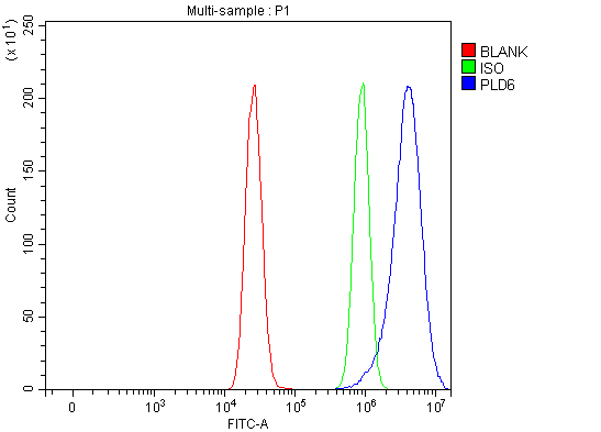
(Figure 5. IF analysis of PLD6 using anti-PLD6 antibody (AAA127683).PLD6 was detected in a paraffin-embedded section of human oesophagus squama cancer tissue. Heat mediated antigen retrieval was performed in EDTA buffer (pH 8.0, epitope retrieval solution). The tissue section was blocked with 10% goat serum. The tissue section was then incubated with 5ug/mL rabbit anti-PLD6 Antibody (AAA127683) overnight at 4 degree C. Cy3 Conjugated Goat Anti-Rabbit IgG (BA1032) was used as secondary antibody at 1:500 dilution and incubated for 30 minutes at 37 degree C. The section was counterstained with DAPI. Visualize using a fluorescence microscope and filter sets appropriate for the label used.)
IF (Immunofluorescence)
(Figure 5. IF analysis of PLD6 using anti-PLD6 antibody (AAA127683).PLD6 was detected in a paraffin-embedded section of human oesophagus squama cancer tissue. Heat mediated antigen retrieval was performed in EDTA buffer (pH 8.0, epitope retrieval solution). The tissue section was blocked with 10% goat serum. The tissue section was then incubated with 5ug/mL rabbit anti-PLD6 Antibody (AAA127683) overnight at 4 degree C. Cy3 Conjugated Goat Anti-Rabbit IgG (BA1032) was used as secondary antibody at 1:500 dilution and incubated for 30 minutes at 37 degree C. The section was counterstained with DAPI. Visualize using a fluorescence microscope and filter sets appropriate for the label used.)
PLD6, Polyclonal Antibody (Cat# AAA127683)
Full Name
Anti-PLD6 Antibody Picoband
Gene Names
PLD6; ZUC
Reactivity
Human
Applications
Western Blot, Immunohistochemistry, Immunofluorescence, Flow Cytometry
Purity
Immunogen affinity purified.
Pricing
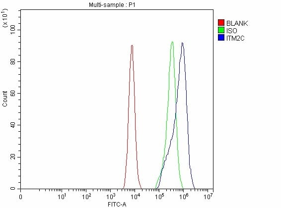
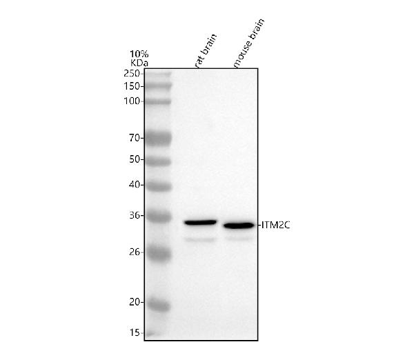
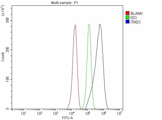 FCM/FACS (Flow Cytometry)
(Figure 3. Flow Cytometry analysis of PC-3 cells using anti-ITM2C antibody (AAA127549).Overlay histogram showing PC-3 cells stained with AAA127549 (Blue line). The cells were fixed with 4% paraformaldehyde and blocked with 10% normal goat serum. And then incubated with rabbit anti-ITM2C Antibody (AAA127549, 1ug/1x106 cells) for 30 min at 20 degree C. DyLight488 conjugated goat anti-rabbit IgG was used as secondary antibody for 30 minutes at 20 degree C. Isotype control antibody (Green line) was rabbit IgG (1ug/1x106) used under the same conditions. Unlabelled sample without incubation with primary antibody and secondary antibody (Red line) was used as a blank control.)
FCM/FACS (Flow Cytometry)
(Figure 3. Flow Cytometry analysis of PC-3 cells using anti-ITM2C antibody (AAA127549).Overlay histogram showing PC-3 cells stained with AAA127549 (Blue line). The cells were fixed with 4% paraformaldehyde and blocked with 10% normal goat serum. And then incubated with rabbit anti-ITM2C Antibody (AAA127549, 1ug/1x106 cells) for 30 min at 20 degree C. DyLight488 conjugated goat anti-rabbit IgG was used as secondary antibody for 30 minutes at 20 degree C. Isotype control antibody (Green line) was rabbit IgG (1ug/1x106) used under the same conditions. Unlabelled sample without incubation with primary antibody and secondary antibody (Red line) was used as a blank control.)
ITM2C, Polyclonal Antibody (Cat# AAA127549)
Full Name
Anti-ITM2C Antibody Picoband
Gene Names
ITM2C; E25; BRI3; E25C; ITM3; BRICD2C
Reactivity
Human, Mouse, Rat
Applications
Flow Cytometry, Western Blot
Purity
Immunogen affinity purified.
Pricing
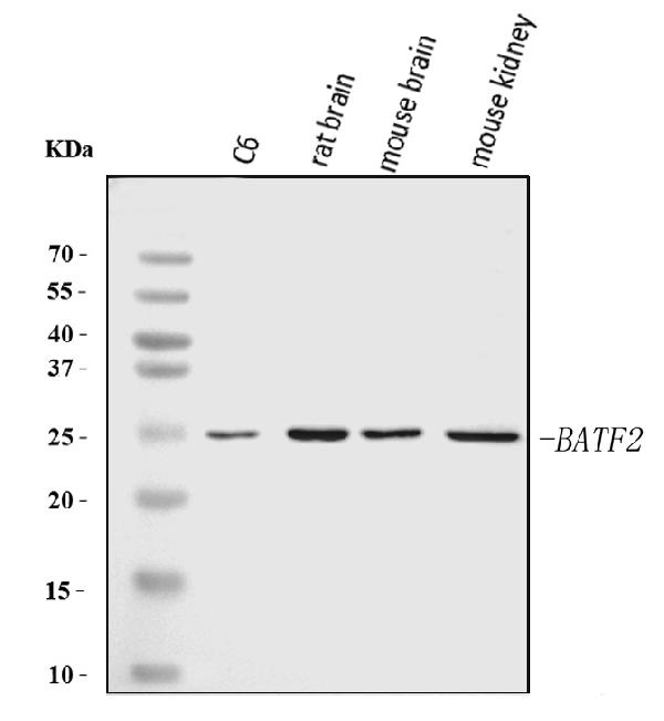
 IHC (Immunohistochemisry)
(Figure 3. IHC analysis of Batf2 using anti-Batf2 antibody (AAA126564).Batf2 was detected in a paraffin-embedded section of rat kidney tissue. Heat mediated antigen retrieval was performed in EDTA buffer (pH 8.0, epitope retrieval solution). The tissue section was blocked with 10% goat serum. The tissue section was then incubated with 2 ug/ml rabbit anti-Batf2 Antibody (AAA126564) overnight at 4 degree C. Peroxidase Conjugated Goat Anti-rabbit IgG was used as secondary antibody and incubated for 30 minutes at 37 degree C. The tissue section was developed using HRP Conjugated Rabbit IgG Super Vision Assay Kit with DAB as the chromogen.)
IHC (Immunohistochemisry)
(Figure 3. IHC analysis of Batf2 using anti-Batf2 antibody (AAA126564).Batf2 was detected in a paraffin-embedded section of rat kidney tissue. Heat mediated antigen retrieval was performed in EDTA buffer (pH 8.0, epitope retrieval solution). The tissue section was blocked with 10% goat serum. The tissue section was then incubated with 2 ug/ml rabbit anti-Batf2 Antibody (AAA126564) overnight at 4 degree C. Peroxidase Conjugated Goat Anti-rabbit IgG was used as secondary antibody and incubated for 30 minutes at 37 degree C. The tissue section was developed using HRP Conjugated Rabbit IgG Super Vision Assay Kit with DAB as the chromogen.)
Batf2, Polyclonal Antibody (Cat# AAA126564)
Full Name
Anti-Batf2 Antibody Picoband
Gene Names
Batf2; 4933430F08Rik
Reactivity
Mouse, Rat
Applications
Immunohistochemistry, Western Blot
Purity
Immunogen affinity purified.
Pricing
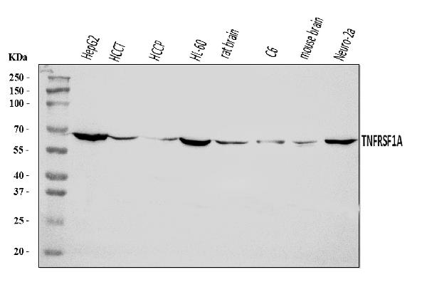
 FCM/FACS (Flow Cytometry)
(Figure 2. Flow Cytometry analysis of U937 cells using anti-TNF Receptor I/TNFRSF1A antibody (AAA126002).Overlay histogram showing U937 cells stained with AAA126002 (Blue line). The cells were blocked with 10% normal goat serum. And then incubated with rabbit anti-TNF Receptor I/TNFRSF1A Antibody (AAA126002, 1 ug/1x10^6 cells) for 30 min at 20 degree C. DyLight488 conjugated goat anti-rabbit IgG was used as secondary antibody for 30 minutes at 20 degree C. Isotype control antibody (Green line) was rabbit IgG (1 ug/1x10^6) used under the same conditions. Unlabelled sample (Red line) was also used as a control.)
FCM/FACS (Flow Cytometry)
(Figure 2. Flow Cytometry analysis of U937 cells using anti-TNF Receptor I/TNFRSF1A antibody (AAA126002).Overlay histogram showing U937 cells stained with AAA126002 (Blue line). The cells were blocked with 10% normal goat serum. And then incubated with rabbit anti-TNF Receptor I/TNFRSF1A Antibody (AAA126002, 1 ug/1x10^6 cells) for 30 min at 20 degree C. DyLight488 conjugated goat anti-rabbit IgG was used as secondary antibody for 30 minutes at 20 degree C. Isotype control antibody (Green line) was rabbit IgG (1 ug/1x10^6) used under the same conditions. Unlabelled sample (Red line) was also used as a control.)
TNF Receptor I/TNFRSF1A, Polyclonal Antibody (Cat# AAA126002)
Full Name
Anti-TNF Receptor I/TNFRSF1A Antibody Picoband
Gene Names
TNFRSF1A; FPF; MS5; p55; p60; TBP1; TNF-R; TNFAR; TNFR1; p55-R; CD120a; TNFR55; TNFR60; TNF-R-I; TNF-R55; TNFR1-d2
Reactivity
Human, Mouse, Rat
Applications
Western Blot, Flow Cytometry
Purity
Immunogen affinity purified.
Pricing
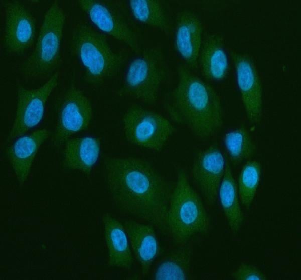
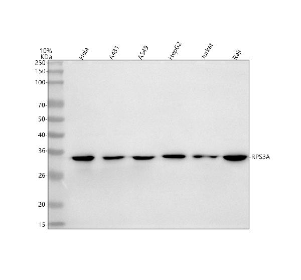
 FCM/FACS (Flow Cytometry)
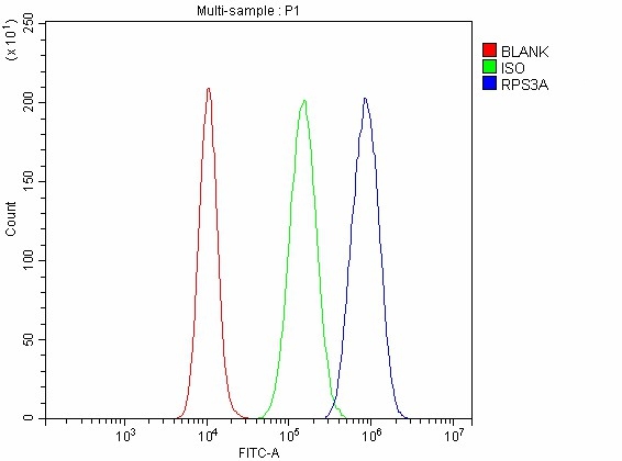
(Figure 3. Flow Cytometry analysis of A431 cells using anti-RPS3A antibody (AAA127281).Overlay histogram showing A431 cells stained with AAA127281 (Blue line). To facilitate intracellular staining, cells were fixed with 4% paraformaldehyde and permeabilized with permeabilization buffer. The cells were blocked with 10% normal goat serum. And then incubated with rabbit anti-RPS3A Antibody (AAA127281, 1ug/1x106 cells) for 30 min at 20 degree C. DyLight488 conjugated goat anti-rabbit IgG was used as secondary antibody for 30 minutes at 20 degree C. Isotype control antibody (Green line) was rabbit IgG (1ug/1x106) used under the same conditions. Unlabelled sample without incubation with primary antibody and secondary antibody (Red line) was used as a blank control.)
FCM/FACS (Flow Cytometry)
(Figure 3. Flow Cytometry analysis of A431 cells using anti-RPS3A antibody (AAA127281).Overlay histogram showing A431 cells stained with AAA127281 (Blue line). To facilitate intracellular staining, cells were fixed with 4% paraformaldehyde and permeabilized with permeabilization buffer. The cells were blocked with 10% normal goat serum. And then incubated with rabbit anti-RPS3A Antibody (AAA127281, 1ug/1x106 cells) for 30 min at 20 degree C. DyLight488 conjugated goat anti-rabbit IgG was used as secondary antibody for 30 minutes at 20 degree C. Isotype control antibody (Green line) was rabbit IgG (1ug/1x106) used under the same conditions. Unlabelled sample without incubation with primary antibody and secondary antibody (Red line) was used as a blank control.)
RPS3A, Polyclonal Antibody (Cat# AAA127281)
Full Name
Anti-RPS3A Antibody Picoband
Gene Names
RPS3A; S3A; FTE1; MFTL
Reactivity
Human, Mouse, Rat
Applications
Flow Cytometry, Western Blot
Purity
Immunogen affinity purified.
Pricing
 FCM/FACS (Flow Cytometry)
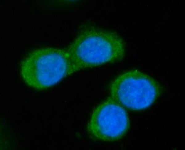
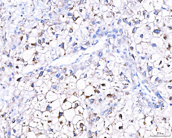
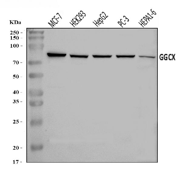
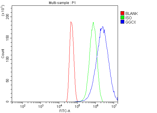
(Figure 4. Flow Cytometry analysis of U251 cells using anti-GGCX antibody (AAA126239).Overlay histogram showing U251 cells stained with AAA126239 (Blue line). The cells were blocked with 10% normal goat serum. And then incubated with rabbit anti-GGCX Antibody (AAA126239, 1 ug/1x10^6 cells) for 30 min at 20 degree C. DyLight488 conjugated goat anti-rabbit IgG was used as secondary antibody for 30 minutes at 20 degree C. Isotype control antibody (Green line) was rabbit IgG (1 ug/1x10^6) used under the same conditions. Unlabelled sample (Red line) was also used as a control.)
FCM/FACS (Flow Cytometry)
(Figure 4. Flow Cytometry analysis of U251 cells using anti-GGCX antibody (AAA126239).Overlay histogram showing U251 cells stained with AAA126239 (Blue line). The cells were blocked with 10% normal goat serum. And then incubated with rabbit anti-GGCX Antibody (AAA126239, 1 ug/1x10^6 cells) for 30 min at 20 degree C. DyLight488 conjugated goat anti-rabbit IgG was used as secondary antibody for 30 minutes at 20 degree C. Isotype control antibody (Green line) was rabbit IgG (1 ug/1x10^6) used under the same conditions. Unlabelled sample (Red line) was also used as a control.)
GGCX, Polyclonal Antibody (Cat# AAA126239)
Full Name
Anti-GGCX Antibody Picoband
Gene Names
GGCX; VKCFD1
Reactivity
Human, Mouse
Applications
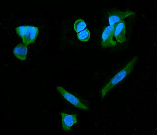
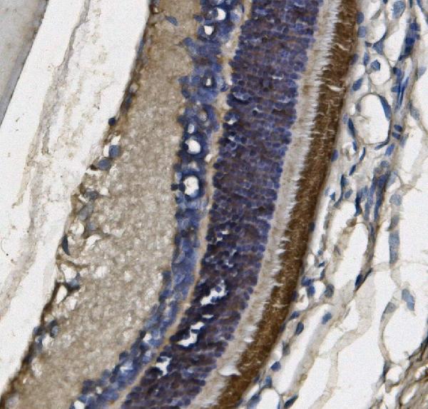
Western Blot, Immunocytochemistry, Immunohistochemistry, Immunofluorescence, Flow Cytometry
Purity
Immunogen affinity purified.
Pricing
 FCM/FACS (Flow Cytometry)
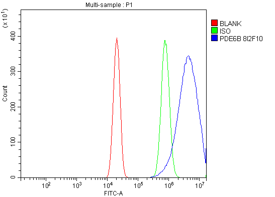
(Figure 4. Flow Cytometry analysis of U2OS cells using anti-PDE6 beta/PDE6B antibody (AAA126913).Overlay histogram showing U2OS cells stained with AAA126913 (Blue line). The cells were blocked with 10% normal goat serum. And then incubated with mouse anti-PDE6 beta/PDE6B Antibody (AAA126913, 1 ug/1x10^6 cells) for 30 min at 20 degree C. DyLight488 conjugated goat anti-mouse IgG was used as secondary antibody for 30 minutes at 20 degree C. Isotype control antibody (Green line) was mouse IgG (1 ug/1x10^6) used under the same conditions. Unlabelled sample (Red line) was also used as a control.)
FCM/FACS (Flow Cytometry)
(Figure 4. Flow Cytometry analysis of U2OS cells using anti-PDE6 beta/PDE6B antibody (AAA126913).Overlay histogram showing U2OS cells stained with AAA126913 (Blue line). The cells were blocked with 10% normal goat serum. And then incubated with mouse anti-PDE6 beta/PDE6B Antibody (AAA126913, 1 ug/1x10^6 cells) for 30 min at 20 degree C. DyLight488 conjugated goat anti-mouse IgG was used as secondary antibody for 30 minutes at 20 degree C. Isotype control antibody (Green line) was mouse IgG (1 ug/1x10^6) used under the same conditions. Unlabelled sample (Red line) was also used as a control.)
PDE6 beta/PDE6B, Monoclonal Antibody (Cat# AAA126913)
Full Name
Anti-PDE6 beta/PDE6B Antibody Picoband (monoclonal, 8I2F10)
Reactivity
Human, Mouse, Rat
Applications
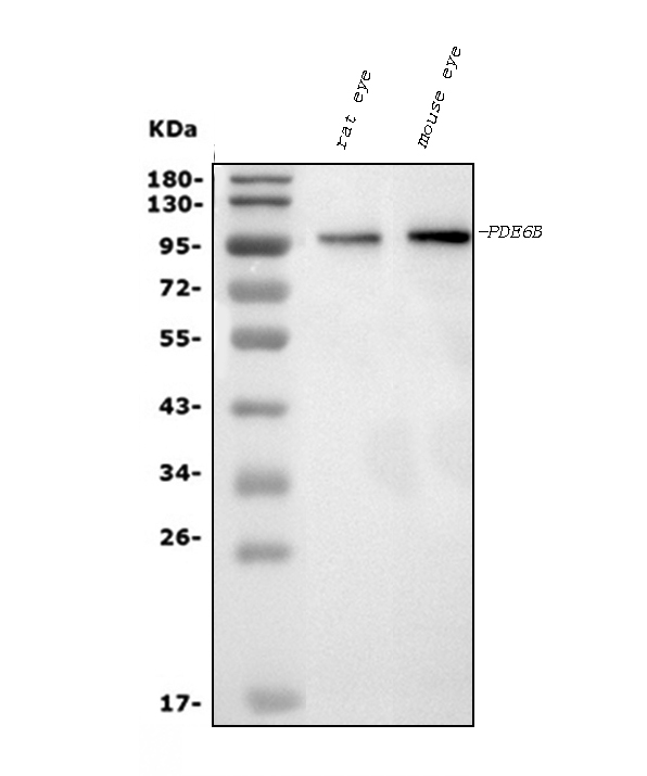
Flow Cytometry, Immunofluorescence, Immunocytochemistry, Immunohistochemistry, Western Blot
Purity
Immunogen affinity purified.
Pricing
 FCM/FACS (Flow Cytometry)
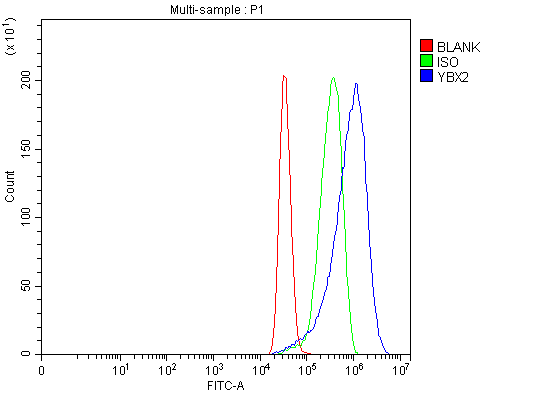
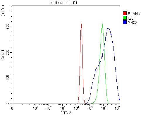
(Figure 5. Flow Cytometry analysis of U2OS cells using anti-MSY2/YBOX2/YBX2 antibody (AAA126529).Overlay histogram showing U2OS cells stained with AAA126529 (Blue line). The cells were blocked with 10% normal goat serum. And then incubated with rabbit anti-MSY2/YBOX2/YBX2 Antibody (AAA126529, 1 ug/1x10^6 cells) for 30 min at 20 degree C. DyLight488 conjugated goat anti-rabbit IgG was used as secondary antibody for 30 minutes at 20 degree C. Isotype control antibody (Green line) was rabbit IgG (1 ug/1x10^6) used under the same conditions. Unlabelled sample (Red line) was also used as a control.)
FCM/FACS (Flow Cytometry)
(Figure 5. Flow Cytometry analysis of U2OS cells using anti-MSY2/YBOX2/YBX2 antibody (AAA126529).Overlay histogram showing U2OS cells stained with AAA126529 (Blue line). The cells were blocked with 10% normal goat serum. And then incubated with rabbit anti-MSY2/YBOX2/YBX2 Antibody (AAA126529, 1 ug/1x10^6 cells) for 30 min at 20 degree C. DyLight488 conjugated goat anti-rabbit IgG was used as secondary antibody for 30 minutes at 20 degree C. Isotype control antibody (Green line) was rabbit IgG (1 ug/1x10^6) used under the same conditions. Unlabelled sample (Red line) was also used as a control.)
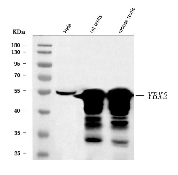
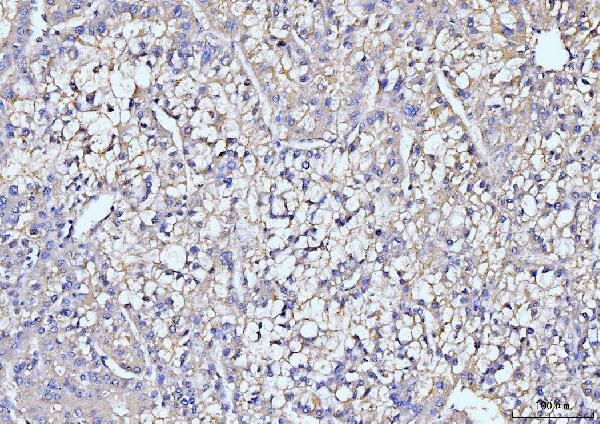
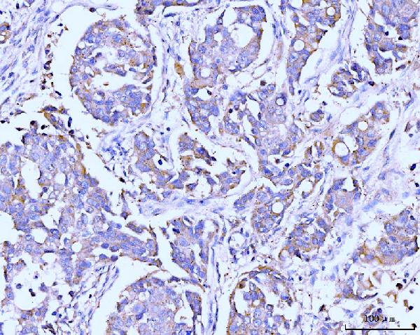
MSY2/YBOX2/YBX2, Polyclonal Antibody (Cat# AAA126529)
Full Name
Anti-MSY2/YBOX2/YBX2 Antibody Picoband
Gene Names
YBX2; DBPC; MSY2; CSDA3; CONTRIN
Reactivity
Human, Mouse, Rat
Applications
Flow Cytometry, Immunohistochemistry, Western Blot
Purity
Immunogen affinity purified.
Pricing
 FCM/FACS (Flow Cytometry)
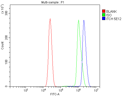
(Figure 5. Flow Cytometry analysis of U937 cells using anti-ITCH/AIP4 antibody (AAA126863).Overlay histogram showing U937 cells stained with AAA126863 (Blue line). The cells were blocked with 10% normal goat serum. And then incubated with mouse anti-ITCH/AIP4 Antibody (AAA126863, 1 ug/1x10^6 cells) for 30 min at 20 degree C. DyLight488 conjugated goat anti-mouse IgG was used as secondary antibody for 30 minutes at 20 degree C. Isotype control antibody (Green line) was mouse IgG (1 ug/1x10^6) used under the same conditions. Unlabelled sample (Red line) was also used as a control.)
FCM/FACS (Flow Cytometry)
(Figure 5. Flow Cytometry analysis of U937 cells using anti-ITCH/AIP4 antibody (AAA126863).Overlay histogram showing U937 cells stained with AAA126863 (Blue line). The cells were blocked with 10% normal goat serum. And then incubated with mouse anti-ITCH/AIP4 Antibody (AAA126863, 1 ug/1x10^6 cells) for 30 min at 20 degree C. DyLight488 conjugated goat anti-mouse IgG was used as secondary antibody for 30 minutes at 20 degree C. Isotype control antibody (Green line) was mouse IgG (1 ug/1x10^6) used under the same conditions. Unlabelled sample (Red line) was also used as a control.)
ITCH/AIP4, Monoclonal Antibody (Cat# AAA126863)
Full Name
Anti-ITCH/AIP4 Antibody Picoband (monoclonal, 5E12)
Gene Names
ITCH; AIF4; AIP4; NAPP1; dJ468O1.1
Reactivity
Human, Mouse, Rat
Applications
Flow Cytometry, Immunohistochemistry, Western Blot
Purity
Immunogen affinity purified.
Pricing
 SDS-PAGE
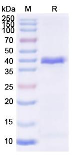
(SDS-PAGE for Recombinant EEEV Spike glycoprotein E2 Protein)
SDS-PAGE
(SDS-PAGE for Recombinant EEEV Spike glycoprotein E2 Protein)
Spike glycoprotein E2, Recombinant Protein (Cat# AAA120778)
Full Name
Recombinant EEEV Spike glycoprotein E2 Protein, N-His
Applications
Western Blot
Purity
>90% as determined by SDS-PAGE.
Pricing
 Application Data
(The purity of STAT5B protein was determined to be >95% by densitometry.Approx. MW 115kDa.)
Application Data
(The purity of STAT5B protein was determined to be >95% by densitometry.Approx. MW 115kDa.)
STAT5B, Recombinant Protein (Cat# AAA73147)
Full Name
STAT5B Protein
Gene Names
STAT5B; STAT5
Applications
SDS-PAGE
Pricing
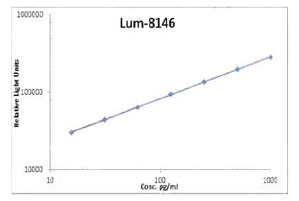 Standard Curve (Sample)
Leptin, CLIA Kit (Cat# AAA318549)
Full Name
Leptin (Human) LumiAb ELISA Kit
Gene Names
LEP; OB; OBS; LEPD
Reactivity
Human
Pricing
 SDS-PAGE
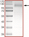
(Purity: Sample Purity Data. For specific information on a given lot, see related technical data sheet.)
SDS-PAGE
(Purity: Sample Purity Data. For specific information on a given lot, see related technical data sheet.)
COVID 19 Spike S1 (T19R, G142D, del157-158, L452R, T478K, D614G, P681R) Coronavirus, Recombinant Protein (Cat# AAA73329)
Full Name
2019-nCoV Spike protein S1 (T19R, G142D, del157-158, L452R, T478K, D614G, P681R)
Pricing
 Amino Acid Info
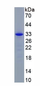
Neuropeptide Q (NPQ), Recombinant Protein (Cat# AAA153745)
Full Name
Recombinant Neuropeptide Q (NPQ)
Gene Names
SPX; C12orf39
Applications
Western Blot
Purity
Greater than 95% by SDS-PAGE
Pricing
RAB7A, Recombinant Protein (Cat# AAA38952)
Full Name
Recombinant Human RAB7A, Member RAS Oncogene Family
Gene Names
RAB7A; RAB7; PRO2706
Purity
Greater than 90% as determined by SDS-PAGE.
Pricing
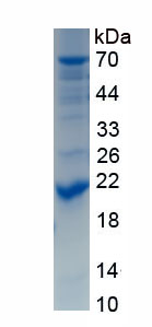
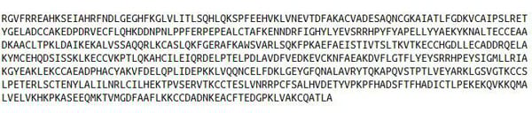 Amino Acid Info
Albumin (ALB), Recombinant Protein (Cat# AAA153418)
Full Name
Recombinant Albumin (ALB)
Applications
Western Blot
Purity
Greater than 90% by SDS-PAGE
Pricing
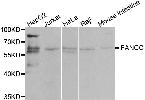
 IF (Immunofluorescence)
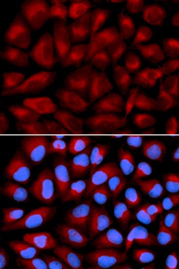
(Immunofluorescence analysis of U2OS cells using FANCC antibody. Blue: DAPI for nuclear staining.)
IF (Immunofluorescence)
(Immunofluorescence analysis of U2OS cells using FANCC antibody. Blue: DAPI for nuclear staining.)
FANCC, Polyclonal Antibody (Cat# AAA36911)
Full Name
FANCC Polyclonal Antibody
Gene Names
FANCC; FA3; FAC; FACC
Reactivity
Human, Mouse, Rat
Applications
Immunofluorescence, Immunohistochemistry, Western Blot
Purity
Affinity Purification
Pricing
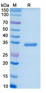 SDS-PAGE
(SDS-PAGE for Recombinant Human PI4KB protein)
SDS-PAGE
(SDS-PAGE for Recombinant Human PI4KB protein)
PI4KB, Recombinant Protein (Cat# AAA120650)
Full Name
Recombinant Human PI4KB Protein, N-His
Gene Names
PI4KB; NPIK; PI4K92; PIK4CB; PI4KBETA; PI4K-BETA; PI4KIIIBETA
Applications
Western Blot
Purity
>90% as determined by SDS-PAGE
Pricing