
Filters
Clonality
Type
Reactivity
Gene Name
Isotype
Host
Application
Clone
10000 results for "Recombinant Proteins" - showing 1500-1550




 FCM (Flow Cytometry)
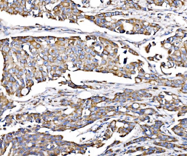
(Figure 11. Flow Cytometry analysis of U937 cells using anti-PMPCA/INPP5 antibody (AAA19611).Overlay histogram showing U937 cells stained with AAA19611 (Blue line). The cells were blocked with 10% normal goat serum. And then incubated with rabbit anti-PMPCA/INPP5 Antibody (AAA19611, 1 ug/1x10^6 cells) for 30 min at 20 degree C. DyLight488 conjugated goat anti-rabbit IgG was used as secondary antibody for 30 minutes at 20 degree C. Isotype control antibody (Green line) was rabbit IgG (1 ug/1x10^6) used under the same conditions. Unlabelled sample (Red line) was also used as a control.)
FCM (Flow Cytometry)
(Figure 11. Flow Cytometry analysis of U937 cells using anti-PMPCA/INPP5 antibody (AAA19611).Overlay histogram showing U937 cells stained with AAA19611 (Blue line). The cells were blocked with 10% normal goat serum. And then incubated with rabbit anti-PMPCA/INPP5 Antibody (AAA19611, 1 ug/1x10^6 cells) for 30 min at 20 degree C. DyLight488 conjugated goat anti-rabbit IgG was used as secondary antibody for 30 minutes at 20 degree C. Isotype control antibody (Green line) was rabbit IgG (1 ug/1x10^6) used under the same conditions. Unlabelled sample (Red line) was also used as a control.)
PMPCA/INPP5, Polyclonal Antibody (Cat# AAA19611)
Full Name
Anti-PMPCA/INPP5 Antibody Picoband
Gene Names
PMPCA; P-55; INPP5E; Alpha-MPP
Reactivity
Human, Mouse, Rat
Applications
Flow Cytometry, Immunofluorescence, Immunohistochemistry, Immunocytochemistry, Western Blot
Purity
Immunogen affinity purified.
Pricing


 IHC (Immunohistchemistry)
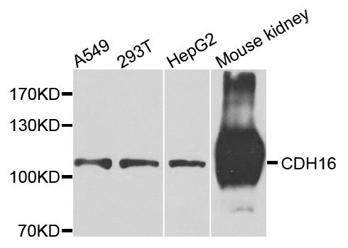
(Immunohistochemistry of paraffin-embedded mouse kidney using CDH16 antibody at dilution of 1:100 (40x lens).)
IHC (Immunohistchemistry)
(Immunohistochemistry of paraffin-embedded mouse kidney using CDH16 antibody at dilution of 1:100 (40x lens).)
CDH16, Polyclonal Antibody (Cat# AAA28211)
Full Name
CDH16 Polyclonal Antibody
Reactivity
Human, Mouse, Rat
Applications
Western Blot, Immunohistochemistry
Purity
Affinity Purification
Pricing



 IHC (Immunohistochemistry)
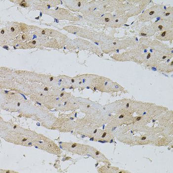
(Immunohistochemistry of paraffin-embedded mouse liver using POP4 antibody at dilution of 1:100 (40x lens).)
IHC (Immunohistochemistry)
(Immunohistochemistry of paraffin-embedded mouse liver using POP4 antibody at dilution of 1:100 (40x lens).)
POP4, Polyclonal Antibody (Cat# AAA28182)
Full Name
POP4 Polyclonal Antibody
Gene Names
POP4; RPP29
Reactivity
Human, Mouse, Rat
Applications
Western Blot
Purity
Affinity Purification
Pricing


 Application Data
(Detection limit for recombinant GST tagged RPS5 is ~3ng/ml as a capture antibody.)
Application Data
(Detection limit for recombinant GST tagged RPS5 is ~3ng/ml as a capture antibody.)
RPS5, Monoclonal Antibody (Cat# AAA14776)
Full Name
RPS5 (40S Ribosomal Protein S5)
Reactivity
Human, Mouse, Rat
Applications
Western Blot, Immunofluorescence
Purity
Affinity Purified
Purified by Protein A affinity chromatography.
Purified by Protein A affinity chromatography.
Pricing


 WB (Western Blot)
(BANF1 monoclonal antibody (M07), clone M2. Western Blot analysis of BANF1 expression in PC-12.)
WB (Western Blot)
(BANF1 monoclonal antibody (M07), clone M2. Western Blot analysis of BANF1 expression in PC-12.)
BANF1, Monoclonal Antibody (Cat# AAA26443)
Full Name
BANF1 (Barrier to Autointegration Factor 1, BAF, BCRP1, D14S1460, MGC111161) (HRP)
Gene Names
BANF1; BAF; NGPS; BCRP1; D14S1460
Applications
Immunofluorescence, Western Blot
Purity
Purified
Pricing



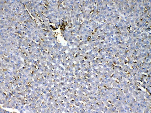 IHC (Immunohistochemistry)
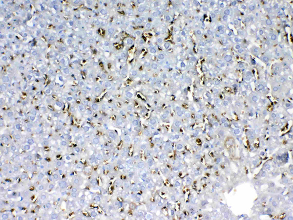
(Figure 8. IHC analysis of HBD using anti-HBD antibody (AAA19142).HBD was detected in paraffin-embedded section of rat liver tissue. Heat mediated antigen retrieval was performed in citrate buffer (pH6, epitope retrieval solution) for 20 mins. The tissue section was blocked with 10% goat serum. The tissue section was then incubated with 1ug/ml rabbit anti-HBD Antibody (AAA19142) overnight at 4 degree C. Biotinylated goat anti-rabbit IgG was used as secondary antibody and incubated for 30 minutes at 37 degree C. The tissue section was developed using Strepavidin-Biotin-Complex (SABC) with DAB as the chromogen.)
IHC (Immunohistochemistry)
(Figure 8. IHC analysis of HBD using anti-HBD antibody (AAA19142).HBD was detected in paraffin-embedded section of rat liver tissue. Heat mediated antigen retrieval was performed in citrate buffer (pH6, epitope retrieval solution) for 20 mins. The tissue section was blocked with 10% goat serum. The tissue section was then incubated with 1ug/ml rabbit anti-HBD Antibody (AAA19142) overnight at 4 degree C. Biotinylated goat anti-rabbit IgG was used as secondary antibody and incubated for 30 minutes at 37 degree C. The tissue section was developed using Strepavidin-Biotin-Complex (SABC) with DAB as the chromogen.)
HBD, Polyclonal Antibody (Cat# AAA19142)
Full Name
Anti-HBD Picoband Antibody
Reactivity
Human, Mouse, Rat
No cross reactivity with other proteins.
No cross reactivity with other proteins.
Applications
Immunohistochemistry, Western Blot
Purity
Immunogen affinity purified
Pricing


 IHC (Immunohistochemistry)
(Figure 8. IHC analysis of DDB1 using anti-DDB1 antibody (AAA11660).DDB1 was detected in frozen section of human placenta tissue. Heat mediated antigen retrieval was performed in citrate buffer (pH6, epitope retrieval solution) for 20 mins. The tissue section was blocked with 10% goat serum. The tissue section was then incubated with 1ug/ml rabbit anti-DDB1 Antibody (AAA11660) overnight at 4 degree C. Biotinylated goat anti-rabbit IgG was used as secondary antibody and incubated for 30 minutes at 37 degree C. The tissue section was developed using Strepavidin-Biotin-Complex (SABC) with DAB as the chromogen.)
IHC (Immunohistochemistry)
(Figure 8. IHC analysis of DDB1 using anti-DDB1 antibody (AAA11660).DDB1 was detected in frozen section of human placenta tissue. Heat mediated antigen retrieval was performed in citrate buffer (pH6, epitope retrieval solution) for 20 mins. The tissue section was blocked with 10% goat serum. The tissue section was then incubated with 1ug/ml rabbit anti-DDB1 Antibody (AAA11660) overnight at 4 degree C. Biotinylated goat anti-rabbit IgG was used as secondary antibody and incubated for 30 minutes at 37 degree C. The tissue section was developed using Strepavidin-Biotin-Complex (SABC) with DAB as the chromogen.)
DDB1, Polyclonal Antibody (Cat# AAA11660)
Full Name
Anti-DDB1 Antibody
Gene Names
DDB1; XPE; DDBA; XAP1; XPCE; XPE-BF; UV-DDB1
Reactivity
Human, Mouse, Rat
Applications
Western Blot, Immunohistochemistry
Purity
Immunogen Affinity Purified
Pricing


 IP (Immunoprecipitation)
(Figure 6. Immunoprecipitating RPL27 in Hela whole cell lysate .Western blot analysis of RPL27 using anti-RPL27 antibody (AAA19932).Lane 1: Hela whole cell lysates (30ug)Lane 2: Rabbit control IgG instead of anti-RPL27 antibody in Hela whole cell lysate.Lane 3: anti-RPL27 antibody (2ug) + Hela whole cell lysate (500ug)After electrophoresis, proteins were transferred to a membrane. Then the membrane was incubated with rabbit anti-RPL27 antigen affinity purified polyclonal antibody (AAA19932) at a dilution of 0.5ug/mL and probed with a goat anti-rabbit IgG-HRP secondary antibody ( cells were fixed with 4% paraformaldehyde and permeabilized with permeabilization buffer. The cells were blocked with 10% normal goat serum. And then incubated with rabbit anti-RPL27 Antibody (AAA19932, 1ug/1x106 cells) for 30 min at 20 degree C. DyLight488 conjugated goat anti-rabbit IgG was used as secondary antibody for 30 minutes at 20 degree C. Isotype control antibody (Green line) was rabbit IgG (1ug/1x106) used under the same conditions. Unlabelled sample without incubation with primary antibody and secondary antibody (Red line) was used as a blank control.)
IP (Immunoprecipitation)
(Figure 6. Immunoprecipitating RPL27 in Hela whole cell lysate .Western blot analysis of RPL27 using anti-RPL27 antibody (AAA19932).Lane 1: Hela whole cell lysates (30ug)Lane 2: Rabbit control IgG instead of anti-RPL27 antibody in Hela whole cell lysate.Lane 3: anti-RPL27 antibody (2ug) + Hela whole cell lysate (500ug)After electrophoresis, proteins were transferred to a membrane. Then the membrane was incubated with rabbit anti-RPL27 antigen affinity purified polyclonal antibody (AAA19932) at a dilution of 0.5ug/mL and probed with a goat anti-rabbit IgG-HRP secondary antibody ( cells were fixed with 4% paraformaldehyde and permeabilized with permeabilization buffer. The cells were blocked with 10% normal goat serum. And then incubated with rabbit anti-RPL27 Antibody (AAA19932, 1ug/1x106 cells) for 30 min at 20 degree C. DyLight488 conjugated goat anti-rabbit IgG was used as secondary antibody for 30 minutes at 20 degree C. Isotype control antibody (Green line) was rabbit IgG (1ug/1x106) used under the same conditions. Unlabelled sample without incubation with primary antibody and secondary antibody (Red line) was used as a blank control.)
RPL27, Polyclonal Antibody (Cat# AAA19932)
Full Name
Anti-RPL27 Antibody Picoband
Gene Names
RPL27; L27
Reactivity
Human, Mouse, Rat
Applications
Western Blot, Immunoprecipitation, Flow Cytometry
Purity
Immunogen affinity purified.
Pricing


 FCM (Flow Cytometry)
(Figure 10. Flow Cytometry analysis of NIH/3T3 cells using anti-Hcn2 antibody (AAA19781).Overlay histogram showing NIH/3T3 cells stained with AAA19781 (Blue line). The cells were fixed with 4% paraformaldehyde and blocked with 10% normal goat serum. And then incubated with rabbit anti-Hcn2 Antibody (AAA19781, 1ug/1x106 cells) for 30 min at 20 degree C. DyLight488 conjugated goat anti-rabbit IgG was used as secondary antibody for 30 minutes at 20 degree C. Isotype control antibody (Green line) was rabbit IgG (1ug/1x106) used under the same conditions. Unlabelled sample (Red line) was also used as a control.)
FCM (Flow Cytometry)
(Figure 10. Flow Cytometry analysis of NIH/3T3 cells using anti-Hcn2 antibody (AAA19781).Overlay histogram showing NIH/3T3 cells stained with AAA19781 (Blue line). The cells were fixed with 4% paraformaldehyde and blocked with 10% normal goat serum. And then incubated with rabbit anti-Hcn2 Antibody (AAA19781, 1ug/1x106 cells) for 30 min at 20 degree C. DyLight488 conjugated goat anti-rabbit IgG was used as secondary antibody for 30 minutes at 20 degree C. Isotype control antibody (Green line) was rabbit IgG (1ug/1x106) used under the same conditions. Unlabelled sample (Red line) was also used as a control.)
Hcn2, Polyclonal Antibody (Cat# AAA19781)
Full Name
Anti-Hcn2 Antibody Picoband
Gene Names
Hcn2; HAC1; trls; BCNG2
Reactivity
Mouse, Rat
Applications
Western Blot, Immunohistochemistry, Flow Cytometry
Purity
Immunogen affinity purified.
Pricing
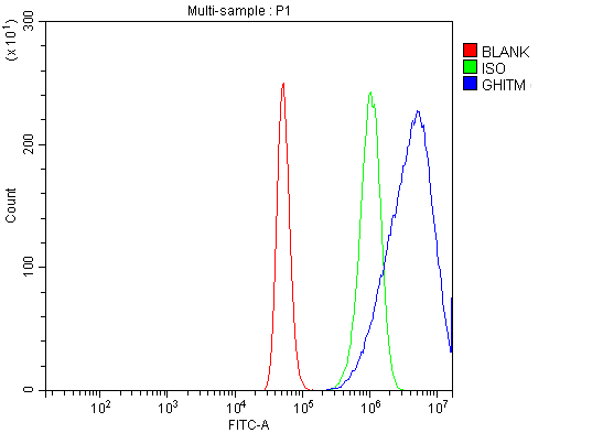


 IF (Immunofluorescence)
(Figure 6. IF analysis of GHITM using anti-GHITM antibody (AAA19621).GHITM was detected in an immunocytochemical section of HEP3B cells. Enzyme antigen retrieval was performed using IHC enzyme antigen retrieval reagent (AR0022) for 15 mins. The cells were blocked with 10% goat serum. And then incubated with 5 ug/mL rabbit anti-GHITM Antibody (AAA19621) overnight at 4 degree C. DyLight488 Conjugated Goat Anti-Rabbit IgG was used as secondary antibody at 1:100 dilution and incubated for 30 minutes at 37 degree C. The section was counterstained with DAPI. Visualize using a fluorescence microscope and filter sets appropriate for the label used.)
IF (Immunofluorescence)
(Figure 6. IF analysis of GHITM using anti-GHITM antibody (AAA19621).GHITM was detected in an immunocytochemical section of HEP3B cells. Enzyme antigen retrieval was performed using IHC enzyme antigen retrieval reagent (AR0022) for 15 mins. The cells were blocked with 10% goat serum. And then incubated with 5 ug/mL rabbit anti-GHITM Antibody (AAA19621) overnight at 4 degree C. DyLight488 Conjugated Goat Anti-Rabbit IgG was used as secondary antibody at 1:100 dilution and incubated for 30 minutes at 37 degree C. The section was counterstained with DAPI. Visualize using a fluorescence microscope and filter sets appropriate for the label used.)
GHITM, Polyclonal Antibody (Cat# AAA19621)
Full Name
Anti-GHITM Antibody Picoband
Gene Names
GHITM; DERP2; MICS1; My021; PTD010; TMBIM5; HSPC282
Reactivity
Human, Mouse, Rat
Applications
Western Blot, Immunohistochemistry, Immunocytochemistry, Immunofluorescence, Flow Cytometry
Purity
Immunogen affinity purified.
Pricing


 WB (Western Blot)
(SAV1 monoclonal antibody (M02), Western Blot analysis of SAV1 expression in Hela NE.)
WB (Western Blot)
(SAV1 monoclonal antibody (M02), Western Blot analysis of SAV1 expression in Hela NE.)
SAV1, Monoclonal Antibody (Cat# AAA24945)
Full Name
SAV1 (Protein Salvador Homolog 1, SAV, 45kD WW Domain Protein, WW45, hWW45, WWP4) (Biotin)
Gene Names
SAV1; SAV; WW45; WWP4
Reactivity
Human
Applications
Immunofluorescence, Immunoprecipitation, Western Blot
Purity
Purified by Protein A Affinity Chromatography.
Pricing


 IF (Immunofluorescence)
(Immunofluorescence analysis of mouse pancreas using PNLIPRP1 antibody at dilution of 1:100. Blue: DAPI for nuclear staining.)
IF (Immunofluorescence)
(Immunofluorescence analysis of mouse pancreas using PNLIPRP1 antibody at dilution of 1:100. Blue: DAPI for nuclear staining.)
PNLIPRP1, Polyclonal Antibody (Cat# AAA28353)
Full Name
PNLIPRP1 Rabbit pAb
Gene Names
PNLIPRP1; PLRP1
Reactivity
Human, Mouse, Rat
Applications
Western Blot, Immunohistochemistry, Immunofluorescence
Purity
Affinity purification
Pricing


 WB (Western Blot)
(DNAJC10 monoclonal antibody. Western Blot analysis of DNAJC10 expression in HeLa.)
WB (Western Blot)
(DNAJC10 monoclonal antibody. Western Blot analysis of DNAJC10 expression in HeLa.)
DNAJC10, Monoclonal Antibody (Cat# AAA24489)
Full Name
DNAJC10 (DnaJ Homolog Subfamily C Member 10, ER-resident Protein ERdj5, Macrothioredoxin, MTHr, ERDJ5, UNQ495/PRO1012, DKFZp434J1813, MGC104194) APC
Gene Names
DNAJC10; JPDI; MTHr; ERdj5; PDIA19
Reactivity
Human, Mouse, Rat
Applications
Immunofluorescence, Immunohistochemistry, Western Blot
Purity
Purified by Protein A Affinity Chromatography.
Pricing


 WB (Western Blot)
(SMAD3 monoclonal antibody (M01), clone 2C12. Western Blot analysis of SMAD3 expression in Hs 181.Tes.)
WB (Western Blot)
(SMAD3 monoclonal antibody (M01), clone 2C12. Western Blot analysis of SMAD3 expression in Hs 181.Tes.)
SMAD3, Monoclonal Antibody (Cat# AAA26562)
Full Name
SMAD3 (SMAD Family Member 3, DKFZp586N0721, DKFZp686J10186, HSPC193, HsT17436, JV15-2, MADH3, MGC60396) (PE)
Gene Names
SMAD3; LDS3; LDS1C; MADH3; JV15-2; HSPC193; HsT17436
Applications
Immunohistochemistry, Western Blot
Purity
Purified
Pricing


 FCM (Flow Cytometry)
(Figure 6. Flow Cytometry analysis of HCT116 cells using anti-SRPRB antibody (AAA19985).Overlay histogram showing HCT116 cells stained with AAA19985 (Blue line). To facilitate intracellular staining, cells were fixed with 4% paraformaldehyde and permeabilized with permeabilization buffer. The cells were blocked with 10% normal goat serum. And then incubated with rabbit anti-SRPRB Antibody (AAA19985, 1ug/1x106 cells) for 30 min at 20 degree C. DyLight488 conjugated goat anti-rabbit IgG was used as secondary antibody for 30 minutes at 20 degree C. Isotype control antibody (Green line) was rabbit IgG (1ug/1x106) used under the same conditions. Unlabelled sample without incubation with primary antibody and secondary antibody (Red line) was used as a blank control.)
FCM (Flow Cytometry)
(Figure 6. Flow Cytometry analysis of HCT116 cells using anti-SRPRB antibody (AAA19985).Overlay histogram showing HCT116 cells stained with AAA19985 (Blue line). To facilitate intracellular staining, cells were fixed with 4% paraformaldehyde and permeabilized with permeabilization buffer. The cells were blocked with 10% normal goat serum. And then incubated with rabbit anti-SRPRB Antibody (AAA19985, 1ug/1x106 cells) for 30 min at 20 degree C. DyLight488 conjugated goat anti-rabbit IgG was used as secondary antibody for 30 minutes at 20 degree C. Isotype control antibody (Green line) was rabbit IgG (1ug/1x106) used under the same conditions. Unlabelled sample without incubation with primary antibody and secondary antibody (Red line) was used as a blank control.)
SRPRB, Polyclonal Antibody (Cat# AAA19985)
Full Name
Anti-SRPRB Antibody Picoband
Gene Names
SRPRB; APMCF1
Reactivity
Human
Applications
Western Blot, Immunohistochemistry, Immunocytochemistry, Immunofluorescence, Flow Cytometry
Purity
Immunogen affinity purified.
Pricing

 FCM (Flow Cytometry)
(Figure 9. Flow Cytometry analysis of MCF-7 cells using anti-MIPEP antibody (AAA19856).Overlay histogram showing MCF-7 cells stained with AAA19856 (Blue line). To facilitate intracellular staining, cells were fixed with 4% paraformaldehyde and permeabilized with permeabilization buffer. The cells were blocked with 10% normal goat serum. And then incubated with rabbit anti-MIPEP Antibody (AAA19856, 1ug/1x106 cells) for 30 min at 20 degree C. DyLight488 conjugated goat anti-rabbit IgG was used as secondary antibody for 30 minutes at 20 degree C. Isotype control antibody (Green line) was rabbit IgG (1ug/1x106) used under the same conditions. Unlabelled sample (Red line) was also used as a control.)
FCM (Flow Cytometry)
(Figure 9. Flow Cytometry analysis of MCF-7 cells using anti-MIPEP antibody (AAA19856).Overlay histogram showing MCF-7 cells stained with AAA19856 (Blue line). To facilitate intracellular staining, cells were fixed with 4% paraformaldehyde and permeabilized with permeabilization buffer. The cells were blocked with 10% normal goat serum. And then incubated with rabbit anti-MIPEP Antibody (AAA19856, 1ug/1x106 cells) for 30 min at 20 degree C. DyLight488 conjugated goat anti-rabbit IgG was used as secondary antibody for 30 minutes at 20 degree C. Isotype control antibody (Green line) was rabbit IgG (1ug/1x106) used under the same conditions. Unlabelled sample (Red line) was also used as a control.)
MIPEP, Polyclonal Antibody (Cat# AAA19856)
Full Name
Anti-MIPEP Antibody Picoband
Gene Names
MIPEP; MIP; HMIP
Reactivity
Human, Mouse, Rat
Applications
Western Blot, Immunohistochemistry, Flow Cytometry
Purity
Immunogen affinity purified.
Pricing
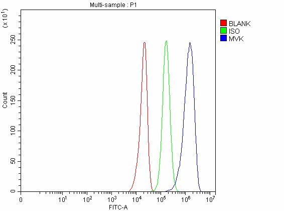

 WB (Western Blot)
(Figure 6. Western blot analysis of MVK using anti-MVK antibody (AAA19776).Electrophoresis was performed on a 5-20% SDS-PAGE gel at 70V (Stacking gel)/90V (Resolving gel) for 2-3 hours. The sample well of each lane was loaded with 30 ug of sample under reducing conditions.Lane 1: human HepG2 whole cell lysates,Lane 2: human MCF-7 whole cell lysates.After electrophoresis, proteins were transferred to a nitrocellulose membrane at 150 mA for 50-90 minutes. Blocked the membrane with 5% non-fat milk/TBS for 1.5 hour at RT. The membrane was incubated with rabbit anti-MVK antigen affinity purified polyclonal antibody (AAA19776) at 0.5ug/mL overnight at 4 degree C, then washed with TBS-0.1%Tween 3 times with 5 minutes each and probed with a goat anti-rabbit IgG-DyLight 647 Conjugated secondary antibody at a dilution of 1:2000 for 1.5 hour at RT. A specific band was detected for MVK at approximately 42 kDa. The expected band size for MVK is at 42 kDa.)
WB (Western Blot)
(Figure 6. Western blot analysis of MVK using anti-MVK antibody (AAA19776).Electrophoresis was performed on a 5-20% SDS-PAGE gel at 70V (Stacking gel)/90V (Resolving gel) for 2-3 hours. The sample well of each lane was loaded with 30 ug of sample under reducing conditions.Lane 1: human HepG2 whole cell lysates,Lane 2: human MCF-7 whole cell lysates.After electrophoresis, proteins were transferred to a nitrocellulose membrane at 150 mA for 50-90 minutes. Blocked the membrane with 5% non-fat milk/TBS for 1.5 hour at RT. The membrane was incubated with rabbit anti-MVK antigen affinity purified polyclonal antibody (AAA19776) at 0.5ug/mL overnight at 4 degree C, then washed with TBS-0.1%Tween 3 times with 5 minutes each and probed with a goat anti-rabbit IgG-DyLight 647 Conjugated secondary antibody at a dilution of 1:2000 for 1.5 hour at RT. A specific band was detected for MVK at approximately 42 kDa. The expected band size for MVK is at 42 kDa.)
MVK, Polyclonal Antibody (Cat# AAA19776)
Full Name
Anti-MVK Antibody Picoband
Gene Names
MVK; MK; LRBP; MVLK; POROK3
Reactivity
Human
Applications
Western Blot, Immunohistochemistry, Immunocytochemistry, Immunofluorescence, Flow Cytometry
Purity
Immunogen affinity purified.
Pricing
 Standard Curve (Sample)
IGF2, ELISA Kit (Cat# AAA17880)
Full Name
Rat IGF2 ELISA Kit
Gene Names
Igf2; IGFII; RNIGF2
Reactivity
Rat
Applications
Sandwich ELISA
Pricing


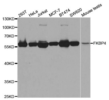
 IF (Immunofluorescence)
(Immunofluorescence analysis of U2OS cells using FKBP4 antibody. Blue: DAPI for nuclear staining.)
IF (Immunofluorescence)
(Immunofluorescence analysis of U2OS cells using FKBP4 antibody. Blue: DAPI for nuclear staining.)
FKBP4, Polyclonal Antibody (Cat# AAA28189)
Full Name
FKBP4 Polyclonal Antibody
Gene Names
FKBP4; HBI; p52; Hsp56; FKBP51; FKBP52; FKBP59; PPIase
Reactivity
Human, Mouse
Applications
Western Blot, Immunohistochemistry, Immunofluorescence
Purity
Affinity Purification
Pricing


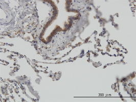

 Application Data
(Detection limit for recombinant GST tagged STIP1 is approximately 0.03ng/ml as a capture antibody.)
Application Data
(Detection limit for recombinant GST tagged STIP1 is approximately 0.03ng/ml as a capture antibody.)
STIP1, Monoclonal Antibody (Cat# AAA25917)
Full Name
STIP1 (Stress-Induced-Phosphoprotein 1, HOP, IEF-SSP-3521, P60, STI1, STI1L) (AP)
Gene Names
STIP1; HOP; P60; STI1; STI1L; HEL-S-94n; IEF-SSP-3521
Applications
Immunohistochemistry, Western Blot
Purity
Purified
Pricing


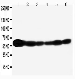

 ICC (Immunocytochemistry)
(Anti-SHC antibody, AAA11580, ICCICC: A549 Cell)
ICC (Immunocytochemistry)
(Anti-SHC antibody, AAA11580, ICCICC: A549 Cell)
SHC, Polyclonal Antibody (Cat# AAA11580)
Full Name
Anti-SHC antibody
Gene Names
SHC1; SHC; SHCA
Reactivity
Human, Mouse, Rat
Applications
Western Blot, Immunohistochemistry, Immunohistochemistry, Immunocytochemistry
Purity
Immunogen affinity purified.
Pricing


 WB (Western Blot)
(Figure 7. Western blot analysis of PLS1 using anti-PLS1 antibody (AAA19893).Electrophoresis was performed on a 5-20% SDS-PAGE gel at 70V (Stacking gel)/90V (Resolving gel) for 2-3 hours. The sample well of each lane was loaded with 30 ug of sample under reducing conditions.Lane 1: human A431 whole cell lysates,Lane 2: human RT4 whole cell lysates,Lane 3: human A549 whole cell lysates,Lane 4: human Caco-2 whole cell lysates,Lane 5: mouse small intestine tissue lysates,Lane 6: rat stomach tissue lysates.After electrophoresis, proteins were transferred to a nitrocellulose membrane at 150 mA for 50-90 minutes. Blocked the membrane with 5% non-fat milk/TBS for 1.5 hour at RT. The membrane was incubated with rabbit anti-PLS1 antigen affinity purified polyclonal antibody (AAA19893) at 0.5ug/mL overnight at 4 degree C, then washed with TBS-0.1%Tween 3 times with 5 minutes each and probed with a goat anti-rabbit IgG-DyLight 647 Conjugated secondary antibody at a dilution of 1:2000 for 1.5 hour at RT. A specific band was detected for PLS1 at approximately 70 kDa. The expected band size for PLS1 is at 70 kDa.)
WB (Western Blot)
(Figure 7. Western blot analysis of PLS1 using anti-PLS1 antibody (AAA19893).Electrophoresis was performed on a 5-20% SDS-PAGE gel at 70V (Stacking gel)/90V (Resolving gel) for 2-3 hours. The sample well of each lane was loaded with 30 ug of sample under reducing conditions.Lane 1: human A431 whole cell lysates,Lane 2: human RT4 whole cell lysates,Lane 3: human A549 whole cell lysates,Lane 4: human Caco-2 whole cell lysates,Lane 5: mouse small intestine tissue lysates,Lane 6: rat stomach tissue lysates.After electrophoresis, proteins were transferred to a nitrocellulose membrane at 150 mA for 50-90 minutes. Blocked the membrane with 5% non-fat milk/TBS for 1.5 hour at RT. The membrane was incubated with rabbit anti-PLS1 antigen affinity purified polyclonal antibody (AAA19893) at 0.5ug/mL overnight at 4 degree C, then washed with TBS-0.1%Tween 3 times with 5 minutes each and probed with a goat anti-rabbit IgG-DyLight 647 Conjugated secondary antibody at a dilution of 1:2000 for 1.5 hour at RT. A specific band was detected for PLS1 at approximately 70 kDa. The expected band size for PLS1 is at 70 kDa.)
PLS1, Polyclonal Antibody (Cat# AAA19893)
Full Name
Anti-PLS1 Antibody Picoband
Reactivity
Human, Mouse, Rat
Applications
Western Blot, Immunohistochemistry, Immunocytochemistry, Immunofluorescence, Flow Cytometry
Purity
Immunogen affinity purified.
Pricing


 FCM (Flow Cytometry)
(Figure 11. Flow Cytometry analysis of U2OS cells using anti-SBNO1 antibody (AAA19633).Overlay histogram showing U2OS cells stained with AAA19633 (Blue line). The cells were blocked with 10% normal goat serum. And then incubated with rabbit anti-SBNO1 Antibody (AAA19633, 1 ug/1x10^6 cells) for 30 min at 20 degree C. DyLight488 conjugated goat anti-rabbit IgG was used as secondary antibody for 30 minutes at 20 degree C. Isotype control antibody (Green line) was rabbit IgG (1 ug/1x10^6) used under the same conditions. Unlabelled sample (Red line) was also used as a control.)
FCM (Flow Cytometry)
(Figure 11. Flow Cytometry analysis of U2OS cells using anti-SBNO1 antibody (AAA19633).Overlay histogram showing U2OS cells stained with AAA19633 (Blue line). The cells were blocked with 10% normal goat serum. And then incubated with rabbit anti-SBNO1 Antibody (AAA19633, 1 ug/1x10^6 cells) for 30 min at 20 degree C. DyLight488 conjugated goat anti-rabbit IgG was used as secondary antibody for 30 minutes at 20 degree C. Isotype control antibody (Green line) was rabbit IgG (1 ug/1x10^6) used under the same conditions. Unlabelled sample (Red line) was also used as a control.)
SBNO1, Polyclonal Antibody (Cat# AAA19633)
Full Name
Anti-SBNO1 Antibody Picoband
Gene Names
SBNO1; Sno; MOP3
Reactivity
Human, Mouse, Rat
Applications
Western Blot, Immunohistochemistry, Immunocytochemistry, Immunofluorescence, Flow Cytometry
Purity
Immunogen affinity purified.
Pricing


 IF (Immunofluorescence)
(Immunofluorescence analysis of A549 cells using PEX14 antibody. Blue: DAPI for nuclear staining.)
IF (Immunofluorescence)
(Immunofluorescence analysis of A549 cells using PEX14 antibody. Blue: DAPI for nuclear staining.)
PEX14, Polyclonal Antibody (Cat# AAA28157)
Full Name
PEX14
Gene Names
PEX14; NAPP2; PBD13A; Pex14p; dJ734G22.2
Reactivity
Human, Mouse, Rat
Applications
Western Blot, Immunohistochemistry
Pricing


 WB (Western Blot)
(RAB33B monoclonal antibody. Western Blot analysis of RAB33B expression in NIH/3T3.)
WB (Western Blot)
(RAB33B monoclonal antibody. Western Blot analysis of RAB33B expression in NIH/3T3.)
RAB33B, Monoclonal Antibody (Cat# AAA25220)
Full Name
RAB33B (Ras-related Protein Rab-33B, SMC2) (FITC)
Gene Names
RAB33B; SMC2
Reactivity
Human, Mouse, Rat
Applications
Western Blot
Purity
Purified by Protein A Affinity Chromatography.
Pricing


 IHC (Immunohistchemistry)
(Immunohistochemistry of paraffin-embedded mouse stomach using SULT2A1 Antibody at dilution of 1:200 (40x lens).)
IHC (Immunohistchemistry)
(Immunohistochemistry of paraffin-embedded mouse stomach using SULT2A1 Antibody at dilution of 1:200 (40x lens).)
SULT2A1, Polyclonal Antibody (Cat# AAA28271)
Full Name
SULT2A1 Polyclonal Antibody
Gene Names
SULT2A1; HST; ST2; STD; hSTa; DHEAS; ST2A1; ST2A3; DHEA-ST
Reactivity
Human, Mouse, Rat
Applications
Western Blot, Immunohistochemistry
Purity
Affinity Purification
Pricing

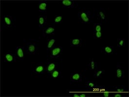


 WB (Western Blot)
(DDX54 monoclonal antibody, Western Blot analysis of DDX54 expression in HeLa.)
WB (Western Blot)
(DDX54 monoclonal antibody, Western Blot analysis of DDX54 expression in HeLa.)
DDX54, Monoclonal Antibody (Cat# AAA25076)
Full Name
DDX54 (ATP-dependent RNA Helicase DDX54, ATP-dependent RNA Helicase DP97, DEAD Box RNA Helicase 97kD, DEAD Box Protein 54, MGC2835) (FITC)
Gene Names
DDX54; DP97
Reactivity
Human
Applications
Immunofluorescence, Immunohistochemistry, Western Blot
Purity
Purified by Protein A Affinity Chromatography.
Pricing


 FCM (Flow Cytometry)
(Figure 10. Flow Cytometry analysis of K562 cells using anti-PACSIN1 antibody (AAA19863).Overlay histogram showing K562 cells stained with AAA19863 (Blue line). To facilitate intracellular staining, cells were fixed with 4% paraformaldehyde and permeabilized with permeabilization buffer. The cells were blocked with 10% normal goat serum. And then incubated with rabbit anti-PACSIN1 Antibody (AAA19863, 1ug/1x106 cells) for 30 min at 20 degree C. DyLight488 conjugated goat anti-rabbit IgG was used as secondary antibody for 30 minutes at 20 degree C. Isotype control antibody (Green line) was rabbit IgG (1ug/1x106) used under the same conditions. Unlabelled sample (Red line) was also used as a control.)
FCM (Flow Cytometry)
(Figure 10. Flow Cytometry analysis of K562 cells using anti-PACSIN1 antibody (AAA19863).Overlay histogram showing K562 cells stained with AAA19863 (Blue line). To facilitate intracellular staining, cells were fixed with 4% paraformaldehyde and permeabilized with permeabilization buffer. The cells were blocked with 10% normal goat serum. And then incubated with rabbit anti-PACSIN1 Antibody (AAA19863, 1ug/1x106 cells) for 30 min at 20 degree C. DyLight488 conjugated goat anti-rabbit IgG was used as secondary antibody for 30 minutes at 20 degree C. Isotype control antibody (Green line) was rabbit IgG (1ug/1x106) used under the same conditions. Unlabelled sample (Red line) was also used as a control.)
PACSIN1, Polyclonal Antibody (Cat# AAA19863)
Full Name
Anti-PACSIN1 Antibody Picoband
Gene Names
PACSIN1; SDPI
Reactivity
Human, Mouse, Rat
Applications
Western Blot, Immunohistochemistry, Immunocytochemistry, Immunofluorescence, Flow Cytometry
Purity
Immunogen affinity purified.
Pricing


 IF (Immunofluorescence)
(Figure 9. IF analysis of SARS2 using anti-SARS2 antibody (AAA19598).SARS2 was detected in an immunocytochemical section of A549 cells. Enzyme antigen retrieval was performed using IHC enzyme antigen retrieval reagent (AR0022) for 15 mins. The cells were blocked with 10% goat serum. And then incubated with 5 ug/mL rabbit anti-SARS2 Antibody (AAA19598) overnight at 4 degree C. Cy3 Conjugated Goat Anti-Rabbit IgG (BA1032) was used as secondary antibody at 1:100 dilution and incubated for 30 minutes at 37 degree C. The section was counterstained with DAPI. Visualize using a fluorescence microscope and filter sets appropriate for the label used.)
IF (Immunofluorescence)
(Figure 9. IF analysis of SARS2 using anti-SARS2 antibody (AAA19598).SARS2 was detected in an immunocytochemical section of A549 cells. Enzyme antigen retrieval was performed using IHC enzyme antigen retrieval reagent (AR0022) for 15 mins. The cells were blocked with 10% goat serum. And then incubated with 5 ug/mL rabbit anti-SARS2 Antibody (AAA19598) overnight at 4 degree C. Cy3 Conjugated Goat Anti-Rabbit IgG (BA1032) was used as secondary antibody at 1:100 dilution and incubated for 30 minutes at 37 degree C. The section was counterstained with DAPI. Visualize using a fluorescence microscope and filter sets appropriate for the label used.)
SARS2, Polyclonal Antibody (Cat# AAA19598)
Full Name
Anti-SARS2 Antibody Picoband
Gene Names
SARS2; SYS; SARS; SERS; SARSM; SerRS; SerRSmt; mtSerRS
Reactivity
Human
Applications
Western Blot, Immunohistochemistry, Immunocytochemistry, Immunofluorescence
Purity
Immunogen affinity purified.
Pricing


 WB (Western Blot)
(LEPRE1 monoclonal antibody, Western Blot analysis of LEPRE1 expression in HeLa.)
WB (Western Blot)
(LEPRE1 monoclonal antibody, Western Blot analysis of LEPRE1 expression in HeLa.)
GROS1, Monoclonal Antibody (Cat# AAA24228)
Full Name
GROS1 (Growth Suppressor 1, Leucine- and Proline-enriched Proteoglycan 1, LEPRE1, Leprecan-1, MGC117314, OI8, Prolyl 3-hydroxylase 1, P3H1, PSEC0109) (AP)
Gene Names
P3H1; OI8; GROS1; LEPRE1
Reactivity
Human
Applications
Immunohistochemistry, Western Blot
Purity
Purified by Protein A Affinity Chromatography.
Pricing
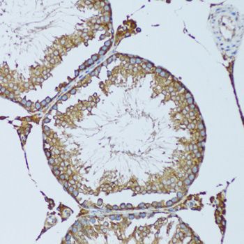


 IHC (Immunohistchemistry)
(Immunohistochemistry of paraffin-embedded mouse spinal cord using RPN1 antibody at dilution of 1:100 (40x lens).)
IHC (Immunohistchemistry)
(Immunohistochemistry of paraffin-embedded mouse spinal cord using RPN1 antibody at dilution of 1:100 (40x lens).)
RPN1, Polyclonal Antibody (Cat# AAA28249)
Full Name
RPN1 Polyclonal Antibody
Gene Names
RPN1; OST1; RBPH1
Reactivity
Human, Mouse, Rat
Applications
Western Blot, Immunohistochemistry
Purity
Affinity Purification
Pricing


 IF (Immunofluorescence)
(Immunofluorescence analysis of U-2 OS cells using RALGDS antibody at dilution of 1:100. Blue: DAPI for nuclear staining.)
IF (Immunofluorescence)
(Immunofluorescence analysis of U-2 OS cells using RALGDS antibody at dilution of 1:100. Blue: DAPI for nuclear staining.)
RALGDS, Polyclonal Antibody (Cat# AAA28359)
Full Name
RALGDS Rabbit pAb
Gene Names
RALGDS; RGF; RGDS; RalGEF
Reactivity
Human, Mouse, Rat
Applications
Western Blot, Immunohistochemistry, Immunofluorescence
Purity
Affinity purification
Pricing

 FCM (Flow Cytometry)
(Figure 7. Flow Cytometry analysis of MCF-7 cells using anti-OXSM antibody (AAA19976).Overlay histogram showing MCF-7 cells stained with AAA19976 (Blue line). To facilitate intracellular staining, cells were fixed with 4% paraformaldehyde and permeabilized with permeabilization buffer. The cells were blocked with 10% normal goat serum. And then incubated with rabbit anti-OXSM Antibody (AAA19976, 1ug/1x106 cells) for 30 min at 20 degree C. DyLight488 conjugated goat anti-rabbit IgG was used as secondary antibody for 30 minutes at 20 degree C. Isotype control antibody (Green line) was rabbit IgG (1ug/1x106) used under the same conditions. Unlabelled sample without incubation with primary antibody and secondary antibody (Red line) was used as a blank control.)
FCM (Flow Cytometry)
(Figure 7. Flow Cytometry analysis of MCF-7 cells using anti-OXSM antibody (AAA19976).Overlay histogram showing MCF-7 cells stained with AAA19976 (Blue line). To facilitate intracellular staining, cells were fixed with 4% paraformaldehyde and permeabilized with permeabilization buffer. The cells were blocked with 10% normal goat serum. And then incubated with rabbit anti-OXSM Antibody (AAA19976, 1ug/1x106 cells) for 30 min at 20 degree C. DyLight488 conjugated goat anti-rabbit IgG was used as secondary antibody for 30 minutes at 20 degree C. Isotype control antibody (Green line) was rabbit IgG (1ug/1x106) used under the same conditions. Unlabelled sample without incubation with primary antibody and secondary antibody (Red line) was used as a blank control.)
OXSM, Polyclonal Antibody (Cat# AAA19976)
Full Name
Anti-OXSM Antibody Picoband
Gene Names
OXSM; KS; KASI; FASN2D
Reactivity
Human, Rat
Applications
Western Blot, Immunohistochemistry, Immunocytochemistry, Immunofluorescence, Flow Cytometry
Purity
Immunogen affinity purified.
Pricing


 FCM (Flow Cytometry)
(Figure 8. Flow Cytometry analysis of HepG2 cells using anti-PGC antibody (AAA19725).Overlay histogram showing HepG2 cells stained with AAA19725 (Blue line). The cells were fixed with 4% paraformaldehyde and blocked with 10% normal goat serum. And then incubated with rabbit anti-PGC Antibody (AAA19725, 1ug/1x106 cells) for 30 min at 20 degree C. DyLight488 conjugated goat anti-rabbit IgG was used as secondary antibody for 30 minutes at 20 degree C. Isotype control antibody (Green line) was rabbit IgG (1ug/1x106) used under the same conditions. Unlabelled sample (Red line) was also used as a control.)
FCM (Flow Cytometry)
(Figure 8. Flow Cytometry analysis of HepG2 cells using anti-PGC antibody (AAA19725).Overlay histogram showing HepG2 cells stained with AAA19725 (Blue line). The cells were fixed with 4% paraformaldehyde and blocked with 10% normal goat serum. And then incubated with rabbit anti-PGC Antibody (AAA19725, 1ug/1x106 cells) for 30 min at 20 degree C. DyLight488 conjugated goat anti-rabbit IgG was used as secondary antibody for 30 minutes at 20 degree C. Isotype control antibody (Green line) was rabbit IgG (1ug/1x106) used under the same conditions. Unlabelled sample (Red line) was also used as a control.)
PGC, Polyclonal Antibody (Cat# AAA19725)
Full Name
Anti-PGC Antibody Picoband
Gene Names
PGC; PEPC; PGII
Reactivity
Human, Mouse, Rat
Applications
Immunofluorescence, Immunohistochemistry, Western Blot, Flow Cytometry
Purity
Immunogen affinity purified.
Pricing

 FCM (Flow Cytometry)
(Figure 10. Flow Cytometry analysis of A549 cells using anti-SHKBP1 antibody (AAA19991).Overlay histogram showing A549 cells stained with AAA19991 (Blue line). To facilitate intracellular staining, cells were fixed with 4% paraformaldehyde and permeabilized with permeabilization buffer. The cells were blocked with 10% normal goat serum. And then incubated with rabbit anti-SHKBP1 Antibody (AAA19991, 1ug/1x106 cells) for 30 min at 20 degree C. DyLight488 conjugated goat anti-rabbit IgG was used as secondary antibody for 30 minutes at 20 degree C. Isotype control antibody (Green line) was rabbit IgG (1ug/1x106) used under the same conditions. Unlabelled sample (Red line) was also used as a control.)
FCM (Flow Cytometry)
(Figure 10. Flow Cytometry analysis of A549 cells using anti-SHKBP1 antibody (AAA19991).Overlay histogram showing A549 cells stained with AAA19991 (Blue line). To facilitate intracellular staining, cells were fixed with 4% paraformaldehyde and permeabilized with permeabilization buffer. The cells were blocked with 10% normal goat serum. And then incubated with rabbit anti-SHKBP1 Antibody (AAA19991, 1ug/1x106 cells) for 30 min at 20 degree C. DyLight488 conjugated goat anti-rabbit IgG was used as secondary antibody for 30 minutes at 20 degree C. Isotype control antibody (Green line) was rabbit IgG (1ug/1x106) used under the same conditions. Unlabelled sample (Red line) was also used as a control.)
SHKBP1, Polyclonal Antibody (Cat# AAA19991)
Full Name
Anti-SHKBP1 Antibody Picoband
Gene Names
SHKBP1; Sb1; PP203
Reactivity
Human
Applications
Western Blot, Immunohistochemistry, Flow Cytometry
Purity
Immunogen affinity purified.
Pricing
 IHC (Immunohistochemistry)
(Figure 8. IHC analysis of MELTF using anti-MELTF antibody (AAA19980).MELTF was detected in a paraffin-embedded section of rat testis tissue. Heat mediated antigen retrieval was performed in EDTA buffer (pH 8.0, epitope retrieval solution). The tissue section was blocked with 10% goat serum. The tissue section was then incubated with 2ug/ml rabbit anti-MELTF Antibody (AAA19980) overnight at 4 degree C. Peroxidase Conjugated Goat Anti-rabbit IgG was used as secondary antibody and incubated for 30 minutes at 37 degree C. The tissue section was developed using HRP Conjugated Rabbit IgG Super Vision Assay Kit ( epitope retrieval solution). The tissue section was blocked with 10% goat serum. The tissue section was then incubated with 2ug/ml rabbit anti-MELTF Antibody (AAA19980) overnight at 4 degree C. Peroxidase Conjugated Goat Anti-rabbit IgG was used as secondary antibody and incubated for 30 minutes at 37 degree C. The tissue section was developed using HRP Conjugated Rabbit IgG Super Vision Assay Kit ( epitope retrieval solution). The tissue section was blocked with 10% goat serum. The tissue section was then incubated with 2ug/ml rabbit anti-MELTF Antibody (AAA19980) overnight at 4 degree C. Peroxidase Conjugated Goat Anti-rabbit IgG was used as secondary antibody and incubated for 30 minutes at 37 degree C. The tissue section was developed using HRP Conjugated Rabbit IgG Super Vision Assay Kit ( epitope retrieval solution). The tissue section was blocked with 10% goat serum. The tissue section was then incubated with 2ug/ml rabbit anti-MELTF Antibody (AAA19980) overnight at 4 degree C. Peroxidase Conjugated Goat Anti-rabbit IgG was used as secondary antibody and incubated for 30 minutes at 37 degree C. The tissue section was developed using HRP Conjugated Rabbit IgG Super Vision Assay Kit ( epitope retrieval solution). The tissue section was blocked with 10% goat serum. The tissue section was then incubated with 2ug/ml rabbit anti-MELTF Antibody (AAA19980) overnight at 4 degree C. Peroxidase Conjugated Goat Anti-rabbit IgG was used as secondary antibody and incubated for 30 minutes at 37 degree C. The tissue section was developed using HRP Conjugated Rabbit IgG Super Vision Assay Kit ( epitope retrieval solution). The tissue section was blocked with 10% goat serum. The tissue section was then incubated with 2ug/ml rabbit anti-MELTF Antibody (AAA19980) overnight at 4 degree C. Peroxidase Conjugated Goat Anti-rabbit IgG was used as secondary antibody and incubated for 30 minutes at 37 degree C. The tissue section was developed using HRP Conjugated Rabbit IgG Super Vision Assay Kit (Lane 2: rat C6 whole cell lysates,Lane 3: mouse skin tissue lysates.After electrophoresis, proteins were transferred to a nitrocellulose membrane at 150 mA for 50-90 minutes. Blocked the membrane with 5% non-fat milk/TBS for 1.5 hour at RT. The membrane was incubated with rabbit anti-MELTF antigen affinity purified polyclonal antibody (#AAA19980) at 0.5ug/mL overnight at 4 degree C, then washed with TBS-0.1%Tween 3 times with 5 minutes each and probed with a goat anti-rabbit IgG-HRP secondary antibody at a dilution of 1:5000 for 1.5 hour at RT. The signal is developed using an Enhanced Chemiluminescent detection (ECL) kit (Lane 2: human A431 whole cell lysates,Lane 3: human A549 whole cell lysates,Lane 4: human PC-3 whole cell lysates.After electrophoresis, proteins were transferred to a nitrocellulose membrane at 150 mA for 50-90 minutes. Blocked the membrane with 5% non-fat milk/TBS for 1.5 hour at RT. The membrane was incubated with rabbit anti-MELTF antigen affinity purified polyclonal antibody (#AAA19980) at 0.5ug/mL overnight at 4 degree C, then washed with TBS-0.1%Tween 3 times with 5 minutes each and probed with a goat anti-rabbit IgG-HRP secondary antibody at a dilution of 1:5000 for 1.5 hour at RT. The signal is developed using an Enhanced Chemiluminescent detection (ECL) kit with Tanon 5200 system. A specific band was detected for MELTF at approximately 90 kDa. The expected band size for MELTF is at 80 kDa.)
IHC (Immunohistochemistry)
(Figure 8. IHC analysis of MELTF using anti-MELTF antibody (AAA19980).MELTF was detected in a paraffin-embedded section of rat testis tissue. Heat mediated antigen retrieval was performed in EDTA buffer (pH 8.0, epitope retrieval solution). The tissue section was blocked with 10% goat serum. The tissue section was then incubated with 2ug/ml rabbit anti-MELTF Antibody (AAA19980) overnight at 4 degree C. Peroxidase Conjugated Goat Anti-rabbit IgG was used as secondary antibody and incubated for 30 minutes at 37 degree C. The tissue section was developed using HRP Conjugated Rabbit IgG Super Vision Assay Kit ( epitope retrieval solution). The tissue section was blocked with 10% goat serum. The tissue section was then incubated with 2ug/ml rabbit anti-MELTF Antibody (AAA19980) overnight at 4 degree C. Peroxidase Conjugated Goat Anti-rabbit IgG was used as secondary antibody and incubated for 30 minutes at 37 degree C. The tissue section was developed using HRP Conjugated Rabbit IgG Super Vision Assay Kit ( epitope retrieval solution). The tissue section was blocked with 10% goat serum. The tissue section was then incubated with 2ug/ml rabbit anti-MELTF Antibody (AAA19980) overnight at 4 degree C. Peroxidase Conjugated Goat Anti-rabbit IgG was used as secondary antibody and incubated for 30 minutes at 37 degree C. The tissue section was developed using HRP Conjugated Rabbit IgG Super Vision Assay Kit ( epitope retrieval solution). The tissue section was blocked with 10% goat serum. The tissue section was then incubated with 2ug/ml rabbit anti-MELTF Antibody (AAA19980) overnight at 4 degree C. Peroxidase Conjugated Goat Anti-rabbit IgG was used as secondary antibody and incubated for 30 minutes at 37 degree C. The tissue section was developed using HRP Conjugated Rabbit IgG Super Vision Assay Kit ( epitope retrieval solution). The tissue section was blocked with 10% goat serum. The tissue section was then incubated with 2ug/ml rabbit anti-MELTF Antibody (AAA19980) overnight at 4 degree C. Peroxidase Conjugated Goat Anti-rabbit IgG was used as secondary antibody and incubated for 30 minutes at 37 degree C. The tissue section was developed using HRP Conjugated Rabbit IgG Super Vision Assay Kit ( epitope retrieval solution). The tissue section was blocked with 10% goat serum. The tissue section was then incubated with 2ug/ml rabbit anti-MELTF Antibody (AAA19980) overnight at 4 degree C. Peroxidase Conjugated Goat Anti-rabbit IgG was used as secondary antibody and incubated for 30 minutes at 37 degree C. The tissue section was developed using HRP Conjugated Rabbit IgG Super Vision Assay Kit (Lane 2: rat C6 whole cell lysates,Lane 3: mouse skin tissue lysates.After electrophoresis, proteins were transferred to a nitrocellulose membrane at 150 mA for 50-90 minutes. Blocked the membrane with 5% non-fat milk/TBS for 1.5 hour at RT. The membrane was incubated with rabbit anti-MELTF antigen affinity purified polyclonal antibody (#AAA19980) at 0.5ug/mL overnight at 4 degree C, then washed with TBS-0.1%Tween 3 times with 5 minutes each and probed with a goat anti-rabbit IgG-HRP secondary antibody at a dilution of 1:5000 for 1.5 hour at RT. The signal is developed using an Enhanced Chemiluminescent detection (ECL) kit (Lane 2: human A431 whole cell lysates,Lane 3: human A549 whole cell lysates,Lane 4: human PC-3 whole cell lysates.After electrophoresis, proteins were transferred to a nitrocellulose membrane at 150 mA for 50-90 minutes. Blocked the membrane with 5% non-fat milk/TBS for 1.5 hour at RT. The membrane was incubated with rabbit anti-MELTF antigen affinity purified polyclonal antibody (#AAA19980) at 0.5ug/mL overnight at 4 degree C, then washed with TBS-0.1%Tween 3 times with 5 minutes each and probed with a goat anti-rabbit IgG-HRP secondary antibody at a dilution of 1:5000 for 1.5 hour at RT. The signal is developed using an Enhanced Chemiluminescent detection (ECL) kit with Tanon 5200 system. A specific band was detected for MELTF at approximately 90 kDa. The expected band size for MELTF is at 80 kDa.)
MELTF, Polyclonal Antibody (Cat# AAA19980)
Full Name
Anti-MELTF Antibody Picoband
Gene Names
MFI2; MTF1; CD228; MAP97
Reactivity
Human, Mouse, Rat
Applications
Western Blot, Immunohistochemistry
Purity
Immunogen affinity purified.
Pricing


 FCM (Flow Cytometry)
(Figure 8. Flow Cytometry analysis of MCF-7 cells using anti-PAPSS2 antibody (AAA19850).Overlay histogram showing MCF-7 cells stained with AAA19850 (Blue line). To facilitate intracellular staining, cells were fixed with 4% paraformaldehyde and permeabilized with permeabilization buffer. The cells were blocked with 10% normal goat serum. And then incubated with rabbit anti-PAPSS2 Antibody (AAA19850, 1ug/1x106 cells) for 30 min at 20 degree C. DyLight488 conjugated goat anti-rabbit IgG was used as secondary antibody for 30 minutes at 20 degree C. Isotype control antibody (Green line) was rabbit IgG (1ug/1x106) used under the same conditions. Unlabelled sample (Red line) was also used as a control.)
FCM (Flow Cytometry)
(Figure 8. Flow Cytometry analysis of MCF-7 cells using anti-PAPSS2 antibody (AAA19850).Overlay histogram showing MCF-7 cells stained with AAA19850 (Blue line). To facilitate intracellular staining, cells were fixed with 4% paraformaldehyde and permeabilized with permeabilization buffer. The cells were blocked with 10% normal goat serum. And then incubated with rabbit anti-PAPSS2 Antibody (AAA19850, 1ug/1x106 cells) for 30 min at 20 degree C. DyLight488 conjugated goat anti-rabbit IgG was used as secondary antibody for 30 minutes at 20 degree C. Isotype control antibody (Green line) was rabbit IgG (1ug/1x106) used under the same conditions. Unlabelled sample (Red line) was also used as a control.)
PAPSS2, Polyclonal Antibody (Cat# AAA19850)
Full Name
Anti-PAPSS2 Antibody Picoband
Gene Names
PAPSS2; SK2; BCYM4; ATPSK2
Reactivity
Human, Mouse, Rat
Applications
Immunofluorescence, Immunohistochemistry, Western Blot, Flow Cytometry
Purity
Immunogen affinity purified.
Pricing

 FCM (Flow Cytometry)
(Figure 10. Flow Cytometry analysis of U20S cells using anti-TMEM87A antibody (AAA19997).Overlay histogram showing U20S cells stained with AAA19997 (Blue line). To facilitate intracellular staining, cells were fixed with 4% paraformaldehyde and permeabilized with permeabilization buffer. The cells were blocked with 10% normal goat serum. And then incubated with rabbit anti-TMEM87A Antibody (AAA19997, 1ug/1x106 cells) for 30 min at 20 degree C. PE conjugated goat anti-rabbit IgG (5-10ug/1x106 cells) was used as secondary antibody for 30 minutes at 20 degree C. Isotype control antibody (Green line) was rabbit IgG (1ug/1x106) used under the same conditions. Unlabelled sample without incubation with primary antibody and secondary antibody (Red line) was used as a blank control.)
FCM (Flow Cytometry)
(Figure 10. Flow Cytometry analysis of U20S cells using anti-TMEM87A antibody (AAA19997).Overlay histogram showing U20S cells stained with AAA19997 (Blue line). To facilitate intracellular staining, cells were fixed with 4% paraformaldehyde and permeabilized with permeabilization buffer. The cells were blocked with 10% normal goat serum. And then incubated with rabbit anti-TMEM87A Antibody (AAA19997, 1ug/1x106 cells) for 30 min at 20 degree C. PE conjugated goat anti-rabbit IgG (5-10ug/1x106 cells) was used as secondary antibody for 30 minutes at 20 degree C. Isotype control antibody (Green line) was rabbit IgG (1ug/1x106) used under the same conditions. Unlabelled sample without incubation with primary antibody and secondary antibody (Red line) was used as a blank control.)
TMEM87A, Polyclonal Antibody (Cat# AAA19997)
Full Name
Anti-TMEM87A Antibody Picoband
Reactivity
Human, Mouse, Rat
Applications
Western Blot, Immunohistochemistry, Immunocytochemistry, Immunofluorescence, Flow Cytometry
Purity
Immunogen affinity purified.
Pricing

 FCM (Flow Cytometry)
(Figure 7. Flow Cytometry analysis of PC-3 cells using anti-POC1A antibody (AAA19908).Overlay histogram showing PC-3 cells stained with AAA19908 (Blue line). To facilitate intracellular staining, cells were fixed with 4% paraformaldehyde and permeabilized with permeabilization buffer. The cells were blocked with 10% normal goat serum. And then incubated with rabbit anti-POC1A Antibody (AAA19908, 1ug/1x106 cells) for 30 min at 20 degree C. DyLight488 conjugated goat anti-rabbit IgG was used as secondary antibody for 30 minutes at 20 degree C. Isotype control antibody (Green line) was rabbit IgG (1ug/1x106) used under the same conditions. Unlabelled sample without incubation with primary antibody and secondary antibody (Red line) was used as a blank control.)
FCM (Flow Cytometry)
(Figure 7. Flow Cytometry analysis of PC-3 cells using anti-POC1A antibody (AAA19908).Overlay histogram showing PC-3 cells stained with AAA19908 (Blue line). To facilitate intracellular staining, cells were fixed with 4% paraformaldehyde and permeabilized with permeabilization buffer. The cells were blocked with 10% normal goat serum. And then incubated with rabbit anti-POC1A Antibody (AAA19908, 1ug/1x106 cells) for 30 min at 20 degree C. DyLight488 conjugated goat anti-rabbit IgG was used as secondary antibody for 30 minutes at 20 degree C. Isotype control antibody (Green line) was rabbit IgG (1ug/1x106) used under the same conditions. Unlabelled sample without incubation with primary antibody and secondary antibody (Red line) was used as a blank control.)
POC1A, Polyclonal Antibody (Cat# AAA19908)
Full Name
Anti-POC1A Antibody Picoband
Gene Names
POC1A; PIX2; SOFT; WDR51A
Reactivity
Human, Mouse, Rat
Applications
Western Blot, Immunohistochemistry, Immunocytochemistry, Immunofluorescence, Flow Cytometry
Purity
Immunogen affinity purified.
Pricing

 FCM (Flow Cytometry)
(Figure 7. Flow Cytometry analysis of Caco-2 cells using anti-MATK antibody (AAA19767).Overlay histogram showing Caco-2 cells stained with AAA19767 (Blue line). To facilitate intracellular staining, cells were fixed with 4% paraformaldehyde and permeabilized with permeabilization buffer. The cells were blocked with 10% normal goat serum. And then incubated with rabbit anti-MATK Antibody (AAA19767, 1ug/1x106 cells) for 30 min at 20 degree C. DyLight488 conjugated goat anti-rabbit IgG was used as secondary antibody for 30 minutes at 20 degree C. Isotype control antibody (Green line) was rabbit IgG (1ug/1x106) used under the same conditions. Unlabelled sample without incubation with primary antibody and secondary antibody (Red line) was used as a blank control.)
FCM (Flow Cytometry)
(Figure 7. Flow Cytometry analysis of Caco-2 cells using anti-MATK antibody (AAA19767).Overlay histogram showing Caco-2 cells stained with AAA19767 (Blue line). To facilitate intracellular staining, cells were fixed with 4% paraformaldehyde and permeabilized with permeabilization buffer. The cells were blocked with 10% normal goat serum. And then incubated with rabbit anti-MATK Antibody (AAA19767, 1ug/1x106 cells) for 30 min at 20 degree C. DyLight488 conjugated goat anti-rabbit IgG was used as secondary antibody for 30 minutes at 20 degree C. Isotype control antibody (Green line) was rabbit IgG (1ug/1x106) used under the same conditions. Unlabelled sample without incubation with primary antibody and secondary antibody (Red line) was used as a blank control.)
MATK, Polyclonal Antibody (Cat# AAA19767)
Full Name
Anti-MATK Antibody Picoband
Gene Names
MATK; CHK; CTK; HYL; Lsk; HYLTK; HHYLTK
Reactivity
Human, Mouse, Rat
Applications
Western Blot, Immunohistochemistry, Immunocytochemistry, Immunofluorescence, Flow Cytometry
Purity
Immunogen affinity purified.
Pricing


 IF (Immunofluorescence)
(Figure 9. IF analysis of CAD using anti-CAD antibody (AAA19398).CAD was detected in an immunocytochemical section of MCF-7 cells. Enzyme antigen retrieval was performed using IHC enzyme antigen retrieval reagent (AR0022) for 15 mins. The cells were blocked with 10% goat serum. And then incubated with 5 ug/mL rabbit anti-CAD Antibody (AAA19398) overnight at 4 degree C. DyLight488 Conjugated Goat Anti-Rabbit IgG was used as secondary antibody at 1:100 dilution and incubated for 30 minutes at 37 degree C. The section was counterstained with DAPI. Visualize using a fluorescence microscope and filter sets appropriate for the label used.)
IF (Immunofluorescence)
(Figure 9. IF analysis of CAD using anti-CAD antibody (AAA19398).CAD was detected in an immunocytochemical section of MCF-7 cells. Enzyme antigen retrieval was performed using IHC enzyme antigen retrieval reagent (AR0022) for 15 mins. The cells were blocked with 10% goat serum. And then incubated with 5 ug/mL rabbit anti-CAD Antibody (AAA19398) overnight at 4 degree C. DyLight488 Conjugated Goat Anti-Rabbit IgG was used as secondary antibody at 1:100 dilution and incubated for 30 minutes at 37 degree C. The section was counterstained with DAPI. Visualize using a fluorescence microscope and filter sets appropriate for the label used.)
CAD, Polyclonal Antibody (Cat# AAA19398)
Full Name
Anti-CAD Antibody Picoband
Reactivity
Human, Mouse, Rat
Applications
Western Blot, Immunohistochemistry, Immunocytochemistry, Immunofluorescence
Purity
Immunogen affinity purified.
Pricing


 WB (Western Blot)
(DDX54 monoclonal antibody, Western Blot analysis of DDX54 expression in HeLa.)
WB (Western Blot)
(DDX54 monoclonal antibody, Western Blot analysis of DDX54 expression in HeLa.)
DDX54, Monoclonal Antibody (Cat# AAA24484)
Full Name
DDX54 (ATP-dependent RNA Helicase DDX54, ATP-dependent RNA Helicase DP97, DEAD Box RNA Helicase 97kD, DEAD Box Protein 54, MGC2835) APC
Gene Names
DDX54; DP97
Reactivity
Human
Applications
Immunofluorescence, Immunohistochemistry, Western Blot
Purity
Purified by Protein A Affinity Chromatography.
Pricing


 WB (Western Blot)
(LEPRE1 monoclonal antibody, Western Blot analysis of LEPRE1 expression in HeLa.)
WB (Western Blot)
(LEPRE1 monoclonal antibody, Western Blot analysis of LEPRE1 expression in HeLa.)
GROS1, Monoclonal Antibody (Cat# AAA25410)
Full Name
GROS1 (Growth Suppressor 1, Leucine- and Proline-enriched Proteoglycan 1, LEPRE1, Leprecan-1, MGC117314, OI8, Prolyl 3-hydroxylase 1, P3H1, PSEC0109) (HRP)
Gene Names
P3H1; OI8; GROS1; LEPRE1
Reactivity
Human
Applications
Immunohistochemistry, Western Blot
Purity
Purified by Protein A Affinity Chromatography.
Pricing


 WB (Western Blot)
(Western Blot analysis of SMAD3 expression in transfected 293T cell line by SMAD3 monoclonal antibody (M07), clone 1B7.Lane 1: SMAD3 transfected lysate(48 KDa).Lane 2: Non-transfected lysate.)
WB (Western Blot)
(Western Blot analysis of SMAD3 expression in transfected 293T cell line by SMAD3 monoclonal antibody (M07), clone 1B7.Lane 1: SMAD3 transfected lysate(48 KDa).Lane 2: Non-transfected lysate.)
SMAD3, Monoclonal Antibody (Cat# AAA25983)
Full Name
SMAD3 (SMAD Family Member 3, DKFZp586N0721, DKFZp686J10186, HSPC193, HsT17436, JV15-2, MADH3, MGC60396) (AP)
Gene Names
SMAD3; LDS3; LDS1C; MADH3; JV15-2; HSPC193; HsT17436
Applications
Western Blot
Purity
Purified
Pricing


 WB (Western Blot)
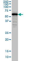
(SMAD4 monoclonal antibody. Western Blot analysis of SMAD4 expression in IMR-32.)
WB (Western Blot)
(SMAD4 monoclonal antibody. Western Blot analysis of SMAD4 expression in IMR-32.)
SMAD4, Monoclonal Antibody (Cat# AAA24955)
Full Name
SMAD4 (Mothers Against Decapentaplegic Homolog 4, SMAD 4, Mothers Against DPP Homolog 4, Deletion Target In Pancreatic Carcinoma 4, MAD Homolog 4, SMAD Family Member 4, hSMAD4, DPC4, MADH4) (Biotin)
Gene Names
SMAD4; JIP; DPC4; MADH4; MYHRS
Reactivity
Human
Applications
Immunofluorescence, Western Blot
Purity
Purified by Protein A Affinity Chromatography.
Pricing


 Application Data
(Analysis of Protein Array containing more than 19, 000 full-length human proteins using Mouse Emerin Monoclonal Antibody (EMD/2168) Z- and S- Score: The Z-score represents the strength of a signal that a monoclonal antibody (MAb) (in combination with a fluorescently-tagged anti-IgG secondary antibody) produces when binding to a particular protein on the HuProtTM array. Z-scores are described in units of standard deviations (SD's) above the mean value of all signals generated on that array. If targets on HuProtTM are arranged in descending order of the Z-score, the S-score is the difference (also in units of SD's) between the Z-score. S-score therefore represents the relative target specificity of a MAb to its intended target. A MAb is considered to specific to its intended target, if the MAb has an S-score of at least 2.5. For example, if a MAb binds to protein X with a Z-score of 43 and to protein Y with a Z-score of 14, then the S-score for the binding of that MAb to protein X is equal to 29.)
Application Data
(Analysis of Protein Array containing more than 19, 000 full-length human proteins using Mouse Emerin Monoclonal Antibody (EMD/2168) Z- and S- Score: The Z-score represents the strength of a signal that a monoclonal antibody (MAb) (in combination with a fluorescently-tagged anti-IgG secondary antibody) produces when binding to a particular protein on the HuProtTM array. Z-scores are described in units of standard deviations (SD's) above the mean value of all signals generated on that array. If targets on HuProtTM are arranged in descending order of the Z-score, the S-score is the difference (also in units of SD's) between the Z-score. S-score therefore represents the relative target specificity of a MAb to its intended target. A MAb is considered to specific to its intended target, if the MAb has an S-score of at least 2.5. For example, if a MAb binds to protein X with a Z-score of 43 and to protein Y with a Z-score of 14, then the S-score for the binding of that MAb to protein X is equal to 29.)
Emerin, Monoclonal Antibody (Cat# AAA23899)
Full Name
Emerin (Papillary Thyroid Carcinoma and EDMD Marker)
Gene Names
EMD; STA; EDMD; LEMD5
Reactivity
Human. Others not known.
Applications
Immunohistochemistry
Pricing

 FCM (Flow Cytometry)
(Figure 6. Flow Cytometry analysis of A549 cells using anti-NIT2 antibody (AAA19853).Overlay histogram showing A549 cells stained with AAA19853 (Blue line). To facilitate intracellular staining, cells were fixed with 4% paraformaldehyde and permeabilized with permeabilization buffer. The cells were blocked with 10% normal goat serum. And then incubated with rabbit anti-NIT2 Antibody (AAA19853, 1ug/1x106 cells) for 30 min at 20 degree C. DyLight488 conjugated goat anti-rabbit IgG was used as secondary antibody for 30 minutes at 20 degree C. Isotype control antibody (Green line) was rabbit IgG (1ug/1x106) used under the same conditions. Unlabelled sample (Red line) was also used as a control.)
FCM (Flow Cytometry)
(Figure 6. Flow Cytometry analysis of A549 cells using anti-NIT2 antibody (AAA19853).Overlay histogram showing A549 cells stained with AAA19853 (Blue line). To facilitate intracellular staining, cells were fixed with 4% paraformaldehyde and permeabilized with permeabilization buffer. The cells were blocked with 10% normal goat serum. And then incubated with rabbit anti-NIT2 Antibody (AAA19853, 1ug/1x106 cells) for 30 min at 20 degree C. DyLight488 conjugated goat anti-rabbit IgG was used as secondary antibody for 30 minutes at 20 degree C. Isotype control antibody (Green line) was rabbit IgG (1ug/1x106) used under the same conditions. Unlabelled sample (Red line) was also used as a control.)
NIT2, Polyclonal Antibody (Cat# AAA19853)
Full Name
Anti-NIT2 Antibody Picoband
Gene Names
NIT2; HEL-S-8a
Reactivity
Human, Mouse, Rat
Applications
Western Blot, Immunohistochemistry, Flow Cytometry
Purity
Immunogen affinity purified.
Pricing


 FCM (Flow Cytometry)
(Figure 9. Flow Cytometry analysis of HeLa cells using anti-SND1 antibody (AAA19699).Overlay histogram showing HeLa cells stained with AAA19699 (Blue line). The cells were blocked with 10% normal goat serum. And then incubated with mouse anti-SND1 Antibody (AAA19699, 1 ug/1x10^6 cells) for 30 min at 20 degree C. DyLight488 conjugated goat anti-mouse IgG was used as secondary antibody for 30 minutes at 20 degree C. Isotype control antibody (Green line) was mouse IgG (1 ug/1x10^6) used under the same conditions. Unlabelled sample (Red line) was also used as a control.)
FCM (Flow Cytometry)
(Figure 9. Flow Cytometry analysis of HeLa cells using anti-SND1 antibody (AAA19699).Overlay histogram showing HeLa cells stained with AAA19699 (Blue line). The cells were blocked with 10% normal goat serum. And then incubated with mouse anti-SND1 Antibody (AAA19699, 1 ug/1x10^6 cells) for 30 min at 20 degree C. DyLight488 conjugated goat anti-mouse IgG was used as secondary antibody for 30 minutes at 20 degree C. Isotype control antibody (Green line) was mouse IgG (1 ug/1x10^6) used under the same conditions. Unlabelled sample (Red line) was also used as a control.)
SND1, Monoclonal Antibody (Cat# AAA19699)
Full Name
Anti-SND1 Antibody Picoband (monoclonal, 5F5E9)
Gene Names
SND1; p100; TDRD11
Reactivity
Human, Mouse, Rat
Applications
Western Blot, Immunohistochemistry, Immunocytochemistry, Immunofluorescence, Flow Cytometry
Purity
Immunogen affinity purified.
Pricing
 Standard Curve (Sample)
Periostin, ELISA Kit (Cat# AAA17869)
Full Name
Human Periostin ELISA Kit
Gene Names
POSTN; PN; OSF2; OSF-2; PDLPOSTN; periostin
Reactivity
Human
Applications
Sandwich ELISA
Pricing
 SDS-PAGE
(3ug by SDS-PAGE under reducing condition and visualized by coomassie blue stain.)
SDS-PAGE
(3ug by SDS-PAGE under reducing condition and visualized by coomassie blue stain.)
PDIA6, Recombinant Protein (Cat# AAA11803)
Full Name
PDIA6, 20-440aa, Human, His tag, E Coli
Gene Names
PDIA6; P5; ERP5; TXNDC7
Applications
SDS-PAGE
Purity
> 90% by SDS-PAGE
Pricing