
Filters
Clonality
Type
Reactivity
Gene Name
Isotype
Host
Application
Clone
10000 results for "Recombinant Proteins" - showing 1650-1700


 FCM (Flow Cytometry)
(Figure 6. Flow Cytometry analysis of THP-1 cells using anti-PRMT8 antibody (AAA19321).Overlay histogram showing THP-1 cells stained with AAA19321 (Blue line). The cells were blocked with 10% normal goat serum. And then incubated with rabbit anti-PRMT8 Antibody (AAA19321,1μg/1x106 cells) for 30 min at 20 degree C. DyLight®488 conjugated goat anti-rabbit IgG (5-10μg/1x106 cells) was used as secondary antibody for 30 minutes at 20 degree C. Isotype control antibody (Green line) was rabbit IgG (1μg/1x106) used under the same conditions. Unlabelled sample (Red line) was also used as a control.)
FCM (Flow Cytometry)
(Figure 6. Flow Cytometry analysis of THP-1 cells using anti-PRMT8 antibody (AAA19321).Overlay histogram showing THP-1 cells stained with AAA19321 (Blue line). The cells were blocked with 10% normal goat serum. And then incubated with rabbit anti-PRMT8 Antibody (AAA19321,1μg/1x106 cells) for 30 min at 20 degree C. DyLight®488 conjugated goat anti-rabbit IgG (5-10μg/1x106 cells) was used as secondary antibody for 30 minutes at 20 degree C. Isotype control antibody (Green line) was rabbit IgG (1μg/1x106) used under the same conditions. Unlabelled sample (Red line) was also used as a control.)
PRMT8, Polyclonal Antibody (Cat# AAA19321)
Full Name
Anti-PRMT8 Antibody
Gene Names
PRMT8; HRMT1L3; HRMT1L4
Reactivity
Human, Rat
Applications
Western Blot, Immunohistochemistry, Flow Cytometry, Direct ELISA
Purity
Immunogen affinity purified.
Pricing


 FCM (Flow Cytometry)
(Figure 12. Flow Cytometry analysis of U937 cells using anti-MRPS25 antibody (AAA19639).Overlay histogram showing U937 cells stained with AAA19639 (Blue line). The cells were blocked with 10% normal goat serum. And then incubated with rabbit anti-MRPS25 Antibody (AAA19639, 1 ug/1x10^6 cells) for 30 min at 20 degree C. DyLight488 conjugated goat anti-rabbit IgG was used as secondary antibody for 30 minutes at 20 degree C. Isotype control antibody (Green line) was rabbit IgG (1 ug/1x10^6) used under the same conditions. Unlabelled sample (Red line) was also used as a control.)
FCM (Flow Cytometry)
(Figure 12. Flow Cytometry analysis of U937 cells using anti-MRPS25 antibody (AAA19639).Overlay histogram showing U937 cells stained with AAA19639 (Blue line). The cells were blocked with 10% normal goat serum. And then incubated with rabbit anti-MRPS25 Antibody (AAA19639, 1 ug/1x10^6 cells) for 30 min at 20 degree C. DyLight488 conjugated goat anti-rabbit IgG was used as secondary antibody for 30 minutes at 20 degree C. Isotype control antibody (Green line) was rabbit IgG (1 ug/1x10^6) used under the same conditions. Unlabelled sample (Red line) was also used as a control.)
MRPS25, Polyclonal Antibody (Cat# AAA19639)
Full Name
Anti-MRPS25 Antibody Picoband
Gene Names
MRPS25; RPMS25; MRP-S25
Reactivity
Human, Mouse, Rat
Applications
Flow Cytometry, Immunofluorescence, Immunohistochemistry, Immunocytochemistry, Western Blot
Purity
Immunogen affinity purified.
Pricing


 FCM (Flow Cytometry)
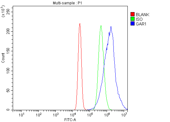
(Figure 11. Flow Cytometry analysis of K562 cells using anti-NOLA1/GAR1 antibody (AAA19579).Overlay histogram showing K562 cells stained with AAA19579 (Blue line). The cells were blocked with 10% normal goat serum. And then incubated with rabbit anti-NOLA1/GAR1 Antibody (AAA19579, 1 ug/1x10^6 cells) for 30 min at 20 degree C. DyLight488 conjugated goat anti-rabbit IgG was used as secondary antibody for 30 minutes at 20 degree C. Isotype control antibody (Green line) was rabbit IgG (1 ug/1x10^6) used under the same conditions. Unlabelled sample (Red line) was also used as a control.)
FCM (Flow Cytometry)
(Figure 11. Flow Cytometry analysis of K562 cells using anti-NOLA1/GAR1 antibody (AAA19579).Overlay histogram showing K562 cells stained with AAA19579 (Blue line). The cells were blocked with 10% normal goat serum. And then incubated with rabbit anti-NOLA1/GAR1 Antibody (AAA19579, 1 ug/1x10^6 cells) for 30 min at 20 degree C. DyLight488 conjugated goat anti-rabbit IgG was used as secondary antibody for 30 minutes at 20 degree C. Isotype control antibody (Green line) was rabbit IgG (1 ug/1x10^6) used under the same conditions. Unlabelled sample (Red line) was also used as a control.)
NOLA1/GAR1, Polyclonal Antibody (Cat# AAA19579)
Full Name
Anti-NOLA1/GAR1 Antibody Picoband
Gene Names
GAR1; NOLA1
Reactivity
Human
Applications
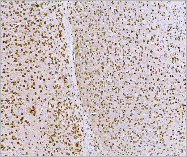
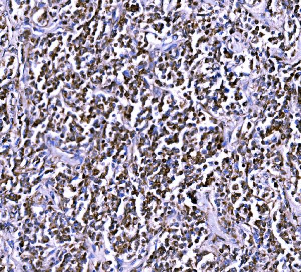
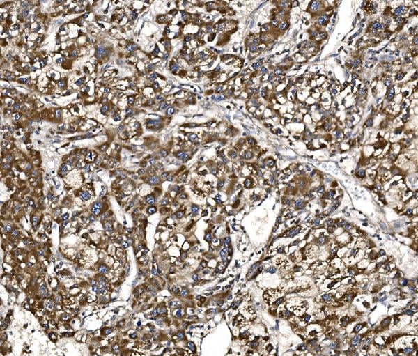
Western Blot, Immunohistochemistry, Immunocytochemistry, Immunofluorescence, Flow Cytometry
Purity
Immunogen affinity purified.
Pricing







 FCM (Flow Cytometry)
(Figure 14. Flow Cytometry analysis of PC-3 cells using anti-BCKDHA antibody (AAA19523).Overlay histogram showing PC-3 cells stained with AAA19523 (Blue line). The cells were blocked with 10% normal goat serum. And then incubated with rabbit anti-BCKDHA Antibody (AAA19523, 1 ug/1x106 cells) for 30 min at 20 degree C. DyLight488 conjugated goat anti-rabbit IgG (BA1127, 5-10 ug/1x106 cells) was used as secondary antibody for 30 minutes at 20 degree C. Isotype control antibody (Green line) was rabbit IgG (1 ug/1x106) used under the same conditions. Unlabelled sample (Red line) was also used as a control.)
FCM (Flow Cytometry)
(Figure 14. Flow Cytometry analysis of PC-3 cells using anti-BCKDHA antibody (AAA19523).Overlay histogram showing PC-3 cells stained with AAA19523 (Blue line). The cells were blocked with 10% normal goat serum. And then incubated with rabbit anti-BCKDHA Antibody (AAA19523, 1 ug/1x106 cells) for 30 min at 20 degree C. DyLight488 conjugated goat anti-rabbit IgG (BA1127, 5-10 ug/1x106 cells) was used as secondary antibody for 30 minutes at 20 degree C. Isotype control antibody (Green line) was rabbit IgG (1 ug/1x106) used under the same conditions. Unlabelled sample (Red line) was also used as a control.)
BCKDHA, Polyclonal Antibody (Cat# AAA19523)
Full Name
Anti-BCKDHA Antibody Picoband
Gene Names
BCKDHA; MSU; MSUD1; OVD1A; BCKDE1A
Reactivity
Human, Mouse, Rat
Applications
Western Blot, Immunohistochemistry, Immunocytochemistry, Immunofluorescence, Flow Cytometry
Purity
Immunogen affinity purified.
Pricing

 FCM (Flow Cytometry)
(Figure 9. Flow Cytometry analysis of MCF-7 cells using anti-C18orf22/RBFA antibody (AAA19984).Overlay histogram showing MCF-7 cells stained with AAA19984 (Blue line). To facilitate intracellular staining, cells were fixed with 4% paraformaldehyde and permeabilized with permeabilization buffer. The cells were blocked with 10% normal goat serum. And then incubated with rabbit anti-C18orf22/RBFA Antibody (AAA19984, 1ug/1x106 cells) for 30 min at 20 degree C. DyLight488 conjugated goat anti-rabbit IgG was used as secondary antibody for 30 minutes at 20 degree C. Isotype control antibody (Green line) was rabbit IgG (1ug/1x106) used under the same conditions. Unlabelled sample without incubation with primary antibody and secondary antibody (Red line) was used as a blank control.)
FCM (Flow Cytometry)
(Figure 9. Flow Cytometry analysis of MCF-7 cells using anti-C18orf22/RBFA antibody (AAA19984).Overlay histogram showing MCF-7 cells stained with AAA19984 (Blue line). To facilitate intracellular staining, cells were fixed with 4% paraformaldehyde and permeabilized with permeabilization buffer. The cells were blocked with 10% normal goat serum. And then incubated with rabbit anti-C18orf22/RBFA Antibody (AAA19984, 1ug/1x106 cells) for 30 min at 20 degree C. DyLight488 conjugated goat anti-rabbit IgG was used as secondary antibody for 30 minutes at 20 degree C. Isotype control antibody (Green line) was rabbit IgG (1ug/1x106) used under the same conditions. Unlabelled sample without incubation with primary antibody and secondary antibody (Red line) was used as a blank control.)
C18orf22/RBFA, Polyclonal Antibody (Cat# AAA19984)
Full Name
Anti-C18orf22/RBFA Antibody Picoband
Gene Names
RBFA; HsT169; C18orf22
Reactivity
Human
Applications
Western Blot, Immunohistochemistry, Immunocytochemistry, Immunofluorescence, Flow Cytometry
Purity
Immunogen affinity purified.
Pricing


 FCM (Flow Cytometry)
(Figure 6. Flow Cytometry analysis of U87 cells using anti-IFIT5 antibody (AAA19582).Overlay histogram showing U87 cells stained with AAA19582 (Blue line). The cells were blocked with 10% normal goat serum. And then incubated with rabbit anti-IFIT5 Antibody (AAA19582, 1 ug/1x10^6 cells) for 30 min at 20 degree C. DyLight488 conjugated goat anti-rabbit IgG was used as secondary antibody for 30 minutes at 20 degree C. Isotype control antibody (Green line) was rabbit IgG (1 ug/1x10^6) used under the same conditions. Unlabelled sample (Red line) was also used as a control.)
FCM (Flow Cytometry)
(Figure 6. Flow Cytometry analysis of U87 cells using anti-IFIT5 antibody (AAA19582).Overlay histogram showing U87 cells stained with AAA19582 (Blue line). The cells were blocked with 10% normal goat serum. And then incubated with rabbit anti-IFIT5 Antibody (AAA19582, 1 ug/1x10^6 cells) for 30 min at 20 degree C. DyLight488 conjugated goat anti-rabbit IgG was used as secondary antibody for 30 minutes at 20 degree C. Isotype control antibody (Green line) was rabbit IgG (1 ug/1x10^6) used under the same conditions. Unlabelled sample (Red line) was also used as a control.)
IFIT5, Polyclonal Antibody (Cat# AAA19582)
Full Name
Anti-IFIT5 Antibody Picoband
Gene Names
IFIT5; P58; RI58
Reactivity
Human
Applications
Western Blot, Immunohistochemistry, Immunocytochemistry, Immunofluorescence, Flow Cytometry
Purity
Immunogen affinity purified.
Pricing


 FCM (Flow Cytometry)
(Figure 10. Flow Cytometry analysis of SiHa cells using anti-MLST8 antibody (AAA19842).Overlay histogram showing SiHa cells stained with AAA19842 (Blue line). To facilitate intracellular staining, cells were fixed with 4% paraformaldehyde and permeabilized with permeabilization buffer. The cells were blocked with 10% normal goat serum. And then incubated with rabbit anti-MLST8 Antibody (AAA19842, 1ug/1x106 cells) for 30 min at 20 degree C. DyLight488 conjugated goat anti-rabbit IgG was used as secondary antibody for 30 minutes at 20 degree C. Isotype control antibody (Green line) was rabbit IgG (1ug/1x106) used under the same conditions. Unlabelled sample (Red line) was also used as a control.)
FCM (Flow Cytometry)
(Figure 10. Flow Cytometry analysis of SiHa cells using anti-MLST8 antibody (AAA19842).Overlay histogram showing SiHa cells stained with AAA19842 (Blue line). To facilitate intracellular staining, cells were fixed with 4% paraformaldehyde and permeabilized with permeabilization buffer. The cells were blocked with 10% normal goat serum. And then incubated with rabbit anti-MLST8 Antibody (AAA19842, 1ug/1x106 cells) for 30 min at 20 degree C. DyLight488 conjugated goat anti-rabbit IgG was used as secondary antibody for 30 minutes at 20 degree C. Isotype control antibody (Green line) was rabbit IgG (1ug/1x106) used under the same conditions. Unlabelled sample (Red line) was also used as a control.)
MLST8, Polyclonal Antibody (Cat# AAA19842)
Full Name
Anti-MLST8 Antibody Picoband
Gene Names
MLST8; GBL; LST8; POP3; WAT1; GbetaL
Reactivity
Human, Mouse, Rat
Applications
Immunofluorescence, Immunohistochemistry, Immunocytochemistry, Western Blot, Flow Cytometry
Purity
Immunogen affinity purified.
Pricing

 FCM (Flow Cytometry)
(Figure 9. Flow Cytometry analysis of HepG2 cells using anti-GIPC1 antibody (AAA19828).Overlay histogram showing HepG2 cells stained with AAA19828 (Blue line). The cells were fixed with 4% paraformaldehyde and blocked with 10% normal goat serum. And then incubated with rabbit anti-GIPC1 Antibody (AAA19828, 1ug/1x106 cells) for 30 min at 20 degree C. DyLight488 conjugated goat anti-rabbit IgG was used as secondary antibody for 30 minutes at 20 degree C. Isotype control antibody (Green line) was rabbit IgG (1ug/1x106) used under the same conditions. Unlabelled sample without incubation with primary antibody and secondary antibody (Red line) was used as a blank control.)
FCM (Flow Cytometry)
(Figure 9. Flow Cytometry analysis of HepG2 cells using anti-GIPC1 antibody (AAA19828).Overlay histogram showing HepG2 cells stained with AAA19828 (Blue line). The cells were fixed with 4% paraformaldehyde and blocked with 10% normal goat serum. And then incubated with rabbit anti-GIPC1 Antibody (AAA19828, 1ug/1x106 cells) for 30 min at 20 degree C. DyLight488 conjugated goat anti-rabbit IgG was used as secondary antibody for 30 minutes at 20 degree C. Isotype control antibody (Green line) was rabbit IgG (1ug/1x106) used under the same conditions. Unlabelled sample without incubation with primary antibody and secondary antibody (Red line) was used as a blank control.)
GIPC1, Polyclonal Antibody (Cat# AAA19828)
Full Name
Anti-GIPC1 Antibody Picoband
Gene Names
GIPC1; NIP; GIPC; IIP-1; TIP-2; SEMCAP; C19orf3; Hs.6454; GLUT1CBP; RGS19IP1; SYNECTIN; SYNECTIIN
Reactivity
Human, Mouse, Rat
Applications
Western Blot, Immunohistochemistry, Immunocytochemistry, Immunofluorescence, Flow Cytometry
Purity
Immunogen affinity purified.
Pricing


 IF (Immunofluorescence)
(Figure 11. IF analysis of SCO1 using anti-SCO1 antibody (AAA19474).SCO1 was detected in a paraffin-embedded section of human prostate cancer tissue. Heat mediated antigen retrieval was performed in EDTA buffer (pH 8.0, epitope retrieval solution). The tissue section was blocked with 10% goat serum. The tissue section was then incubated with 5 ug/mL rabbit anti-SCO1 Antibody (AAA19474) overnight at 4 degree C. DyLight550 Conjugated Goat Anti-Rabbit IgG (BA1135) was used as secondary antibody at 1:500 dilution and incubated for 30 minutes at 37 degree C. The section was counterstained with DAPI. Visualize using a fluorescence microscope and filter sets appropriate for the label used.)
IF (Immunofluorescence)
(Figure 11. IF analysis of SCO1 using anti-SCO1 antibody (AAA19474).SCO1 was detected in a paraffin-embedded section of human prostate cancer tissue. Heat mediated antigen retrieval was performed in EDTA buffer (pH 8.0, epitope retrieval solution). The tissue section was blocked with 10% goat serum. The tissue section was then incubated with 5 ug/mL rabbit anti-SCO1 Antibody (AAA19474) overnight at 4 degree C. DyLight550 Conjugated Goat Anti-Rabbit IgG (BA1135) was used as secondary antibody at 1:500 dilution and incubated for 30 minutes at 37 degree C. The section was counterstained with DAPI. Visualize using a fluorescence microscope and filter sets appropriate for the label used.)
SCO1, Polyclonal Antibody (Cat# AAA19474)
Full Name
Anti-SCO1 Antibody Picoband
Gene Names
SCO1; SCOD1
Reactivity
Human
Applications
Flow Cytometry, Immunofluorescence, Immunohistochemistry, Western Blot
Purity
Immunogen affinity purified.
Pricing


 IF (Immunofluorescence)
(Figure 10. IF analysis of NDUFAF1 using anti-NDUFAF1 antibody (AAA19616).NDUFAF1 was detected in an immunocytochemical section of T47D cells. Enzyme antigen retrieval was performed using IHC enzyme antigen retrieval reagent (AR0022) for 15 mins. The cells were blocked with 10% goat serum. And then incubated with 5 ug/mL rabbit anti-NDUFAF1 Antibody (AAA19616) overnight at 4 degree C. DyLight488 Conjugated Goat Anti-Rabbit IgG was used as secondary antibody at 1:100 dilution and incubated for 30 minutes at 37 degree C. The section was counterstained with DAPI. Visualize using a fluorescence microscope and filter sets appropriate for the label used.)
IF (Immunofluorescence)
(Figure 10. IF analysis of NDUFAF1 using anti-NDUFAF1 antibody (AAA19616).NDUFAF1 was detected in an immunocytochemical section of T47D cells. Enzyme antigen retrieval was performed using IHC enzyme antigen retrieval reagent (AR0022) for 15 mins. The cells were blocked with 10% goat serum. And then incubated with 5 ug/mL rabbit anti-NDUFAF1 Antibody (AAA19616) overnight at 4 degree C. DyLight488 Conjugated Goat Anti-Rabbit IgG was used as secondary antibody at 1:100 dilution and incubated for 30 minutes at 37 degree C. The section was counterstained with DAPI. Visualize using a fluorescence microscope and filter sets appropriate for the label used.)
NDUFAF1, Polyclonal Antibody (Cat# AAA19616)
Full Name
Anti-NDUFAF1 Antibody Picoband
Gene Names
NDUFAF1; CGI65; CIA30; CGI-65
Reactivity
Human, Mouse, Rat
Applications
Immunofluorescence, Immunohistochemistry, Immunocytochemistry, Western Blot
Purity
Immunogen affinity purified.
Pricing


 FCM (Flow Cytometry)
(Figure 10. Flow Cytometry analysis of JK cells using anti-MTHFD1 antibody (AAA19442).Overlay histogram showing JK cells stained with AAA19442 (Blue line). The cells were blocked with 10% normal goat serum. And then incubated with rabbit anti-MTHFD1 Antibody (AAA19442, 1 ug/1x10^6 cells) for 30 min at 20 degree C. DyLight488 conjugated goat anti-rabbit IgG was used as secondary antibody for 30 minutes at 20 degree C. Isotype control antibody (Green line) was rabbit IgG (1 ug/1x10^6) used under the same conditions. Unlabelled sample (Red line) was also used as a control.)
FCM (Flow Cytometry)
(Figure 10. Flow Cytometry analysis of JK cells using anti-MTHFD1 antibody (AAA19442).Overlay histogram showing JK cells stained with AAA19442 (Blue line). The cells were blocked with 10% normal goat serum. And then incubated with rabbit anti-MTHFD1 Antibody (AAA19442, 1 ug/1x10^6 cells) for 30 min at 20 degree C. DyLight488 conjugated goat anti-rabbit IgG was used as secondary antibody for 30 minutes at 20 degree C. Isotype control antibody (Green line) was rabbit IgG (1 ug/1x10^6) used under the same conditions. Unlabelled sample (Red line) was also used as a control.)
MTHFD1, Polyclonal Antibody (Cat# AAA19442)
Full Name
Anti-MTHFD1 Antibody Picoband
Gene Names
MTHFD1; MTHFC; MTHFD
Reactivity
Human, Mouse, Rat
Applications
Western Blot, Immunohistochemistry, Immunocytochemistry, Immunofluorescence, Flow Cytometry
Purity
Immunogen affinity purified.
Pricing


 FCM (Flow Cytometry)
(Figure 10. Flow Cytometry analysis of RAW264.7 cells using anti-GNB3 antibody (AAA19456).Overlay histogram showing RAW264.7 cells stained with AAA19456 (Blue line). The cells were blocked with 10% normal goat serum. And then incubated with rabbit anti-GNB3 Antibody (AAA19456, 1 ug/1x10^6 cells) for 30 min at 20 degree C. DyLight488 conjugated goat anti-rabbit IgG was used as secondary antibody for 30 minutes at 20 degree C. Isotype control antibody (Green line) was rabbit IgG (1 ug/1x10^6) used under the same conditions. Unlabelled sample (Red line) was also used as a control.)
FCM (Flow Cytometry)
(Figure 10. Flow Cytometry analysis of RAW264.7 cells using anti-GNB3 antibody (AAA19456).Overlay histogram showing RAW264.7 cells stained with AAA19456 (Blue line). The cells were blocked with 10% normal goat serum. And then incubated with rabbit anti-GNB3 Antibody (AAA19456, 1 ug/1x10^6 cells) for 30 min at 20 degree C. DyLight488 conjugated goat anti-rabbit IgG was used as secondary antibody for 30 minutes at 20 degree C. Isotype control antibody (Green line) was rabbit IgG (1 ug/1x10^6) used under the same conditions. Unlabelled sample (Red line) was also used as a control.)
GNB3, Polyclonal Antibody (Cat# AAA19456)
Full Name
Anti-GNB3 Antibody Picoband
Reactivity
Human, Mouse, Rat
Applications
Flow Cytometry, Immunofluorescence, Immunohistochemistry, Immunocytochemistry, Western Blot
Purity
Immunogen affinity purified.
Pricing


 IHC (Immunohistchemistry)
(Immunohistochemistry of paraffin-embedded mouse kidney using CETN3 antibody at dilution of 1:100 (40x lens).)
IHC (Immunohistchemistry)
(Immunohistochemistry of paraffin-embedded mouse kidney using CETN3 antibody at dilution of 1:100 (40x lens).)
CETN3, Polyclonal Antibody (Cat# AAA28193)
Full Name
CETN3 Polyclonal Antibody
Gene Names
CETN3; CEN3; CDC31
Reactivity
Human, Mouse, Rat
Applications
Western Blot
Purity
Affinity Purification
Pricing


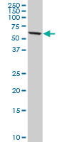
 Application Data
(Detection limit for recombinant GST tagged DDX56 is 0.1 ng/ml as a capture antibody.)
Application Data
(Detection limit for recombinant GST tagged DDX56 is 0.1 ng/ml as a capture antibody.)
DDX56, Monoclonal Antibody (Cat# AAA26253)
Full Name
DDX56 (DEAD (Asp-Glu-Ala-Asp) Box Polypeptide 56, DDX21, DDX26, NOH61) (Biotin)
Gene Names
DDX56; DDX21; DDX26; NOH61
Applications
Western Blot
Purity
Purified
Pricing


 IF (Immunofluorescence)
(Immunofluorescence analysis of U2OS cells using RPL19 Rabbit pAb at dilution of 1:200 (40x lens). Blue: DAPI for nuclear staining.)
IF (Immunofluorescence)
(Immunofluorescence analysis of U2OS cells using RPL19 Rabbit pAb at dilution of 1:200 (40x lens). Blue: DAPI for nuclear staining.)
RPL19, Polyclonal Antibody (Cat# AAA28389)
Full Name
RPL19 Rabbit pAb
Gene Names
RPL19; L19
Reactivity
Human, Mouse, Rat
Applications
Western Blot, Immunohistochemistry
Purity
Affinity purification
Pricing


 FCM (Flow Cytometry)
(Flow cytometry analysis of Hsp105alpha in Hep3B cell line, staining at 2-5ug for 1x10⁶cells (red line). The secondary antibody used goat anti-mouse IgG Alexa fluor 488 conjugate. Isotype control antibody was mouse IgG (black line).)
FCM (Flow Cytometry)
(Flow cytometry analysis of Hsp105alpha in Hep3B cell line, staining at 2-5ug for 1x10⁶cells (red line). The secondary antibody used goat anti-mouse IgG Alexa fluor 488 conjugate. Isotype control antibody was mouse IgG (black line).)
HSP105alpha, Monoclonal Antibody (Cat# AAA11727)
Full Name
HSP105alpha antibody
Gene Names
HSPH1; HSP105; HSP105A; HSP105B; NY-CO-25
Reactivity
Human
Applications
Western Blot, Flow Cytometry, Immunocytochemistry
Purity
By protein-G affinity chromatography
Pricing

 IP (Immunoprecipitation)
(Figure 7. Immunoprecipitating IDH3B in U251 whole cell lysate.Western blot analysis of IDH3B using anti-IDH3B antibody (AAA19926).Lane 1: U251 whole cell lysates (30ug)Lane 2: Rabbit control IgG instead of anti-IDH3B antibody in U251 whole cell lysate.Lane 3: anti-IDH3B antibody (2ug) + U251 whole cell lysate (500ug)After electrophoresis, proteins were transferred to a membrane. Then the membrane was incubated with rabbit anti-IDH3B antigen affinity purified polyclonal antibody (AAA19926) at a dilution of 0.5ug/mL and probed with a goat anti-rabbit IgG-HRP secondary antibody ( cells were fixed with 4% paraformaldehyde and permeabilized with permeabilization buffer. The cells were blocked with 10% normal goat serum. And then incubated with rabbit anti-IDH3B Antibody (AAA19926, 1ug/1x106 cells) for 30 min at 20 degree C. DyLight488 conjugated goat anti-rabbit IgG was used as secondary antibody for 30 minutes at 20 degree C. Isotype control antibody (Green line) was rabbit IgG (1ug/1x106) used under the same conditions. Unlabelled sample (Red line) was also used as a control.)
IP (Immunoprecipitation)
(Figure 7. Immunoprecipitating IDH3B in U251 whole cell lysate.Western blot analysis of IDH3B using anti-IDH3B antibody (AAA19926).Lane 1: U251 whole cell lysates (30ug)Lane 2: Rabbit control IgG instead of anti-IDH3B antibody in U251 whole cell lysate.Lane 3: anti-IDH3B antibody (2ug) + U251 whole cell lysate (500ug)After electrophoresis, proteins were transferred to a membrane. Then the membrane was incubated with rabbit anti-IDH3B antigen affinity purified polyclonal antibody (AAA19926) at a dilution of 0.5ug/mL and probed with a goat anti-rabbit IgG-HRP secondary antibody ( cells were fixed with 4% paraformaldehyde and permeabilized with permeabilization buffer. The cells were blocked with 10% normal goat serum. And then incubated with rabbit anti-IDH3B Antibody (AAA19926, 1ug/1x106 cells) for 30 min at 20 degree C. DyLight488 conjugated goat anti-rabbit IgG was used as secondary antibody for 30 minutes at 20 degree C. Isotype control antibody (Green line) was rabbit IgG (1ug/1x106) used under the same conditions. Unlabelled sample (Red line) was also used as a control.)
IDH3B, Polyclonal Antibody (Cat# AAA19926)
Full Name
Anti-IDH3B Antibody Picoband
Gene Names
IDH3B; RP46; H-IDHB
Reactivity
Human, Mouse, Rat
Applications
Flow Cytometry, Immunoprecipitation, Immunohistochemistry, Western Blot
Purity
Immunogen affinity purified.
Pricing

 FCM (Flow Cytometry)
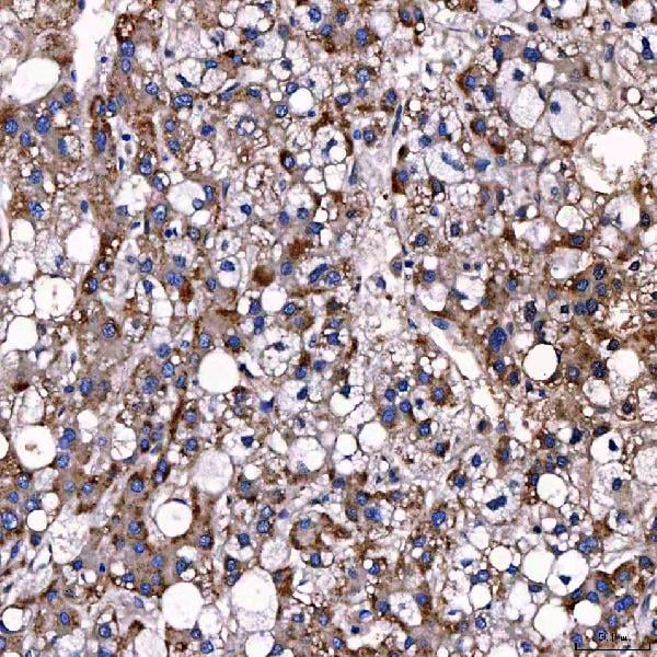
(Figure 8. Flow Cytometry analysis of HepG2 cells using anti-LARG/ARHGEF12 antibody (AAA19877).Overlay histogram showing HepG2 cells stained with AAA19877 (Blue line). To facilitate intracellular staining, cells were fixed with 4% paraformaldehyde and permeabilized with permeabilization buffer. The cells were blocked with 10% normal goat serum. And then incubated with rabbit anti-LARG/ARHGEF12 Antibody (AAA19877, 1ug/1x106 cells) for 30 min at 20 degree C. DyLight488 conjugated goat anti-rabbit IgG was used as secondary antibody for 30 minutes at 20 degree C. Isotype control antibody (Green line) was rabbit IgG (1ug/1x106) used under the same conditions. Unlabelled sample (Red line) was also used as a control.)
FCM (Flow Cytometry)
(Figure 8. Flow Cytometry analysis of HepG2 cells using anti-LARG/ARHGEF12 antibody (AAA19877).Overlay histogram showing HepG2 cells stained with AAA19877 (Blue line). To facilitate intracellular staining, cells were fixed with 4% paraformaldehyde and permeabilized with permeabilization buffer. The cells were blocked with 10% normal goat serum. And then incubated with rabbit anti-LARG/ARHGEF12 Antibody (AAA19877, 1ug/1x106 cells) for 30 min at 20 degree C. DyLight488 conjugated goat anti-rabbit IgG was used as secondary antibody for 30 minutes at 20 degree C. Isotype control antibody (Green line) was rabbit IgG (1ug/1x106) used under the same conditions. Unlabelled sample (Red line) was also used as a control.)
LARG/ARHGEF12, Polyclonal Antibody (Cat# AAA19877)
Full Name
Anti-LARG/ARHGEF12 Antibody Picoband
Gene Names
ARHGEF12; LARG; PRO2792
Reactivity
Human, Mouse, Rat
Applications
Flow Cytometry, Immunofluorescence, Immunohistochemistry, Immunocytochemistry, Western Blot
Purity
Immunogen affinity purified.
Pricing




 FCM (Flow Cytometry)
(Figure 9. Flow Cytometry analysis of HepG2 cells using anti-TTC7B antibody (AAA19626).Overlay histogram showing HepG2 cells stained with AAA19626 (Blue line). The cells were blocked with 10% normal goat serum. And then incubated with rabbit anti-TTC7B Antibody (AAA19626, 1 ug/1x10^6 cells) for 30 min at 20 degree C. DyLight488 conjugated goat anti-rabbit IgG was used as secondary antibody for 30 minutes at 20 degree C. Isotype control antibody (Green line) was rabbit IgG (1 ug/1x10^6) used under the same conditions. Unlabelled sample (Red line) was also used as a control.)
FCM (Flow Cytometry)
(Figure 9. Flow Cytometry analysis of HepG2 cells using anti-TTC7B antibody (AAA19626).Overlay histogram showing HepG2 cells stained with AAA19626 (Blue line). The cells were blocked with 10% normal goat serum. And then incubated with rabbit anti-TTC7B Antibody (AAA19626, 1 ug/1x10^6 cells) for 30 min at 20 degree C. DyLight488 conjugated goat anti-rabbit IgG was used as secondary antibody for 30 minutes at 20 degree C. Isotype control antibody (Green line) was rabbit IgG (1 ug/1x10^6) used under the same conditions. Unlabelled sample (Red line) was also used as a control.)
TTC7B, Polyclonal Antibody (Cat# AAA19626)
Full Name
Anti-TTC7B Antibody Picoband
Gene Names
TTC7B; TTC7L1; c14_5685
Reactivity
Human, Mouse, Rat
Applications
Western Blot, Immunohistochemistry, Flow Cytometry
Purity
Immunogen affinity purified.
Pricing
 WB (Western Blot)
(Western blot analysis of extracts of various cell lines, using SCN3B antibody (AAA28227) at 1:1000 dilution.Secondary antibody: HRP Goat Anti-Rabbit IgG (H+L) at 1:10000 dilution.Lysates/proteins: 25ug per lane.Blocking buffer: 3% nonfat dry milk in TBST.Detection: ECL Basic Kit.Exposure time: 5s.)
WB (Western Blot)
(Western blot analysis of extracts of various cell lines, using SCN3B antibody (AAA28227) at 1:1000 dilution.Secondary antibody: HRP Goat Anti-Rabbit IgG (H+L) at 1:10000 dilution.Lysates/proteins: 25ug per lane.Blocking buffer: 3% nonfat dry milk in TBST.Detection: ECL Basic Kit.Exposure time: 5s.)
SCN3B, Polyclonal Antibody (Cat# AAA28227)
Full Name
SCN3B Polyclonal Antibody
Gene Names
SCN3B; SCNB3; ATFB16; BRGDA7; HSA243396
Reactivity
Human, Mouse
Applications
Western Blot
Purity
Affinity Purification
Pricing


 Application Data
(Detection limit for recombinant GST tagged DNAJB6 is ~1ng/ml as a capture antibody.)
Application Data
(Detection limit for recombinant GST tagged DNAJB6 is ~1ng/ml as a capture antibody.)
DNAJB6, Monoclonal Antibody (Cat# AAA25669)
Full Name
DNAJB6 (HSJ2, MRJ, MSJ1, DnaJ Homolog Subfamily B Member 6, HHDJ1, Heat Shock Protein J2, HSJ-2, MSJ-1, MGC1152, MGC117297, DKFZp566D0824, FLJ42837) (PE)
Gene Names
DNAJB6; DJ4; MRJ; DnaJ; HSJ2; HHDJ1; HSJ-2; MSJ-1; LGMD1D; LGMD1E; LGMDD1
Reactivity
Human
Applications
Immunofluorescence, Western Blot
Purity
Purified by Protein A Affinity Chromatography.
Pricing

 IF (Immunofluorescence)
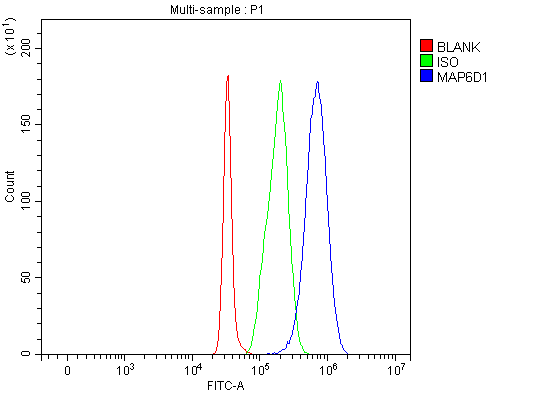
(Figure 7. IF analysis of MAP6D1 using anti-MAP6D1 antibody (AAA20004).MAP6D1 was detected in an immunocytochemical section of A549 cells. Enzyme antigen retrieval was performed using IHC enzyme antigen retrieval reagent ( cells were fixed with 4% paraformaldehyde and permeabilized with permeabilization buffer. The cells were blocked with 10% normal goat serum. And then incubated with rabbit anti-MAP6D1 Antibody (AAA20004, 1ug/1x106 cells) for 30 min at 20 degree C. DyLight488 conjugated goat anti-rabbit IgG was used as secondary antibody for 30 minutes at 20 degree C. Isotype control antibody (Green line) was rabbit IgG (1ug/1x106) used under the same conditions. Unlabelled sample (Red line) was also used as a control.)
IF (Immunofluorescence)
(Figure 7. IF analysis of MAP6D1 using anti-MAP6D1 antibody (AAA20004).MAP6D1 was detected in an immunocytochemical section of A549 cells. Enzyme antigen retrieval was performed using IHC enzyme antigen retrieval reagent ( cells were fixed with 4% paraformaldehyde and permeabilized with permeabilization buffer. The cells were blocked with 10% normal goat serum. And then incubated with rabbit anti-MAP6D1 Antibody (AAA20004, 1ug/1x106 cells) for 30 min at 20 degree C. DyLight488 conjugated goat anti-rabbit IgG was used as secondary antibody for 30 minutes at 20 degree C. Isotype control antibody (Green line) was rabbit IgG (1ug/1x106) used under the same conditions. Unlabelled sample (Red line) was also used as a control.)
MAP6D1, Polyclonal Antibody (Cat# AAA20004)
Full Name
Anti-MAP6D1 Antibody Picoband
Gene Names
MAP6D1; SL21; MAPO6D1
Reactivity
Human, Mouse, Rat
Applications
Western Blot, Immunohistochemistry, Immunocytochemistry, Immunofluorescence, Flow Cytometry
Purity
Immunogen affinity purified.
Pricing


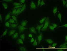


 WB (Western Blot)
(DNAJC10 monoclonal antibody. Western Blot analysis of DNAJC10 expression in HeLa.)
WB (Western Blot)
(DNAJC10 monoclonal antibody. Western Blot analysis of DNAJC10 expression in HeLa.)
DNAJC10, Monoclonal Antibody (Cat# AAA25670)
Full Name
DNAJC10 (DnaJ Homolog Subfamily C Member 10, ER-resident Protein ERdj5, Macrothioredoxin, MTHr, ERDJ5, UNQ495/PRO1012, DKFZp434J1813, MGC104194) (PE)
Gene Names
DNAJC10; JPDI; MTHr; ERdj5; PDIA19
Reactivity
Human, Mouse, Rat
Applications
Immunofluorescence, Immunohistochemistry, Western Blot
Purity
Purified by Protein A Affinity Chromatography.
Pricing

 FCM (Flow Cytometry)
(Figure 6. Flow Cytometry analysis of HEL cells using anti-STXBP6 antibody (AAA19956).Overlay histogram showing HEL cells stained with AAA19956 (Blue line). To facilitate intracellular staining, cells were fixed with 4% paraformaldehyde and permeabilized with permeabilization buffer. The cells were blocked with 10% normal goat serum. And then incubated with rabbit anti-STXBP6 Antibody (AAA19956, 1ug/1x106 cells) for 30 min at 20 degree C. DyLight488 conjugated goat anti-rabbit IgG was used as secondary antibody for 30 minutes at 20 degree C. Isotype control antibody (Green line) was rabbit IgG (1ug/1x106) used under the same conditions. Unlabelled sample (Red line) was also used as a control.)
FCM (Flow Cytometry)
(Figure 6. Flow Cytometry analysis of HEL cells using anti-STXBP6 antibody (AAA19956).Overlay histogram showing HEL cells stained with AAA19956 (Blue line). To facilitate intracellular staining, cells were fixed with 4% paraformaldehyde and permeabilized with permeabilization buffer. The cells were blocked with 10% normal goat serum. And then incubated with rabbit anti-STXBP6 Antibody (AAA19956, 1ug/1x106 cells) for 30 min at 20 degree C. DyLight488 conjugated goat anti-rabbit IgG was used as secondary antibody for 30 minutes at 20 degree C. Isotype control antibody (Green line) was rabbit IgG (1ug/1x106) used under the same conditions. Unlabelled sample (Red line) was also used as a control.)
STXBP6, Polyclonal Antibody (Cat# AAA19956)
Full Name
Anti-STXBP6 Antibody Picoband
Gene Names
STXBP6; amisyn; HSPC156
Reactivity
Human, Mouse, Rat
Applications
Western Blot, Immunohistochemistry, Flow Cytometry
Purity
Immunogen affinity purified.
Pricing

 FCM (Flow Cytometry)
(Figure 10. Flow Cytometry analysis of A549 cells using anti-SLK antibody (AAA19754).Overlay histogram showing A549 cells stained with AAA19754 (Blue line). To facilitate intracellular staining, cells were fixed with 4% paraformaldehyde and permeabilized with permeabilization buffer. The cells were blocked with 10% normal goat serum. And then incubated with rabbit anti-SLK Antibody (AAA19754, 1ug/1x106 cells) for 30 min at 20 degree C. DyLight488 conjugated goat anti-rabbit IgG was used as secondary antibody for 30 minutes at 20 degree C. Isotype control antibody (Green line) was rabbit IgG (1ug/1x106) used under the same conditions. Unlabelled sample (Red line) was also used as a control.)
FCM (Flow Cytometry)
(Figure 10. Flow Cytometry analysis of A549 cells using anti-SLK antibody (AAA19754).Overlay histogram showing A549 cells stained with AAA19754 (Blue line). To facilitate intracellular staining, cells were fixed with 4% paraformaldehyde and permeabilized with permeabilization buffer. The cells were blocked with 10% normal goat serum. And then incubated with rabbit anti-SLK Antibody (AAA19754, 1ug/1x106 cells) for 30 min at 20 degree C. DyLight488 conjugated goat anti-rabbit IgG was used as secondary antibody for 30 minutes at 20 degree C. Isotype control antibody (Green line) was rabbit IgG (1ug/1x106) used under the same conditions. Unlabelled sample (Red line) was also used as a control.)
SLK, Polyclonal Antibody (Cat# AAA19754)
Full Name
Anti-SLK Antibody Picoband
Gene Names
SLK; LOSK; STK2; se20-9; bA16H23.1
Reactivity
Human, Mouse
Applications
Western Blot, Immunohistochemistry, Flow Cytometry
Purity
Immunogen affinity purified.
Pricing


 FCM (Flow Cytometry)
(Figure 6. Flow Cytometry analysis of MCF-7 cells using anti-MCM7 antibody (AAA19689).Overlay histogram showing MCF-7 cells stained with AAA19689 (Blue line). The cells were blocked with 10% normal goat serum. And then incubated with mouse anti-MCM7 Antibody (AAA19689, 1 ug/1x10^6 cells) for 30 min at 20 degree C. DyLight488 conjugated goat anti-mouse IgG was used as secondary antibody for 30 minutes at 20 degree C. Isotype control antibody (Green line) was mouse IgG (1 ug/1x10^6) used under the same conditions. Unlabelled sample (Red line) was also used as a control.)
FCM (Flow Cytometry)
(Figure 6. Flow Cytometry analysis of MCF-7 cells using anti-MCM7 antibody (AAA19689).Overlay histogram showing MCF-7 cells stained with AAA19689 (Blue line). The cells were blocked with 10% normal goat serum. And then incubated with mouse anti-MCM7 Antibody (AAA19689, 1 ug/1x10^6 cells) for 30 min at 20 degree C. DyLight488 conjugated goat anti-mouse IgG was used as secondary antibody for 30 minutes at 20 degree C. Isotype control antibody (Green line) was mouse IgG (1 ug/1x10^6) used under the same conditions. Unlabelled sample (Red line) was also used as a control.)
MCM7, Monoclonal Antibody (Cat# AAA19689)
Full Name
Anti-MCM7 Antibody Picoband (monoclonal, 3H11)
Gene Names
MCM7; MCM2; CDC47; P85MCM; P1CDC47; PNAS146; P1.1-MCM3
Reactivity
Human, Rat
Applications
Western Blot, Immunohistochemistry, Immunocytochemistry, Immunofluorescence, Flow Cytometry
Purity
Immunogen affinity purified.
Pricing


 IF (Immunofluorescence)
(Figure 14. IF analysis of TTLL2 using anti-TTLL2 antibody (AAA19641).TTLL2 was detected in a paraffin-embedded section of rat testis tissue. Heat mediated antigen retrieval was performed in EDTA buffer (pH 8.0, epitope retrieval solution). The tissue section was blocked with 10% goat serum. The tissue section was then incubated with 5 ug/mL rabbit anti-TTLL2 Antibody (AAA19641) overnight at 4 degree C. DyLight550 Conjugated Goat Anti-Rabbit IgG (BA1135) was used as secondary antibody at 1:100 dilution and incubated for 30 minutes at 37 degree C. The section was counterstained with DAPI. Visualize using a fluorescence microscope and filter sets appropriate for the label used.)
IF (Immunofluorescence)
(Figure 14. IF analysis of TTLL2 using anti-TTLL2 antibody (AAA19641).TTLL2 was detected in a paraffin-embedded section of rat testis tissue. Heat mediated antigen retrieval was performed in EDTA buffer (pH 8.0, epitope retrieval solution). The tissue section was blocked with 10% goat serum. The tissue section was then incubated with 5 ug/mL rabbit anti-TTLL2 Antibody (AAA19641) overnight at 4 degree C. DyLight550 Conjugated Goat Anti-Rabbit IgG (BA1135) was used as secondary antibody at 1:100 dilution and incubated for 30 minutes at 37 degree C. The section was counterstained with DAPI. Visualize using a fluorescence microscope and filter sets appropriate for the label used.)
TTLL2, Polyclonal Antibody (Cat# AAA19641)
Full Name
Anti-TTLL2 Antibody Picoband
Gene Names
TTLL2; C6orf104; NYD-TSPG; dJ366N23.3
Reactivity
Human, Mouse, Rat
Applications
Immunofluorescence, Immunohistochemistry, Western Blot
Purity
Immunogen affinity purified.
Pricing


 WB (Western Blot)
(LDB3 monoclonal antibody Western Blot analysis of LDB3 expression in A-431.)
WB (Western Blot)
(LDB3 monoclonal antibody Western Blot analysis of LDB3 expression in A-431.)
LDB3, Monoclonal Antibody (Cat# AAA25437)
Full Name
LDB3 (LIM Domain-binding Protein 3, Protein Cypher, Z-band Alternatively Spliced PDZ-motif Protein, KIAA0613, ZASP) (HRP)
Gene Names
LDB3; MFM4; ZASP; CMD1C; CMH24; CMPD3; LVNC3; CYPHER; LDB3Z1; LDB3Z4; ORACLE; PDLIM6
Reactivity
Human
Applications
Western Blot
Purity
Purified by Protein A Affinity Chromatography.
Pricing

 FCM (Flow Cytometry)
(Figure 10. Flow Cytometry analysis of Hela cells using anti-RPL18 antibody (AAA19881).Overlay histogram showing Hela cells stained with AAA19881 (Blue line). To facilitate intracellular staining, cells were fixed with 4% paraformaldehyde and permeabilized with permeabilization buffer. The cells were blocked with 10% normal goat serum. And then incubated with rabbit anti-RPL18 Antibody (AAA19881, 1ug/1x106 cells) for 30 min at 20 degree C. DyLight488 conjugated goat anti-rabbit IgG was used as secondary antibody for 30 minutes at 20 degree C. Isotype control antibody (Green line) was rabbit IgG (1ug/1x106) used under the same conditions. Unlabelled sample (Red line) was also used as a control.)
FCM (Flow Cytometry)
(Figure 10. Flow Cytometry analysis of Hela cells using anti-RPL18 antibody (AAA19881).Overlay histogram showing Hela cells stained with AAA19881 (Blue line). To facilitate intracellular staining, cells were fixed with 4% paraformaldehyde and permeabilized with permeabilization buffer. The cells were blocked with 10% normal goat serum. And then incubated with rabbit anti-RPL18 Antibody (AAA19881, 1ug/1x106 cells) for 30 min at 20 degree C. DyLight488 conjugated goat anti-rabbit IgG was used as secondary antibody for 30 minutes at 20 degree C. Isotype control antibody (Green line) was rabbit IgG (1ug/1x106) used under the same conditions. Unlabelled sample (Red line) was also used as a control.)
RPL18, Polyclonal Antibody (Cat# AAA19881)
Full Name
Anti-RPL18 Antibody Picoband
Gene Names
RPL18; L18
Reactivity
Human, Mouse, Rat
Applications
Western Blot, Immunohistochemistry, Immunocytochemistry, Immunofluorescence, Flow Cytometry
Purity
Immunogen affinity purified.
Pricing


 WB (Western Blot)
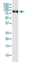
(Western Blot analysis of OPA1 expression in transfected 293T cell line by OPA1 monoclonal antibody (M06), clone 8A32.Lane 1: OPA1 transfected lysate (Predicted MW: 111.6 KDa).Lane 2: Non-transfected lysate.)
WB (Western Blot)
(Western Blot analysis of OPA1 expression in transfected 293T cell line by OPA1 monoclonal antibody (M06), clone 8A32.Lane 1: OPA1 transfected lysate (Predicted MW: 111.6 KDa).Lane 2: Non-transfected lysate.)
OPA1, Monoclonal Antibody (Cat# AAA26419)
Full Name
OPA1 (Pptic Atrophy 1 (Autosomal Dominant), FLJ12460, KIAA0567, MGM1, NPG, NTG, LargeG) (FITC)
Gene Names
OPA1; NPG; NTG; MGM1; BERHS; largeG; MTDPS14
Applications
Western Blot
Purity
Purified
Pricing


 FCM (Flow Cytometry)
(Figure 6. Flow Cytometry analysis of JK cells using anti-GSPT1,2 antibody (AAA19586).Overlay histogram showing JK cells stained with AAA19586 (Blue line). The cells were blocked with 10% normal goat serum. And then incubated with rabbit anti-GSPT1,2 Antibody (AAA19586, 1 ug/1x10^6 cells) for 30 min at 20 degree C. DyLight488 conjugated goat anti-rabbit IgG was used as secondary antibody for 30 minutes at 20 degree C. Isotype control antibody (Green line) was rabbit IgG (1 ug/1x10^6) used under the same conditions. Unlabelled sample (Red line) was also used as a control.)
FCM (Flow Cytometry)
(Figure 6. Flow Cytometry analysis of JK cells using anti-GSPT1,2 antibody (AAA19586).Overlay histogram showing JK cells stained with AAA19586 (Blue line). The cells were blocked with 10% normal goat serum. And then incubated with rabbit anti-GSPT1,2 Antibody (AAA19586, 1 ug/1x10^6 cells) for 30 min at 20 degree C. DyLight488 conjugated goat anti-rabbit IgG was used as secondary antibody for 30 minutes at 20 degree C. Isotype control antibody (Green line) was rabbit IgG (1 ug/1x10^6) used under the same conditions. Unlabelled sample (Red line) was also used as a control.)
GSPT1,2, Polyclonal Antibody (Cat# AAA19586)
Full Name
Anti-GSPT1,2 Antibody Picoband
Gene Names
GSPT1; GST1; ETF3A; eRF3a; 551G9.2
Reactivity
Human, Mouse, Rat
Applications
Western Blot, Immunohistochemistry, Immunocytochemistry, Immunofluorescence, Flow Cytometry
Purity
Immunogen affinity purified.
Pricing


 FCM (Flow Cytometry)
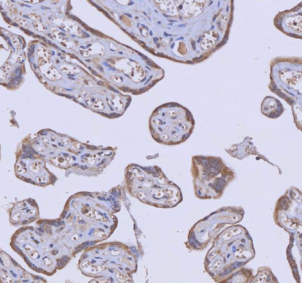
(Figure 8. Flow Cytometry analysis of SiHa cells using anti-WDR44 antibody (AAA19624).Overlay histogram showing SiHa cells stained with AAA19624 (Blue line). The cells were blocked with 10% normal goat serum. And then incubated with rabbit anti-WDR44 Antibody (AAA19624, 1 ug/1x10^6 cells) for 30 min at 20 degree C. DyLight488 conjugated goat anti-rabbit IgG was used as secondary antibody for 30 minutes at 20 degree C. Isotype control antibody (Green line) was rabbit IgG (1 ug/1x10^6) used under the same conditions. Unlabelled sample (Red line) was also used as a control.)
FCM (Flow Cytometry)
(Figure 8. Flow Cytometry analysis of SiHa cells using anti-WDR44 antibody (AAA19624).Overlay histogram showing SiHa cells stained with AAA19624 (Blue line). The cells were blocked with 10% normal goat serum. And then incubated with rabbit anti-WDR44 Antibody (AAA19624, 1 ug/1x10^6 cells) for 30 min at 20 degree C. DyLight488 conjugated goat anti-rabbit IgG was used as secondary antibody for 30 minutes at 20 degree C. Isotype control antibody (Green line) was rabbit IgG (1 ug/1x10^6) used under the same conditions. Unlabelled sample (Red line) was also used as a control.)
WDR44, Polyclonal Antibody (Cat# AAA19624)
Full Name
Anti-WDR44 Antibody Picoband
Gene Names
WDR44; RPH11; RAB11BP
Reactivity
Human
Applications
Western Blot, Immunohistochemistry, Flow Cytometry
Purity
Immunogen affinity purified.
Pricing


 IHC (Immunohistochemistry)
(Immunohistochemistry of paraffin-embedded mouse pancreas using DOK1 Antibody at dilution of 1:100 (40x lens).)
IHC (Immunohistochemistry)
(Immunohistochemistry of paraffin-embedded mouse pancreas using DOK1 Antibody at dilution of 1:100 (40x lens).)
DOK1, Polyclonal Antibody (Cat# AAA28181)
Full Name
DOK1 Polyclonal Antibody
Gene Names
DOK1; P62DOK
Reactivity
Human, Mouse, Rat
Applications
Western Blot, Immunohistochemistry, Immunofluorescence
Purity
Affinity Purification
Pricing


 IHC (Immunohistchemistry)
(Immunohistochemistry of paraffin-embedded human liver cancer using SMARCE1 Antibody at dilution of 1:200 (40x lens).)
IHC (Immunohistchemistry)
(Immunohistochemistry of paraffin-embedded human liver cancer using SMARCE1 Antibody at dilution of 1:200 (40x lens).)
SMARCE1, Polyclonal Antibody (Cat# AAA28107)
Full Name
SMARCE1 Polyclonal Antibody
Gene Names
SMARCE1; BAF57
Reactivity
Human, Mouse
Applications
Western Blot, Immunohistochemistry, Immunofluorescence, Immunoprecipitation, Chromatin Immunoprecipitation
Purity
Affinity Purification
Pricing


 FCM (Flow Cytometry)
(Figure 9. Flow Cytometry analysis of K562 cells using anti-MCM6 antibody (AAA19372).Overlay histogram showing K562 cells stained with AAA19372 (Blue line). The cells were blocked with 10% normal goat serum. And then incubated with mouse anti- MCM6 Antibody (AAA19372, 1μg/1x106 cells) for 30 min at 20 degree C. DyLight®488 conjugated goat anti-mouse IgG (BA1126, 5-10μg/1x106 cells) was used as secondary antibody for 30 minutes at 20 degree C. Isotype control antibody (Green line) was mouse IgG (1μg/1x106) used under the same conditions. Unlabelled sample (Red line) was also used as a control.)
FCM (Flow Cytometry)
(Figure 9. Flow Cytometry analysis of K562 cells using anti-MCM6 antibody (AAA19372).Overlay histogram showing K562 cells stained with AAA19372 (Blue line). The cells were blocked with 10% normal goat serum. And then incubated with mouse anti- MCM6 Antibody (AAA19372, 1μg/1x106 cells) for 30 min at 20 degree C. DyLight®488 conjugated goat anti-mouse IgG (BA1126, 5-10μg/1x106 cells) was used as secondary antibody for 30 minutes at 20 degree C. Isotype control antibody (Green line) was mouse IgG (1μg/1x106) used under the same conditions. Unlabelled sample (Red line) was also used as a control.)
MCM6, Monoclonal Antibody (Cat# AAA19372)
Full Name
Anti-MCM6 Antibody (monoclonal, 10I9)
Gene Names
MCM6; Mis5; P105MCM; MCG40308
Reactivity
Human
Applications
Western Blot, Immunohistochemistry, Immunocytochemistry, Immunofluorescence, Flow Cytometry
Purity
Immunogen affinity purified.
Pricing


 FCM (Flow Cytometry)
(Figure 9. Flow Cytometry analysis of Hela cells using anti-PACSIN2 antibody (AAA19809).Overlay histogram showing Hela cells stained with AAA19809 (Blue line). To facilitate intracellular staining, cells were fixed with 4% paraformaldehyde and permeabilized with permeabilization buffer. The cells were blocked with 10% normal goat serum. And then incubated with rabbit anti-PACSIN2 Antibody (AAA19809, 1ug/1x106 cells) for 30 min at 20 degree C. DyLight488 conjugated goat anti-rabbit IgG was used as secondary antibody for 30 minutes at 20 degree C. Isotype control antibody (Green line) was rabbit IgG (1ug/1x106) used under the same conditions. Unlabelled sample without incubation with primary antibody and secondary antibody (Red line) was used as a blank control.)
FCM (Flow Cytometry)
(Figure 9. Flow Cytometry analysis of Hela cells using anti-PACSIN2 antibody (AAA19809).Overlay histogram showing Hela cells stained with AAA19809 (Blue line). To facilitate intracellular staining, cells were fixed with 4% paraformaldehyde and permeabilized with permeabilization buffer. The cells were blocked with 10% normal goat serum. And then incubated with rabbit anti-PACSIN2 Antibody (AAA19809, 1ug/1x106 cells) for 30 min at 20 degree C. DyLight488 conjugated goat anti-rabbit IgG was used as secondary antibody for 30 minutes at 20 degree C. Isotype control antibody (Green line) was rabbit IgG (1ug/1x106) used under the same conditions. Unlabelled sample without incubation with primary antibody and secondary antibody (Red line) was used as a blank control.)
PACSIN2, Polyclonal Antibody (Cat# AAA19809)
Full Name
Anti-PACSIN2 Antibody Picoband
Gene Names
PACSIN2; SDPII
Reactivity
Human, Monkey, Mouse, Rat
Applications
Western Blot, Immunohistochemistry, Immunocytochemistry, Immunofluorescence, Flow Cytometry
Purity
Immunogen affinity purified.
Pricing


 IHC (Immunohistochemistry)
(Figure 8. IHC analysis of RPL29 using anti-RPL29 antibody (AAA19561).RPL29 was detected in a paraffin-embedded section of human gall bladder adenosquamous carcinoma tissue. Heat mediated antigen retrieval was performed in EDTA buffer (pH 8.0, epitope retrieval solution). The tissue section was blocked with 10% goat serum. The tissue section was then incubated with 2 ug/ml rabbit anti-RPL29 Antibody (AAA19561) overnight at 4 degree C. Biotinylated goat anti-rabbit IgG was used as secondary antibody and incubated for 30 minutes at 37 degree C. The tissue section was developed using Strepavidin-Biotin-Complex (SABC) with DAB as the chromogen.)
IHC (Immunohistochemistry)
(Figure 8. IHC analysis of RPL29 using anti-RPL29 antibody (AAA19561).RPL29 was detected in a paraffin-embedded section of human gall bladder adenosquamous carcinoma tissue. Heat mediated antigen retrieval was performed in EDTA buffer (pH 8.0, epitope retrieval solution). The tissue section was blocked with 10% goat serum. The tissue section was then incubated with 2 ug/ml rabbit anti-RPL29 Antibody (AAA19561) overnight at 4 degree C. Biotinylated goat anti-rabbit IgG was used as secondary antibody and incubated for 30 minutes at 37 degree C. The tissue section was developed using Strepavidin-Biotin-Complex (SABC) with DAB as the chromogen.)
RPL29, Polyclonal Antibody (Cat# AAA19561)
Full Name
Anti-RPL29 Antibody Picoband
Gene Names
RPL29; HIP; L29; HUMRPL29; RPL29P10; RPL29_3_370
Reactivity
Human, Mouse, Rat, Monkey
Applications
Western Blot, Immunohistochemistry, Immunocytochemistry, Immunofluorescence, Flow Cytometry
Purity
Immunogen affinity purified.
Pricing


 FCM (Flow Cytometry)
(Figure 6. Flow Cytometry analysis of RT4 cells using anti-RFX3 antibody (AAA19892).Overlay histogram showing RT4 cells stained with AAA19892 (Blue line). To facilitate intracellular staining, cells were fixed with 4% paraformaldehyde and permeabilized with permeabilization buffer. The cells were blocked with 10% normal goat serum. And then incubated with rabbit anti-RFX3 Antibody (AAA19892, 1ug/1x106 cells) for 30 min at 20 degree C. DyLight488 conjugated goat anti-rabbit IgG was used as secondary antibody for 30 minutes at 20 degree C. Isotype control antibody (Green line) was rabbit IgG (1ug/1x106) used under the same conditions. Unlabelled sample without incubation with primary antibody and secondary antibody (Red line) was used as a blank control.)
FCM (Flow Cytometry)
(Figure 6. Flow Cytometry analysis of RT4 cells using anti-RFX3 antibody (AAA19892).Overlay histogram showing RT4 cells stained with AAA19892 (Blue line). To facilitate intracellular staining, cells were fixed with 4% paraformaldehyde and permeabilized with permeabilization buffer. The cells were blocked with 10% normal goat serum. And then incubated with rabbit anti-RFX3 Antibody (AAA19892, 1ug/1x106 cells) for 30 min at 20 degree C. DyLight488 conjugated goat anti-rabbit IgG was used as secondary antibody for 30 minutes at 20 degree C. Isotype control antibody (Green line) was rabbit IgG (1ug/1x106) used under the same conditions. Unlabelled sample without incubation with primary antibody and secondary antibody (Red line) was used as a blank control.)
RFX3, Polyclonal Antibody (Cat# AAA19892)
Full Name
Anti-RFX3 Antibody Picoband
Reactivity
Human, Mouse, Rat
Applications
Western Blot, Immunohistochemistry, Immunocytochemistry, Immunofluorescence, Flow Cytometry
Purity
Immunogen affinity purified.
Pricing




 FCM (Flow Cytometry)
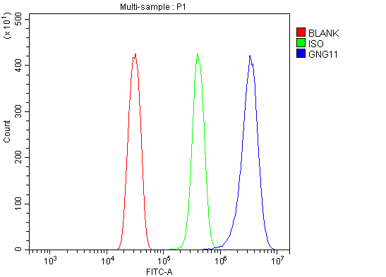
(Figure 12. Flow Cytometry analysis of HEPA1-6 cells using anti-GNG11 antibody (AAA19638).Overlay histogram showing HEPA1-6 cells stained with AAA19638 (Blue line).The cells were blocked with 10% normal goat serum. And then incubated with rabbit anti-GNG11 Antibody (AAA19638, 1ug/1x10^6 cells) for 30 min at 20 degree C. DyLight488 conjugated goat anti-rabbit IgG was used as secondary antibody for 30 minutes at 20 degree C. Isotype control antibody (Green line) was rabbit IgG (1ug/1x10^6) used under the same conditions. Unlabelled sample (Red line) was also used as a control.)
FCM (Flow Cytometry)
(Figure 12. Flow Cytometry analysis of HEPA1-6 cells using anti-GNG11 antibody (AAA19638).Overlay histogram showing HEPA1-6 cells stained with AAA19638 (Blue line).The cells were blocked with 10% normal goat serum. And then incubated with rabbit anti-GNG11 Antibody (AAA19638, 1ug/1x10^6 cells) for 30 min at 20 degree C. DyLight488 conjugated goat anti-rabbit IgG was used as secondary antibody for 30 minutes at 20 degree C. Isotype control antibody (Green line) was rabbit IgG (1ug/1x10^6) used under the same conditions. Unlabelled sample (Red line) was also used as a control.)
GNG11, Polyclonal Antibody (Cat# AAA19638)
Full Name
Anti-GNG11 Antibody Picoband
Gene Names
GNG11; GNGT11
Reactivity
Human, Mouse, Rat
Applications
Western Blot, Immunohistochemistry, Flow Cytometry
Purity
Immunogen affinity purified.
Pricing


 FCM (Flow Cytometry)
(Figure 6. Flow Cytometry analysis of 293T cells using anti-PYCR1 antibody (AAA19858).Overlay histogram showing 293T cells stained with AAA19858 (Blue line). To facilitate intracellular staining, cells were fixed with 4% paraformaldehyde and permeabilized with permeabilization buffer. The cells were blocked with 10% normal goat serum. And then incubated with rabbit anti-PYCR1 Antibody (AAA19858, 1ug/1x106 cells) for 30 min at 20 degree C. DyLight488 conjugated goat anti-rabbit IgG was used as secondary antibody for 30 minutes at 20 degree C. Isotype control antibody (Green line) was rabbit IgG (1ug/1x106) used under the same conditions. Unlabelled sample (Red line) was also used as a control.)
FCM (Flow Cytometry)
(Figure 6. Flow Cytometry analysis of 293T cells using anti-PYCR1 antibody (AAA19858).Overlay histogram showing 293T cells stained with AAA19858 (Blue line). To facilitate intracellular staining, cells were fixed with 4% paraformaldehyde and permeabilized with permeabilization buffer. The cells were blocked with 10% normal goat serum. And then incubated with rabbit anti-PYCR1 Antibody (AAA19858, 1ug/1x106 cells) for 30 min at 20 degree C. DyLight488 conjugated goat anti-rabbit IgG was used as secondary antibody for 30 minutes at 20 degree C. Isotype control antibody (Green line) was rabbit IgG (1ug/1x106) used under the same conditions. Unlabelled sample (Red line) was also used as a control.)
PYCR1, Polyclonal Antibody (Cat# AAA19858)
Full Name
Anti-PYCR1 Antibody Picoband
Gene Names
PYCR1; P5C; P5CR; PRO3; PYCR; PIG45; PP222; ARCL2B; ARCL3B
Reactivity
Human
Applications
Western Blot, Immunohistochemistry, Immunocytochemistry, Immunofluorescence, Flow Cytometry
Purity
Immunogen affinity purified.
Pricing


 FCM (Flow Cytometry)
(Figure 8. Flow Cytometry analysis of Jurkat cells using anti-SUCLG1 antibody (AAA19867).Overlay histogram showing Jurkat cells stained with AAA19867 (Blue line). To facilitate intracellular staining, cells were fixed with 4% paraformaldehyde and permeabilized with permeabilization buffer. The cells were blocked with 10% normal goat serum. And then incubated with rabbit anti-SUCLG1 Antibody (AAA19867, 1ug/1x106 cells) for 30 min at 20 degree C. DyLight488 conjugated goat anti-rabbit IgG was used as secondary antibody for 30 minutes at 20 degree C. Isotype control antibody (Green line) was rabbit IgG (1ug/1x106) used under the same conditions. Unlabelled sample without incubation with primary antibody and secondary antibody (Red line) was used as a blank control.)
FCM (Flow Cytometry)
(Figure 8. Flow Cytometry analysis of Jurkat cells using anti-SUCLG1 antibody (AAA19867).Overlay histogram showing Jurkat cells stained with AAA19867 (Blue line). To facilitate intracellular staining, cells were fixed with 4% paraformaldehyde and permeabilized with permeabilization buffer. The cells were blocked with 10% normal goat serum. And then incubated with rabbit anti-SUCLG1 Antibody (AAA19867, 1ug/1x106 cells) for 30 min at 20 degree C. DyLight488 conjugated goat anti-rabbit IgG was used as secondary antibody for 30 minutes at 20 degree C. Isotype control antibody (Green line) was rabbit IgG (1ug/1x106) used under the same conditions. Unlabelled sample without incubation with primary antibody and secondary antibody (Red line) was used as a blank control.)
SUCLG1, Polyclonal Antibody (Cat# AAA19867)
Full Name
Anti-SUCLG1 Antibody Picoband
Gene Names
SUCLG1; GALPHA; MTDPS9; SUCLA1
Reactivity
Human, Rat
Applications
Western Blot, Immunohistochemistry, Immunocytochemistry, Immunofluorescence, Flow Cytometry
Purity
Immunogen affinity purified.
Pricing


 FCM (Flow Cytometry)
(Figure 9. Flow Cytometry analysis of U2OS cells using anti-SCML1 antibody (AAA19625).Overlay histogram showing U2OS cells stained with AAA19625 (Blue line). The cells were blocked with 10% normal goat serum. And then incubated with rabbit anti-SCML1 Antibody (AAA19625, 1 ug/1x10^6 cells) for 30 min at 20 degree C. DyLight488 conjugated goat anti-rabbit IgG was used as secondary antibody for 30 minutes at 20 degree C. Isotype control antibody (Green line) was rabbit IgG (1 ug/1x10^6) used under the same conditions. Unlabelled sample (Red line) was also used as a control.)
FCM (Flow Cytometry)
(Figure 9. Flow Cytometry analysis of U2OS cells using anti-SCML1 antibody (AAA19625).Overlay histogram showing U2OS cells stained with AAA19625 (Blue line). The cells were blocked with 10% normal goat serum. And then incubated with rabbit anti-SCML1 Antibody (AAA19625, 1 ug/1x10^6 cells) for 30 min at 20 degree C. DyLight488 conjugated goat anti-rabbit IgG was used as secondary antibody for 30 minutes at 20 degree C. Isotype control antibody (Green line) was rabbit IgG (1 ug/1x10^6) used under the same conditions. Unlabelled sample (Red line) was also used as a control.)
SCML1, Polyclonal Antibody (Cat# AAA19625)
Full Name
Anti-SCML1 Antibody Picoband
Reactivity
Human
Applications
Western Blot, Immunohistochemistry, Immunocytochemistry, Immunofluorescence, Flow Cytometry
Purity
Immunogen affinity purified.
Pricing

 FCM (Flow Cytometry)
(Figure 8. Flow Cytometry analysis of 293T cells using anti-NCOA5 antibody (AAA19894).Overlay histogram showing 293T cells stained with AAA19894 (Blue line). To facilitate intracellular staining, cells were fixed with 4% paraformaldehyde and permeabilized with permeabilization buffer. The cells were blocked with 10% normal goat serum. And then incubated with rabbit anti-NCOA5 Antibody (AAA19894, 1ug/1x106 cells) for 30 min at 20 degree C. DyLight488 conjugated goat anti-rabbit IgG was used as secondary antibody for 30 minutes at 20 degree C. Isotype control antibody (Green line) was rabbit IgG (1ug/1x106) used under the same conditions. Unlabelled sample without incubation with primary antibody and secondary antibody (Red line) was used as a blank control.)
FCM (Flow Cytometry)
(Figure 8. Flow Cytometry analysis of 293T cells using anti-NCOA5 antibody (AAA19894).Overlay histogram showing 293T cells stained with AAA19894 (Blue line). To facilitate intracellular staining, cells were fixed with 4% paraformaldehyde and permeabilized with permeabilization buffer. The cells were blocked with 10% normal goat serum. And then incubated with rabbit anti-NCOA5 Antibody (AAA19894, 1ug/1x106 cells) for 30 min at 20 degree C. DyLight488 conjugated goat anti-rabbit IgG was used as secondary antibody for 30 minutes at 20 degree C. Isotype control antibody (Green line) was rabbit IgG (1ug/1x106) used under the same conditions. Unlabelled sample without incubation with primary antibody and secondary antibody (Red line) was used as a blank control.)
NCOA5, Polyclonal Antibody (Cat# AAA19894)
Full Name
Anti-NCOA5 Antibody Picoband
Gene Names
NCOA5; CIA; bA465L10.6
Reactivity
Human, Mouse, Rat
Applications
Western Blot, Immunohistochemistry, Immunocytochemistry, Immunofluorescence, Flow Cytometry
Purity
Immunogen affinity purified.
Pricing
 IHC (Immunohistochemistry)
(Figure 10. IHC analysis of ISYNA1 using anti-ISYNA1 antibody (AAA19907).ISYNA1 was detected in a paraffin-embedded section of rat testis tissue. Heat mediated antigen retrieval was performed in EDTA buffer (pH 8.0, epitope retrieval solution). The tissue section was blocked with 10% goat serum. The tissue section was then incubated with 2ug/ml rabbit anti-ISYNA1 Antibody (AAA19907) overnight at 4 degree C. Peroxidase Conjugated Goat Anti-rabbit IgG was used as secondary antibody and incubated for 30 minutes at 37 degree C. The tissue section was developed using HRP Conjugated Rabbit IgG Super Vision Assay Kit ( epitope retrieval solution). The tissue section was blocked with 10% goat serum. The tissue section was then incubated with 2ug/ml rabbit anti-ISYNA1 Antibody (AAA19907) overnight at 4 degree C. Peroxidase Conjugated Goat Anti-rabbit IgG was used as secondary antibody and incubated for 30 minutes at 37 degree C. The tissue section was developed using HRP Conjugated Rabbit IgG Super Vision Assay Kit ( epitope retrieval solution). The tissue section was blocked with 10% goat serum. The tissue section was then incubated with 2ug/ml rabbit anti-ISYNA1 Antibody (AAA19907) overnight at 4 degree C. Peroxidase Conjugated Goat Anti-rabbit IgG was used as secondary antibody and incubated for 30 minutes at 37 degree C. The tissue section was developed using HRP Conjugated Rabbit IgG Super Vision Assay Kit ( epitope retrieval solution). The tissue section was blocked with 10% goat serum. The tissue section was then incubated with 2ug/ml rabbit anti-ISYNA1 Antibody (AAA19907) overnight at 4 degree C. Peroxidase Conjugated Goat Anti-rabbit IgG was used as secondary antibody and incubated for 30 minutes at 37 degree C. The tissue section was developed using HRP Conjugated Rabbit IgG Super Vision Assay Kit ( epitope retrieval solution). The tissue section was blocked with 10% goat serum. The tissue section was then incubated with 2ug/ml rabbit anti-ISYNA1 Antibody (AAA19907) overnight at 4 degree C. Peroxidase Conjugated Goat Anti-rabbit IgG was used as secondary antibody and incubated for 30 minutes at 37 degree C. The tissue section was developed using HRP Conjugated Rabbit IgG Super Vision Assay Kit ( epitope retrieval solution). The tissue section was blocked with 10% goat serum. The tissue section was then incubated with 2ug/ml rabbit anti-ISYNA1 Antibody (AAA19907) overnight at 4 degree C. Peroxidase Conjugated Goat Anti-rabbit IgG was used as secondary antibody and incubated for 30 minutes at 37 degree C. The tissue section was developed using HRP Conjugated Rabbit IgG Super Vision Assay Kit ( epitope retrieval solution). The tissue section was blocked with 10% goat serum. The tissue section was then incubated with 2ug/ml rabbit anti-ISYNA1 Antibody (AAA19907) overnight at 4 degree C. Peroxidase Conjugated Goat Anti-rabbit IgG was used as secondary antibody and incubated for 30 minutes at 37 degree C. The tissue section was developed using HRP Conjugated Rabbit IgG Super Vision Assay Kit ( epitope retrieval solution). The tissue section was blocked with 10% goat serum. The tissue section was then incubated with 2ug/ml rabbit anti-ISYNA1 Antibody (AAA19907) overnight at 4 degree C. Peroxidase Conjugated Goat Anti-rabbit IgG was used as secondary antibody and incubated for 30 minutes at 37 degree C. The tissue section was developed using HRP Conjugated Rabbit IgG Super Vision Assay Kit ( epitope retrieval solution). The tissue section was blocked with 10% goat serum. The tissue section was then incubated with 2ug/ml rabbit anti-ISYNA1 Antibody (AAA19907) overnight at 4 degree C. Peroxidase Conjugated Goat Anti-rabbit IgG was used as secondary antibody and incubated for 30 minutes at 37 degree C. The tissue section was developed using HRP Conjugated Rabbit IgG Super Vision Assay Kit (Lane 2: human K562 whole cell lysates,Lane 3: human 293T whole cell lysates,Lane 4: human HEL whole cell lysates,Lane 5: rat testis tissue lysates,Lane 6: mouse testis tissue lysates.After electrophoresis, proteins were transferred to a nitrocellulose membrane at 150 mA for 50-90 minutes. Blocked the membrane with 5% non-fat milk/TBS for 1.5 hour at RT. The membrane was incubated with rabbit anti-ISYNA1 antigen affinity purified polyclonal antibody (#AAA19907) at 0.25ug/mL overnight at 4 degree C, then washed with TBS-0.1%Tween 3 times with 5 minutes each and probed with a goat anti-rabbit IgG-HRP secondary antibody at a dilution of 1:5000 for 1.5 hour at RT. The signal is developed using an Enhanced Chemiluminescent detection (ECL) kit with Tanon 5200 system. A specific band was detected for ISYNA1 at approximately 61 kDa. The expected band size for ISYNA1 is at 61 kDa.)
IHC (Immunohistochemistry)
(Figure 10. IHC analysis of ISYNA1 using anti-ISYNA1 antibody (AAA19907).ISYNA1 was detected in a paraffin-embedded section of rat testis tissue. Heat mediated antigen retrieval was performed in EDTA buffer (pH 8.0, epitope retrieval solution). The tissue section was blocked with 10% goat serum. The tissue section was then incubated with 2ug/ml rabbit anti-ISYNA1 Antibody (AAA19907) overnight at 4 degree C. Peroxidase Conjugated Goat Anti-rabbit IgG was used as secondary antibody and incubated for 30 minutes at 37 degree C. The tissue section was developed using HRP Conjugated Rabbit IgG Super Vision Assay Kit ( epitope retrieval solution). The tissue section was blocked with 10% goat serum. The tissue section was then incubated with 2ug/ml rabbit anti-ISYNA1 Antibody (AAA19907) overnight at 4 degree C. Peroxidase Conjugated Goat Anti-rabbit IgG was used as secondary antibody and incubated for 30 minutes at 37 degree C. The tissue section was developed using HRP Conjugated Rabbit IgG Super Vision Assay Kit ( epitope retrieval solution). The tissue section was blocked with 10% goat serum. The tissue section was then incubated with 2ug/ml rabbit anti-ISYNA1 Antibody (AAA19907) overnight at 4 degree C. Peroxidase Conjugated Goat Anti-rabbit IgG was used as secondary antibody and incubated for 30 minutes at 37 degree C. The tissue section was developed using HRP Conjugated Rabbit IgG Super Vision Assay Kit ( epitope retrieval solution). The tissue section was blocked with 10% goat serum. The tissue section was then incubated with 2ug/ml rabbit anti-ISYNA1 Antibody (AAA19907) overnight at 4 degree C. Peroxidase Conjugated Goat Anti-rabbit IgG was used as secondary antibody and incubated for 30 minutes at 37 degree C. The tissue section was developed using HRP Conjugated Rabbit IgG Super Vision Assay Kit ( epitope retrieval solution). The tissue section was blocked with 10% goat serum. The tissue section was then incubated with 2ug/ml rabbit anti-ISYNA1 Antibody (AAA19907) overnight at 4 degree C. Peroxidase Conjugated Goat Anti-rabbit IgG was used as secondary antibody and incubated for 30 minutes at 37 degree C. The tissue section was developed using HRP Conjugated Rabbit IgG Super Vision Assay Kit ( epitope retrieval solution). The tissue section was blocked with 10% goat serum. The tissue section was then incubated with 2ug/ml rabbit anti-ISYNA1 Antibody (AAA19907) overnight at 4 degree C. Peroxidase Conjugated Goat Anti-rabbit IgG was used as secondary antibody and incubated for 30 minutes at 37 degree C. The tissue section was developed using HRP Conjugated Rabbit IgG Super Vision Assay Kit ( epitope retrieval solution). The tissue section was blocked with 10% goat serum. The tissue section was then incubated with 2ug/ml rabbit anti-ISYNA1 Antibody (AAA19907) overnight at 4 degree C. Peroxidase Conjugated Goat Anti-rabbit IgG was used as secondary antibody and incubated for 30 minutes at 37 degree C. The tissue section was developed using HRP Conjugated Rabbit IgG Super Vision Assay Kit ( epitope retrieval solution). The tissue section was blocked with 10% goat serum. The tissue section was then incubated with 2ug/ml rabbit anti-ISYNA1 Antibody (AAA19907) overnight at 4 degree C. Peroxidase Conjugated Goat Anti-rabbit IgG was used as secondary antibody and incubated for 30 minutes at 37 degree C. The tissue section was developed using HRP Conjugated Rabbit IgG Super Vision Assay Kit ( epitope retrieval solution). The tissue section was blocked with 10% goat serum. The tissue section was then incubated with 2ug/ml rabbit anti-ISYNA1 Antibody (AAA19907) overnight at 4 degree C. Peroxidase Conjugated Goat Anti-rabbit IgG was used as secondary antibody and incubated for 30 minutes at 37 degree C. The tissue section was developed using HRP Conjugated Rabbit IgG Super Vision Assay Kit (Lane 2: human K562 whole cell lysates,Lane 3: human 293T whole cell lysates,Lane 4: human HEL whole cell lysates,Lane 5: rat testis tissue lysates,Lane 6: mouse testis tissue lysates.After electrophoresis, proteins were transferred to a nitrocellulose membrane at 150 mA for 50-90 minutes. Blocked the membrane with 5% non-fat milk/TBS for 1.5 hour at RT. The membrane was incubated with rabbit anti-ISYNA1 antigen affinity purified polyclonal antibody (#AAA19907) at 0.25ug/mL overnight at 4 degree C, then washed with TBS-0.1%Tween 3 times with 5 minutes each and probed with a goat anti-rabbit IgG-HRP secondary antibody at a dilution of 1:5000 for 1.5 hour at RT. The signal is developed using an Enhanced Chemiluminescent detection (ECL) kit with Tanon 5200 system. A specific band was detected for ISYNA1 at approximately 61 kDa. The expected band size for ISYNA1 is at 61 kDa.)
ISYNA1, Polyclonal Antibody (Cat# AAA19907)
Full Name
Anti-ISYNA1 Antibody Picoband
Gene Names
ISYNA1; IPS; INO1; INOS; IPS 1; IPS-1
Reactivity
Human, Mouse, Rat
Applications
Western Blot, Immunohistochemistry
Purity
Immunogen affinity purified.
Pricing
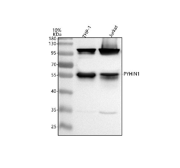
 FCM (Flow Cytometry)
(Figure 7. Flow Cytometry analysis of JK cells using anti-PYHIN1 antibody (AAA19944).Overlay histogram showing JK cells stained with AAA19944 (Blue line). To facilitate intracellular staining, cells were fixed with 4% paraformaldehyde and permeabilized with permeabilization buffer. The cells were blocked with 10% normal goat serum. And then incubated with rabbit anti-PYHIN1 Antibody (AAA19944, 1ug/1x106 cells) for 30 min at 20 degree C. DyLight488 conjugated goat anti-rabbit IgG was used as secondary antibody for 30 minutes at 20 degree C. Isotype control antibody (Green line) was rabbit IgG (1ug/1x106) used under the same conditions. Unlabelled sample without incubation with primary antibody and secondary antibody (Red line) was used as a blank control.)
FCM (Flow Cytometry)
(Figure 7. Flow Cytometry analysis of JK cells using anti-PYHIN1 antibody (AAA19944).Overlay histogram showing JK cells stained with AAA19944 (Blue line). To facilitate intracellular staining, cells were fixed with 4% paraformaldehyde and permeabilized with permeabilization buffer. The cells were blocked with 10% normal goat serum. And then incubated with rabbit anti-PYHIN1 Antibody (AAA19944, 1ug/1x106 cells) for 30 min at 20 degree C. DyLight488 conjugated goat anti-rabbit IgG was used as secondary antibody for 30 minutes at 20 degree C. Isotype control antibody (Green line) was rabbit IgG (1ug/1x106) used under the same conditions. Unlabelled sample without incubation with primary antibody and secondary antibody (Red line) was used as a blank control.)
PYHIN1, Polyclonal Antibody (Cat# AAA19944)
Full Name
Anti-PYHIN1 Antibody Picoband
Gene Names
PYHIN1; IFIX
Reactivity
Human, Mouse, Rat
Applications
Western Blot, Immunohistochemistry, Immunocytochemistry, Immunofluorescence, Flow Cytometry
Purity
Immunogen affinity purified.
Pricing

 FCM (Flow Cytometry)
(Figure 8. Flow Cytometry analysis of HepG2 cells using anti-ORC2 antibody (AAA19812).Overlay histogram showing HepG2 cells stained with AAA19812 (Blue line). To facilitate intracellular staining, cells were fixed with 4% paraformaldehyde and permeabilized with permeabilization buffer. The cells were blocked with 10% normal goat serum. And then incubated with rabbit anti-ORC2 Antibody (AAA19812, 1ug/1x106 cells) for 30 min at 20 degree C. DyLight488 conjugated goat anti-rabbit IgG was used as secondary antibody for 30 minutes at 20 degree C. Isotype control antibody (Green line) was rabbit IgG (1ug/1x106) used under the same conditions. Unlabelled sample (Red line) was also used as a control.)
FCM (Flow Cytometry)
(Figure 8. Flow Cytometry analysis of HepG2 cells using anti-ORC2 antibody (AAA19812).Overlay histogram showing HepG2 cells stained with AAA19812 (Blue line). To facilitate intracellular staining, cells were fixed with 4% paraformaldehyde and permeabilized with permeabilization buffer. The cells were blocked with 10% normal goat serum. And then incubated with rabbit anti-ORC2 Antibody (AAA19812, 1ug/1x106 cells) for 30 min at 20 degree C. DyLight488 conjugated goat anti-rabbit IgG was used as secondary antibody for 30 minutes at 20 degree C. Isotype control antibody (Green line) was rabbit IgG (1ug/1x106) used under the same conditions. Unlabelled sample (Red line) was also used as a control.)
ORC2, Polyclonal Antibody (Cat# AAA19812)
Full Name
Anti-ORC2 Antibody Picoband
Gene Names
ORC2; ORC2L
Reactivity
Human, Mouse, Rat
Applications
Western Blot, Immunohistochemistry, Immunocytochemistry, Immunofluorescence, Flow Cytometry
Purity
Immunogen affinity purified.
Pricing


 FCM (Flow Cytometry)
(Figure 7. Flow Cytometry analysis of SiHa cells using anti-CEP250 antibody (AAA19580).Overlay histogram showing SiHa cells stained with AAA19580 (Blue line). The cells were blocked with 10% normal goat serum. And then incubated with rabbit anti-CEP250 Antibody (AAA19580, 1 ug/1x10^6 cells) for 30 min at 20 degree C. DyLight488 conjugated goat anti-rabbit IgG was used as secondary antibody for 30 minutes at 20 degree C. Isotype control antibody (Green line) was rabbit IgG (1 ug/1x10^6) used under the same conditions. Unlabelled sample (Red line) was also used as a control.)
FCM (Flow Cytometry)
(Figure 7. Flow Cytometry analysis of SiHa cells using anti-CEP250 antibody (AAA19580).Overlay histogram showing SiHa cells stained with AAA19580 (Blue line). The cells were blocked with 10% normal goat serum. And then incubated with rabbit anti-CEP250 Antibody (AAA19580, 1 ug/1x10^6 cells) for 30 min at 20 degree C. DyLight488 conjugated goat anti-rabbit IgG was used as secondary antibody for 30 minutes at 20 degree C. Isotype control antibody (Green line) was rabbit IgG (1 ug/1x10^6) used under the same conditions. Unlabelled sample (Red line) was also used as a control.)
CEP250, Polyclonal Antibody (Cat# AAA19580)
Full Name
Anti-CEP250 Antibody Picoband
Gene Names
CEP250; CEP2; CNAP1; C-NAP1
Reactivity
Human, Mouse, Rat
Applications
Flow Cytometry, Immunohistochemistry, Western Blot
Purity
Immunogen affinity purified.
Pricing
Hypoxia-Inducible Factor-1 Alpha, Recombinant Protein (Cat# AAA10841)
Full Name
Recombinant Human Hypoxia-Inducible Factor-1 Alpha
Gene Names
HIF1A; HIF1; MOP1; PASD8; HIF-1A; bHLHe78; HIF-1alpha; HIF1-ALPHA
Purity
Greater than 90.0% as determined by SDS-PAGE.
Pricing


 IHC (Immunohistchemistry)
(Figure 6. IHC analysis of MED15 using anti-MED15 antibody (AAA19169).MED15 was detected in paraffin-embedded section of rat brain tissue. Heat mediated antigen retrieval was performed in citrate buffer (pH6, epitope retrieval solution) for 20 mins. The tissue section was blocked with 10% goat serum. The tissue section was then incubated with 1ug/ml rabbit anti-MED15 Antibody (AAA19169) overnight at 4 degree C. Biotinylated goat anti-rabbit IgG was used as secondary antibody and incubated for 30 minutes at 37 degree C. The tissue section was developed using Strepavidin-Biotin-Complex (SABC) with DAB as the chromogen.)
IHC (Immunohistchemistry)
(Figure 6. IHC analysis of MED15 using anti-MED15 antibody (AAA19169).MED15 was detected in paraffin-embedded section of rat brain tissue. Heat mediated antigen retrieval was performed in citrate buffer (pH6, epitope retrieval solution) for 20 mins. The tissue section was blocked with 10% goat serum. The tissue section was then incubated with 1ug/ml rabbit anti-MED15 Antibody (AAA19169) overnight at 4 degree C. Biotinylated goat anti-rabbit IgG was used as secondary antibody and incubated for 30 minutes at 37 degree C. The tissue section was developed using Strepavidin-Biotin-Complex (SABC) with DAB as the chromogen.)
MED15/Pcqap, Polyclonal Antibody (Cat# AAA19169)
Full Name
Anti-MED15/Pcqap Picoband Antibody
Gene Names
MED15; TIG1; CAG7A; CTG7A; PCQAP; TIG-1; TNRC7; ARC105
Reactivity
Human, Mouse, Rat
No cross reactivity with other proteins.
No cross reactivity with other proteins.
Applications
Immunohistochemistry, Western Blot
Purity
Immunogen affinity purified
Pricing
 Standard Curve (Sample)
CD86, ELISA Kit (Cat# AAA17898)
Full Name
Mouse CD86 ELISA Kit
Gene Names
Cd86; B7; B70; MB7; B7-2; B7.2; CLS1; Ly58; ETC-1; Ly-58; MB7-2; Cd28l2; TS/A-2
Reactivity
Mouse
Applications
Sandwich ELISA
Pricing