
Filters
Clonality
Type
Reactivity
Gene Name
Isotype
Host
Application
Clone
10000 results for "Recombinant Proteins" - showing 1600-1650


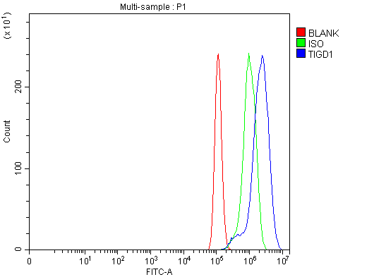 FCM (Flow Cytometry)
(Figure 9. Flow Cytometry analysis of PC-3 cells using anti-TIGD1 antibody (AAA19643).Overlay histogram showing PC-3 cells stained with AAA19643 (Blue line). The cells were blocked with 10% normal goat serum. And then incubated with rabbit anti-TIGD1 Antibody (AAA19643, 1 ug/1x10^6 cells) for 30 min at 20 degree C. DyLight488 conjugated goat anti-rabbit IgG was used as secondary antibody for 30 minutes at 20 degree C. Isotype control antibody (Green line) was rabbit IgG (1 ug/1x10^6) used under the same conditions. Unlabelled sample (Red line) was also used as a control.)
FCM (Flow Cytometry)
(Figure 9. Flow Cytometry analysis of PC-3 cells using anti-TIGD1 antibody (AAA19643).Overlay histogram showing PC-3 cells stained with AAA19643 (Blue line). The cells were blocked with 10% normal goat serum. And then incubated with rabbit anti-TIGD1 Antibody (AAA19643, 1 ug/1x10^6 cells) for 30 min at 20 degree C. DyLight488 conjugated goat anti-rabbit IgG was used as secondary antibody for 30 minutes at 20 degree C. Isotype control antibody (Green line) was rabbit IgG (1 ug/1x10^6) used under the same conditions. Unlabelled sample (Red line) was also used as a control.)
TIGD1, Polyclonal Antibody (Cat# AAA19643)
Full Name
Anti-TIGD1 Antibody Picoband
Gene Names
TIGD1; EEYORE
Reactivity
Human, Mouse, Rat
Applications
Western Blot, Immunohistochemistry, Immunocytochemistry, Immunofluorescence, Flow Cytometry
Purity
Immunogen affinity purified.
Pricing


 IF (Immunofluorescence)
(Immunofluorescence analysis of A549 cells using ULK4 antibody. Blue: DAPI for nuclear staining.)
IF (Immunofluorescence)
(Immunofluorescence analysis of A549 cells using ULK4 antibody. Blue: DAPI for nuclear staining.)
ULK4, Polyclonal Antibody (Cat# AAA28144)
Full Name
ULK4
Gene Names
ULK4; FAM7C1; REC01035
Reactivity
Human
Applications
Western Blot, Immunohistochemistry
Pricing

 FCM (Flow Cytometry)
(Figure 6. Flow Cytometry analysis of JK cells using anti-MOCS2 antibody (AAA19885).Overlay histogram showing JK cells stained with AAA19885 (Blue line). To facilitate intracellular staining, cells were fixed with 4% paraformaldehyde and permeabilized with permeabilization buffer. The cells were blocked with 10% normal goat serum. And then incubated with rabbit anti-MOCS2 Antibody (AAA19885, 1ug/1x106 cells) for 30 min at 20 degree C. DyLight488 conjugated goat anti-rabbit IgG was used as secondary antibody for 30 minutes at 20 degree C. Isotype control antibody (Green line) was rabbit IgG (1ug/1x106) used under the same conditions. Unlabelled sample (Red line) was also used as a control.)
FCM (Flow Cytometry)
(Figure 6. Flow Cytometry analysis of JK cells using anti-MOCS2 antibody (AAA19885).Overlay histogram showing JK cells stained with AAA19885 (Blue line). To facilitate intracellular staining, cells were fixed with 4% paraformaldehyde and permeabilized with permeabilization buffer. The cells were blocked with 10% normal goat serum. And then incubated with rabbit anti-MOCS2 Antibody (AAA19885, 1ug/1x106 cells) for 30 min at 20 degree C. DyLight488 conjugated goat anti-rabbit IgG was used as secondary antibody for 30 minutes at 20 degree C. Isotype control antibody (Green line) was rabbit IgG (1ug/1x106) used under the same conditions. Unlabelled sample (Red line) was also used as a control.)
MOCS2, Polyclonal Antibody (Cat# AAA19885)
Full Name
Anti-MOCS2 Antibody Picoband
Gene Names
MOCS2; MPTS; MCBPE; MOCO1; MOCODB
Reactivity
Human
Applications
Western Blot, Immunohistochemistry, Flow Cytometry
Purity
Immunogen affinity purified.
Pricing


 FCM (Flow Cytometry)
(Figure 7. Flow Cytometry analysis of SiHa cells using anti-Tropomyosin 2/TPM2 antibody (AAA19572).Overlay histogram showing SiHa cells stained with AAA19572 (Blue line). The cells were blocked with 10% normal goat serum. And then incubated with rabbit anti-Tropomyosin 2/TPM2 Antibody (AAA19572, 1 ug/1x10^6 cells) for 30 min at 20 degree C. DyLight488 conjugated goat anti-rabbit IgG was used as secondary antibody for 30 minutes at 20 degree C. Isotype control antibody (Green line) was rabbit IgG (1 ug/1x10^6) used under the same conditions. Unlabelled sample (Red line) was also used as a control.)
FCM (Flow Cytometry)
(Figure 7. Flow Cytometry analysis of SiHa cells using anti-Tropomyosin 2/TPM2 antibody (AAA19572).Overlay histogram showing SiHa cells stained with AAA19572 (Blue line). The cells were blocked with 10% normal goat serum. And then incubated with rabbit anti-Tropomyosin 2/TPM2 Antibody (AAA19572, 1 ug/1x10^6 cells) for 30 min at 20 degree C. DyLight488 conjugated goat anti-rabbit IgG was used as secondary antibody for 30 minutes at 20 degree C. Isotype control antibody (Green line) was rabbit IgG (1 ug/1x10^6) used under the same conditions. Unlabelled sample (Red line) was also used as a control.)
Tripeptidyl peptidase II/TPPII/TPP2, Polyclonal Antibody (Cat# AAA19572)
Full Name
Anti-Tripeptidyl peptidase II/TPPII/TPP2 Antibody Picoband
Gene Names
TPP2; TPP-2; TPPII; TPP-II
Reactivity
Human, Mouse, Rat
Applications
Western Blot, Immunohistochemistry, Immunocytochemistry, Immunofluorescence, Flow Cytometry
Purity
Immunogen affinity purified.
Pricing


 FCM (Flow Cytometry)
(Figure 6. Flow Cytometry analysis of Daudi cells using anti-NLRP7 antibody (AAA19590).Overlay histogram showing Daudi cells stained with AAA19590 (Blue line). The cells were blocked with 10% normal goat serum. And then incubated with rabbit anti-NLRP7 Antibody (AAA19590, 1 ug/1x10^6 cells) for 30 min at 20 degree C. DyLight488 conjugated goat anti-rabbit IgG was used as secondary antibody for 30 minutes at 20 degree C. Isotype control antibody (Green line) was rabbit IgG (1 ug/1x10^6) used under the same conditions. Unlabelled sample (Red line) was also used as a control.)
FCM (Flow Cytometry)
(Figure 6. Flow Cytometry analysis of Daudi cells using anti-NLRP7 antibody (AAA19590).Overlay histogram showing Daudi cells stained with AAA19590 (Blue line). The cells were blocked with 10% normal goat serum. And then incubated with rabbit anti-NLRP7 Antibody (AAA19590, 1 ug/1x10^6 cells) for 30 min at 20 degree C. DyLight488 conjugated goat anti-rabbit IgG was used as secondary antibody for 30 minutes at 20 degree C. Isotype control antibody (Green line) was rabbit IgG (1 ug/1x10^6) used under the same conditions. Unlabelled sample (Red line) was also used as a control.)
NLRP7, Polyclonal Antibody (Cat# AAA19590)
Full Name
Anti-NLRP7 Antibody Picoband
Gene Names
NLRP7; HYDM; PAN7; NALP7; NOD12; PYPAF3; CLR19.4
Reactivity
Human
Applications
Flow Cytometry, Immunohistochemistry, Western Blot
Purity
Immunogen affinity purified.
Pricing

 FCM (Flow Cytometry)
(Figure 7. Flow Cytometry analysis of HepG2 cells using anti-HSPA1L antibody (AAA19810).Overlay histogram showing HepG2 cells stained with AAA19810 (Blue line). To facilitate intracellular staining, cells were fixed with 4% paraformaldehyde and permeabilized with permeabilization buffer. The cells were blocked with 10% normal goat serum. And then incubated with rabbit anti-HSPA1L Antibody (AAA19810, 1ug/1x106 cells) for 30 min at 20 degree C. DyLight488 conjugated goat anti-rabbit IgG was used as secondary antibody for 30 minutes at 20 degree C. Isotype control antibody (Green line) was rabbit IgG (1ug/1x106) used under the same conditions. Unlabelled sample (Red line) was also used as a control.)
FCM (Flow Cytometry)
(Figure 7. Flow Cytometry analysis of HepG2 cells using anti-HSPA1L antibody (AAA19810).Overlay histogram showing HepG2 cells stained with AAA19810 (Blue line). To facilitate intracellular staining, cells were fixed with 4% paraformaldehyde and permeabilized with permeabilization buffer. The cells were blocked with 10% normal goat serum. And then incubated with rabbit anti-HSPA1L Antibody (AAA19810, 1ug/1x106 cells) for 30 min at 20 degree C. DyLight488 conjugated goat anti-rabbit IgG was used as secondary antibody for 30 minutes at 20 degree C. Isotype control antibody (Green line) was rabbit IgG (1ug/1x106) used under the same conditions. Unlabelled sample (Red line) was also used as a control.)
HSPA1L, Polyclonal Antibody (Cat# AAA19810)
Full Name
Anti-HSPA1L Antibody Picoband
Gene Names
HSPA1L; HSP70T; hum70t; HSP70-1L; HSP70-HOM
Reactivity
Human, Mouse, Rat
Applications
Western Blot, Immunohistochemistry, Immunocytochemistry, Immunofluorescence, Flow Cytometry
Purity
Immunogen affinity purified.
Pricing


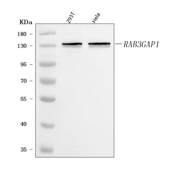
 FCM (Flow Cytometry)
(Figure 11. Flow Cytometry analysis of HL-60 cells using anti-RAB3GAP1 antibody (AAA19824).Overlay histogram showing HL-60 cells stained with AAA19824 (Blue line). To facilitate intracellular staining, cells were fixed with 4% paraformaldehyde and permeabilized with permeabilization buffer. The cells were blocked with 10% normal goat serum. And then incubated with rabbit anti-RAB3GAP1 Antibody (AAA19824, 1ug/1x106 cells) for 30 min at 20 degree C. DyLight488 conjugated goat anti-rabbit IgG (BA1127, 5-10ug/1x106 cells) was used as secondary antibody for 30 minutes at 20 degree C. Isotype control antibody (Green line) was rabbit IgG (1ug/1x106) used under the same conditions. Unlabelled sample without incubation with primary antibody and secondary antibody (Red line) was used as a blank control.)
FCM (Flow Cytometry)
(Figure 11. Flow Cytometry analysis of HL-60 cells using anti-RAB3GAP1 antibody (AAA19824).Overlay histogram showing HL-60 cells stained with AAA19824 (Blue line). To facilitate intracellular staining, cells were fixed with 4% paraformaldehyde and permeabilized with permeabilization buffer. The cells were blocked with 10% normal goat serum. And then incubated with rabbit anti-RAB3GAP1 Antibody (AAA19824, 1ug/1x106 cells) for 30 min at 20 degree C. DyLight488 conjugated goat anti-rabbit IgG (BA1127, 5-10ug/1x106 cells) was used as secondary antibody for 30 minutes at 20 degree C. Isotype control antibody (Green line) was rabbit IgG (1ug/1x106) used under the same conditions. Unlabelled sample without incubation with primary antibody and secondary antibody (Red line) was used as a blank control.)
RAB3GAP1, Polyclonal Antibody (Cat# AAA19824)
Full Name
Anti-RAB3GAP1 Antibody Picoband
Gene Names
RAB3GAP1; P130; WARBM1; RAB3GAP; RAB3GAP130
Reactivity
Human
Applications
Flow Cytometry, Immunofluorescence, Immunohistochemistry, Immunocytochemistry, Western Blot
Purity
Immunogen affinity purified.
Pricing
 IHC (Immunohistochemistry)
(Figure 15. IHC analysis of SCAMP3 using anti-SCAMP3 antibody (AAA19900).SCAMP3 was detected in a paraffin-embedded section of rat ovary tissue. Heat mediated antigen retrieval was performed in EDTA buffer (pH 8.0, epitope retrieval solution). The tissue section was blocked with 10% goat serum. The tissue section was then incubated with 2ug/ml rabbit anti-SCAMP3 Antibody (AAA19900) overnight at 4 degree C. Peroxidase Conjugated Goat Anti-rabbit IgG was used as secondary antibody and incubated for 30 minutes at 37 degree C. The tissue section was developed using HRP Conjugated Rabbit IgG Super Vision Assay Kit ( epitope retrieval solution). The tissue section was blocked with 10% goat serum. The tissue section was then incubated with 2ug/ml rabbit anti-SCAMP3 Antibody (AAA19900) overnight at 4 degree C. Peroxidase Conjugated Goat Anti-rabbit IgG was used as secondary antibody and incubated for 30 minutes at 37 degree C. The tissue section was developed using HRP Conjugated Rabbit IgG Super Vision Assay Kit ( epitope retrieval solution). The tissue section was blocked with 10% goat serum. The tissue section was then incubated with 2ug/ml rabbit anti-SCAMP3 Antibody (AAA19900) overnight at 4 degree C. Peroxidase Conjugated Goat Anti-rabbit IgG was used as secondary antibody and incubated for 30 minutes at 37 degree C. The tissue section was developed using HRP Conjugated Rabbit IgG Super Vision Assay Kit ( epitope retrieval solution). The tissue section was blocked with 10% goat serum. The tissue section was then incubated with 2ug/ml rabbit anti-SCAMP3 Antibody (AAA19900) overnight at 4 degree C. Peroxidase Conjugated Goat Anti-rabbit IgG was used as secondary antibody and incubated for 30 minutes at 37 degree C. The tissue section was developed using HRP Conjugated Rabbit IgG Super Vision Assay Kit ( epitope retrieval solution). The tissue section was blocked with 10% goat serum. The tissue section was then incubated with 2ug/ml rabbit anti-SCAMP3 Antibody (AAA19900) overnight at 4 degree C. Peroxidase Conjugated Goat Anti-rabbit IgG was used as secondary antibody and incubated for 30 minutes at 37 degree C. The tissue section was developed using HRP Conjugated Rabbit IgG Super Vision Assay Kit ( epitope retrieval solution). The tissue section was blocked with 10% goat serum. The tissue section was then incubated with 2ug/ml rabbit anti-SCAMP3 Antibody (AAA19900) overnight at 4 degree C. Peroxidase Conjugated Goat Anti-rabbit IgG was used as secondary antibody and incubated for 30 minutes at 37 degree C. The tissue section was developed using HRP Conjugated Rabbit IgG Super Vision Assay Kit ( epitope retrieval solution). The tissue section was blocked with 10% goat serum. The tissue section was then incubated with 2ug/ml rabbit anti-SCAMP3 Antibody (AAA19900) overnight at 4 degree C. Peroxidase Conjugated Goat Anti-rabbit IgG was used as secondary antibody and incubated for 30 minutes at 37 degree C. The tissue section was developed using HRP Conjugated Rabbit IgG Super Vision Assay Kit ( epitope retrieval solution). The tissue section was blocked with 10% goat serum. The tissue section was then incubated with 2ug/ml rabbit anti-SCAMP3 Antibody (AAA19900) overnight at 4 degree C. Peroxidase Conjugated Goat Anti-rabbit IgG was used as secondary antibody and incubated for 30 minutes at 37 degree C. The tissue section was developed using HRP Conjugated Rabbit IgG Super Vision Assay Kit ( epitope retrieval solution). The tissue section was blocked with 10% goat serum. The tissue section was then incubated with 2ug/ml rabbit anti-SCAMP3 Antibody (AAA19900) overnight at 4 degree C. Peroxidase Conjugated Goat Anti-rabbit IgG was used as secondary antibody and incubated for 30 minutes at 37 degree C. The tissue section was developed using HRP Conjugated Rabbit IgG Super Vision Assay Kit ( epitope retrieval solution). The tissue section was blocked with 10% goat serum. The tissue section was then incubated with 2ug/ml rabbit anti-SCAMP3 Antibody (AAA19900) overnight at 4 degree C. Peroxidase Conjugated Goat Anti-rabbit IgG was used as secondary antibody and incubated for 30 minutes at 37 degree C. The tissue section was developed using HRP Conjugated Rabbit IgG Super Vision Assay Kit ( epitope retrieval solution). The tissue section was blocked with 10% goat serum. The tissue section was then incubated with 2ug/ml rabbit anti-SCAMP3 Antibody (AAA19900) overnight at 4 degree C. Peroxidase Conjugated Goat Anti-rabbit IgG was used as secondary antibody and incubated for 30 minutes at 37 degree C. The tissue section was developed using HRP Conjugated Rabbit IgG Super Vision Assay Kit ( epitope retrieval solution). The tissue section was blocked with 10% goat serum. The tissue section was then incubated with 2ug/ml rabbit anti-SCAMP3 Antibody (AAA19900) overnight at 4 degree C. Peroxidase Conjugated Goat Anti-rabbit IgG was used as secondary antibody and incubated for 30 minutes at 37 degree C. The tissue section was developed using HRP Conjugated Rabbit IgG Super Vision Assay Kit ( epitope retrieval solution). The tissue section was blocked with 10% goat serum. The tissue section was then incubated with 2ug/ml rabbit anti-SCAMP3 Antibody (AAA19900) overnight at 4 degree C. Peroxidase Conjugated Goat Anti-rabbit IgG was used as secondary antibody and incubated for 30 minutes at 37 degree C. The tissue section was developed using HRP Conjugated Rabbit IgG Super Vision Assay Kit ( epitope retrieval solution). The tissue section was blocked with 10% goat serum. The tissue section was then incubated with 2ug/ml rabbit anti-SCAMP3 Antibody (AAA19900) overnight at 4 degree C. Peroxidase Conjugated Goat Anti-rabbit IgG was used as secondary antibody and incubated for 30 minutes at 37 degree C. The tissue section was developed using HRP Conjugated Rabbit IgG Super Vision Assay Kit (Lane 2: human A549 whole cell lysates,Lane 3: human K562 whole cell lysates,Lane 4: human 293T whole cell lysates.After electrophoresis, proteins were transferred to a nitrocellulose membrane at 150 mA for 50-90 minutes. Blocked the membrane with 5% non-fat milk/TBS for 1.5 hour at RT. The membrane was incubated with rabbit anti-SCAMP3 antigen affinity purified polyclonal antibody (#AAA19900) at 0.5ug/mL overnight at 4 degree C, then washed with TBS-0.1%Tween 3 times with 5 minutes each and probed with a goat anti-rabbit IgG-HRP secondary antibody at a dilution of 1:5000 for 1.5 hour at RT. The signal is developed using an Enhanced Chemiluminescent detection (ECL) kit with Tanon 5200 system. A specific band was detected for SCAMP3 at approximately 36 kDa. The expected band size for SCAMP3 is at 38 kDa.)
IHC (Immunohistochemistry)
(Figure 15. IHC analysis of SCAMP3 using anti-SCAMP3 antibody (AAA19900).SCAMP3 was detected in a paraffin-embedded section of rat ovary tissue. Heat mediated antigen retrieval was performed in EDTA buffer (pH 8.0, epitope retrieval solution). The tissue section was blocked with 10% goat serum. The tissue section was then incubated with 2ug/ml rabbit anti-SCAMP3 Antibody (AAA19900) overnight at 4 degree C. Peroxidase Conjugated Goat Anti-rabbit IgG was used as secondary antibody and incubated for 30 minutes at 37 degree C. The tissue section was developed using HRP Conjugated Rabbit IgG Super Vision Assay Kit ( epitope retrieval solution). The tissue section was blocked with 10% goat serum. The tissue section was then incubated with 2ug/ml rabbit anti-SCAMP3 Antibody (AAA19900) overnight at 4 degree C. Peroxidase Conjugated Goat Anti-rabbit IgG was used as secondary antibody and incubated for 30 minutes at 37 degree C. The tissue section was developed using HRP Conjugated Rabbit IgG Super Vision Assay Kit ( epitope retrieval solution). The tissue section was blocked with 10% goat serum. The tissue section was then incubated with 2ug/ml rabbit anti-SCAMP3 Antibody (AAA19900) overnight at 4 degree C. Peroxidase Conjugated Goat Anti-rabbit IgG was used as secondary antibody and incubated for 30 minutes at 37 degree C. The tissue section was developed using HRP Conjugated Rabbit IgG Super Vision Assay Kit ( epitope retrieval solution). The tissue section was blocked with 10% goat serum. The tissue section was then incubated with 2ug/ml rabbit anti-SCAMP3 Antibody (AAA19900) overnight at 4 degree C. Peroxidase Conjugated Goat Anti-rabbit IgG was used as secondary antibody and incubated for 30 minutes at 37 degree C. The tissue section was developed using HRP Conjugated Rabbit IgG Super Vision Assay Kit ( epitope retrieval solution). The tissue section was blocked with 10% goat serum. The tissue section was then incubated with 2ug/ml rabbit anti-SCAMP3 Antibody (AAA19900) overnight at 4 degree C. Peroxidase Conjugated Goat Anti-rabbit IgG was used as secondary antibody and incubated for 30 minutes at 37 degree C. The tissue section was developed using HRP Conjugated Rabbit IgG Super Vision Assay Kit ( epitope retrieval solution). The tissue section was blocked with 10% goat serum. The tissue section was then incubated with 2ug/ml rabbit anti-SCAMP3 Antibody (AAA19900) overnight at 4 degree C. Peroxidase Conjugated Goat Anti-rabbit IgG was used as secondary antibody and incubated for 30 minutes at 37 degree C. The tissue section was developed using HRP Conjugated Rabbit IgG Super Vision Assay Kit ( epitope retrieval solution). The tissue section was blocked with 10% goat serum. The tissue section was then incubated with 2ug/ml rabbit anti-SCAMP3 Antibody (AAA19900) overnight at 4 degree C. Peroxidase Conjugated Goat Anti-rabbit IgG was used as secondary antibody and incubated for 30 minutes at 37 degree C. The tissue section was developed using HRP Conjugated Rabbit IgG Super Vision Assay Kit ( epitope retrieval solution). The tissue section was blocked with 10% goat serum. The tissue section was then incubated with 2ug/ml rabbit anti-SCAMP3 Antibody (AAA19900) overnight at 4 degree C. Peroxidase Conjugated Goat Anti-rabbit IgG was used as secondary antibody and incubated for 30 minutes at 37 degree C. The tissue section was developed using HRP Conjugated Rabbit IgG Super Vision Assay Kit ( epitope retrieval solution). The tissue section was blocked with 10% goat serum. The tissue section was then incubated with 2ug/ml rabbit anti-SCAMP3 Antibody (AAA19900) overnight at 4 degree C. Peroxidase Conjugated Goat Anti-rabbit IgG was used as secondary antibody and incubated for 30 minutes at 37 degree C. The tissue section was developed using HRP Conjugated Rabbit IgG Super Vision Assay Kit ( epitope retrieval solution). The tissue section was blocked with 10% goat serum. The tissue section was then incubated with 2ug/ml rabbit anti-SCAMP3 Antibody (AAA19900) overnight at 4 degree C. Peroxidase Conjugated Goat Anti-rabbit IgG was used as secondary antibody and incubated for 30 minutes at 37 degree C. The tissue section was developed using HRP Conjugated Rabbit IgG Super Vision Assay Kit ( epitope retrieval solution). The tissue section was blocked with 10% goat serum. The tissue section was then incubated with 2ug/ml rabbit anti-SCAMP3 Antibody (AAA19900) overnight at 4 degree C. Peroxidase Conjugated Goat Anti-rabbit IgG was used as secondary antibody and incubated for 30 minutes at 37 degree C. The tissue section was developed using HRP Conjugated Rabbit IgG Super Vision Assay Kit ( epitope retrieval solution). The tissue section was blocked with 10% goat serum. The tissue section was then incubated with 2ug/ml rabbit anti-SCAMP3 Antibody (AAA19900) overnight at 4 degree C. Peroxidase Conjugated Goat Anti-rabbit IgG was used as secondary antibody and incubated for 30 minutes at 37 degree C. The tissue section was developed using HRP Conjugated Rabbit IgG Super Vision Assay Kit ( epitope retrieval solution). The tissue section was blocked with 10% goat serum. The tissue section was then incubated with 2ug/ml rabbit anti-SCAMP3 Antibody (AAA19900) overnight at 4 degree C. Peroxidase Conjugated Goat Anti-rabbit IgG was used as secondary antibody and incubated for 30 minutes at 37 degree C. The tissue section was developed using HRP Conjugated Rabbit IgG Super Vision Assay Kit ( epitope retrieval solution). The tissue section was blocked with 10% goat serum. The tissue section was then incubated with 2ug/ml rabbit anti-SCAMP3 Antibody (AAA19900) overnight at 4 degree C. Peroxidase Conjugated Goat Anti-rabbit IgG was used as secondary antibody and incubated for 30 minutes at 37 degree C. The tissue section was developed using HRP Conjugated Rabbit IgG Super Vision Assay Kit (Lane 2: human A549 whole cell lysates,Lane 3: human K562 whole cell lysates,Lane 4: human 293T whole cell lysates.After electrophoresis, proteins were transferred to a nitrocellulose membrane at 150 mA for 50-90 minutes. Blocked the membrane with 5% non-fat milk/TBS for 1.5 hour at RT. The membrane was incubated with rabbit anti-SCAMP3 antigen affinity purified polyclonal antibody (#AAA19900) at 0.5ug/mL overnight at 4 degree C, then washed with TBS-0.1%Tween 3 times with 5 minutes each and probed with a goat anti-rabbit IgG-HRP secondary antibody at a dilution of 1:5000 for 1.5 hour at RT. The signal is developed using an Enhanced Chemiluminescent detection (ECL) kit with Tanon 5200 system. A specific band was detected for SCAMP3 at approximately 36 kDa. The expected band size for SCAMP3 is at 38 kDa.)
SCAMP3, Polyclonal Antibody (Cat# AAA19900)
Full Name
Anti-SCAMP3 Antibody Picoband
Gene Names
SCAMP3; C1orf3
Reactivity
Human, Rat
Applications
Immunofluorescence, Immunohistochemistry, Immunocytochemistry, Western Blot, Flow Cytometry
Purity
Immunogen affinity purified.
Pricing

 FCM (Flow Cytometry)
(Figure 7. Flow Cytometry analysis of HepG2 cells using anti-GBF1 antibody (AAA19761).Overlay histogram showing HepG2 cells stained with AAA19761 (Blue line). To facilitate intracellular staining, cells were fixed with 4% paraformaldehyde and permeabilized with permeabilization buffer. The cells were blocked with 10% normal goat serum. And then incubated with rabbit anti-GBF1 Antibody (AAA19761, 1ug/1x106 cells) for 30 min at 20 degree C. DyLight488 conjugated goat anti-rabbit IgG was used as secondary antibody for 30 minutes at 20 degree C. Isotype control antibody (Green line) was rabbit IgG (1ug/1x106) used under the same conditions. Unlabelled sample without incubation with primary antibody and secondary antibody (Red line) was used as a blank control.)
FCM (Flow Cytometry)
(Figure 7. Flow Cytometry analysis of HepG2 cells using anti-GBF1 antibody (AAA19761).Overlay histogram showing HepG2 cells stained with AAA19761 (Blue line). To facilitate intracellular staining, cells were fixed with 4% paraformaldehyde and permeabilized with permeabilization buffer. The cells were blocked with 10% normal goat serum. And then incubated with rabbit anti-GBF1 Antibody (AAA19761, 1ug/1x106 cells) for 30 min at 20 degree C. DyLight488 conjugated goat anti-rabbit IgG was used as secondary antibody for 30 minutes at 20 degree C. Isotype control antibody (Green line) was rabbit IgG (1ug/1x106) used under the same conditions. Unlabelled sample without incubation with primary antibody and secondary antibody (Red line) was used as a blank control.)
GBF1, Polyclonal Antibody (Cat# AAA19761)
Full Name
Anti-GBF1 Antibody Picoband
Gene Names
GBF1; ARF1GEF
Reactivity
Human, Mouse, Rat
Applications
Western Blot, Immunohistochemistry, Immunocytochemistry, Immunofluorescence, Flow Cytometry
Purity
Immunogen affinity purified.
Pricing


 FCM (Flow Cytometry)
(Figure 9. Flow Cytometry analysis of Daudi cells using anti-ZMYND8 antibody (AAA19575).Overlay histogram showing Daudi cells stained with AAA19575 (Blue line). The cells were blocked with 10% normal goat serum. And then incubated with rabbit anti-ZMYND8 Antibody (AAA19575, 1 ug/1x10^6 cells) for 30 min at 20 degree C. DyLight488 conjugated goat anti-rabbit IgG was used as secondary antibody for 30 minutes at 20 degree C. Isotype control antibody (Green line) was rabbit IgG (1 ug/1x10^6) used under the same conditions. Unlabelled sample (Red line) was also used as a control.)
FCM (Flow Cytometry)
(Figure 9. Flow Cytometry analysis of Daudi cells using anti-ZMYND8 antibody (AAA19575).Overlay histogram showing Daudi cells stained with AAA19575 (Blue line). The cells were blocked with 10% normal goat serum. And then incubated with rabbit anti-ZMYND8 Antibody (AAA19575, 1 ug/1x10^6 cells) for 30 min at 20 degree C. DyLight488 conjugated goat anti-rabbit IgG was used as secondary antibody for 30 minutes at 20 degree C. Isotype control antibody (Green line) was rabbit IgG (1 ug/1x10^6) used under the same conditions. Unlabelled sample (Red line) was also used as a control.)
ZMYND8, Polyclonal Antibody (Cat# AAA19575)
Full Name
Anti-ZMYND8 Antibody Picoband
Gene Names
ZMYND8; RACK7; PRKCBP1; PRO2893
Reactivity
Human, Mouse, Rat
Applications
Western Blot, Immunohistochemistry, Immunocytochemistry, Immunofluorescence, Flow Cytometry
Purity
Immunogen affinity purified.
Pricing


 FCM (Flow Cytometry)
(Figure 11. Flow Cytometry analysis of HL-60 cells using anti-MCM6 antibody (AAA19704).Overlay histogram showing HL-60 cells stained with AAA19704 (Blue line). The cells were blocked with 10% normal goat serum. And then incubated with mouse anti-MCM6 Antibody (AAA19704, 1 ug/1x10^6 cells) for 30 min at 20 degree C. DyLight488 conjugated goat anti-mouse IgG was used as secondary antibody for 30 minutes at 20 degree C. Isotype control antibody (Green line) was mouse IgG (1 ug/1x10^6) used under the same conditions. Unlabelled sample (Red line) was also used as a control.)
FCM (Flow Cytometry)
(Figure 11. Flow Cytometry analysis of HL-60 cells using anti-MCM6 antibody (AAA19704).Overlay histogram showing HL-60 cells stained with AAA19704 (Blue line). The cells were blocked with 10% normal goat serum. And then incubated with mouse anti-MCM6 Antibody (AAA19704, 1 ug/1x10^6 cells) for 30 min at 20 degree C. DyLight488 conjugated goat anti-mouse IgG was used as secondary antibody for 30 minutes at 20 degree C. Isotype control antibody (Green line) was mouse IgG (1 ug/1x10^6) used under the same conditions. Unlabelled sample (Red line) was also used as a control.)
MCM6, Monoclonal Antibody (Cat# AAA19704)
Full Name
Anti-MCM6 Antibody Picoband (monoclonal, 3F13C4)
Gene Names
MCM6; Mis5; P105MCM; MCG40308
Reactivity
Human, Mouse, Rat
Applications
Western Blot, Immunohistochemistry, Immunocytochemistry, Immunofluorescence, Flow Cytometry
Purity
Immunogen affinity purified.
Pricing
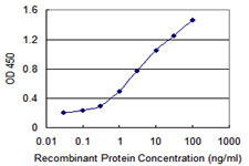


 WB (Western Blot)
(DDX54 monoclonal antibody, Western Blot analysis of DDX54 expression in HeLa.)
WB (Western Blot)
(DDX54 monoclonal antibody, Western Blot analysis of DDX54 expression in HeLa.)
DDX54, Monoclonal Antibody (Cat# AAA25665)
Full Name
DDX54 (ATP-dependent RNA Helicase DDX54, ATP-dependent RNA Helicase DP97, DEAD Box RNA Helicase 97kD, DEAD Box Protein 54, MGC2835) (PE)
Gene Names
DDX54; DP97
Reactivity
Human
Applications
Immunofluorescence, Immunohistochemistry, Western Blot
Purity
Purified by Protein A Affinity Chromatography.
Pricing


 FCM (Flow Cytometry)
(Figure 12. Flow Cytometry analysis of U937 cells using anti-DYNLL1/PIN antibody (AAA19276).Overlay histogram showing U937 cells stained with AAA19276 (Blue line). The cells were blocked with 10% normal goat serum. And then incubated with rabbit anti-DYNLL1/PIN Antibody (AAA19276, 1μg/1x106 cells) for 30 min at 20 degree C. DyLight®488 conjugated goat anti-rabbit IgG (5-10μg/1x106 cells) was used as secondary antibody for 30 minutes at 20 degree C. Isotype control antibody (Green line) was rabbit IgG (1μg/1x106) used under the same conditions. Unlabelled sample (Red line) was also used as a control.)
FCM (Flow Cytometry)
(Figure 12. Flow Cytometry analysis of U937 cells using anti-DYNLL1/PIN antibody (AAA19276).Overlay histogram showing U937 cells stained with AAA19276 (Blue line). The cells were blocked with 10% normal goat serum. And then incubated with rabbit anti-DYNLL1/PIN Antibody (AAA19276, 1μg/1x106 cells) for 30 min at 20 degree C. DyLight®488 conjugated goat anti-rabbit IgG (5-10μg/1x106 cells) was used as secondary antibody for 30 minutes at 20 degree C. Isotype control antibody (Green line) was rabbit IgG (1μg/1x106) used under the same conditions. Unlabelled sample (Red line) was also used as a control.)
DYNLL1/PIN, Polyclonal Antibody (Cat# AAA19276)
Full Name
Anti-DYNLL1/PIN Antibody
Gene Names
DYNLL1; LC8; PIN; DLC1; DLC8; LC8a; DNCL1; hdlc1; DNCLC1
Reactivity
Human, Mouse, Rat
Applications
Western Blot, Immunohistochemistry, Immunocytochemistry, Immunofluorescence, Flow Cytometry, Direct ELISA
Purity
Immunogen affinity purified.
Pricing

 Activity
(AapCas12b nuclease activity assayed using a fluorogenic substrate and 40nM of enzyme as per the Cas12 Activity Assay Protocol provided below.)
Activity
(AapCas12b nuclease activity assayed using a fluorogenic substrate and 40nM of enzyme as per the Cas12 Activity Assay Protocol provided below.)
 Standard Curve (Sample)
HER2, ELISA Kit (Cat# AAA17893)
Full Name
Human HER2 ELISA Kit
Gene Names
ERBB2; NEU; NGL; HER2; TKR1; CD340; HER-2; MLN 19; HER-2/neu
Reactivity
Human
Applications
Sandwich ELISA
Pricing


 IF (Immunofluorescence)
(Immunofluorescence of monoclonal antibody to RPL19 on HeLa cell. [antibody concentration 10ug/ml])
IF (Immunofluorescence)
(Immunofluorescence of monoclonal antibody to RPL19 on HeLa cell. [antibody concentration 10ug/ml])
RPL19, Monoclonal Antibody (Cat# AAA25229)
Full Name
RPL19 (60S Ribosomal Protein L19, DKFZp779D216, FLJ27452, HGNC:10312, MGC71997) (FITC)
Gene Names
RPL19; L19
Reactivity
Human, Mouse, Rat
Applications
Immunofluorescence, Immunohistochemistry, Western Blot
Purity
Purified by Protein A Affinity Chromatography.
Pricing


 Application Data
(Detection limit for recombinant GST tagged DDX56 is 0.1 ng/ml as a capture antibody.)
Application Data
(Detection limit for recombinant GST tagged DDX56 is 0.1 ng/ml as a capture antibody.)
DDX56, Monoclonal Antibody (Cat# AAA26386)
Full Name
DDX56 (DEAD (Asp-Glu-Ala-Asp) Box Polypeptide 56, DDX21, DDX26, NOH61) (FITC)
Gene Names
DDX56; DDX21; DDX26; NOH61
Applications
Western Blot
Purity
Purified
Pricing


 WB (Western Blot)
(RAB7B monoclonal antibody. Western Blot analysis of RAB7B expression in A-431.)
WB (Western Blot)
(RAB7B monoclonal antibody. Western Blot analysis of RAB7B expression in A-431.)
RAB7B, Monoclonal Antibody (Cat# AAA25515)
Full Name
RAB7B (Ras-related Protein Rab-7b, RAB7) (HRP)
Gene Names
RAB7B; RAB7
Reactivity
Human
Applications
Immunoprecipitation, Western Blot
Purity
Purified by Protein A Affinity Chromatography.
Pricing

 IP (Immunoprecipitation)
(Figure 7. Immunoprecipitating GLIPR2 in U251 whole cell lysate .Western blot analysis of GLIPR2 using anti-GLIPR2 antibody (AAA19941).Lane 1: U251 whole cell lysates (30ug)Lane 2: Rabbit control IgG instead of anti-GLIPR2 antibody in U251 whole cell lysate.Lane 3: anti-GLIPR2 antibody (2ug) + U251 whole cell lysate (500ug)After electrophoresis, proteins were transferred to a membrane. Then the membrane was incubated with rabbit anti-GLIPR2 antigen affinity purified polyclonal antibody (AAA19941) at a dilution of 0.5ug/mL and probed with a goat anti-rabbit IgG-HRP secondary antibody ( cells were fixed with 4% paraformaldehyde and permeabilized with permeabilization buffer. The cells were blocked with 10% normal goat serum. And then incubated with rabbit anti-GLIPR2 Antibody (AAA19941, 1ug/1x106 cells) for 30 min at 20 degree C. DyLight488 conjugated goat anti-rabbit IgG was used as secondary antibody for 30 minutes at 20 degree C. Isotype control antibody (Green line) was rabbit IgG (1ug/1x106) used under the same conditions. Unlabelled sample without incubation with primary antibody and secondary antibody (Red line) was used as a blank control.)
IP (Immunoprecipitation)
(Figure 7. Immunoprecipitating GLIPR2 in U251 whole cell lysate .Western blot analysis of GLIPR2 using anti-GLIPR2 antibody (AAA19941).Lane 1: U251 whole cell lysates (30ug)Lane 2: Rabbit control IgG instead of anti-GLIPR2 antibody in U251 whole cell lysate.Lane 3: anti-GLIPR2 antibody (2ug) + U251 whole cell lysate (500ug)After electrophoresis, proteins were transferred to a membrane. Then the membrane was incubated with rabbit anti-GLIPR2 antigen affinity purified polyclonal antibody (AAA19941) at a dilution of 0.5ug/mL and probed with a goat anti-rabbit IgG-HRP secondary antibody ( cells were fixed with 4% paraformaldehyde and permeabilized with permeabilization buffer. The cells were blocked with 10% normal goat serum. And then incubated with rabbit anti-GLIPR2 Antibody (AAA19941, 1ug/1x106 cells) for 30 min at 20 degree C. DyLight488 conjugated goat anti-rabbit IgG was used as secondary antibody for 30 minutes at 20 degree C. Isotype control antibody (Green line) was rabbit IgG (1ug/1x106) used under the same conditions. Unlabelled sample without incubation with primary antibody and secondary antibody (Red line) was used as a blank control.)
GLIPR2, Polyclonal Antibody (Cat# AAA19941)
Full Name
Anti-GLIPR2 Antibody Picoband
Gene Names
GLIPR2; GAPR1; GAPR-1; C9orf19
Reactivity
Human, Mouse, Rat
Applications
Flow Cytometry, Immunoprecipitation, Immunohistochemistry, Western Blot
Purity
Immunogen affinity purified.
Pricing


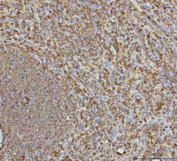


 IF (Immunofluorescence)
(Figure 8. IF analysis of VPS53 using anti-VPS53 antibody (AAA19576).VPS53 was detected in a paraffin-embedded section of human breast cancer tissue. Heat mediated antigen retrieval was performed in EDTA buffer (pH 8.0, epitope retrieval solution). The tissue section was blocked with 10% goat serum. The tissue section was then incubated with 5 ug/mL rabbit anti-VPS53 Antibody (AAA19576) overnight at 4 degree C. DyLight550 Conjugated Goat Anti-Rabbit IgG (BA1135) was used as secondary antibody at 1:500 dilution and incubated for 30 minutes at 37 degree C. The section was counterstained with DAPI. Visualize using a fluorescence microscope and filter sets appropriate for the label used.)
IF (Immunofluorescence)
(Figure 8. IF analysis of VPS53 using anti-VPS53 antibody (AAA19576).VPS53 was detected in a paraffin-embedded section of human breast cancer tissue. Heat mediated antigen retrieval was performed in EDTA buffer (pH 8.0, epitope retrieval solution). The tissue section was blocked with 10% goat serum. The tissue section was then incubated with 5 ug/mL rabbit anti-VPS53 Antibody (AAA19576) overnight at 4 degree C. DyLight550 Conjugated Goat Anti-Rabbit IgG (BA1135) was used as secondary antibody at 1:500 dilution and incubated for 30 minutes at 37 degree C. The section was counterstained with DAPI. Visualize using a fluorescence microscope and filter sets appropriate for the label used.)
VPS53, Polyclonal Antibody (Cat# AAA19576)
Full Name
Anti-VPS53 Antibody Picoband
Gene Names
VPS53; HCCS1; PCH2E; hVps53L; pp13624
Reactivity
Human, Mouse, Rat
Applications
Western Blot, Immunohistochemistry, Immunocytochemistry, Immunofluorescence, Flow Cytometry
Purity
Immunogen affinity purified.
Pricing
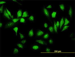


 Application Data
(Detection limit for recombinant GST tagged RPS5 is ~3ng/ml as a capture antibody.)
Application Data
(Detection limit for recombinant GST tagged RPS5 is ~3ng/ml as a capture antibody.)
RPS5, Monoclonal Antibody (Cat# AAA25232)
Full Name
RPS5 (40S Ribosomal Protein S5) (FITC)
Gene Names
RPS5; S5
Reactivity
Human, Mouse, Rat
Applications
Immunofluorescence, Western Blot
Purity
Purified by Protein A Affinity Chromatography.
Pricing


 WB (Western Blot)
(HSPA1B monoclonal antibody (M05), clone 2D11. Western Blot analysis of HSPA1B expression in PC-12.)
WB (Western Blot)
(HSPA1B monoclonal antibody (M05), clone 2D11. Western Blot analysis of HSPA1B expression in PC-12.)
HSPA1B, Monoclonal Antibody (Cat# AAA26245)
Full Name
HSPA1B (Heat Shock 70kD Protein 1B, FLJ54328, HSP70-1B, HSP70-2, HSPA1A) (Biotin)
Gene Names
HSPA1A; HSP72; HSPA1; HSP70I; HSP70-1; HSP70.1; HSP70-1A; HEL-S-103
Applications
Immunohistochemistry, Western Blot
Purity
Purified
Pricing


 FCM (Flow Cytometry)
(Figure 11. Flow Cytometry analysis of THP-1 cells using anti-EPRS1/PARS antibody (AAA19268).Overlay histogram showing THP-1 cells stained with AAA19268 (Blue line). The cells were blocked with 10% normal goat serum. And then incubated with rabbit anti-EPRS1/PARS Antibody (AAA19268, 1μg/1x106 cells) for 30 min at 20 degree C. DyLight®488 conjugated goat anti-rabbit IgG (5-10μg/1x106 cells) was used as secondary antibody for 30 minutes at 20 degree C. Isotype control antibody (Green line) was rabbit IgG (1μg/1x106) used under the same conditions. Unlabelled sample (Red line) was also used as a control.)
FCM (Flow Cytometry)
(Figure 11. Flow Cytometry analysis of THP-1 cells using anti-EPRS1/PARS antibody (AAA19268).Overlay histogram showing THP-1 cells stained with AAA19268 (Blue line). The cells were blocked with 10% normal goat serum. And then incubated with rabbit anti-EPRS1/PARS Antibody (AAA19268, 1μg/1x106 cells) for 30 min at 20 degree C. DyLight®488 conjugated goat anti-rabbit IgG (5-10μg/1x106 cells) was used as secondary antibody for 30 minutes at 20 degree C. Isotype control antibody (Green line) was rabbit IgG (1μg/1x106) used under the same conditions. Unlabelled sample (Red line) was also used as a control.)
EPRS1/PARS, Polyclonal Antibody (Cat# AAA19268)
Full Name
Anti-EPRS1/PARS Antibody
Gene Names
EPRS; EARS; PARS; QARS; QPRS; PIG32; GLUPRORS
Reactivity
Human, Mouse, Rat
Applications
Western Blot, Immunohistochemistry, Immunocytochemistry, Immunofluorescence, Flow Cytometry, Direct ELISA
Purity
Immunogen affinity purified.
Pricing

 FCM (Flow Cytometry)
(Figure 7. Flow Cytometry analysis of MCF-7 cells using anti-NUP37 antibody (AAA19966).Overlay histogram showing MCF-7 cells stained with AAA19966 (Blue line). To facilitate intracellular staining, cells were fixed with 4% paraformaldehyde and permeabilized with permeabilization buffer. The cells were blocked with 10% normal goat serum. And then incubated with rabbit anti-NUP37 Antibody (AAA19966, 1ug/1x106 cells) for 30 min at 20 degree C. DyLight488 conjugated goat anti-rabbit IgG was used as secondary antibody for 30 minutes at 20 degree C. Isotype control antibody (Green line) was rabbit IgG (1ug/1x106) used under the same conditions. Unlabelled sample (Red line) was also used as a control.)
FCM (Flow Cytometry)
(Figure 7. Flow Cytometry analysis of MCF-7 cells using anti-NUP37 antibody (AAA19966).Overlay histogram showing MCF-7 cells stained with AAA19966 (Blue line). To facilitate intracellular staining, cells were fixed with 4% paraformaldehyde and permeabilized with permeabilization buffer. The cells were blocked with 10% normal goat serum. And then incubated with rabbit anti-NUP37 Antibody (AAA19966, 1ug/1x106 cells) for 30 min at 20 degree C. DyLight488 conjugated goat anti-rabbit IgG was used as secondary antibody for 30 minutes at 20 degree C. Isotype control antibody (Green line) was rabbit IgG (1ug/1x106) used under the same conditions. Unlabelled sample (Red line) was also used as a control.)
NUP37, Polyclonal Antibody (Cat# AAA19966)
Full Name
Anti-NUP37 Antibody Picoband
Gene Names
NUP37; p37
Reactivity
Human, Rat
Applications
Immunofluorescence, Immunohistochemistry, Immunocytochemistry, Western Blot, Flow Cytometry
Purity
Immunogen affinity purified.
Pricing


 WB (Western Blot)
(SAV1 monoclonal antibody (M02), Western Blot analysis of SAV1 expression in Hela NE.)
WB (Western Blot)
(SAV1 monoclonal antibody (M02), Western Blot analysis of SAV1 expression in Hela NE.)
SAV1, Monoclonal Antibody (Cat# AAA25241)
Full Name
SAV1 (Protein Salvador Homolog 1, SAV, 45kD WW Domain Protein, WW45, hWW45, WWP4) (FITC)
Gene Names
SAV1; SAV; WW45; WWP4
Reactivity
Human
Applications
Immunofluorescence, Immunoprecipitation, Western Blot
Purity
Purified by Protein A Affinity Chromatography.
Pricing


 WB (Western Blot)
(LEPRE1 monoclonal antibody, Western Blot analysis of LEPRE1 expression in HeLa.)
WB (Western Blot)
(LEPRE1 monoclonal antibody, Western Blot analysis of LEPRE1 expression in HeLa.)
GROS1, Monoclonal Antibody (Cat# AAA25705)
Full Name
GROS1 (Growth Suppressor 1, Leucine- and Proline-enriched Proteoglycan 1, LEPRE1, Leprecan-1, MGC117314, OI8, Prolyl 3-hydroxylase 1, P3H1, PSEC0109) (PE)
Gene Names
P3H1; OI8; GROS1; LEPRE1
Reactivity
Human
Applications
Immunofluorescence, Immunohistochemistry, Western Blot
Purity
Purified by Protein A Affinity Chromatography.
Pricing


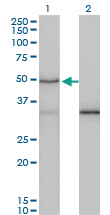

 WB (Western Blot)
(SMAD3 monoclonal antibody. Western Blot analysis of SMAD3 expression in human colon.)
WB (Western Blot)
(SMAD3 monoclonal antibody. Western Blot analysis of SMAD3 expression in human colon.)
SMAD3, Monoclonal Antibody (Cat# AAA25840)
Full Name
SMAD3 (Mothers Against Decapentaplegic Homolog 3, SMAD 3, Mothers Against DPP Homolog 3, MAD Homolog 3, Mad3, hMAD-3, SMAD Family Member 3, JV15-2, hSMAD3, MADH3, DKFZp586N0721, DKFZp686J10186, HSPC193, HsT17436, MGC60396) (PE)
Gene Names
SMAD3; LDS3; LDS1C; MADH3; JV15-2; HSPC193; HsT17436
Reactivity
Human, Rat
Applications
Immunohistochemistry, Western Blot
Purity
Purified by Protein A Affinity Chromatography.
Pricing
 IHC (Immunohistchemistry)
(Figure 6. IHC analysis of SEC61G using anti-SEC61G antibody (AAA19974).SEC61G was detected in a paraffin-embedded section of rat colon tissue. Heat mediated antigen retrieval was performed in EDTA buffer (pH 8.0, epitope retrieval solution). The tissue section was blocked with 10% goat serum. The tissue section was then incubated with 2ug/ml rabbit anti-SEC61G Antibody (AAA19974) overnight at 4 degree C. Peroxidase Conjugated Goat Anti-rabbit IgG was used as secondary antibody and incubated for 30 minutes at 37 degree C. The tissue section was developed using HRP Conjugated Rabbit IgG Super Vision Assay Kit ( epitope retrieval solution). The tissue section was blocked with 10% goat serum. The tissue section was then incubated with 2ug/ml rabbit anti-SEC61G Antibody (AAA19974) overnight at 4 degree C. Peroxidase Conjugated Goat Anti-rabbit IgG was used as secondary antibody and incubated for 30 minutes at 37 degree C. The tissue section was developed using HRP Conjugated Rabbit IgG Super Vision Assay Kit ( epitope retrieval solution). The tissue section was blocked with 10% goat serum. The tissue section was then incubated with 2ug/ml rabbit anti-SEC61G Antibody (AAA19974) overnight at 4 degree C. Peroxidase Conjugated Goat Anti-rabbit IgG was used as secondary antibody and incubated for 30 minutes at 37 degree C. The tissue section was developed using HRP Conjugated Rabbit IgG Super Vision Assay Kit ( epitope retrieval solution). The tissue section was blocked with 10% goat serum. The tissue section was then incubated with 2ug/ml rabbit anti-SEC61G Antibody (AAA19974) overnight at 4 degree C. Peroxidase Conjugated Goat Anti-rabbit IgG was used as secondary antibody and incubated for 30 minutes at 37 degree C. The tissue section was developed using HRP Conjugated Rabbit IgG Super Vision Assay Kit ( epitope retrieval solution). The tissue section was blocked with 10% goat serum. The tissue section was then incubated with 2ug/ml rabbit anti-SEC61G Antibody (AAA19974) overnight at 4 degree C. Peroxidase Conjugated Goat Anti-rabbit IgG was used as secondary antibody and incubated for 30 minutes at 37 degree C. The tissue section was developed using HRP Conjugated Rabbit IgG Super Vision Assay Kit (Lane 2: human U87 whole cell lysates,Lane 3: human Hela whole cell lysates,Lane 4: human U251 whole cell lysates,Lane 5: rat liver tissue lysates.After electrophoresis, proteins were transferred to a nitrocellulose membrane at 150 mA for 50-90 minutes. Blocked the membrane with 5% non-fat milk/TBS for 1.5 hour at RT. The membrane was incubated with rabbit anti-SEC61G antigen affinity purified polyclonal antibody (#AAA19974) at 0.5ug/mL overnight at 4 degree C, then washed with TBS-0.1%Tween 3 times with 5 minutes each and probed with a goat anti-rabbit IgG-HRP secondary antibody at a dilution of 1:5000 for 1.5 hour at RT. The signal is developed using an Enhanced Chemiluminescent detection (ECL) kit with Tanon 5200 system. A specific band was detected for SEC61G at approximately 12 kDa. The expected band size for SEC61G is at 8 kDa.)
IHC (Immunohistchemistry)
(Figure 6. IHC analysis of SEC61G using anti-SEC61G antibody (AAA19974).SEC61G was detected in a paraffin-embedded section of rat colon tissue. Heat mediated antigen retrieval was performed in EDTA buffer (pH 8.0, epitope retrieval solution). The tissue section was blocked with 10% goat serum. The tissue section was then incubated with 2ug/ml rabbit anti-SEC61G Antibody (AAA19974) overnight at 4 degree C. Peroxidase Conjugated Goat Anti-rabbit IgG was used as secondary antibody and incubated for 30 minutes at 37 degree C. The tissue section was developed using HRP Conjugated Rabbit IgG Super Vision Assay Kit ( epitope retrieval solution). The tissue section was blocked with 10% goat serum. The tissue section was then incubated with 2ug/ml rabbit anti-SEC61G Antibody (AAA19974) overnight at 4 degree C. Peroxidase Conjugated Goat Anti-rabbit IgG was used as secondary antibody and incubated for 30 minutes at 37 degree C. The tissue section was developed using HRP Conjugated Rabbit IgG Super Vision Assay Kit ( epitope retrieval solution). The tissue section was blocked with 10% goat serum. The tissue section was then incubated with 2ug/ml rabbit anti-SEC61G Antibody (AAA19974) overnight at 4 degree C. Peroxidase Conjugated Goat Anti-rabbit IgG was used as secondary antibody and incubated for 30 minutes at 37 degree C. The tissue section was developed using HRP Conjugated Rabbit IgG Super Vision Assay Kit ( epitope retrieval solution). The tissue section was blocked with 10% goat serum. The tissue section was then incubated with 2ug/ml rabbit anti-SEC61G Antibody (AAA19974) overnight at 4 degree C. Peroxidase Conjugated Goat Anti-rabbit IgG was used as secondary antibody and incubated for 30 minutes at 37 degree C. The tissue section was developed using HRP Conjugated Rabbit IgG Super Vision Assay Kit ( epitope retrieval solution). The tissue section was blocked with 10% goat serum. The tissue section was then incubated with 2ug/ml rabbit anti-SEC61G Antibody (AAA19974) overnight at 4 degree C. Peroxidase Conjugated Goat Anti-rabbit IgG was used as secondary antibody and incubated for 30 minutes at 37 degree C. The tissue section was developed using HRP Conjugated Rabbit IgG Super Vision Assay Kit (Lane 2: human U87 whole cell lysates,Lane 3: human Hela whole cell lysates,Lane 4: human U251 whole cell lysates,Lane 5: rat liver tissue lysates.After electrophoresis, proteins were transferred to a nitrocellulose membrane at 150 mA for 50-90 minutes. Blocked the membrane with 5% non-fat milk/TBS for 1.5 hour at RT. The membrane was incubated with rabbit anti-SEC61G antigen affinity purified polyclonal antibody (#AAA19974) at 0.5ug/mL overnight at 4 degree C, then washed with TBS-0.1%Tween 3 times with 5 minutes each and probed with a goat anti-rabbit IgG-HRP secondary antibody at a dilution of 1:5000 for 1.5 hour at RT. The signal is developed using an Enhanced Chemiluminescent detection (ECL) kit with Tanon 5200 system. A specific band was detected for SEC61G at approximately 12 kDa. The expected band size for SEC61G is at 8 kDa.)
SEC61G, Polyclonal Antibody (Cat# AAA19974)
Full Name
Anti-SEC61G Antibody Picoband
Gene Names
SEC61G; SSS1
Reactivity
Human, Mouse, Rat
Applications
Western Blot, Immunohistochemistry
Purity
Immunogen affinity purified.
Pricing


 FCM (Flow Cytometry)
(Figure 6. Flow Cytometry analysis of U20S cells using anti-RIMBP2 antibody (AAA19999).Overlay histogram showing U20S cells stained with AAA19999 (Blue line). To facilitate intracellular staining, cells were fixed with 4% paraformaldehyde and permeabilized with permeabilization buffer. The cells were blocked with 10% normal goat serum. And then incubated with rabbit anti-RIMBP2 Antibody (AAA19999, 1ug/1x106 cells) for 30 min at 20 degree C. PE conjugated goat anti-rabbit IgG (5-10ug/1x106 cells) was used as secondary antibody for 30 minutes at 20 degree C. Isotype control antibody (Green line) was rabbit IgG (1ug/1x106) used under the same conditions. Unlabelled sample without incubation with primary antibody and secondary antibody (Red line) was used as a blank control.)
FCM (Flow Cytometry)
(Figure 6. Flow Cytometry analysis of U20S cells using anti-RIMBP2 antibody (AAA19999).Overlay histogram showing U20S cells stained with AAA19999 (Blue line). To facilitate intracellular staining, cells were fixed with 4% paraformaldehyde and permeabilized with permeabilization buffer. The cells were blocked with 10% normal goat serum. And then incubated with rabbit anti-RIMBP2 Antibody (AAA19999, 1ug/1x106 cells) for 30 min at 20 degree C. PE conjugated goat anti-rabbit IgG (5-10ug/1x106 cells) was used as secondary antibody for 30 minutes at 20 degree C. Isotype control antibody (Green line) was rabbit IgG (1ug/1x106) used under the same conditions. Unlabelled sample without incubation with primary antibody and secondary antibody (Red line) was used as a blank control.)
RIMBP2, Polyclonal Antibody (Cat# AAA19999)
Full Name
Anti-RIMBP2 Antibody Picoband
Gene Names
RIMBP2; RBP2; RIM-BP2; PPP1R133
Reactivity
Human, Mouse, Rat
Applications
Flow Cytometry, Immunofluorescence, Immunohistochemistry, Western Blot
Purity
Immunogen affinity purified.
Pricing


 IHC (Immunohistochemistry)
(Immunohistochemistry of paraffin-embedded mouse spleen using STAT4 antibody at dilution of 1:100 (40x lens).)
IHC (Immunohistochemistry)
(Immunohistochemistry of paraffin-embedded mouse spleen using STAT4 antibody at dilution of 1:100 (40x lens).)
STAT4, Polyclonal Antibody (Cat# AAA28146)
Full Name
STAT4 Polyclonal Antibody
Gene Names
STAT4; SLEB11
Reactivity
Human, Mouse, Rat
Applications
Western Blot, Immunohistochemistry, Immunofluorescence
Purity
Affinity Purification
Pricing


 Application Data
(Detection limit for recombinant GST tagged STIP1 is 0.03 ng/ml as a capture antibody.)
Application Data
(Detection limit for recombinant GST tagged STIP1 is 0.03 ng/ml as a capture antibody.)
STIP1, Monoclonal Antibody (Cat# AAA26015)
Full Name
STIP1 (Stress-Induced-Phosphoprotein 1, HOP, IEF-SSP-3521, P60, STI1, STI1L) (AP)
Gene Names
STIP1; HOP; P60; STI1; STI1L; HEL-S-94n; IEF-SSP-3521
Applications
Western Blot
Purity
Purified
Pricing


 IHC (Immunohistochemistry)
(Figure 7. IHC analysis of GCHFR using anti-GCHFR antibody (AAA19322).GCHFR was detected in paraffin-embedded section of mouse liver tissue. Heat mediated antigen retrieval was performed in EDTA buffer (pH8. 0, epitope retrieval solution). The tissue section was blocked with 10% goat serum. The tissue section was then incubated with 2μg/ml rabbit anti-GCHFR Antibody (AAA19322) overnight at 4 degree C. Biotinylated goat anti-rabbit IgG was used as secondary antibody and incubated for 30 minutes at 37 degree C. The tissue section was developed using Strepavidin-Biotin-Complex (SABC) (Catalog # with DAB as the chromogen.)
IHC (Immunohistochemistry)
(Figure 7. IHC analysis of GCHFR using anti-GCHFR antibody (AAA19322).GCHFR was detected in paraffin-embedded section of mouse liver tissue. Heat mediated antigen retrieval was performed in EDTA buffer (pH8. 0, epitope retrieval solution). The tissue section was blocked with 10% goat serum. The tissue section was then incubated with 2μg/ml rabbit anti-GCHFR Antibody (AAA19322) overnight at 4 degree C. Biotinylated goat anti-rabbit IgG was used as secondary antibody and incubated for 30 minutes at 37 degree C. The tissue section was developed using Strepavidin-Biotin-Complex (SABC) (Catalog # with DAB as the chromogen.)
GCHFR, Polyclonal Antibody (Cat# AAA19322)
Full Name
Anti-GCHFR Antibody
Gene Names
GCHFR; P35; GFRP; HsT16933
Reactivity
Human, Mouse, Rat
Applications
Western Blot, Immunohistochemistry, Immunocytochemistry, Immunofluorescence, Flow Cytometry, Direct ELISA
Purity
Immunogen affinity purified.
Pricing

 FCM (Flow Cytometry)
(Figure 11. Flow Cytometry analysis of HepG2 cells using anti-PELI3 antibody (AAA19930).Overlay histogram showing HepG2 cells stained with AAA19930 (Blue line). To facilitate intracellular staining, cells were fixed with 4% paraformaldehyde and permeabilized with permeabilization buffer. The cells were blocked with 10% normal goat serum. And then incubated with rabbit anti-PELI3 Antibody (AAA19930, 1ug/1x106 cells) for 30 min at 20 degree C. DyLight488 conjugated goat anti-rabbit IgG (BA1127, 5-10ug/1x106 cells) was used as secondary antibody for 30 minutes at 20 degree C. Isotype control antibody (Green line) was rabbit IgG (1ug/1x106) used under the same conditions. Unlabelled sample (Red line) was also used as a control.)
FCM (Flow Cytometry)
(Figure 11. Flow Cytometry analysis of HepG2 cells using anti-PELI3 antibody (AAA19930).Overlay histogram showing HepG2 cells stained with AAA19930 (Blue line). To facilitate intracellular staining, cells were fixed with 4% paraformaldehyde and permeabilized with permeabilization buffer. The cells were blocked with 10% normal goat serum. And then incubated with rabbit anti-PELI3 Antibody (AAA19930, 1ug/1x106 cells) for 30 min at 20 degree C. DyLight488 conjugated goat anti-rabbit IgG (BA1127, 5-10ug/1x106 cells) was used as secondary antibody for 30 minutes at 20 degree C. Isotype control antibody (Green line) was rabbit IgG (1ug/1x106) used under the same conditions. Unlabelled sample (Red line) was also used as a control.)
PELI3, Polyclonal Antibody (Cat# AAA19930)
Full Name
Anti-PELI3 Antibody Picoband
Reactivity
Human, Mouse, Rat
Applications
Western Blot, Immunohistochemistry, Immunocytochemistry, Immunofluorescence, Flow Cytometry
Purity
Immunogen affinity purified.
Pricing

 FCM (Flow Cytometry)
(Figure 9. Flow Cytometry analysis of 293T cells using anti-NSUN2 antibody (AAA19802).Overlay histogram showing 293T cells stained with AAA19802 (Blue line). To facilitate intracellular staining, cells were fixed with 4% paraformaldehyde and permeabilized with permeabilization buffer. The cells were blocked with 10% normal goat serum. And then incubated with rabbit anti-NSUN2 Antibody (AAA19802, 1ug/1x106 cells) for 30 min at 20 degree C. DyLight488 conjugated goat anti-rabbit IgG was used as secondary antibody for 30 minutes at 20 degree C. Isotype control antibody (Green line) was rabbit IgG (1ug/1x106) used under the same conditions. Unlabelled sample (Red line) was also used as a control.)
FCM (Flow Cytometry)
(Figure 9. Flow Cytometry analysis of 293T cells using anti-NSUN2 antibody (AAA19802).Overlay histogram showing 293T cells stained with AAA19802 (Blue line). To facilitate intracellular staining, cells were fixed with 4% paraformaldehyde and permeabilized with permeabilization buffer. The cells were blocked with 10% normal goat serum. And then incubated with rabbit anti-NSUN2 Antibody (AAA19802, 1ug/1x106 cells) for 30 min at 20 degree C. DyLight488 conjugated goat anti-rabbit IgG was used as secondary antibody for 30 minutes at 20 degree C. Isotype control antibody (Green line) was rabbit IgG (1ug/1x106) used under the same conditions. Unlabelled sample (Red line) was also used as a control.)
NSUN2, Polyclonal Antibody (Cat# AAA19802)
Full Name
Anti-NSUN2 Antibody Picoband
Gene Names
NSUN2; MISU; MRT5; SAKI; TRM4
Reactivity
Human, Mouse, Rat
Applications
Western Blot, Immunohistochemistry, Flow Cytometry
Purity
Immunogen affinity purified.
Pricing


 FCM (Flow Cytometry)
(Figure 14. Flow Cytometry analysis of RT4 cells using anti-KPNB1 antibody (AAA19692).Overlay histogram showing RT4 cells stained with AAA19692 (Blue line). The cells were blocked with 10% normal goat serum. And then incubated with mouse anti-KPNB1 Antibody (AAA19692, 1 ug/1x106 cells) for 30 min at 20 degree C. DyLight488 conjugated goat anti-mouse IgG (BA1126, 5-10 ug/1x106 cells) was used as secondary antibody for 30 minutes at 20 degree C. Isotype control antibody (Green line) was mouse IgG (1 ug/1x106) used under the same conditions. Unlabelled sample (Red line) was also used as a control.)
FCM (Flow Cytometry)
(Figure 14. Flow Cytometry analysis of RT4 cells using anti-KPNB1 antibody (AAA19692).Overlay histogram showing RT4 cells stained with AAA19692 (Blue line). The cells were blocked with 10% normal goat serum. And then incubated with mouse anti-KPNB1 Antibody (AAA19692, 1 ug/1x106 cells) for 30 min at 20 degree C. DyLight488 conjugated goat anti-mouse IgG (BA1126, 5-10 ug/1x106 cells) was used as secondary antibody for 30 minutes at 20 degree C. Isotype control antibody (Green line) was mouse IgG (1 ug/1x106) used under the same conditions. Unlabelled sample (Red line) was also used as a control.)
KPNB1, Monoclonal Antibody (Cat# AAA19692)
Full Name
Anti-KPNB1 Antibody Picoband (monoclonal, 3I11F2)
Gene Names
KPNB1; IMB1; IPO1; IPOB; Impnb; NTF97
Reactivity
Human, Mouse, Rat
Applications
Western Blot, Immunohistochemistry, Immunocytochemistry, Immunofluorescence, Flow Cytometry
Purity
Immunogen affinity purified.
Pricing
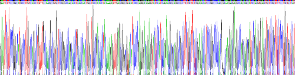
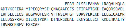
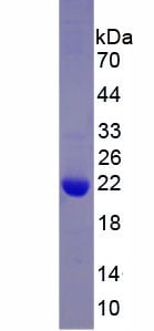 SDS-PAGE
(SDS-PAGE)
SDS-PAGE
(SDS-PAGE)
Growth Hormone (GH), Recombinant Protein (Cat# AAA152136)
Full Name
Eukaryotic Growth Hormone (GH)
Gene Names
GH1; GH; GHB1
Applications
Western Blot
Purity
Greater than 80% by SDS-PAGE
Pricing


 FCM (Flow Cytometry)
(Figure 6. Flow Cytometry analysis of MCF-7 cells using anti-TIMM29 antibody (AAA19648).Overlay histogram showing MCF-7 cells stained with AAA19648 (Blue line). The cells were blocked with 10% normal goat serum. And then incubated with rabbit anti-TIMM29 Antibody (AAA19648, 1 ug/1x10^6 cells) for 30 min at 20 degree C. DyLight488 conjugated goat anti-rabbit IgG was used as secondary antibody for 30 minutes at 20 degree C. Isotype control antibody (Green line) was rabbit IgG (1 ug/1x10^6) used under the same conditions. Unlabelled sample (Red line) was also used as a control.)
FCM (Flow Cytometry)
(Figure 6. Flow Cytometry analysis of MCF-7 cells using anti-TIMM29 antibody (AAA19648).Overlay histogram showing MCF-7 cells stained with AAA19648 (Blue line). The cells were blocked with 10% normal goat serum. And then incubated with rabbit anti-TIMM29 Antibody (AAA19648, 1 ug/1x10^6 cells) for 30 min at 20 degree C. DyLight488 conjugated goat anti-rabbit IgG was used as secondary antibody for 30 minutes at 20 degree C. Isotype control antibody (Green line) was rabbit IgG (1 ug/1x10^6) used under the same conditions. Unlabelled sample (Red line) was also used as a control.)
TIMM29, Polyclonal Antibody (Cat# AAA19648)
Full Name
Anti-TIMM29 Antibody Picoband
Reactivity
Human
Applications
Western Blot, Immunohistochemistry, Immunocytochemistry, Immunofluorescence, Flow Cytometry
Purity
Immunogen affinity purified.
Pricing

 FCM (Flow Cytometry)
(Figure 6. Flow Cytometry analysis of SiHa cells using anti-GLG1 antibody (AAA19888).Overlay histogram showing SiHa cells stained with AAA19888 (Blue line). To facilitate intracellular staining, cells were fixed with 4% paraformaldehyde and permeabilized with permeabilization buffer. The cells were blocked with 10% normal goat serum. And then incubated with rabbit anti-GLG1 Antibody (AAA19888, 1ug/1x106 cells) for 30 min at 20 degree C. DyLight488 conjugated goat anti-rabbit IgG was used as secondary antibody for 30 minutes at 20 degree C. Isotype control antibody (Green line) was rabbit IgG (1ug/1x106) used under the same conditions. Unlabelled sample without incubation with primary antibody and secondary antibody (Red line) was used as a blank control.)
FCM (Flow Cytometry)
(Figure 6. Flow Cytometry analysis of SiHa cells using anti-GLG1 antibody (AAA19888).Overlay histogram showing SiHa cells stained with AAA19888 (Blue line). To facilitate intracellular staining, cells were fixed with 4% paraformaldehyde and permeabilized with permeabilization buffer. The cells were blocked with 10% normal goat serum. And then incubated with rabbit anti-GLG1 Antibody (AAA19888, 1ug/1x106 cells) for 30 min at 20 degree C. DyLight488 conjugated goat anti-rabbit IgG was used as secondary antibody for 30 minutes at 20 degree C. Isotype control antibody (Green line) was rabbit IgG (1ug/1x106) used under the same conditions. Unlabelled sample without incubation with primary antibody and secondary antibody (Red line) was used as a blank control.)
GLG1, Polyclonal Antibody (Cat# AAA19888)
Full Name
Anti-GLG1 Antibody Picoband
Gene Names
GLG1; CFR-1; ESL-1; MG160; MG-160
Reactivity
Human, Mouse, Rat
Applications
Western Blot, Immunohistochemistry, Flow Cytometry
Purity
Immunogen affinity purified.
Pricing


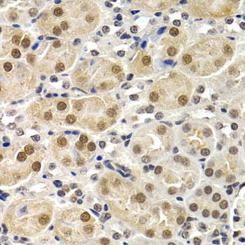


 IF (Immunofluorescence)
(Immunofluorescence analysis of HeLa cells using ID3 antibody.)
IF (Immunofluorescence)
(Immunofluorescence analysis of HeLa cells using ID3 antibody.)
ID3, Polyclonal Antibody (Cat# AAA28175)
Full Name
ID3 Polyclonal Antibody
Gene Names
ID3; HEIR-1; bHLHb25
Reactivity
Human, Mouse, Rat
Applications
Western Blot, Immunohistochemistry, Immunofluorescence
Purity
Affinity Purification
Pricing


 FCM (Flow Cytometry)
(Figure 8. Flow Cytometry analysis of HepG2 cells using anti-ILDR1 antibody (AAA19928).Overlay histogram showing HepG2 cells stained with AAA19928 (Blue line). To facilitate intracellular staining, cells were fixed with 4% paraformaldehyde and permeabilized with permeabilization buffer. The cells were blocked with 10% normal goat serum. And then incubated with rabbit anti-ILDR1 Antibody (AAA19928, 1ug/1x106 cells) for 30 min at 20 degree C. DyLight488 conjugated goat anti-rabbit IgG was used as secondary antibody for 30 minutes at 20 degree C. Isotype control antibody (Green line) was rabbit IgG (1ug/1x106) used under the same conditions. Unlabelled sample (Red line) was also used as a control.)
FCM (Flow Cytometry)
(Figure 8. Flow Cytometry analysis of HepG2 cells using anti-ILDR1 antibody (AAA19928).Overlay histogram showing HepG2 cells stained with AAA19928 (Blue line). To facilitate intracellular staining, cells were fixed with 4% paraformaldehyde and permeabilized with permeabilization buffer. The cells were blocked with 10% normal goat serum. And then incubated with rabbit anti-ILDR1 Antibody (AAA19928, 1ug/1x106 cells) for 30 min at 20 degree C. DyLight488 conjugated goat anti-rabbit IgG was used as secondary antibody for 30 minutes at 20 degree C. Isotype control antibody (Green line) was rabbit IgG (1ug/1x106) used under the same conditions. Unlabelled sample (Red line) was also used as a control.)
IDH3A, Polyclonal Antibody (Cat# AAA19928)
Full Name
Anti-IDH3A Antibody Picoband
Reactivity
Human, Mouse, Rat
Applications
Western Blot, Immunohistochemistry, Immunocytochemistry, Immunofluorescence, Flow Cytometry
Purity
Immunogen affinity purified.
Pricing


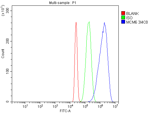 FCM (Flow Cytometry)
(Figure 10. Flow Cytometry analysis of HL-60 cells using anti-MCM6 antibody (AAA19703).Overlay histogram showing HL-60 cells stained with AAA19703 (Blue line). The cells were blocked with 10% normal goat serum. And then incubated with mouse anti-MCM6 Antibody (AAA19703, 1 ug/1x10^6 cells) for 30 min at 20 degree C. DyLight488 conjugated goat anti-mouse IgG was used as secondary antibody for 30 minutes at 20 degree C. Isotype control antibody (Green line) was mouse IgG (1 ug/1x10^6) used under the same conditions. Unlabelled sample (Red line) was also used as a control.)
FCM (Flow Cytometry)
(Figure 10. Flow Cytometry analysis of HL-60 cells using anti-MCM6 antibody (AAA19703).Overlay histogram showing HL-60 cells stained with AAA19703 (Blue line). The cells were blocked with 10% normal goat serum. And then incubated with mouse anti-MCM6 Antibody (AAA19703, 1 ug/1x10^6 cells) for 30 min at 20 degree C. DyLight488 conjugated goat anti-mouse IgG was used as secondary antibody for 30 minutes at 20 degree C. Isotype control antibody (Green line) was mouse IgG (1 ug/1x10^6) used under the same conditions. Unlabelled sample (Red line) was also used as a control.)
MCM6, Monoclonal Antibody (Cat# AAA19703)
Full Name
Anti-MCM6 Antibody Picoband (monoclonal, 3I4C8)
Gene Names
MCM6; Mis5; P105MCM; MCG40308
Reactivity
Human, Mouse, Rat
Applications
Western Blot, Immunohistochemistry, Immunocytochemistry, Immunofluorescence, Flow Cytometry
Purity
Immunogen affinity purified.
Pricing
MHC class I chain-related gene B, Active Protein (Cat# AAA10837)
Full Name
Recombinant Human MHC class I chain-related gene B
Gene Names
MICB; PERB11.2
Purity
Greater than 95.0% as determined by(a) Analysis by RP-HPLC. (b) Analysis by SDS-PAGE.
Pricing


 FCM (Flow Cytometry)
(FACS analysis of ES-2 cells stained with STIP1 monoclonal antibody clone 2E1 (Green) and non-stained ES-2 cells (Black) as negative control.)
FCM (Flow Cytometry)
(FACS analysis of ES-2 cells stained with STIP1 monoclonal antibody clone 2E1 (Green) and non-stained ES-2 cells (Black) as negative control.)
STIP1, Monoclonal Antibody (Cat# AAA26008)
Full Name
STIP1 (Stress-Induced-Phosphoprotein 1, HOP, IEF-SSP-3521, P60, STI1, STI1L) (AP)
Gene Names
STIP1; HOP; P60; STI1; STI1L; HEL-S-94n; IEF-SSP-3521
Applications
Immunofluorescence, Immunohistochemistry, Western Blot
Purity
Purified
Pricing


 Application Data
(Detection limit for recombinant GST tagged PITPNA is 0.1ng/ml as a capture antibody.)
Application Data
(Detection limit for recombinant GST tagged PITPNA is 0.1ng/ml as a capture antibody.)
PITPNA, Monoclonal Antibody (Cat# AAA25206)
Full Name
PITPNA (Phosphatidylinositol Transfer Protein alpha Isoform, PI-TP-alpha, PtdIns Transfer Protein alpha, PtdInsTP alpha, PITPN, MGC99649) (FITC)
Gene Names
PITPNA; PITPN; VIB1A; HEL-S-36; PI-TPalpha
Reactivity
Human, Mouse, Rat
Applications
Western Blot
Purity
Purified by Protein A Affinity Chromatography.
Pricing


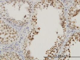


 WB (Western Blot)
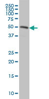
(RASSF8 monoclonal antibody Western Blot analysis of RASSF8 expression in HeLa.)
WB (Western Blot)
(RASSF8 monoclonal antibody Western Blot analysis of RASSF8 expression in HeLa.)
RASSF8, Monoclonal Antibody (Cat# AAA25518)
Full Name
RASSF8 (Ras Association Domain-containing Protein 8, Carcinoma-associated Protein HOJ-1, C12orf2) (HRP)
Gene Names
RASSF8; HOJ1; C12orf2
Reactivity
Human, Mouse, Rat
Applications
Immunohistochemistry, Immunoprecipitation, Western Blot
Purity
Purified by Protein A Affinity Chromatography.
Pricing


 IHC (Immunohistchemistry)
(Figure 6. IHC analysis of Cytoglobin using anti- Cytoglobin antibody (AAA11681).Cytoglobin was detected in paraffin-embedded section of human lung cancer tissues. Heat mediated antigen retrieval was performed in citrate buffer (pH6, epitope retrieval solution) for 20 mins. The tissue section was blocked with 10% goat serum. The tissue section was then incubated with 1ug/ml rabbit anti- Cytoglobin Antibody (AAA11681) overnight at 4 degree C. Biotinylated goat anti-rabbit IgG was used as secondary antibody and incubated for 30 minutes at 37 degree C. The tissue section was developed using Strepavidin-Biotin-Complex (SABC) with DAB as the chromogen.)
IHC (Immunohistchemistry)
(Figure 6. IHC analysis of Cytoglobin using anti- Cytoglobin antibody (AAA11681).Cytoglobin was detected in paraffin-embedded section of human lung cancer tissues. Heat mediated antigen retrieval was performed in citrate buffer (pH6, epitope retrieval solution) for 20 mins. The tissue section was blocked with 10% goat serum. The tissue section was then incubated with 1ug/ml rabbit anti- Cytoglobin Antibody (AAA11681) overnight at 4 degree C. Biotinylated goat anti-rabbit IgG was used as secondary antibody and incubated for 30 minutes at 37 degree C. The tissue section was developed using Strepavidin-Biotin-Complex (SABC) with DAB as the chromogen.)
Cytoglobin, Polyclonal Antibody (Cat# AAA11681)
Full Name
Anti-Cytoglobin Antibody
Gene Names
CYGB; HGB; STAP
Reactivity
Human, Mouse, Rat
Applications
Western Blot, Immunohistochemistry
Purity
Immunogen affinity purified.
Pricing


 FCM (Flow Cytometry)
(Figure 7. Flow Cytometry analysis of HEL cells using anti-LIN41/TRIM71 antibody (AAA19594).Overlay histogram showing HEL cells stained with AAA19594 (Blue line). The cells were blocked with 10% normal goat serum. And then incubated with rabbit anti-LIN41/TRIM71 Antibody (AAA19594, 1 ug/1x10^6 cells) for 30 min at 20 degree C. DyLight488 conjugated goat anti-rabbit IgG was used as secondary antibody for 30 minutes at 20 degree C. Isotype control antibody (Green line) was rabbit IgG (1 ug/1x10^6) used under the same conditions. Unlabelled sample (Red line) was also used as a control.)
FCM (Flow Cytometry)
(Figure 7. Flow Cytometry analysis of HEL cells using anti-LIN41/TRIM71 antibody (AAA19594).Overlay histogram showing HEL cells stained with AAA19594 (Blue line). The cells were blocked with 10% normal goat serum. And then incubated with rabbit anti-LIN41/TRIM71 Antibody (AAA19594, 1 ug/1x10^6 cells) for 30 min at 20 degree C. DyLight488 conjugated goat anti-rabbit IgG was used as secondary antibody for 30 minutes at 20 degree C. Isotype control antibody (Green line) was rabbit IgG (1 ug/1x10^6) used under the same conditions. Unlabelled sample (Red line) was also used as a control.)
LIN41/TRIM71, Polyclonal Antibody (Cat# AAA19594)
Full Name
Anti-LIN41/TRIM71 Antibody Picoband
Gene Names
TRIM71; LIN41; LIN-41
Reactivity
Human, Mouse, Rat
Applications
Western Blot, Immunohistochemistry, Flow Cytometry
Purity
Immunogen affinity purified.
Pricing



 IF (Immunofluorescence)
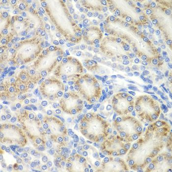
(Immunofluorescence analysis of A549 cells using ASPSCR1 antibody. Blue: DAPI for nuclear staining.)
IF (Immunofluorescence)
(Immunofluorescence analysis of A549 cells using ASPSCR1 antibody. Blue: DAPI for nuclear staining.)
ASPSCR1, Polyclonal Antibody (Cat# AAA28119)
Full Name
ASPSCR1
Gene Names
ASPSCR1; TUG; ASPL; ASPS; RCC17; UBXD9; UBXN9; ASPCR1
Reactivity
Human, Mouse, Rat
Applications
Western Blot, Immunohistochemistry
Pricing


 IP (Immunoprecipitation)
(Immunoprecipitation analysis of 150ug extracts of MCF7 cells using 3ug HIST3H3 antibody. Western blot was performed from the immunoprecipitate using HIST3H3 antibody at a dilition of 1:500.)
IP (Immunoprecipitation)
(Immunoprecipitation analysis of 150ug extracts of MCF7 cells using 3ug HIST3H3 antibody. Western blot was performed from the immunoprecipitate using HIST3H3 antibody at a dilition of 1:500.)
HIST3H3, Polyclonal Antibody (Cat# AAA28284)
Full Name
HIST3H3 Polyclonal Antibody
Gene Names
HIST3H3; H3t; H3.4; H3/g; H3FT
Reactivity
Human, Mouse, Rat, Other (Wide Range)
Applications
Western Blot, Immunohistochemistry, Immunoprecipitation
Purity
Affinity Purification
Pricing


 IHC (Immunohistchemistry)
(Immunohistochemistry of paraffin-embedded human adenomyosis using XRCC5 Antibody at dilution of 1:100 (40x lens).)
IHC (Immunohistchemistry)
(Immunohistochemistry of paraffin-embedded human adenomyosis using XRCC5 Antibody at dilution of 1:100 (40x lens).)
XRCC5, Polyclonal Antibody (Cat# AAA28099)
Full Name
XRCC5 Polyclonal Antibody
Gene Names
XRCC5; KU80; KUB2; Ku86; NFIV; KARP1; KARP-1
Reactivity
Human
Applications
Western Blot, Immunohistochemistry, Immunofluorescence
Purity
Affinity Purification
Pricing