
Filters
Clonality
Type
Reactivity
Gene Name
Isotype
Host
Application
Clone
10000 results for "Recombinant Proteins" - showing 1800-1850
Retinoic Acid Receptor Responder 2, Recombinant Protein (Cat# AAA10868)
Full Name
Recombinant Human Retinoic Acid Receptor Responder 2
Gene Names
RARRES2; TIG2; HP10433
Purity
Greater than 98.0% as determined by: (a) Analysis by RP-HPLC. (b) Analysis by SDS-PAGE.
Pricing
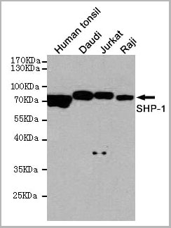
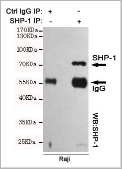 IP (Immunoprecipitation)
(Immunoprecipitation analysis of Raji cell lysates using SHP-1 mouse mAb.)
IP (Immunoprecipitation)
(Immunoprecipitation analysis of Raji cell lysates using SHP-1 mouse mAb.)
SHP-1, Monoclonal Antibody (Cat# AAA14102)
Full Name
Anti-SHP-1 Mouse mAb
Gene Names
PTPN6; HCP; HCPH; SHP1; SHP-1; HPTP1C; PTP-1C; SHP-1L; SH-PTP1
Reactivity
Human
Applications
Western Blot, Immunoprecipitation
Purity
Affinity purified
Pricing
 Application Data
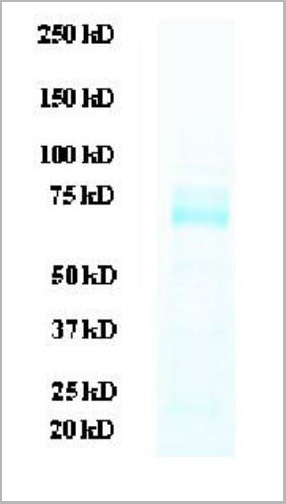
(SDS-PAGE: purified H5 (H5N1) (A/Anhui/1/2005) protein (reducing condition))
Application Data
(SDS-PAGE: purified H5 (H5N1) (A/Anhui/1/2005) protein (reducing condition))
H5 (H5N1) (A/Anhui/1/2005), Recombinant Protein (Cat# AAA13760)
Full Name
H5 (H5N1) (A/Anhui/1/2005) (aa 18-530)
Applications
Western Blot
Purity
> 95% pure by 10% SDS-PAGE gel
Pricing
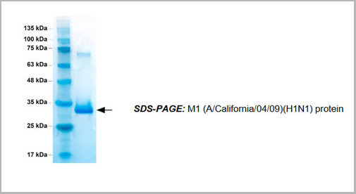 SDS-PAGE
M1 (A/California/04/2009)(H1N1), Recombinant Protein (Cat# AAA13771)
Full Name
M1 (A/California/04/2009)(H1N1)
Applications
Western Blot
Purity
>95% pure by 10% SDS PAGE gel
Pricing
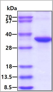 SDS-PAGE
(3ug by SDS-PAGE under reducing condition and visualized by coomassie blue stain.)
SDS-PAGE
(3ug by SDS-PAGE under reducing condition and visualized by coomassie blue stain.)
NQO1, Active Protein (Cat# AAA11735)
Full Name
Recombinant Human NQO1 Protein
Gene Names
NQO1; DTD; QR1; DHQU; DIA4; NMOR1; NMORI
Applications
SDS-PAGE, ELISA
Purity
>95% by SDS-PAGE
Pricing
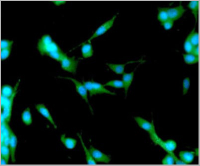 IF (Immunofluorescence)
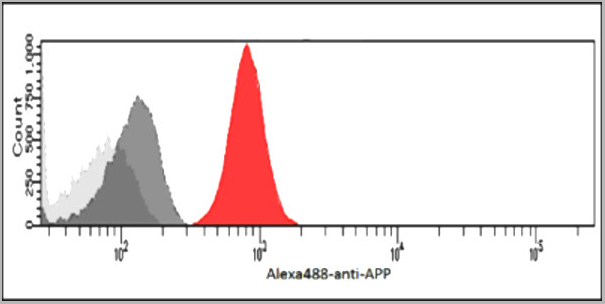
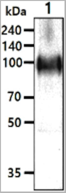
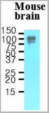
(ICC/IF analysis of APP/Protease Nexin II in U87MG cells. The cell was stained with AAA11729 (1:100). The secondary antibody (green) was used Alexa Fluor 488. DAPI was stained the cell nucleus (blue).)
IF (Immunofluorescence)
(ICC/IF analysis of APP/Protease Nexin II in U87MG cells. The cell was stained with AAA11729 (1:100). The secondary antibody (green) was used Alexa Fluor 488. DAPI was stained the cell nucleus (blue).)
APP, Monoclonal Antibody (Cat# AAA11729)
Full Name
APP antibody
Gene Names
APP; AAA; AD1; PN2; ABPP; APPI; CVAP; ABETA; PN-II; preA4; CTFgamma
Reactivity
Human
Applications
Western Blot, Flow Cytometry, Immunocytochemistry, Immunofluorescence
Purity
By protein-G affinity chromatography
Pricing


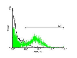 FCM (Flow Cytometry)
(FACS analysis of ES-2 cells stained with STIP1 monoclonal antibody clone 2E11 (Green) and non-stained ES-2 cells (Black) as negative control.)
FCM (Flow Cytometry)
(FACS analysis of ES-2 cells stained with STIP1 monoclonal antibody clone 2E11 (Green) and non-stained ES-2 cells (Black) as negative control.)
STIP1, Monoclonal Antibody (Cat# AAA26541)
Full Name
STIP1 (Stress-Induced-Phosphoprotein 1, HOP, IEF-SSP-3521, P60, STI1, STI1L) (HRP)
Gene Names
STIP1; HOP; P60; STI1; STI1L; HEL-S-94n; IEF-SSP-3521
Applications
Flow Cytometry, Immunofluorescence, Immunohistochemistry, Western Blot
Purity
Purified
Pricing
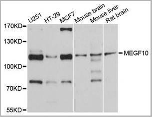 WB (Western Blot)
(Western blot analysis of lysates from U-251 MG cells, using MEGF10 Rabbit pAb (AAA28229) at 1:2000 dilution. Secondary antibody: HRP-conjugated Goat anti-Rabbit IgG (H+L) at 1:10000 dilution. Lysates/proteins: 25?g per lane. Blocking buffer: 3% nonfat dry milk in TBST. Detection: ECL Basic Kit. Exposure time: 90s.)
WB (Western Blot)
(Western blot analysis of lysates from U-251 MG cells, using MEGF10 Rabbit pAb (AAA28229) at 1:2000 dilution. Secondary antibody: HRP-conjugated Goat anti-Rabbit IgG (H+L) at 1:10000 dilution. Lysates/proteins: 25?g per lane. Blocking buffer: 3% nonfat dry milk in TBST. Detection: ECL Basic Kit. Exposure time: 90s.)
MEGF10, Polyclonal Antibody (Cat# AAA28229)
Full Name
MEGF10 Rabbit pAb
Gene Names
MEGF10; EMARDD
Reactivity
Human, Mouse, Rat
Applications
Western Blot
Purity
Affinity purification
Pricing


 WB (Western Blot)
(SMAD1 monoclonal antibody (M03), clone 2E9. Western Blot analysis of SMAD1 expression in IMR-32.)
WB (Western Blot)
(SMAD1 monoclonal antibody (M03), clone 2E9. Western Blot analysis of SMAD1 expression in IMR-32.)
SMAD1, Monoclonal Antibody (Cat# AAA25927)
Full Name
SMAD1 (SMAD Family Member 1, BSP1, JV4-1, JV41, MADH1, MADR1) (AP)
Gene Names
SMAD1; BSP1; JV41; BSP-1; JV4-1; MADH1; MADR1
Applications
Immunohistochemistry, Western Blot
Purity
Purified
Pricing
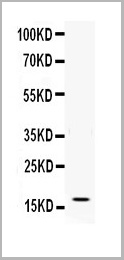 WB (Western Blot)
(Figure. Western blot analysis of IFN gamma using anti- IFN gamma antibody (AAA11517).Electrophoresis was performed on a 5-20% SDS-PAGE gel at 70V (Stacking gel) / 90V (Resolving gel) for 2-3 hours. The sample well of each lane was loaded with 50ug of sample under reducing conditions.Lane: Recombinant Human IFN gamma Protein 0.5ngAfter Electrophoresis, proteins were transferred to a Nitrocellulose membrane at 150mA for 50-90 minutes. Blocked the membrane with 5% Non-fat Milk/ TBS for 1.5 hour at RT. The membrane was incubated with rabbit anti- IFN gamma antigen affinity purified polyclonal antibody at 0.5ug/mL overnight at 4 degree C, then washed with TBS-0.1%Tween 3 times with 5 minutes each and probed with a goat anti-rabbit IgG-HRP secondary antibody at a dilution of 1:10000 for 1.5 hour at RT. The signal is developed using an Enhanced Chemiluminescent detection (ECL) kit with Tanon 5200 system. A specific band was detected for IFN gamma at approximately 17KD. The expected band size for IFN gamma is at 17KD.)
WB (Western Blot)
(Figure. Western blot analysis of IFN gamma using anti- IFN gamma antibody (AAA11517).Electrophoresis was performed on a 5-20% SDS-PAGE gel at 70V (Stacking gel) / 90V (Resolving gel) for 2-3 hours. The sample well of each lane was loaded with 50ug of sample under reducing conditions.Lane: Recombinant Human IFN gamma Protein 0.5ngAfter Electrophoresis, proteins were transferred to a Nitrocellulose membrane at 150mA for 50-90 minutes. Blocked the membrane with 5% Non-fat Milk/ TBS for 1.5 hour at RT. The membrane was incubated with rabbit anti- IFN gamma antigen affinity purified polyclonal antibody at 0.5ug/mL overnight at 4 degree C, then washed with TBS-0.1%Tween 3 times with 5 minutes each and probed with a goat anti-rabbit IgG-HRP secondary antibody at a dilution of 1:10000 for 1.5 hour at RT. The signal is developed using an Enhanced Chemiluminescent detection (ECL) kit with Tanon 5200 system. A specific band was detected for IFN gamma at approximately 17KD. The expected band size for IFN gamma is at 17KD.)
Interferon gamma, Polyclonal Antibody (Cat# AAA11517)
Full Name
Anti-human Interferon gamma antibody
Gene Names
IFNG; IFG; IFI
Reactivity
Human
Applications
Western Blot, Immunohistochemistry, Neutralization Assay, Immunoprecipitation
Purity
Immunogen affinity purified.
Pricing
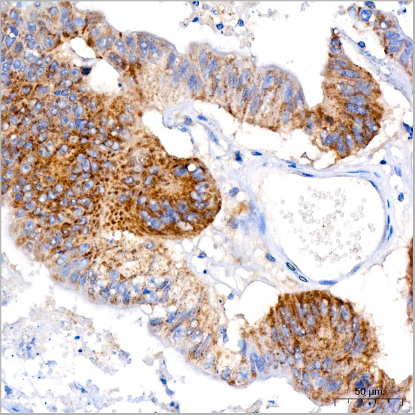
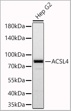
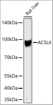
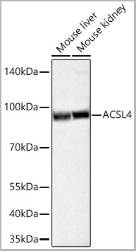
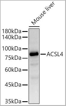
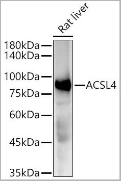
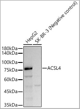
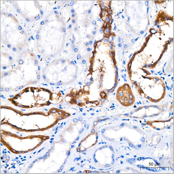 IP (Immunoprecipitation)
(Immunoprecipitation analysis of 300 ug extracts of HepG2 cells using 3 ug ACSL4 antibody (AAA28542). Western blot was performed from the immunoprecipitate using ACSL4 antibody (AAA28542) at a dilution of 1:1000.)
IP (Immunoprecipitation)
(Immunoprecipitation analysis of 300 ug extracts of HepG2 cells using 3 ug ACSL4 antibody (AAA28542). Western blot was performed from the immunoprecipitate using ACSL4 antibody (AAA28542) at a dilution of 1:1000.)
ACSL4, Monoclonal Antibody (Cat# AAA28542)
Full Name
ACSL4 Rabbit mAb
Reactivity
Human
Applications
Western Blot, Immunohistochemistry, Immunofluorescence, Immunocytochemistry, Immunoprecipitation
Purity
Affinity purification
Pricing
Wheat Gliadin, Recombinant Protein (Cat# AAA10885)
Full Name
Recombinant Wheat Gliadin
Applications
Western Blot
Purity
Greater than 95% as determined by SDS-PAGE.
Pricing
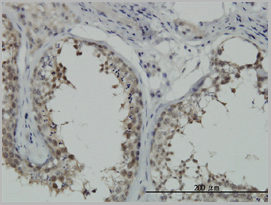
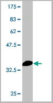
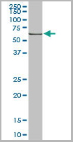 WB (Western Blot)
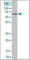
(HSPA1B monoclonal antibody Western Blot analysis of HSPA1B expression in Raw 264.7.)
WB (Western Blot)
(HSPA1B monoclonal antibody Western Blot analysis of HSPA1B expression in Raw 264.7.)
HSPA1A, Monoclonal Antibody (Cat# AAA24832)
Full Name
HSPA1A (Heat Shock 70kD Protein 1A/1B, Heat Shock 70kD Protein 1/2, HSP70.1/HSP70.2, HSP70-1/HSP70-2, HSPA1, HSPA1B) (Biotin)
Gene Names
HSPA1A; HSP72; HSPA1; HSP70I; HSP70-1; HSP70.1; HSP70-1A; HEL-S-103
Reactivity
Human, Mouse, Rat
Applications
Immunofluorescence, Immunohistochemistry, Western Blot
Purity
Purified by Protein A Affinity Chromatography.
Pricing
 ELISA
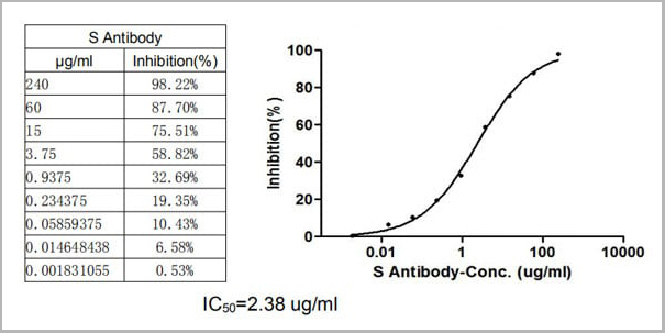
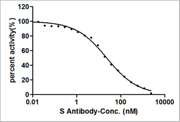
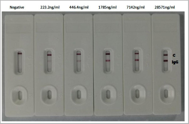
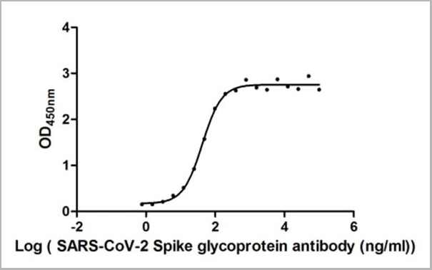
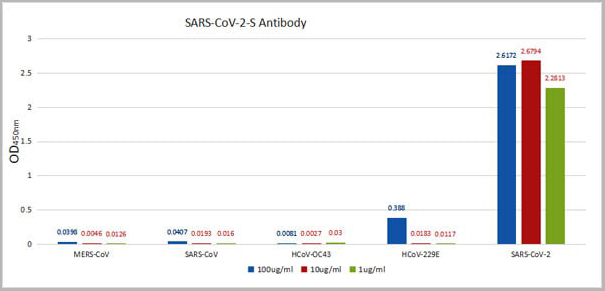
(ELISA: Immobilize various types of SARS proteins at concentration of 2mg/ml on solid substrate, then react with SARS-CoV-2-S Antibody at concentration of 100mg/ml, 10mg/ml and 1mg/ml. It shows the SARS-CoV-2-S Antibody (AAA27036) is specific for SARS-CoV-2-S1-RBD protein, without any cross-reactivity with MERS-CoV, SARS-CoV, HCoV-OC43 or HCoV-229E.)
ELISA
(ELISA: Immobilize various types of SARS proteins at concentration of 2mg/ml on solid substrate, then react with SARS-CoV-2-S Antibody at concentration of 100mg/ml, 10mg/ml and 1mg/ml. It shows the SARS-CoV-2-S Antibody (AAA27036) is specific for SARS-CoV-2-S1-RBD protein, without any cross-reactivity with MERS-CoV, SARS-CoV, HCoV-OC43 or HCoV-229E.)
COVID 19 Spike glycoprotein (S) Coronavirus, Monoclonal Recombinant Antibody (Cat# AAA27036)
Full Name
Human Novel Coronavirus Spike glycoprotein (S) (16-685aa) Antibody
Reactivity
Human Novel Coronavirus (SARS-CoV-2/ 2019-nCoV)
Applications
Colloidal Gold Immunochromatographic Assay, Neutralization Assay
Purity
Affinity-chromatography
Pricing
 WB (Western Blot)
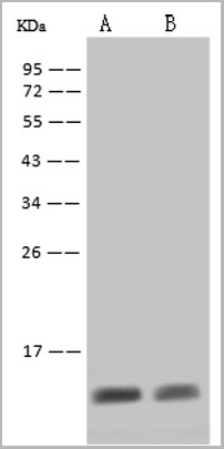
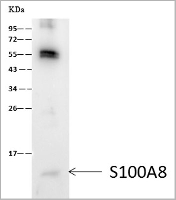
(S100A8 was immunoprecipitated using:Lane A: 0.5 mg HL-60 Whole Cell Lysate 1 uL anti-S100A8 rabbit monoclonal antibody and 60 ug of Immunomagnetic beads Protein A/G.Primary antibody:Anti-S100A8 rabbit monoclonal antibody, at 1:500 dilutionSecondary antibody:Goat Anti-Rabbit IgG (H+L)/HRP at 1/10000 dilution Developed using the ECL technique.Performed under reducing conditions.Predicted band size: 11 kDaObserved band size :11 kDa)
WB (Western Blot)
(S100A8 was immunoprecipitated using:Lane A: 0.5 mg HL-60 Whole Cell Lysate 1 uL anti-S100A8 rabbit monoclonal antibody and 60 ug of Immunomagnetic beads Protein A/G.Primary antibody:Anti-S100A8 rabbit monoclonal antibody, at 1:500 dilutionSecondary antibody:Goat Anti-Rabbit IgG (H+L)/HRP at 1/10000 dilution Developed using the ECL technique.Performed under reducing conditions.Predicted band size: 11 kDaObserved band size :11 kDa)
S100A8, Monoclonal Antibody (Cat# AAA27743)
Full Name
Recombinant Anti-S100A8 Antibody, Rabbit Monoclonal
Gene Names
S100A8; P8; MIF; NIF; CAGA; CFAG; CGLA; L1Ag; MRP8; CP-10; MA387; 60B8AG
Reactivity
Human
Applications
Western Blot, Immunoprecipitation
Purity
Protein A
Pricing
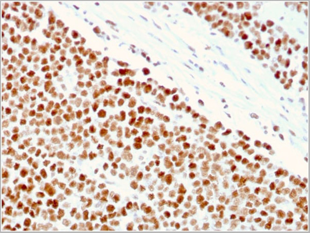
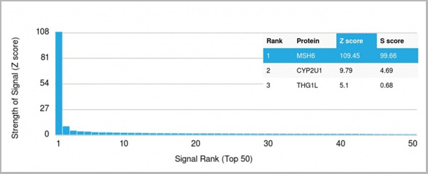 Application Data
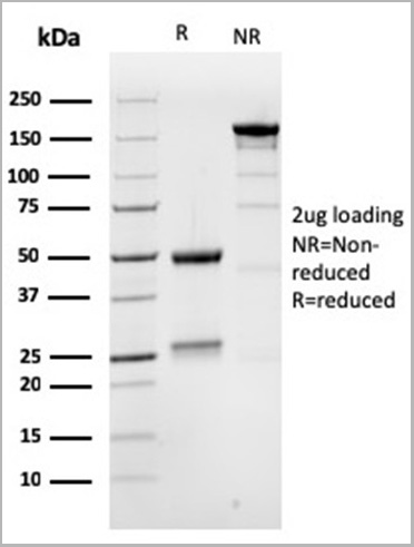
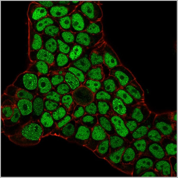
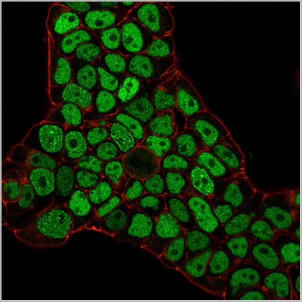
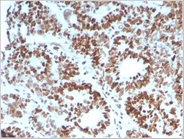
(Analysis of Protein Array containing >19,000 full-length human proteins using MSH6 Mouse Monoclonal Antibody (MSH6/3086) Z- and S- Score: The Z-score represents the strength of a signal that a monoclonal antibody (MAb) (in combination with a fluorescently-tagged anti-IgG secondary antibody) produces when binding to a particular protein on the HuProtTM array. Z-scores are described in units of standard deviations (SD’s) above the mean value of all signals generated on that array. If targets on HuProtTM are arranged in descending order of the Z-score, the S-score is the difference (also in units of SD’s) between the Z-score. S-score therefore represents the relative target specificity of a MAb to its intended target. A MAb is considered to specific to its intended target, if the MAb has an S-score of at least 2.5. For example, if a MAb binds to protein X with a Z-score of 43 and to protein Y with a Z-score of 14, then the S-score for the binding of that MAb to protein X is equal to 29.)
Application Data
(Analysis of Protein Array containing >19,000 full-length human proteins using MSH6 Mouse Monoclonal Antibody (MSH6/3086) Z- and S- Score: The Z-score represents the strength of a signal that a monoclonal antibody (MAb) (in combination with a fluorescently-tagged anti-IgG secondary antibody) produces when binding to a particular protein on the HuProtTM array. Z-scores are described in units of standard deviations (SD’s) above the mean value of all signals generated on that array. If targets on HuProtTM are arranged in descending order of the Z-score, the S-score is the difference (also in units of SD’s) between the Z-score. S-score therefore represents the relative target specificity of a MAb to its intended target. A MAb is considered to specific to its intended target, if the MAb has an S-score of at least 2.5. For example, if a MAb binds to protein X with a Z-score of 43 and to protein Y with a Z-score of 14, then the S-score for the binding of that MAb to protein X is equal to 29.)
MSH6, Monoclonal Antibody (Cat# AAA23917)
Full Name
MSH6 (DNA Mismatch Repair Protein)
Gene Names
MSH6; GTBP; HSAP; p160; GTMBP; HNPCC5
Reactivity
Human
Applications
Flow Cytometry, Immunofluorescence, Western Blot, Immunohistochemistry
Purity
Purified Ab with BSA and Azide at 200ug/ml OR Purified Ab WITHOUT BSA and Azide at 1.0mg/ml
Pricing
Fc-Epsilon RI-Alpha, Recombinant Protein (Cat# AAA10901)
Full Name
Recombinant Human Fc-Epsilon RI-Alpha
Gene Names
FCER1A; FCE1A; FcERI
Purity
Greater than 85.0% as determined by SDS-PAGE.
Pricing
 SDS-PAGE
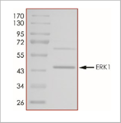
(The purity of ERK1 was determined to be >85% by densitometry.Approx. MW 44kDa.)
SDS-PAGE
(The purity of ERK1 was determined to be >85% by densitometry.Approx. MW 44kDa.)
ERK1, Recombinant Protein (Cat# AAA14252)
Full Name
ERK1, Unactive recombinant protein
Gene Names
MAPK3; ERK1; ERT2; ERK-1; PRKM3; P44ERK1; P44MAPK; HS44KDAP; HUMKER1A; p44-ERK1; p44-MAPK
Applications
Kinase Assay, Western Blot
Purity
>85%
Pricing
 ICC (Immunocytochemistry)
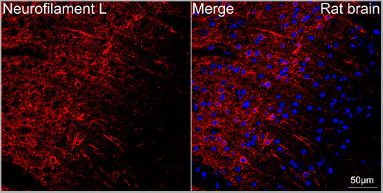
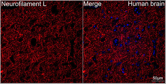
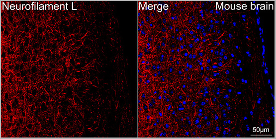
(Confocal imaging of paraffin-embedded Mouse brain tissue using Neurofilament L Rabbit mAb (AAA28541, dilution 1:200) followed by a further incubation with Cy3 Goat Anti-Rabbit IgG (H+L) (AS007, dilution 1:500) (Red). DAPI was used for nuclear staining (Blue). Objective: 40x. Perform microwave antigen retrieval with 0.01 M citrate buffer (pH 6.0) prior to IF staining.)
ICC (Immunocytochemistry)
(Confocal imaging of paraffin-embedded Mouse brain tissue using Neurofilament L Rabbit mAb (AAA28541, dilution 1:200) followed by a further incubation with Cy3 Goat Anti-Rabbit IgG (H+L) (AS007, dilution 1:500) (Red). DAPI was used for nuclear staining (Blue). Objective: 40x. Perform microwave antigen retrieval with 0.01 M citrate buffer (pH 6.0) prior to IF staining.)
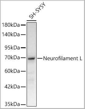
Neurofilament L, Monoclonal Antibody (Cat# AAA28541)
Full Name
Neurofilament L Rabbit mAb
Reactivity
Human
Applications
Western Blot, Immunohistochemistry, Immunofluorescence, Immunocytochemistry
Purity
Affinity purification
Pricing
 IF (Immunofluorescence)
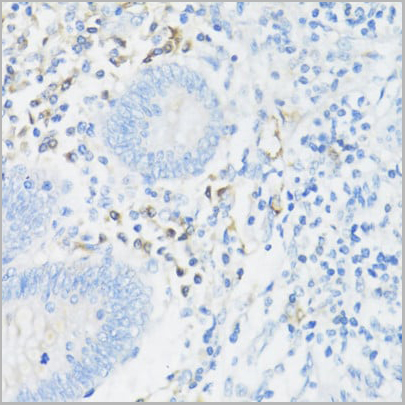
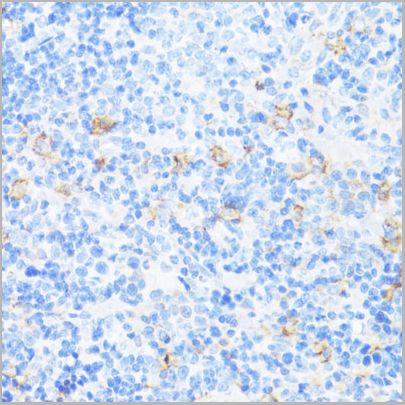
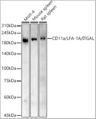
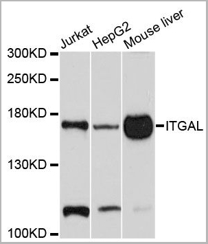
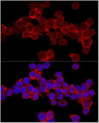
(Confocal immunofluorescence analysis of THP-1 cells using CD11a/LFA-1A/ITGAL Rabbit pAb (AAA10672) at dilution of 1:200. Blue: DAPI for nuclear staining.)
IF (Immunofluorescence)
(Confocal immunofluorescence analysis of THP-1 cells using CD11a/LFA-1A/ITGAL Rabbit pAb (AAA10672) at dilution of 1:200. Blue: DAPI for nuclear staining.)
ITGAL, Polyclonal Antibody (Cat# AAA10672)
Full Name
ITGAL Polyclonal Antibody
Gene Names
ITGAL; CD11A; LFA-1; LFA1A
Reactivity
Human, Mouse, Rat
Applications
Western Blot, Immunohistochemistry, Immunofluorescence, Immunocytochemistry
Purity
Affinity Purification
Pricing
S100 Calcium Binding Protein A12, Recombinant Protein (Cat# AAA10893)
Full Name
Recombinant Human S100 Calcium Binding Protein A12
Gene Names
S100A12; p6; CAGC; CGRP; MRP6; CAAF1; MRP-6; ENRAGE
Pricing
 IF (Immunofluorescence)
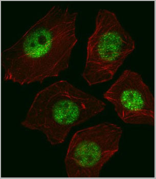
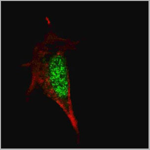
(Fluorescent confocal image of SY5Y cells stained with SOX2 antibody. SY5Y cells were fixed with 4% PFA (20 min), permeabilized with Triton X-100 (0.2%, 30 min). Cells were then incubated with SOX2 primary antibody (1:100, 2 h at room temperature). For secondary antibody, Alexa Fluor® 488 conjugated donkey anti-mouse antibody (green) was used (1:1000, 1h). Note the highly specific localization of the SOX2 mainly to the nucleus.)
IF (Immunofluorescence)
(Fluorescent confocal image of SY5Y cells stained with SOX2 antibody. SY5Y cells were fixed with 4% PFA (20 min), permeabilized with Triton X-100 (0.2%, 30 min). Cells were then incubated with SOX2 primary antibody (1:100, 2 h at room temperature). For secondary antibody, Alexa Fluor® 488 conjugated donkey anti-mouse antibody (green) was used (1:1000, 1h). Note the highly specific localization of the SOX2 mainly to the nucleus.)
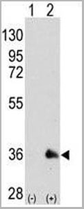
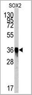
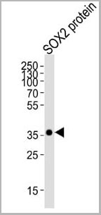
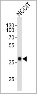
SOX2, Monoclonal Antibody (Cat# AAA28642)
Full Name
SOX2 Antibody
Gene Names
SOX2; ANOP3; MCOPS3
Reactivity
Human
Applications
Western Blot, Immunofluorescence, Immunohistochemistry, Flow Cytometry
Purity
This antibody is purified through a protein G column, followed by dialysis against PBS.
Pricing
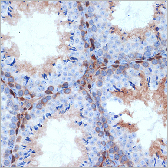
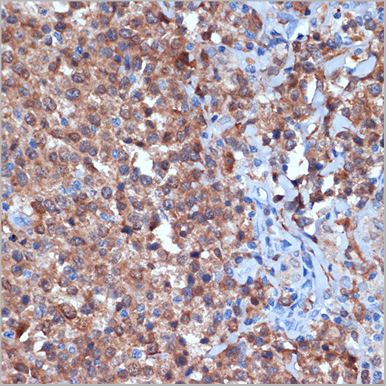
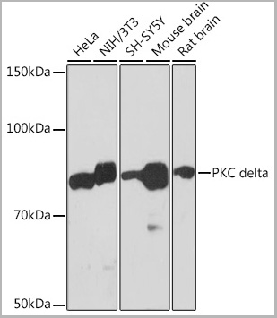
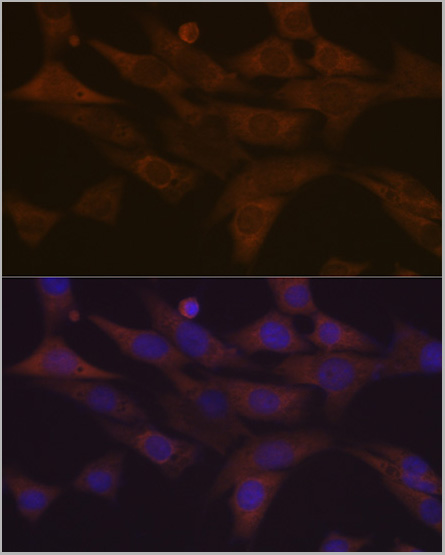 IF (Immunofluorescence)
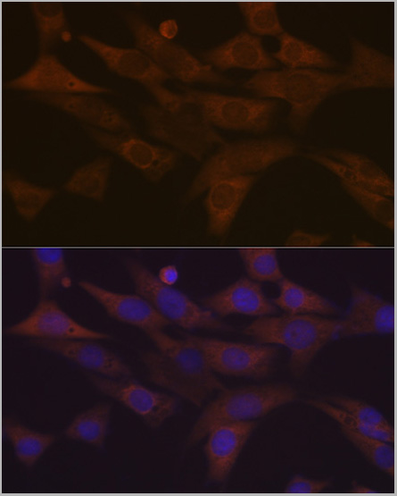
(Immunofluorescence analysis of NIH-3T3 cells using PKC delta Rabbit mAb (AAA28608) at dilution of 1:100 (40x lens). Secondary antibody: Cy3-conjugated Goat anti-Rabbit IgG (H+L) (AS007) at 1:500 dilution. Blue: DAPI for nuclear staining.)
IF (Immunofluorescence)
(Immunofluorescence analysis of NIH-3T3 cells using PKC delta Rabbit mAb (AAA28608) at dilution of 1:100 (40x lens). Secondary antibody: Cy3-conjugated Goat anti-Rabbit IgG (H+L) (AS007) at 1:500 dilution. Blue: DAPI for nuclear staining.)
PKC delta, Monoclonal Antibody (Cat# AAA28608)
Full Name
PKC delta Rabbit mAb
Reactivity
Human
Applications
Western Blot, Immunohistochemistry, Immunofluorescence, Immunocytochemistry
Purity
Affinity purification
Pricing
CCAAT/enhancer binding protein(C/EBP) Alpha, Recombinant Protein (Cat# AAA10839)
Full Name
Recombinant Human CCAAT/enhancer binding protein(C/EBP) Alpha
Gene Names
CEBPA; CEBP; C/EBP-alpha
Purity
Greater than 95.0% as determined by SDS-PAGE.
Pricing
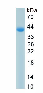
 Bioactivity
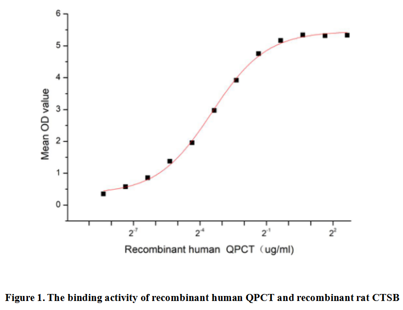
(Glutaminyl Peptide Cyclotransferase (QPCT), also known as Glutaminyl Cyclase, is an enzyme responsible for the biosynthesis of pyroglutamyl peptides. QPCT is present in the pituitary and adrenal glands, where it is important for the generation of the Nterminal pyroglutamyl groups of peptide hormones such as neurotensin and thyrotropin-releasing hormone. Besides, Cathepsin B (CTSB) has been identified as an interactor of QPCT, thus a functional binding ELISA assay was conducted to detect the interaction of recombinant human QPCT and recombinant rat CTSB. Briefly, QPCT was diluted serially in PBS with 0.01% BSA (pH 7.4). Duplicate samples of 100 ul were then transferred to CTSB-coated microtiter wells and incubated for 1h at 37 degree C. Wells were washed with PBST and incubated for 1h with anti-QPCT pAb, then aspirated and washed 3 times. After incubation with HRP labelled secondary antibody for 1h at 37 degree C, wells were aspirated and washed 5 times. With the addition of substrate solution, wells were incubated 15-25 minutes at 37 degree C. Finally, add 50 uL stop solution to the wells and read at 450/630 nm immediately. The binding activity of recombinant human QPCT and recombinant rat CTSB was shown in Figure 1, the EC50 for this effect is 0.09 ug/mL.)
Bioactivity
(Glutaminyl Peptide Cyclotransferase (QPCT), also known as Glutaminyl Cyclase, is an enzyme responsible for the biosynthesis of pyroglutamyl peptides. QPCT is present in the pituitary and adrenal glands, where it is important for the generation of the Nterminal pyroglutamyl groups of peptide hormones such as neurotensin and thyrotropin-releasing hormone. Besides, Cathepsin B (CTSB) has been identified as an interactor of QPCT, thus a functional binding ELISA assay was conducted to detect the interaction of recombinant human QPCT and recombinant rat CTSB. Briefly, QPCT was diluted serially in PBS with 0.01% BSA (pH 7.4). Duplicate samples of 100 ul were then transferred to CTSB-coated microtiter wells and incubated for 1h at 37 degree C. Wells were washed with PBST and incubated for 1h with anti-QPCT pAb, then aspirated and washed 3 times. After incubation with HRP labelled secondary antibody for 1h at 37 degree C, wells were aspirated and washed 5 times. With the addition of substrate solution, wells were incubated 15-25 minutes at 37 degree C. Finally, add 50 uL stop solution to the wells and read at 450/630 nm immediately. The binding activity of recombinant human QPCT and recombinant rat CTSB was shown in Figure 1, the EC50 for this effect is 0.09 ug/mL.)
Glutaminyl Peptide Cyclotransferase (QPCT), Active Protein (Cat# AAA161925)
Full Name
Active Glutaminyl Peptide Cyclotransferase (QPCT)
Gene Names
QPCT; QC; GCT; sQC
Applications
Activity Assay, Cell Culture
Purity
Greater than 90% by SDS-PAGE
Pricing
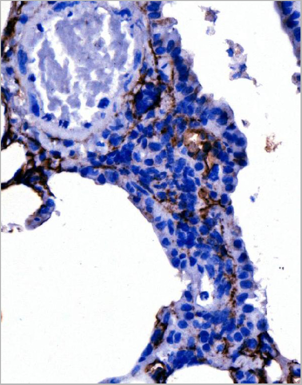
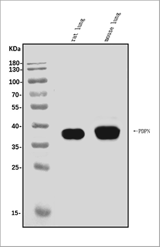
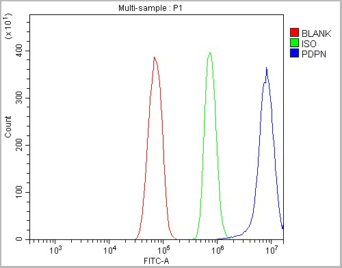 FCM (Flow Cytometry)
(Figure 5. Flow Cytometry analysis of RH35 cells using anti-Podoplanin/gp36/Pdpn antibody (AAA19233).Overlay histogram showing RH35 cells stained with AAA19233 (Blue line). The cells were blocked with 10% normal goat serum. And then incubated with rabbit anti-Podoplanin/gp36/Pdpn Antibody (AAA19233, 1μg/1x106 cells) for 30 min at 20 degree C. DyLight®488 conjugated goat anti-rabbit IgG (5-10μg/1x106 cells) was used as secondary antibody for 30 minutes at 20 degree C. Isotype control antibody (Green line) was rabbit IgG (1μg/1x106) used under the same conditions. Unlabelled sample (Red line) was also used as a control.)
FCM (Flow Cytometry)
(Figure 5. Flow Cytometry analysis of RH35 cells using anti-Podoplanin/gp36/Pdpn antibody (AAA19233).Overlay histogram showing RH35 cells stained with AAA19233 (Blue line). The cells were blocked with 10% normal goat serum. And then incubated with rabbit anti-Podoplanin/gp36/Pdpn Antibody (AAA19233, 1μg/1x106 cells) for 30 min at 20 degree C. DyLight®488 conjugated goat anti-rabbit IgG (5-10μg/1x106 cells) was used as secondary antibody for 30 minutes at 20 degree C. Isotype control antibody (Green line) was rabbit IgG (1μg/1x106) used under the same conditions. Unlabelled sample (Red line) was also used as a control.)
Podoplanin/gp36/Pdpn, Polyclonal Antibody (Cat# AAA19233)
Full Name
Anti-Podoplanin/gp36/Pdpn Antibody
Gene Names
Pdpn; T1a; Gp38; OTS-8; T1alpha; RANDAM-2; T1-alpha
Reactivity
Mouse, Rat
Applications
Western Blot, Immunohistochemistry, Immunocytochemistry, Immunofluorescence, Flow Cytometry, Direct ELISA
Purity
Immunogen affinity purified.
Pricing
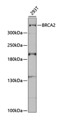
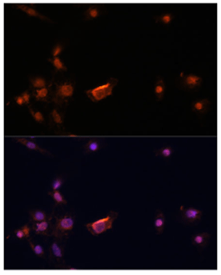 IF (Immunofluorescence)
(Immunofluorescence analysis of C6 cells using BRCA2 antibody (AAA10700) at dilution of 1:100. Blue: DAPI for nuclear staining.)
IF (Immunofluorescence)
(Immunofluorescence analysis of C6 cells using BRCA2 antibody (AAA10700) at dilution of 1:100. Blue: DAPI for nuclear staining.)
BRCA2, Polyclonal Antibody (Cat# AAA10700)
Full Name
BRCA2
Gene Names
BRCA2; FAD; FACD; FAD1; GLM3; BRCC2; FANCD; PNCA2; FANCD1; XRCC11; BROVCA2
Reactivity
Human , Rat
Applications
Western Blot, Immunofluorescence
Purity
Affinity Purification
Pricing
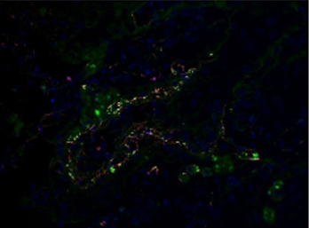


 IHC (Immunohistochemistry)
(IHC Validation of Envelope in COVID-19 Patient Skin (Magro et al., 2020) Detection of SARS-CoV-2 Envelope protein in the blood vessels of COVID-19 patients that were confirmed by PCR. The staining shows Envelope protein expression (green) detected by envelope antibodies (AAA10932, 3 ug/mL) in the endothelial cytoplasms in thrombosed and normal appearing blood vessels with hematoxylin counterstain. The staining was negative in control normal skin/lung (not shown).)
IHC (Immunohistochemistry)
(IHC Validation of Envelope in COVID-19 Patient Skin (Magro et al., 2020) Detection of SARS-CoV-2 Envelope protein in the blood vessels of COVID-19 patients that were confirmed by PCR. The staining shows Envelope protein expression (green) detected by envelope antibodies (AAA10932, 3 ug/mL) in the endothelial cytoplasms in thrombosed and normal appearing blood vessels with hematoxylin counterstain. The staining was negative in control normal skin/lung (not shown).)
COVID 19 Envelope Coronavirus, Polyclonal Antibody (Cat# AAA10932)
Full Name
SARS-CoV-2 (COVID-19, 2019-nCoV) Envelope antibody
Reactivity
Virus
Applications
Immunofluorescence, Immunohistochemistry
Purity
SARS-CoV-2 (COVID-19, 2019-nCoV) Envelope Antibody is affinity chromatography purified via peptide column.
Pricing
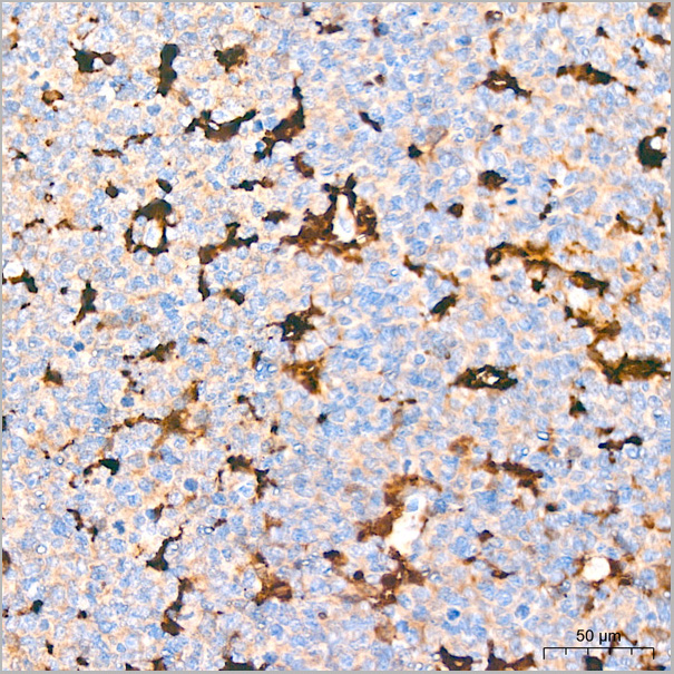
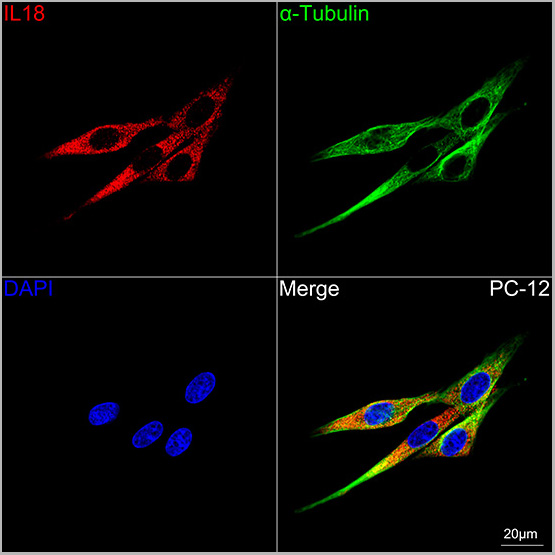
 ICC (Immunocytochemistry)
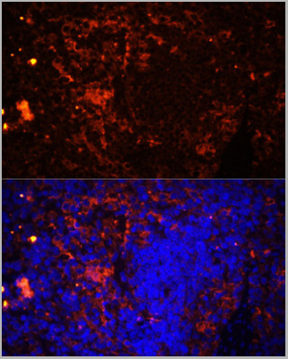
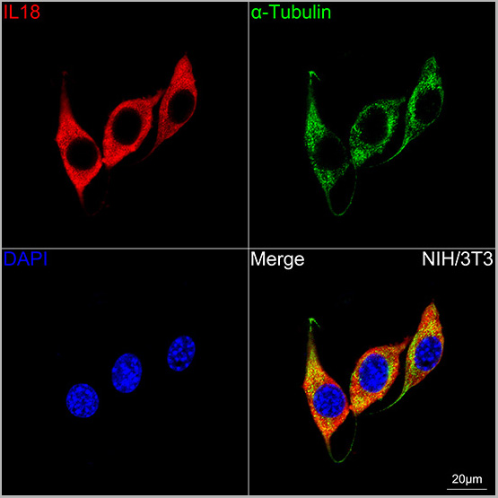
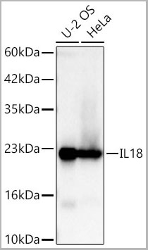
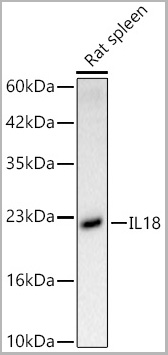
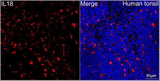
(Confocal imaging of paraffin-embedded Human tonsil tissue using IL18 Rabbit PolymAb® (AAA28566, dilution 1:200) followed by a further incubation with Cy3 Goat Anti-Rabbit IgG (H+L) (AS007, dilution 1:500) (Red). DAPI was used for nuclear staining (Blue). High pressure antigen retrieval performed with 0.01M Citrate Buffer (pH 6.0) prior to IF staining. Objective: 40x.)
ICC (Immunocytochemistry)
(Confocal imaging of paraffin-embedded Human tonsil tissue using IL18 Rabbit PolymAb® (AAA28566, dilution 1:200) followed by a further incubation with Cy3 Goat Anti-Rabbit IgG (H+L) (AS007, dilution 1:500) (Red). DAPI was used for nuclear staining (Blue). High pressure antigen retrieval performed with 0.01M Citrate Buffer (pH 6.0) prior to IF staining. Objective: 40x.)
IL18, Monoclonal Antibody (Cat# AAA28566)
Full Name
IL18 Rabbit PolymAb
Reactivity
Human
Applications
Western Blot, Immunohistochemistry, Immunofluorescence, Immunocytochemistry
Purity
Affinity purification
Pricing
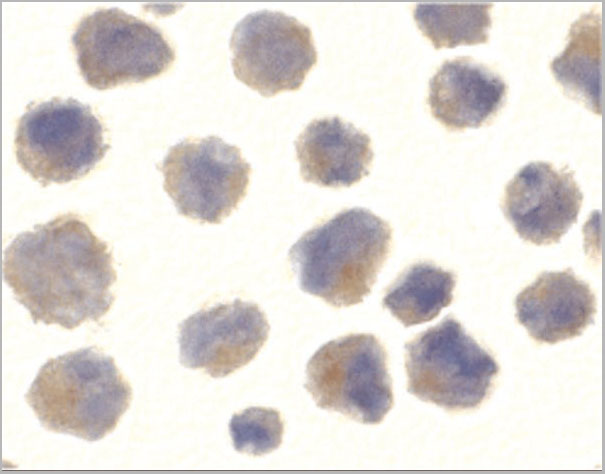
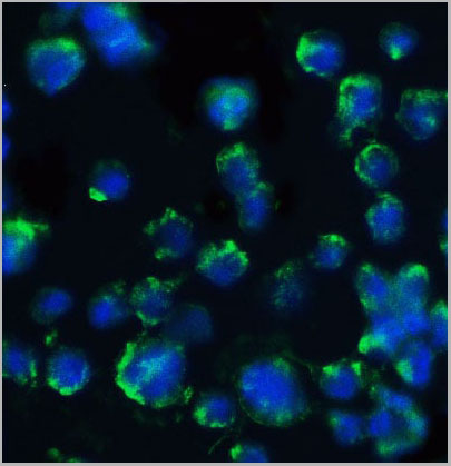
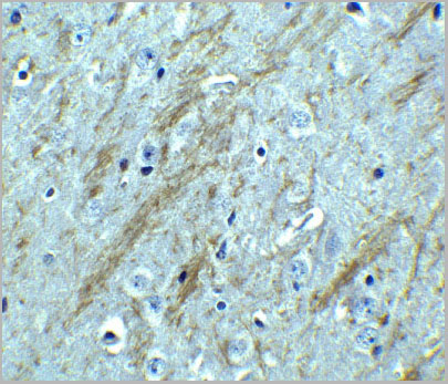
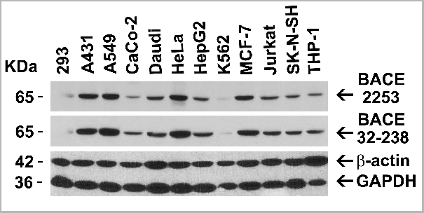
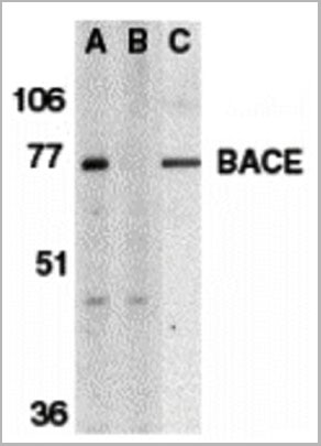
 IF (Immunofluorescence)
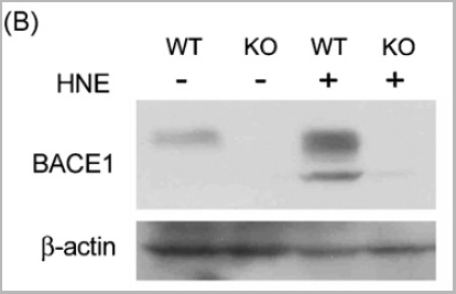
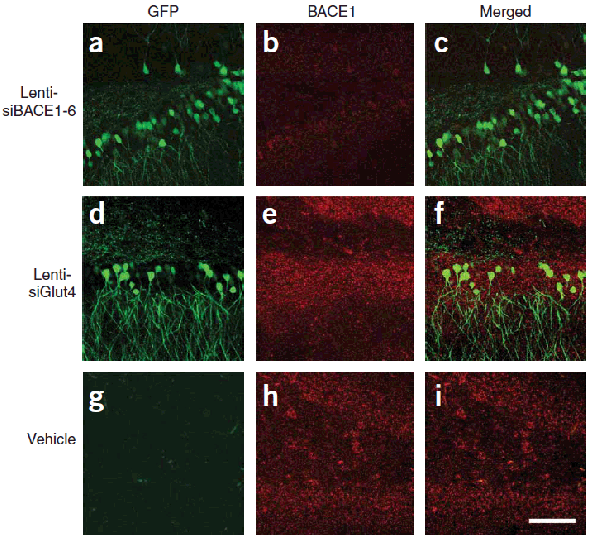
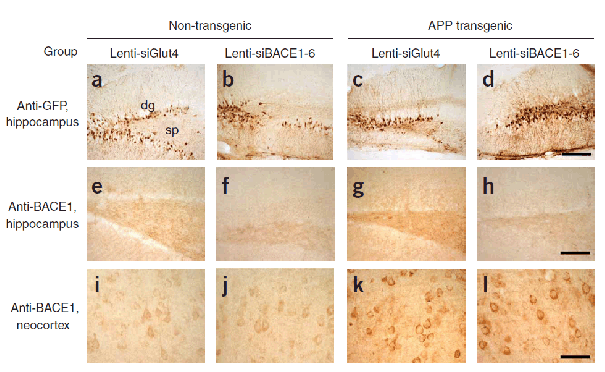
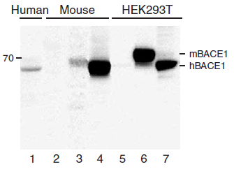
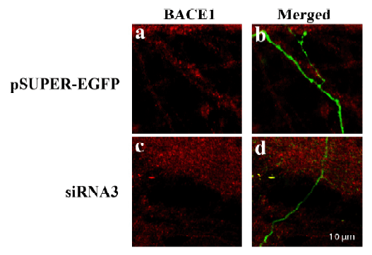
(Figure 10 KD Validation of BACE in DRG (Hyun, 2007)Decreased BACE1 expression in DRG following siRNA3 transfection. DRG neurons were transfected with 1 ug siRNA3 plasmid and incubated for 48 hours in 37°˚C. DRG neurons were stained for BACE1 us¬ing the Anti-BACE antibody (a,b) Neurons transfected with the control plas¬mid pSUPER-EGFP (green) did not display any changes in BACE1 expression (red). (c,d) DRG neurons transfected with siR¬NA3 displayed reduced BACE1 expression in the axon.)
IF (Immunofluorescence)
(Figure 10 KD Validation of BACE in DRG (Hyun, 2007)Decreased BACE1 expression in DRG following siRNA3 transfection. DRG neurons were transfected with 1 ug siRNA3 plasmid and incubated for 48 hours in 37°˚C. DRG neurons were stained for BACE1 us¬ing the Anti-BACE antibody (a,b) Neurons transfected with the control plas¬mid pSUPER-EGFP (green) did not display any changes in BACE1 expression (red). (c,d) DRG neurons transfected with siR¬NA3 displayed reduced BACE1 expression in the axon.)
BACE, Polyclonal Antibody (Cat# AAA10918)
Full Name
BACE Antibody
Gene Names
BACE1; ASP2; BACE; HSPC104
Reactivity
Human, Mouse
Applications
Immunocytochemistry, Immunofluorescence, Immunohistochemistry, Western Blot
Purity
BACE Antibody is affinity chromatography purified via peptide column.
Pricing
 SDS-PAGE
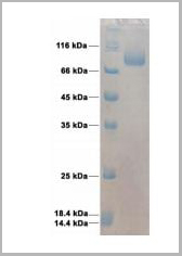
(purified HA (H3N2)(A/ Singapore/INFIMH-16-0019/2016) protein)
SDS-PAGE
(purified HA (H3N2)(A/ Singapore/INFIMH-16-0019/2016) protein)
HA (A/Singapore/INFIMH-16-0019/2016), Recombinant Protein (Cat# AAA13697)
Full Name
HA (A/Singapore/INFIMH-16-0019/2016)(H3N2)(aa 17-529)
Applications
Western Blot
Purity
>95% pure by 10% SDS PAGE gel
Pricing


 FCM (Flow Cytometry)
(FACS analysis of ES-2 cells stained with STIP1 monoclonal antibody clone 2E11 (Green) and non-stained ES-2 cells (Black) as negative control.)
FCM (Flow Cytometry)
(FACS analysis of ES-2 cells stained with STIP1 monoclonal antibody clone 2E11 (Green) and non-stained ES-2 cells (Black) as negative control.)
STIP1, Monoclonal Antibody (Cat# AAA26009)
Full Name
STIP1 (Stress-Induced-Phosphoprotein 1, HOP, IEF-SSP-3521, P60, STI1, STI1L) (AP)
Gene Names
STIP1; HOP; P60; STI1; STI1L; HEL-S-94n; IEF-SSP-3521
Applications
Immunofluorescence, Immunohistochemistry, Western Blot
Purity
Purified
Pricing


 WB (Western Blot)
(Western blot analysis of extracts of various cell lines, using TYR antibody (AAA10704) at 1:1000 dilution.Secondary antibody: HRP Goat Anti-Rabbit IgG (H+L) at 1:10000 dilution.Lysates/proteins: 25ug per lane.Blocking buffer: 3% nonfat dry milk in TBST.)
WB (Western Blot)
(Western blot analysis of extracts of various cell lines, using TYR antibody (AAA10704) at 1:1000 dilution.Secondary antibody: HRP Goat Anti-Rabbit IgG (H+L) at 1:10000 dilution.Lysates/proteins: 25ug per lane.Blocking buffer: 3% nonfat dry milk in TBST.)
TYR, Polyclonal Antibody (Cat# AAA10704)
Full Name
TYR Polyclonal Antibody
Gene Names
TYR; ATN; CMM8; OCA1; OCA1A; OCAIA; SHEP3
Reactivity
Human, Mouse, Rat
Applications
Western Blot, Immunohistochemistry, Immunofluorescence
Purity
Affinity Purification
Pricing
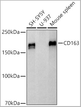
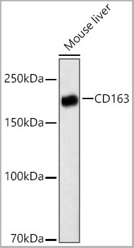
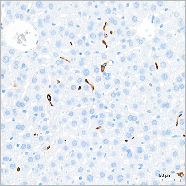 ICC (Immunocytochemistry)
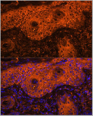
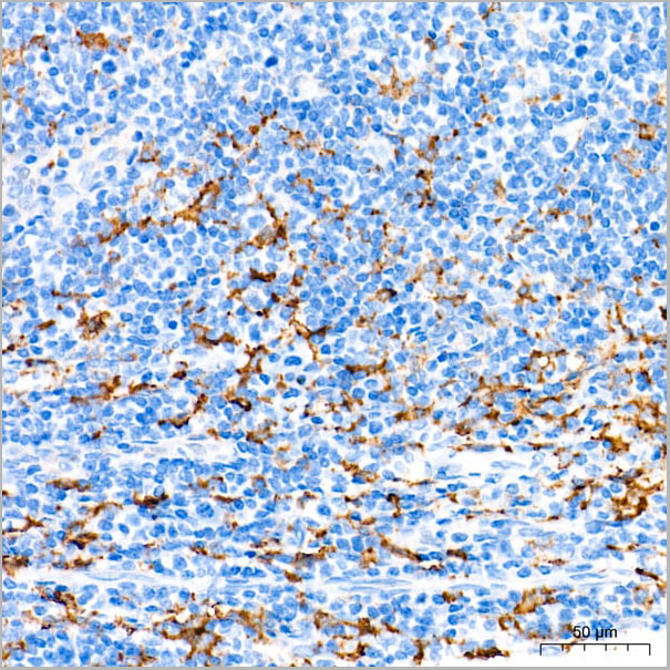
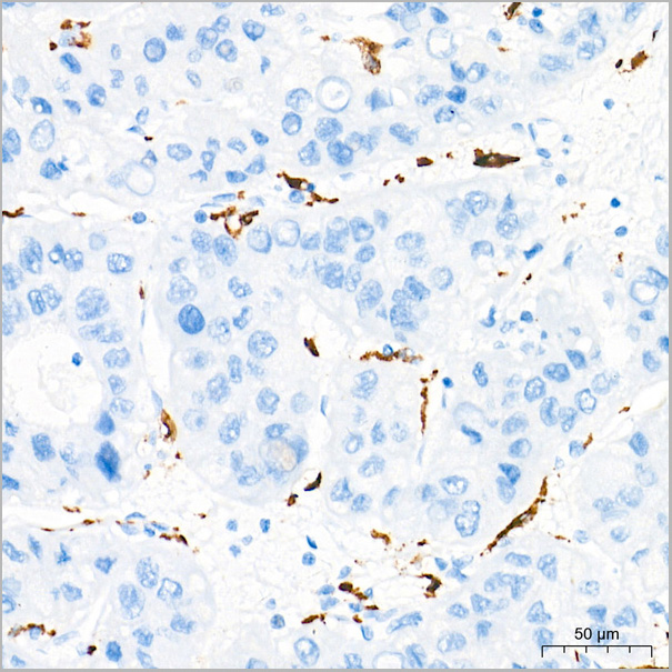
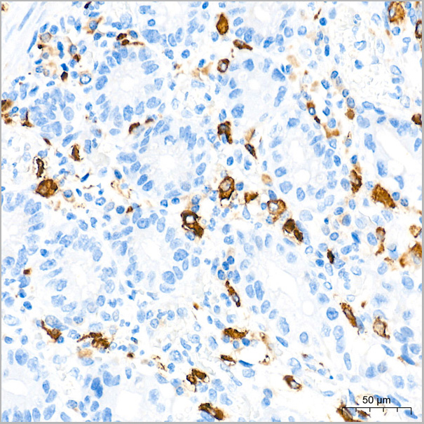
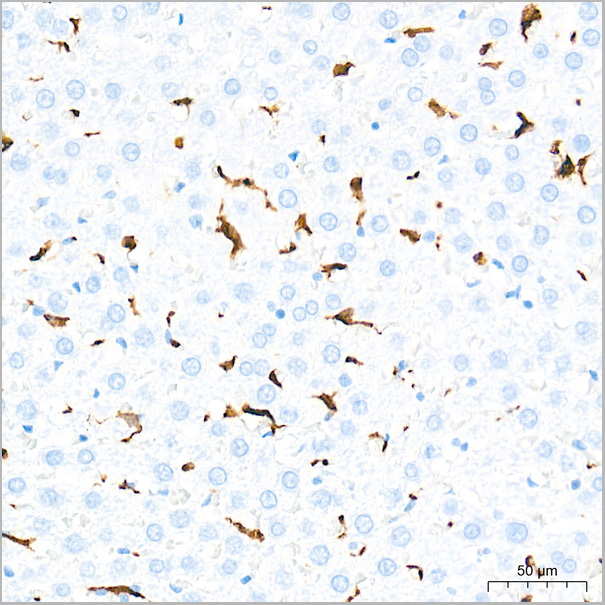
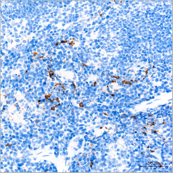
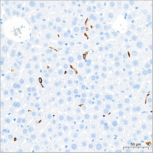
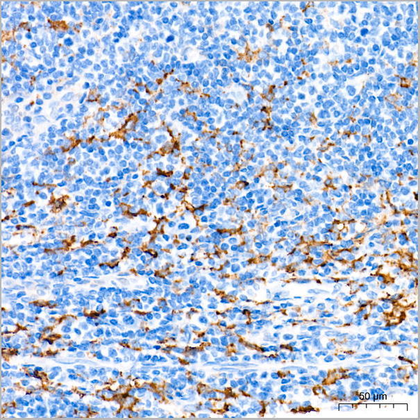
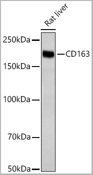
(Confocal imaging of paraffin-embedded Mouse spleen tissue using CD163 Rabbit PolymAb® (AAA28577, dilution 1:4000) followed by a further incubation with Cy3 Goat Anti-Rabbit IgG (H+L) (AS007, dilution 1:500) (Red). DAPI was used for nuclear staining (Blue). High pressure antigen retrieval performed with 0.01M Citrate Buffer (pH 6.0) prior to IF staining. Objective: 40x.)
ICC (Immunocytochemistry)
(Confocal imaging of paraffin-embedded Mouse spleen tissue using CD163 Rabbit PolymAb® (AAA28577, dilution 1:4000) followed by a further incubation with Cy3 Goat Anti-Rabbit IgG (H+L) (AS007, dilution 1:500) (Red). DAPI was used for nuclear staining (Blue). High pressure antigen retrieval performed with 0.01M Citrate Buffer (pH 6.0) prior to IF staining. Objective: 40x.)
CD163, Monoclonal Antibody (Cat# AAA28577)
Full Name
CD163 Rabbit PolymAb
Reactivity
Human, Mouse
Applications
Western Blot, Immunohistochemistry, Immunofluorescence, Immunocytochemistry
Purity
Affinity purification
Pricing
 ELISA
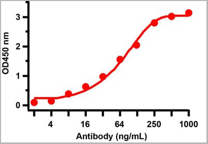
(Figure 2 ELISA TestAntibodies: SARS-CoV-2 Nucleocapsid antibody, AAA11029. An ELISA was performed using human SARS-CoV-2 Nucleocapsid recombinant protein as coating antigen and the SARS-CoV-2 Nucleocapsid antibody, as the capture antibody. Secondary: Goat anti-mouse IgG HRP conjugate at 1:20000 dilution. Detection range is from 2 ng/mL to 1000 ng/mL.)
ELISA
(Figure 2 ELISA TestAntibodies: SARS-CoV-2 Nucleocapsid antibody, AAA11029. An ELISA was performed using human SARS-CoV-2 Nucleocapsid recombinant protein as coating antigen and the SARS-CoV-2 Nucleocapsid antibody, as the capture antibody. Secondary: Goat anti-mouse IgG HRP conjugate at 1:20000 dilution. Detection range is from 2 ng/mL to 1000 ng/mL.)
COVID 19 Nucleocapsid (NP) Coronavirus, Monoclonal Antibody (Cat# AAA11029)
Full Name
SARS-CoV-2 (COVID-19) Nucleoprotein
Reactivity
Virus
Applications
Immunofluorescence
Purity
SARS-CoV-2 (COVID-19, 2019-nCoV) Nucleoprotein antibody is purified from ascites fluid or culture medium by protein A chromoatography or sequential differential precipitations.
Pricing
 Application Data
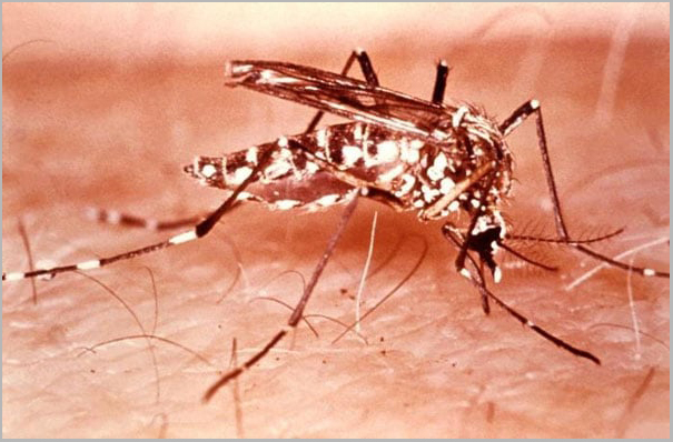
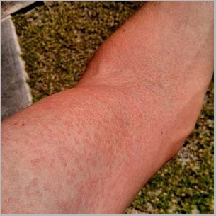
(Zika rash: arm.)
Application Data
(Zika rash: arm.)
Zika Virus NS1, Monoclonal Antibody (Cat# AAA24113)
Full Name
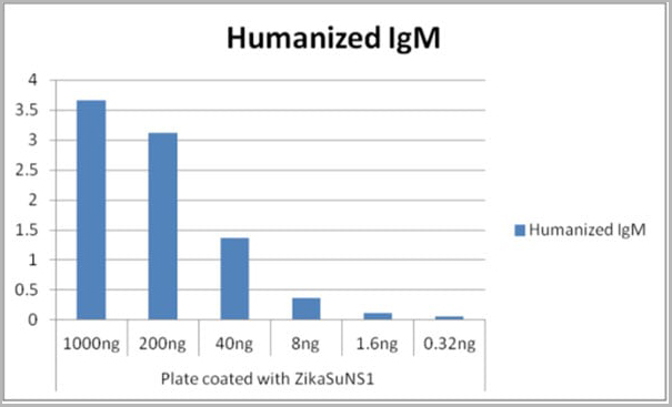
Human Anti-Zika Virus NS1 (Zika Virus Nonstructural Protein)
Applications
ELISA
Purity
>98% (SDS-PAGE). Purified from cell culture supernatant by lectin affinity chromatography.
Pricing
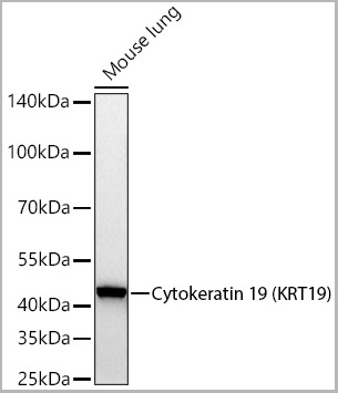
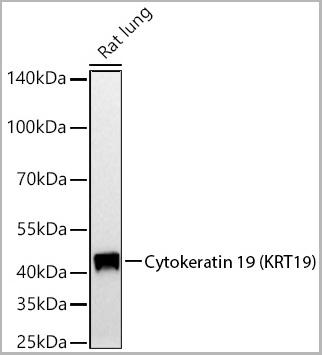
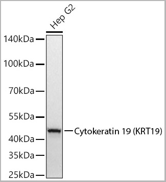
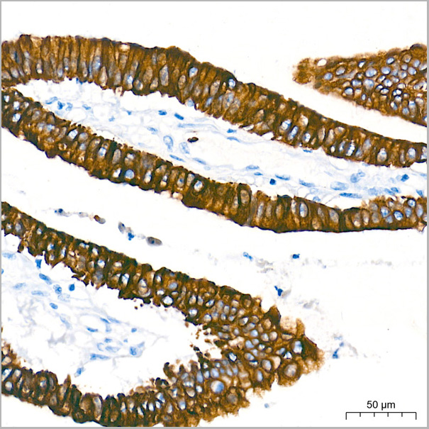 IHC (Immunohistochemistry)
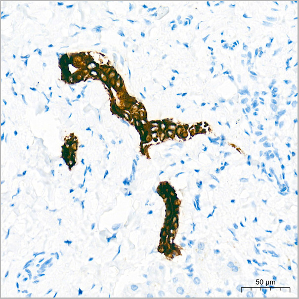
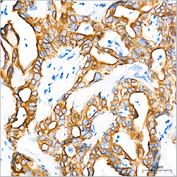
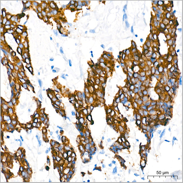
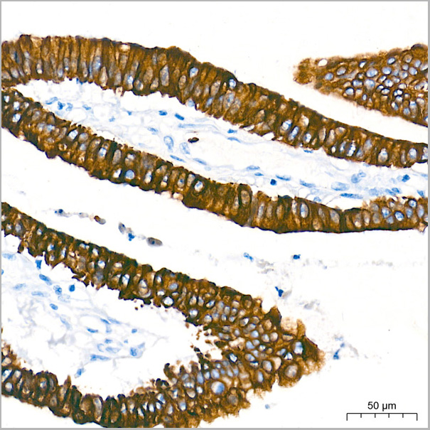
(Immunohistochemistry analysis of paraffin-embedded Human tonsil tissue using Cytokeratin 19 (KRT19) Rabbit PolymAb® (AAA28447) at a dilution of 1:10000 (40x lens). High pressure antigen retrieval performed with 0.01M Tris-EDTA Buffer(pH 9.0) prior to IHC staining.)
IHC (Immunohistochemistry)
(Immunohistochemistry analysis of paraffin-embedded Human tonsil tissue using Cytokeratin 19 (KRT19) Rabbit PolymAb® (AAA28447) at a dilution of 1:10000 (40x lens). High pressure antigen retrieval performed with 0.01M Tris-EDTA Buffer(pH 9.0) prior to IHC staining.)
Cytokeratin 19 (KRT19), Monoclonal Antibody (Cat# AAA28447)
Full Name
Cytokeratin 19 (KRT19) Rabbit PolymAb
Reactivity
Human
Applications
Western Blot, Immunohistochemistry
Purity
Affinity purification
Pricing
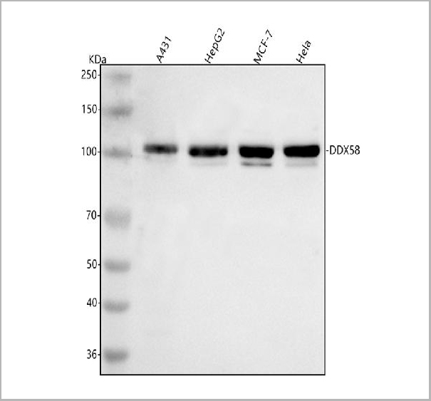
 FCM (Flow Cytometry)
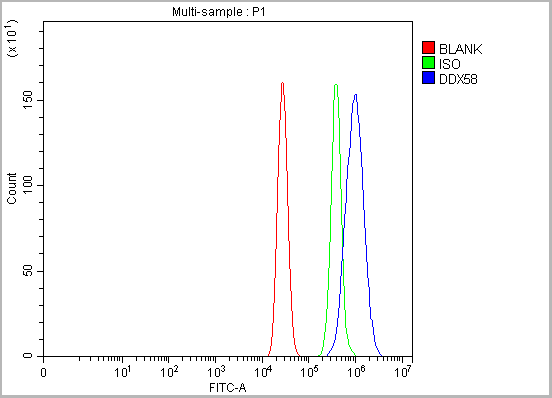
(Figure 12. Flow Cytometry analysis of MCF-7 cells using anti-DDX58/RIG-1 antibody (AAA19726).Overlay histogram showing MCF-7 cells stained with AAA19726 (Blue line). To facilitate intracellular staining, cells were fixed with 4% paraformaldehyde and permeabilized with permeabilization buffer. The cells were blocked with 10% normal goat serum. And then incubated with rabbit anti-DDX58/RIG-1 Antibody (AAA19726, 1ug/1x106 cells) for 30 min at 20 degree C. DyLight®488 conjugated goat anti-rabbit IgG (BA1127, 5-10ug/1x106 cells) was used as secondary antibody for 30 minutes at 20 degree C. Isotype control antibody (Green line) was rabbit IgG (1ug/1x106) used under the same conditions. Unlabelled sample (Red line) was also used as a control.)
FCM (Flow Cytometry)
(Figure 12. Flow Cytometry analysis of MCF-7 cells using anti-DDX58/RIG-1 antibody (AAA19726).Overlay histogram showing MCF-7 cells stained with AAA19726 (Blue line). To facilitate intracellular staining, cells were fixed with 4% paraformaldehyde and permeabilized with permeabilization buffer. The cells were blocked with 10% normal goat serum. And then incubated with rabbit anti-DDX58/RIG-1 Antibody (AAA19726, 1ug/1x106 cells) for 30 min at 20 degree C. DyLight®488 conjugated goat anti-rabbit IgG (BA1127, 5-10ug/1x106 cells) was used as secondary antibody for 30 minutes at 20 degree C. Isotype control antibody (Green line) was rabbit IgG (1ug/1x106) used under the same conditions. Unlabelled sample (Red line) was also used as a control.)
DDX58/RIG-1, Polyclonal Antibody (Cat# AAA19726)
Full Name
Anti-DDX58/RIG-1 Antibody Picoband
Gene Names
DDX58; RIGI; RIG-I; RLR-1
Reactivity
Human
Applications
Western Blot, Immunohistochemistry, Flow Cytometry
Purity
Immunogen affinity purified.
Pricing
 SDS-PAGE
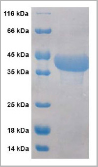
(SDS-PAGE: purified VIsE protein of Borrelia burgdorferi from 293 cells)
SDS-PAGE
(SDS-PAGE: purified VIsE protein of Borrelia burgdorferi from 293 cells)
VlsE protein of Borrelia burgdorferi, Recombinant Protein (Cat# AAA13692)
Full Name
Recombinant VlsE protein of Borrelia burgdorferi
Applications
Western Blot
Purity
>= 95% (by SDS-PAGE)
Pricing
Troponin-C2, Recombinant Protein (Cat# AAA10806)
Full Name
Recombinant Human Troponin-C2
Purity
Greater than 95.0% as determined by SDS-PAGE.
Pricing
 IF (Immunofluorescence)
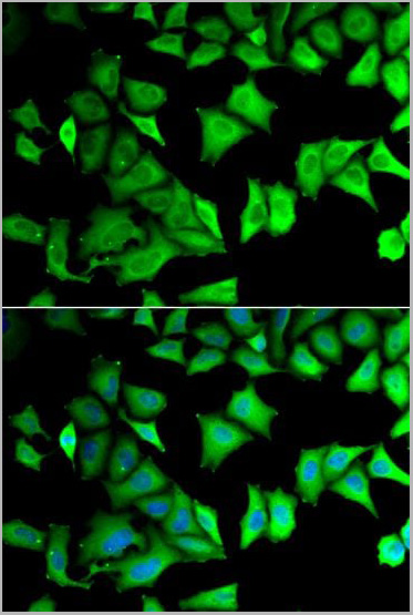
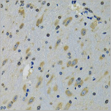
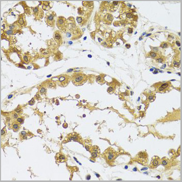
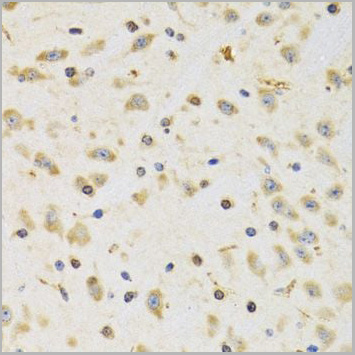
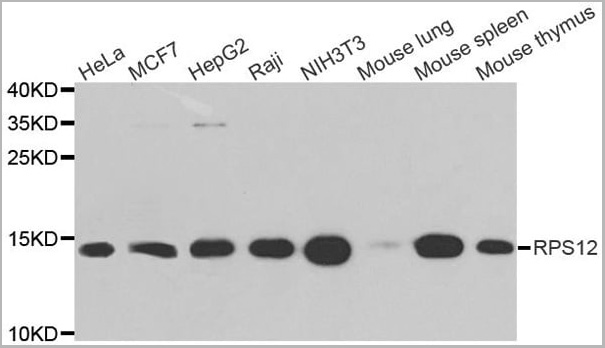
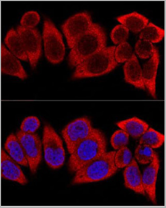
(Confocal immunofluorescence analysis of HeLa cells using RPS12 Polyclonal Antibody (AAA10775) at dilution of 1:400. Blue: DAPI for nuclear staining.)
IF (Immunofluorescence)
(Confocal immunofluorescence analysis of HeLa cells using RPS12 Polyclonal Antibody (AAA10775) at dilution of 1:400. Blue: DAPI for nuclear staining.)
RPS12, Polyclonal Antibody (Cat# AAA10775)
Full Name
RPS12 Polyclonal Antibody
Gene Names
RPS12; S12
Reactivity
Human, Mouse, Rat
Applications
Western Blot, Immunohistochemistry, Immunofluorescence
Purity
Affinity Purification
Pricing
Green Fluorescent Protein, Recombinant Protein (Cat# AAA10867)
Full Name
Recombinant Green Fluorescent Protein
Purity
Greater than 95.0% as determined by SDS-PAGE.
Pricing
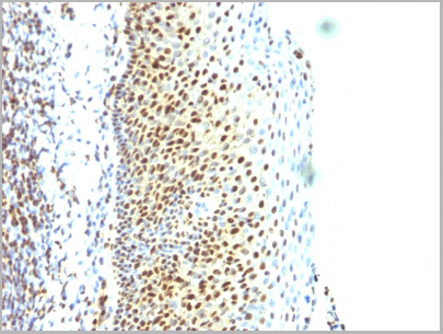
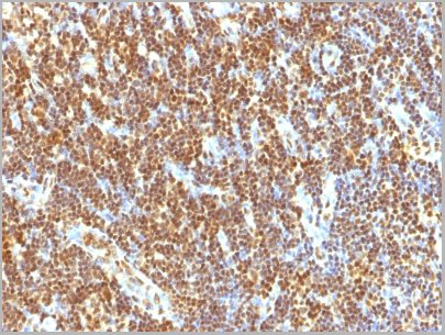
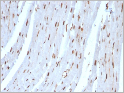 IHC (Immunohistchemistry)
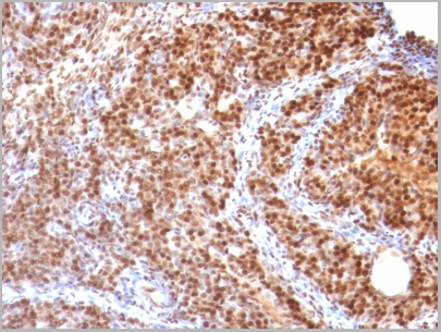
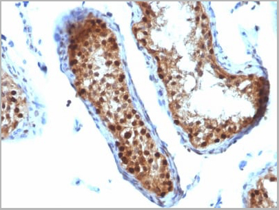
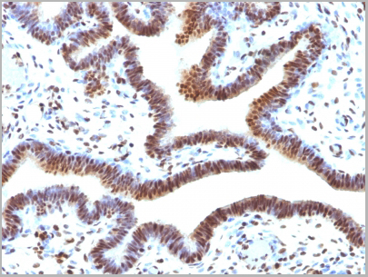
(Formalin-fixed, paraffin-embedded Rat Heart stained with SUMO-2 Monoclonal Antibody (SUMO2/1199))
IHC (Immunohistchemistry)
(Formalin-fixed, paraffin-embedded Rat Heart stained with SUMO-2 Monoclonal Antibody (SUMO2/1199))
SUMO-2, Monoclonal Antibody (Cat# AAA13825)
Full Name
SUMO-2 Mouse Monoclonal Antibody
Gene Names
SUMO3; SMT3A; Smt3B; SMT3H1; SUMO-3
Reactivity
Human, Rat.
Predicted to show a broad species reactivity
Predicted to show a broad species reactivity
Applications
Flow Cytometry, Immunofluorescence, Immunohistochemistry
Pricing
 IP (Immunoprecipitation)
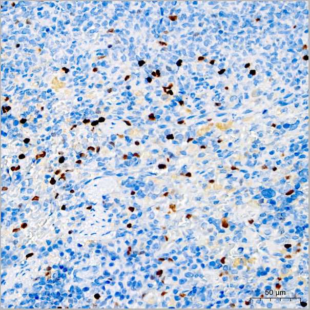
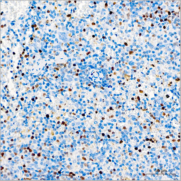
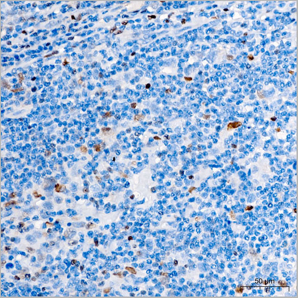
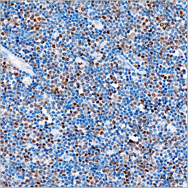
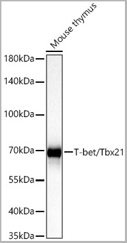
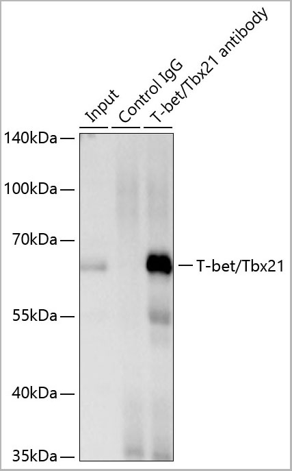
(Immunoprecipitation of T-bet/Tbx21 from 400 µg extracts of NK-92 cells was performed using 1 µg of T-bet/Tbx21 Rabbit PolymAb (AAA28562). Rabbit IgG isotype control (AC042) was used to precipitate the Control IgG sample. IP samples were eluted with 1X reducing Laemmli Buffer. The Input lane represents 10% of the total input. Western blot analysis of immunoprecipitates was conducted using T-bet/Tbx21 Rabbit PolymAb (AAA28562) at a dilution of 1:1000.)
IP (Immunoprecipitation)
(Immunoprecipitation of T-bet/Tbx21 from 400 µg extracts of NK-92 cells was performed using 1 µg of T-bet/Tbx21 Rabbit PolymAb (AAA28562). Rabbit IgG isotype control (AC042) was used to precipitate the Control IgG sample. IP samples were eluted with 1X reducing Laemmli Buffer. The Input lane represents 10% of the total input. Western blot analysis of immunoprecipitates was conducted using T-bet/Tbx21 Rabbit PolymAb (AAA28562) at a dilution of 1:1000.)
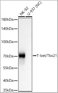
T-bet/Tbx21, Monoclonal Antibody (Cat# AAA28562)
Full Name
T-bet/Tbx21 Rabbit PolymAb
Reactivity
Human
Applications
Western Blot, Immunohistochemistry, Immunoprecipitation
Purity
Affinity purification
Pricing
 IHC (Immunohistchemistry)
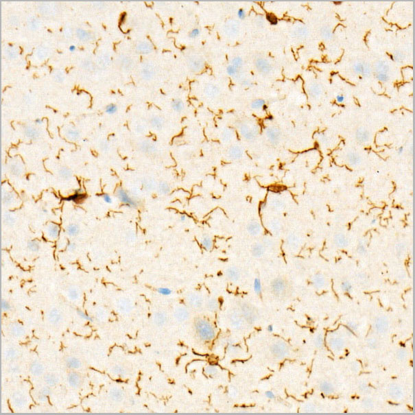
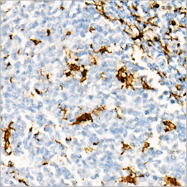
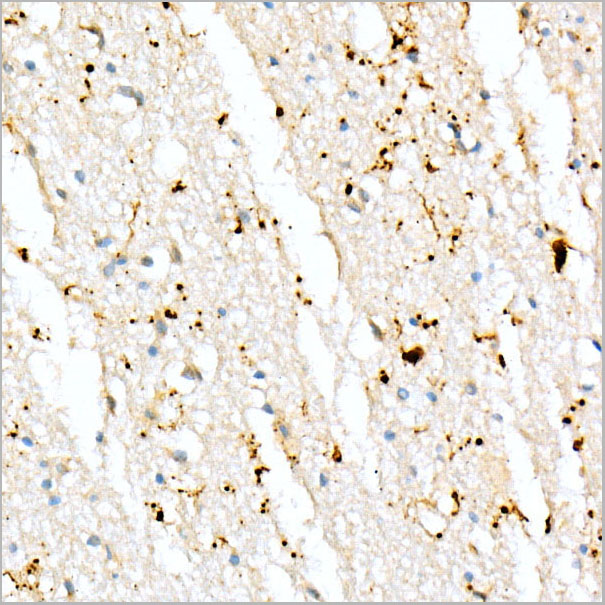
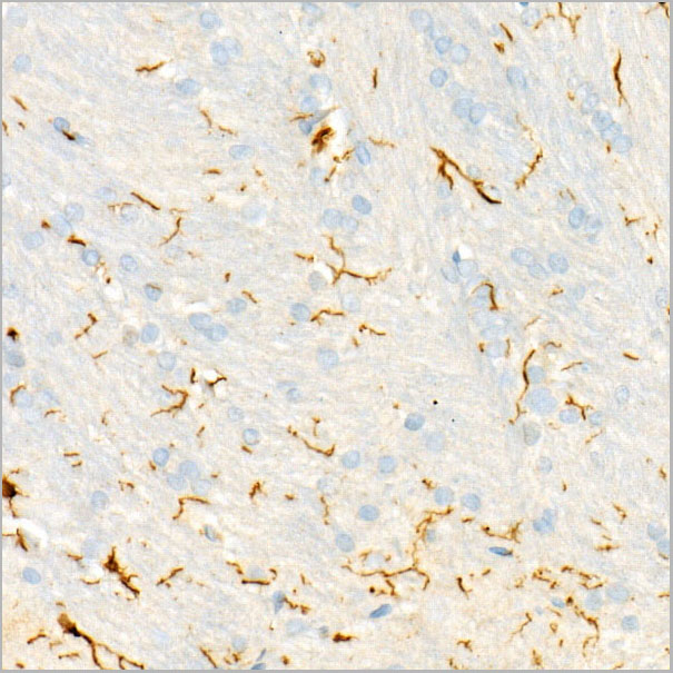
(Immunohistochemistry analysis of paraffin-embedded Rat brain using AIF1/IBA1 Rabbit PolymAb® (AAA28552) at dilution of 1:800 (40x lens). High pressure antigen retrieval performed with 0.01M Citrate Bufferr (pH 6.0) prior to IHC staining.)
IHC (Immunohistchemistry)
(Immunohistochemistry analysis of paraffin-embedded Rat brain using AIF1/IBA1 Rabbit PolymAb® (AAA28552) at dilution of 1:800 (40x lens). High pressure antigen retrieval performed with 0.01M Citrate Bufferr (pH 6.0) prior to IHC staining.)
AIF1/IBA1, Monoclonal Antibody (Cat# AAA28552)
Full Name
AIF1/IBA1 Rabbit PolymAb
Reactivity
Human
Applications
Western Blot, Immunohistochemistry
Purity
Affinity purification
Pricing
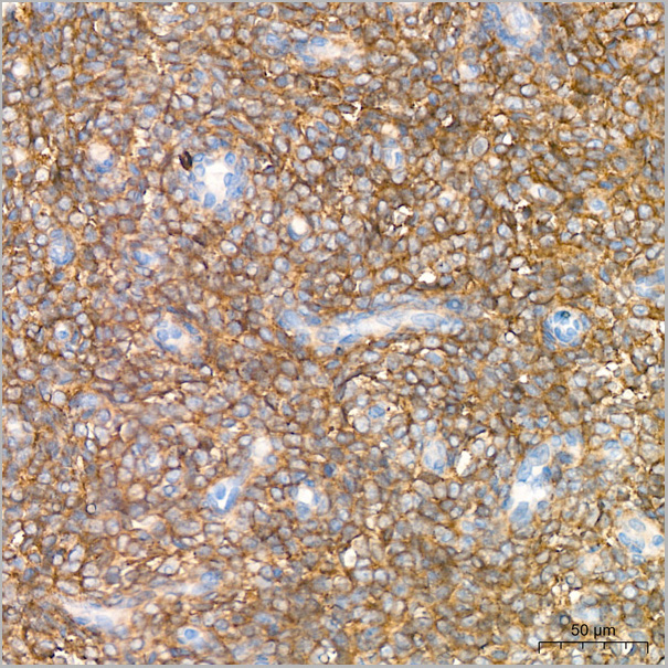
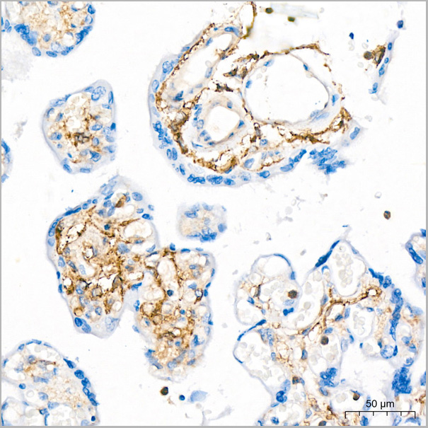
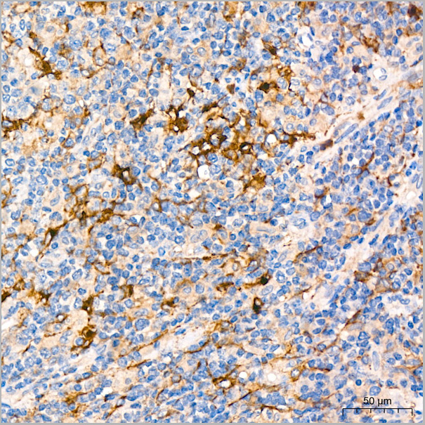
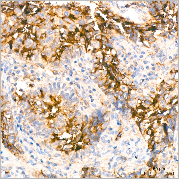
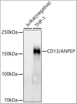
 FCM (Flow Cytometry)
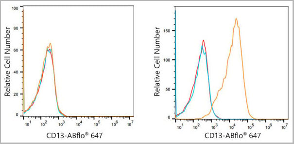
(Flow cytometry:1X10^6 Jurkat cells (negative control,left) and HepG2 cells (right) were surface-stained with CD13/ANPEP Rabbit mAb(AAA28547, 2.5 ug/mL,orange line) or Rabbit IgG isotype control (AC042, 10 ug/mL,blue line),followed by Alexa Fluor 647 conjugated goat anti-mouse pAb(1:600 dilution) staining. Non-fluorescently stained cells were used as blank control (red line).)
FCM (Flow Cytometry)
(Flow cytometry:1X10^6 Jurkat cells (negative control,left) and HepG2 cells (right) were surface-stained with CD13/ANPEP Rabbit mAb(AAA28547, 2.5 ug/mL,orange line) or Rabbit IgG isotype control (AC042, 10 ug/mL,blue line),followed by Alexa Fluor 647 conjugated goat anti-mouse pAb(1:600 dilution) staining. Non-fluorescently stained cells were used as blank control (red line).)
CD13/ANPEP, Monoclonal Antibody (Cat# AAA28547)
Full Name
CD13/ANPEP Rabbit mAb
Reactivity
Human
Applications
Western Blot, Immunohistochemistry, Flow Cytometry
Purity
Affinity purification
Pricing
S100A4, Monoclonal Antibody (Cat# AAA24350)
Full Name
S100A4 (Protein S100-A4, Calvasculin, Metastasin, Placental Calcium-binding Protein, Protein Mts1, S100 Calcium-binding Protein A4, CAPL, MTS1) (AP)
Gene Names
S100A4; 42A; 18A2; CAPL; FSP1; MTS1; P9KA; PEL98
Reactivity
Human, Mouse
Applications
Western Blot
Purity
Purified by Protein A Affinity Chromatography.
Pricing
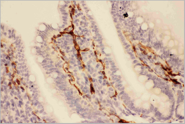
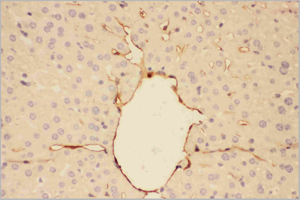
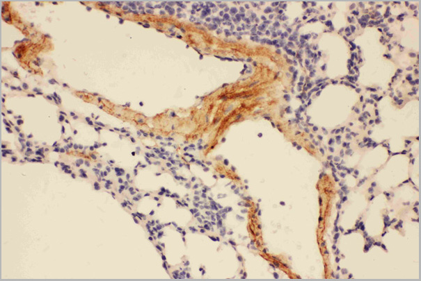
 WB (Western Blot)
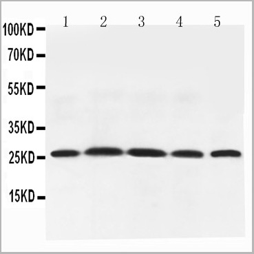
(Figure 6. Western blot analysis of Adiponectin using anti-Adiponectin antibody (AAA11602).Electrophoresis was performed on a 5-20% SDS-PAGE gel at 70V (Stacking gel) / 90V (Resolving gel) for 2-3 hours. The sample well of each lane was loaded with 50ug of sample under reducing conditions.Lane 1: Rat Testis Tissue Lysate,Lane 2: Rat Spleen Tissue Lysate,Lane 3: Rat Skeletal Muscle Tissue Lysate,Lane 4: Rat Cardiac Muscle Tissue Lysate,Lane 5: Rat Liver Tissue Lysate,After Electrophoresis, proteins were transferred to a Nitrocellulose membrane at 150mA for 50-90 minutes. Blocked the membrane with 5% Non-fat Milk/ TBS for 1.5 hour at RT. The membrane was incubated with rabbit anti-Adiponectin antigen affinity purified polyclonal antibody at 0.5ug/mL overnight at 4 degree C, then washed with TBS-0.1%Tween 3 times with 5 minutes each and probed with a goat anti-rabbit IgG-HRP secondary antibody at a dilution of 1:10000 for 1.5 hour at RT. The signal is developed using an Enhanced Chemiluminescent detection (ECL) kit with Tanon 5200 system. A specific band was detected for Adiponectin at approximately 26KD. The expected band size for Adiponectin is at 26KD.)
WB (Western Blot)
(Figure 6. Western blot analysis of Adiponectin using anti-Adiponectin antibody (AAA11602).Electrophoresis was performed on a 5-20% SDS-PAGE gel at 70V (Stacking gel) / 90V (Resolving gel) for 2-3 hours. The sample well of each lane was loaded with 50ug of sample under reducing conditions.Lane 1: Rat Testis Tissue Lysate,Lane 2: Rat Spleen Tissue Lysate,Lane 3: Rat Skeletal Muscle Tissue Lysate,Lane 4: Rat Cardiac Muscle Tissue Lysate,Lane 5: Rat Liver Tissue Lysate,After Electrophoresis, proteins were transferred to a Nitrocellulose membrane at 150mA for 50-90 minutes. Blocked the membrane with 5% Non-fat Milk/ TBS for 1.5 hour at RT. The membrane was incubated with rabbit anti-Adiponectin antigen affinity purified polyclonal antibody at 0.5ug/mL overnight at 4 degree C, then washed with TBS-0.1%Tween 3 times with 5 minutes each and probed with a goat anti-rabbit IgG-HRP secondary antibody at a dilution of 1:10000 for 1.5 hour at RT. The signal is developed using an Enhanced Chemiluminescent detection (ECL) kit with Tanon 5200 system. A specific band was detected for Adiponectin at approximately 26KD. The expected band size for Adiponectin is at 26KD.)
Adiponectin, Polyclonal Antibody (Cat# AAA11602)
Full Name
Anti-Adiponectin antibody
Gene Names
Adipoq; Acdc; Acrp30
Reactivity
Mouse, Rat
Applications
Western Blot, Immunohistochemistry
Purity
Immunogen affinity purified.
Pricing
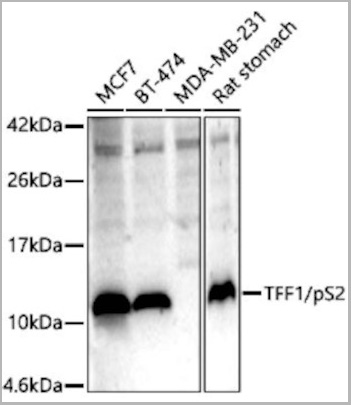
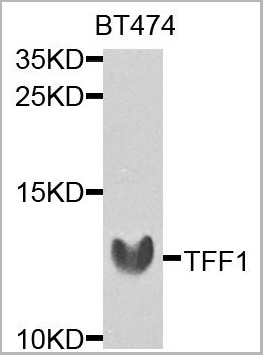
 IHC (Immunohistochemistry)
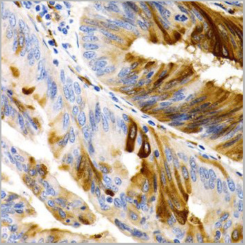
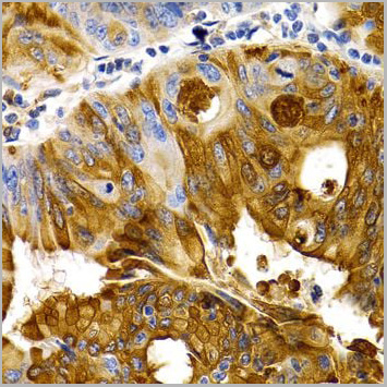
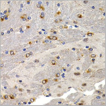
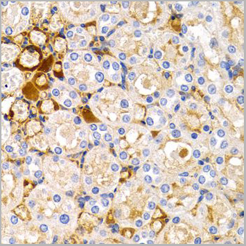
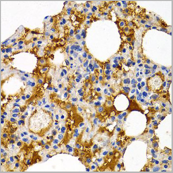
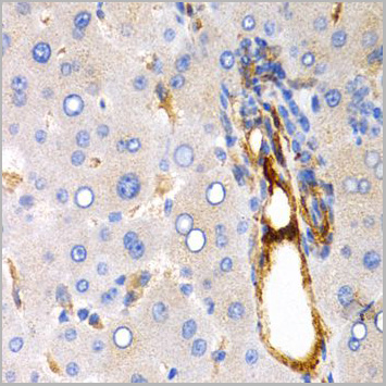
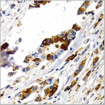
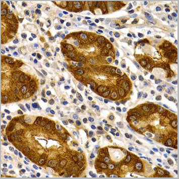
(Immunohistochemistry of paraffin-embedded human stomach using TFF1 antibody at dilution of 1:200 (40x lens).)
IHC (Immunohistochemistry)
(Immunohistochemistry of paraffin-embedded human stomach using TFF1 antibody at dilution of 1:200 (40x lens).)
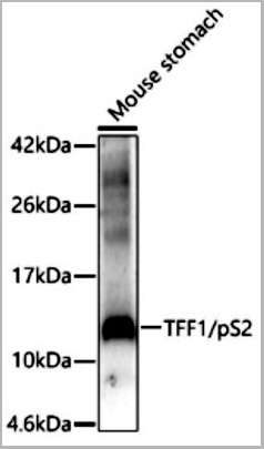
TFF1, Polyclonal Antibody (Cat# AAA10666)
Full Name
TFF1 Polyclonal Antibody
Gene Names
TFF1; pS2; BCEI; HPS2; HP1.A; pNR-2; D21S21
Reactivity
Human, Mouse, Rat
Applications
Western Blot, Immunohistochemistry
Purity
Affinity Purification
Pricing
 IHC (Immunohistochemistry)
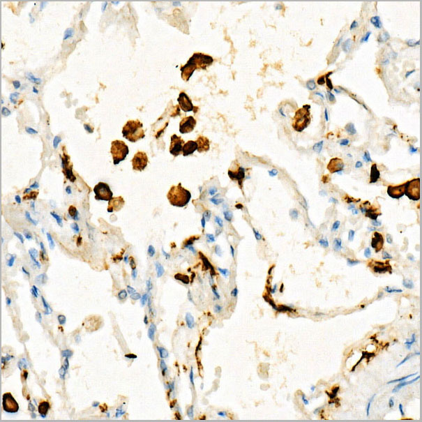
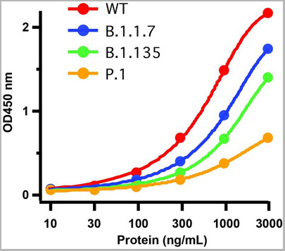
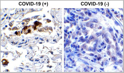
(Immunohistochemistry Validation of SARSCoV-2 (COVID-19) Spike RBD in COVID-19 Patient Lung Immunohistochemical analysis of paraffin-embedded COVID-19 patient lung tissue using anti- SARS-CoV-2 (COVID-19) Spike RBD antibody (AAA11030, 0.5 µg/mL). Tissue was fixed with formaldehyde and blocked with 10% serum for 1 h at RT; antigen retrieval was by heat mediation with a citrate buffer (pH6). Samples were incubated with primary antibody overnight at 4°C. A goat anti-rabbit IgG H&L (HRP) at 1/250 was used as secondary. Counter stained with Hematoxylin. Strong signal of SARS-COV-2 Spike RBD protein was observed in macrophage of COVID-19 patient lung, but not in non-COVID-19 patient lung.)
IHC (Immunohistochemistry)
(Immunohistochemistry Validation of SARSCoV-2 (COVID-19) Spike RBD in COVID-19 Patient Lung Immunohistochemical analysis of paraffin-embedded COVID-19 patient lung tissue using anti- SARS-CoV-2 (COVID-19) Spike RBD antibody (AAA11030, 0.5 µg/mL). Tissue was fixed with formaldehyde and blocked with 10% serum for 1 h at RT; antigen retrieval was by heat mediation with a citrate buffer (pH6). Samples were incubated with primary antibody overnight at 4°C. A goat anti-rabbit IgG H&L (HRP) at 1/250 was used as secondary. Counter stained with Hematoxylin. Strong signal of SARS-COV-2 Spike RBD protein was observed in macrophage of COVID-19 patient lung, but not in non-COVID-19 patient lung.)
COVID 19 Spike RBD Coronavirus, Polyclonal Antibody (Cat# AAA11030)
Full Name
SARS-CoV-2 (COVID-19) Spike RBD Antibody
Gene Names
S; spike glycoprotein
Reactivity
Virus
Applications
Immunofluorescence, Immunohistochemistry, Western Blot
Purity
SARS-CoV-2 (COVID-19) Spike RBD antibody is affinity chromatography purified via peptide column.
Pricing