
Filters
Clonality
Type
Reactivity
Gene Name
Isotype
Host
Application
Clone
10000 results for "Recombinant Proteins" - showing 2100-2150
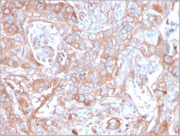
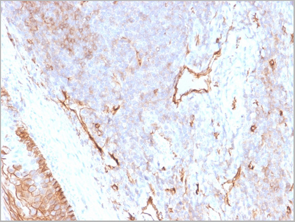
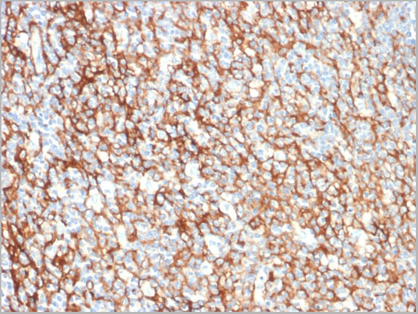
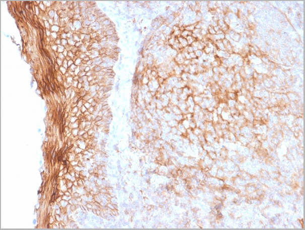
 Application Data
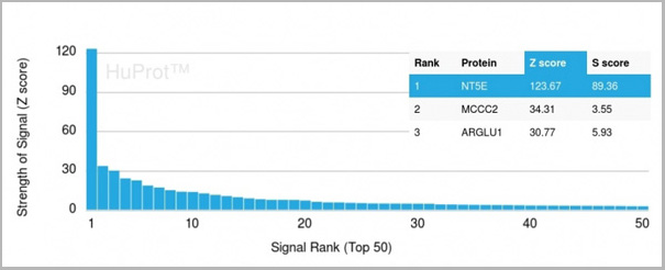
(Analysis of Protein Array containing more than 19,000 full-length human proteins using CD73 Mouse Recombinant Monoclonal Antibody (NT5E/2505) Z- and S- Score: The Z-score represents the strength of a signal that a monoclonal antibody (MAb) (in combination with a fluorescently-tagged anti-IgG secondary antibody) produces when binding to a particular protein on the HuProtTM array. Z-scores are described in units of standard deviations (SD’s) above the mean value of all signals generated on that array. If targets on HuProtTM are arranged in descending order of the Z-score, the S-score is the difference (also in units of SD’s) between the Z-score. S-score therefore represents the relative target specificity of a MAb to its intended target. A MAb is considered to specific to its intended target, if the MAb has an S-score of at least 2.5. For example, if a MAb binds to protein X with a Z-score of 43 and to protein Y with a Z-score of 14, then the S-score for the binding of that MAb to protein X is equal to 29.)
Application Data
(Analysis of Protein Array containing more than 19,000 full-length human proteins using CD73 Mouse Recombinant Monoclonal Antibody (NT5E/2505) Z- and S- Score: The Z-score represents the strength of a signal that a monoclonal antibody (MAb) (in combination with a fluorescently-tagged anti-IgG secondary antibody) produces when binding to a particular protein on the HuProtTM array. Z-scores are described in units of standard deviations (SD’s) above the mean value of all signals generated on that array. If targets on HuProtTM are arranged in descending order of the Z-score, the S-score is the difference (also in units of SD’s) between the Z-score. S-score therefore represents the relative target specificity of a MAb to its intended target. A MAb is considered to specific to its intended target, if the MAb has an S-score of at least 2.5. For example, if a MAb binds to protein X with a Z-score of 43 and to protein Y with a Z-score of 14, then the S-score for the binding of that MAb to protein X is equal to 29.)
CD73, Monoclonal Antibody (Cat# AAA23927)
Full Name
CD73 (Immuno-Oncology Target)
Gene Names
NT5E; NT; eN; NT5; NTE; eNT; CD73; E5NT; CALJA
Reactivity
Human
Applications
Western Blot, Flow Cytometry, Immunofluorescence, Immunohistochemistry
Purity
Purified Ab with BSA and Azide at 200ug/ml OR Purified Ab WITHOUT BSA and Azide at 1.0mg/ml
Pricing

 Bioactivity
(Annexin V (ANXA5) is a multifunctional protein that is highly expressed on the apical surfaces of syncytiotrophoblasts, and plays an important role in haemostatic regulations, maintaining blood fluidity of the placenta. Lower ANXA5 levels have been observed in M2/ANXA5 haplotype carrying chorion. The association found between the maternal carriage of the M2/ANXA5 haplotype and an elevated risk of IUGR and/or PE supports the hypothesis that carrier status of this haplotype and the consequently reduced placental ANXA5 expression might be responsible, at least partially, for the onset of these gestational vascular complications. Annexin V is a calcium-dependent phospholipid binding protein that can be used to bind Phosphatidylserine (PS) during an early apoptosis event where the PS becomes exposed at the cell surface. Jurkat cells were treated with 10 uM camptothecin for 4h, 2*105 cells which were resuspended in binding buffer were stained with 10 ug recombinant human Annexin V-FITC and 10 ul Propidium iodide (PI) for 20min in dark room temperature. The flow cytometry was used to detect the early apoptotic and late apoptotic of camptothecin-treated Jurkat cells (Figure 1), the combination of Annexin V-FITC and propidium iodide allows for the distinction between early apoptotic cells (Annexin V-FITC positive and propidium iodide negative), late apoptotic and/or necrotic cells (Annexin V-FITC and propidium iodide positive), and viable cells (unstained). Thus, the recombinant human Annexin V-FITC can bind Phosphatidylserine (PS) at early apoptosis of Jurkat.)
Bioactivity
(Annexin V (ANXA5) is a multifunctional protein that is highly expressed on the apical surfaces of syncytiotrophoblasts, and plays an important role in haemostatic regulations, maintaining blood fluidity of the placenta. Lower ANXA5 levels have been observed in M2/ANXA5 haplotype carrying chorion. The association found between the maternal carriage of the M2/ANXA5 haplotype and an elevated risk of IUGR and/or PE supports the hypothesis that carrier status of this haplotype and the consequently reduced placental ANXA5 expression might be responsible, at least partially, for the onset of these gestational vascular complications. Annexin V is a calcium-dependent phospholipid binding protein that can be used to bind Phosphatidylserine (PS) during an early apoptosis event where the PS becomes exposed at the cell surface. Jurkat cells were treated with 10 uM camptothecin for 4h, 2*105 cells which were resuspended in binding buffer were stained with 10 ug recombinant human Annexin V-FITC and 10 ul Propidium iodide (PI) for 20min in dark room temperature. The flow cytometry was used to detect the early apoptotic and late apoptotic of camptothecin-treated Jurkat cells (Figure 1), the combination of Annexin V-FITC and propidium iodide allows for the distinction between early apoptotic cells (Annexin V-FITC positive and propidium iodide negative), late apoptotic and/or necrotic cells (Annexin V-FITC and propidium iodide positive), and viable cells (unstained). Thus, the recombinant human Annexin V-FITC can bind Phosphatidylserine (PS) at early apoptosis of Jurkat.)
Annexin V (ANXA5), Active Protein (Cat# AAA161723)
Full Name
Active Annexin V (ANXA5)
Gene Names
ANXA5; PP4; ANX5; ENX2; RPRGL3; HEL-S-7
Applications
Activity Assay, Cell Culture
Purity
Greater than 95% by SDS-PAGE
Pricing
 Bioactivity
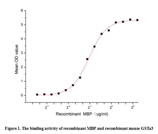
(Maltose binding protein (MBP) encoded by the malE gene in Escherichia coli, is a 44-kD monomeric periplasmic protein. MBP is component of the Escherichia coli maltose/maltodextrin system, which regulates the uptake and catabolism of maltrodextrins as part of the chemotactic response. This protein is used in recombinant protein expression as an affinity and solubility tag. Glutathione S Transferase Alpha 3 (GSTa3) has been identified as an interactor of MBP, thus a functional binding ELISA assay was conducted to detect the interaction of recombinant MBP and recombinant mouse GSTa3. Briefly, biotin-linked MBP were diluted serially in PBS, with 0.01% BSA (pH 7.4). Duplicate samples of 100 ul were then transferred to GSTa3-coated microtiter wells and incubated for 1h at 37 degree C. Wells were washed with PBST 3 times and incubation with Streptavidin-HRP for 30min, then wells were aspirated and washed 5 times. With the addition of substrate solution, wells were incubated 15-25 minutes at 37 degree C. Finally, add 50 ul stop solution to the wells and read at 450 nm immediately. The binding activity of recombinant MBP and recombinant mouse GSTa3 was shown in Figure 1, the EC50 for this effect is 0.46 ug/mL.)
Bioactivity
(Maltose binding protein (MBP) encoded by the malE gene in Escherichia coli, is a 44-kD monomeric periplasmic protein. MBP is component of the Escherichia coli maltose/maltodextrin system, which regulates the uptake and catabolism of maltrodextrins as part of the chemotactic response. This protein is used in recombinant protein expression as an affinity and solubility tag. Glutathione S Transferase Alpha 3 (GSTa3) has been identified as an interactor of MBP, thus a functional binding ELISA assay was conducted to detect the interaction of recombinant MBP and recombinant mouse GSTa3. Briefly, biotin-linked MBP were diluted serially in PBS, with 0.01% BSA (pH 7.4). Duplicate samples of 100 ul were then transferred to GSTa3-coated microtiter wells and incubated for 1h at 37 degree C. Wells were washed with PBST 3 times and incubation with Streptavidin-HRP for 30min, then wells were aspirated and washed 5 times. With the addition of substrate solution, wells were incubated 15-25 minutes at 37 degree C. Finally, add 50 ul stop solution to the wells and read at 450 nm immediately. The binding activity of recombinant MBP and recombinant mouse GSTa3 was shown in Figure 1, the EC50 for this effect is 0.46 ug/mL.)
Maltose Binding Protein (MBP), Active Protein (Cat# AAA161942)
Full Name
Active Maltose Binding Protein (MBP)
Gene Names
malE; ECK4026; JW3994; malJ
Applications
Activity Assay, Cell Culture
Purity
Greater than 95% by SDS-PAGE
Pricing
 Bioactivity
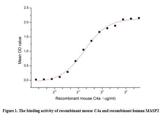
(Complement Component 4a (C4a) is a component of the complement system, which is a cleavage product of the complement C4 protein. C4a has been implicated in various inflammatory and immune responses. It acts as a chemoattractant, recruiting immune cells such as neutrophils and macrophages to the site of inflammation. Additionally, C4a can stimulate the release of pro-inflammatory cytokines and chemokines, further amplifying the immune response. Besides, the binding of MASP2 to C4a is an important step in the lectin pathway of the complement system, thus a functional binding ELISA assay was conducted to detect the interaction of recombinant mouse C4a and recombinant human MASP2. Briefly, C4a was diluted serially in PBS with 0.01% BSA (pH 7.4). Duplicate samples of 100 ul were then transferred to MASP2-coated microtiter wells and incubated for 1h at 37 degree C. Wells were washed with PBST and incubated for 1h with anti-C4a pAb, then aspirated and washed 3 times. After incubation with HRP labelled secondary antibody for 1h at 37 degree C, wells were aspirated and washed 5 times. With the addition of substrate solution, wells were incubated 15-25 minutes at 37 degree C. Finally, add 50 uL stop solution to the wells and read at 450/630 nm immediately. The binding activity of recombinant mouse C4a and recombinant human MASP2 was shown in Figure 1, the EC50 for this effect is 0.21 ug/mL.)
Bioactivity
(Complement Component 4a (C4a) is a component of the complement system, which is a cleavage product of the complement C4 protein. C4a has been implicated in various inflammatory and immune responses. It acts as a chemoattractant, recruiting immune cells such as neutrophils and macrophages to the site of inflammation. Additionally, C4a can stimulate the release of pro-inflammatory cytokines and chemokines, further amplifying the immune response. Besides, the binding of MASP2 to C4a is an important step in the lectin pathway of the complement system, thus a functional binding ELISA assay was conducted to detect the interaction of recombinant mouse C4a and recombinant human MASP2. Briefly, C4a was diluted serially in PBS with 0.01% BSA (pH 7.4). Duplicate samples of 100 ul were then transferred to MASP2-coated microtiter wells and incubated for 1h at 37 degree C. Wells were washed with PBST and incubated for 1h with anti-C4a pAb, then aspirated and washed 3 times. After incubation with HRP labelled secondary antibody for 1h at 37 degree C, wells were aspirated and washed 5 times. With the addition of substrate solution, wells were incubated 15-25 minutes at 37 degree C. Finally, add 50 uL stop solution to the wells and read at 450/630 nm immediately. The binding activity of recombinant mouse C4a and recombinant human MASP2 was shown in Figure 1, the EC50 for this effect is 0.21 ug/mL.)
Complement Component 4a (C4a), Active Protein (Cat# AAA161729)
Full Name
Active Complement Component 4a (C4a)
Applications
Activity Assay, Cell Culture
Purity
Greater than 90% by SDS-PAGE
Pricing
 IP (Immunoprecipitation)
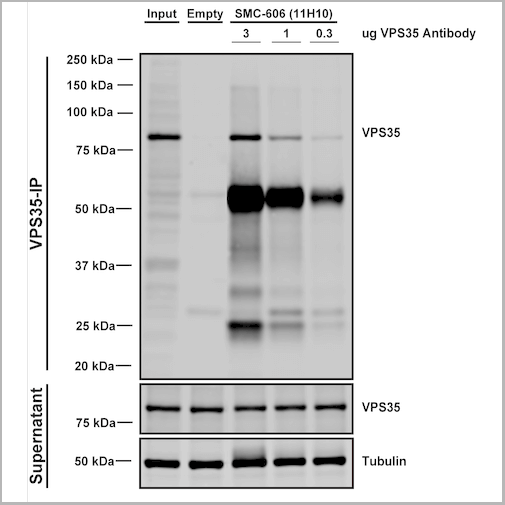
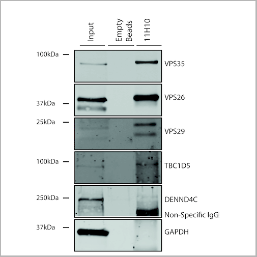
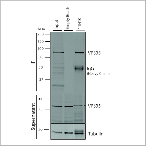
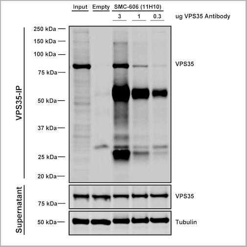
(Immunoprecipitation analysis using Mouse Anti-VPS35 Monoclonal Antibody, Clone 11H10. Tissue: embryonic fibroblast. Species: Mouse. Primary Antibody: Mouse Anti-VPS35 Monoclonal Antibody. Three amounts of (3, 1 and 0.3 ug) were non-covalently coupled to 10uL of A/G sepharose beads for 1 hour at 4 degree C and next incubated with 250ug of MEF lysate for 2 hours at 4 degree C.)
IP (Immunoprecipitation)
(Immunoprecipitation analysis using Mouse Anti-VPS35 Monoclonal Antibody, Clone 11H10. Tissue: embryonic fibroblast. Species: Mouse. Primary Antibody: Mouse Anti-VPS35 Monoclonal Antibody. Three amounts of (3, 1 and 0.3 ug) were non-covalently coupled to 10uL of A/G sepharose beads for 1 hour at 4 degree C and next incubated with 250ug of MEF lysate for 2 hours at 4 degree C.)
VPS35, Monoclonal Antibody (Cat# AAA27708)
Full Name
VPS35 Antibody, Clone 11H10: RPE
Gene Names
VPS35; MEM3; PARK17
Reactivity
Human, Mouse, Rat
Applications
Western Blot, Immunoprecipitation
Purity
Protein G Purified
Pricing
 Bioactivity
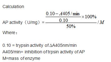
(Aprotinin (AP) is a competitive serine protease inhibitor. Reversibly binds to and blocks the enzymatic active site. Inhibits a range of serine proteases including trypsin, chymotrypsin, kallikrein and plasmin. Inhibits cytopathogenic effect of SARS-CoV-2 and double-stranded RNA formation in SARS-CoV-2-infected cells. The activity of recombinant bovine AP was measured by its ability to inhibit trypsin cleavage of a peptide substrate BAPNA in the assay buffer 200 mM Triethanolamine hydrochloride, 20 mM CaCl2, pH 7.8. The reaction was performed in adding 20 ul 4 mg/mL trypsin diluted by 1mM HCl to 160 ul assay buffer and 20 ul 0.85% (w/v) NaCl and start the reaction by adding 100 ul of 1mg/ml BAPNA. Include a substrate blank containing 160 ul assay buffer, 20 ul 1mM HCl, 20 ul 0.85% (w/v) NaCl and 100 uL of 1mg/ml substrate. Rapidly mixing at 25 degree C, then read at 405 nm in kinetic mode for 5 minutes using a microplate reader controlling the ?A405nm/min=0.08-0.12. The 20 ul different concentrations of recombinant bovine AP was incubated with 20 ul 4 mg/mL trypsin in 160 ul assay buffer at 25 degree C for 10 minutes followed by adding 100 ul substrate, then read at 405 nm in kinetic mode for 5 minutes using a microplate reader. Under these conditions, the enzyme amount of 50% inhibition of trypsin activity per minute is defined as a unit. The specific activity of recombinant bovine AP is >2000 U/mg.)
Bioactivity
(Aprotinin (AP) is a competitive serine protease inhibitor. Reversibly binds to and blocks the enzymatic active site. Inhibits a range of serine proteases including trypsin, chymotrypsin, kallikrein and plasmin. Inhibits cytopathogenic effect of SARS-CoV-2 and double-stranded RNA formation in SARS-CoV-2-infected cells. The activity of recombinant bovine AP was measured by its ability to inhibit trypsin cleavage of a peptide substrate BAPNA in the assay buffer 200 mM Triethanolamine hydrochloride, 20 mM CaCl2, pH 7.8. The reaction was performed in adding 20 ul 4 mg/mL trypsin diluted by 1mM HCl to 160 ul assay buffer and 20 ul 0.85% (w/v) NaCl and start the reaction by adding 100 ul of 1mg/ml BAPNA. Include a substrate blank containing 160 ul assay buffer, 20 ul 1mM HCl, 20 ul 0.85% (w/v) NaCl and 100 uL of 1mg/ml substrate. Rapidly mixing at 25 degree C, then read at 405 nm in kinetic mode for 5 minutes using a microplate reader controlling the ?A405nm/min=0.08-0.12. The 20 ul different concentrations of recombinant bovine AP was incubated with 20 ul 4 mg/mL trypsin in 160 ul assay buffer at 25 degree C for 10 minutes followed by adding 100 ul substrate, then read at 405 nm in kinetic mode for 5 minutes using a microplate reader. Under these conditions, the enzyme amount of 50% inhibition of trypsin activity per minute is defined as a unit. The specific activity of recombinant bovine AP is >2000 U/mg.)
Aprotinin (AP), Active Protein (Cat# AAA161810)
Full Name
Active Aprotinin (AP)
Applications
Activity Assay, Cell Culture
Purity
Greater than 95% by SDS-PAGE
Pricing
 IP (Immunoprecipitation)
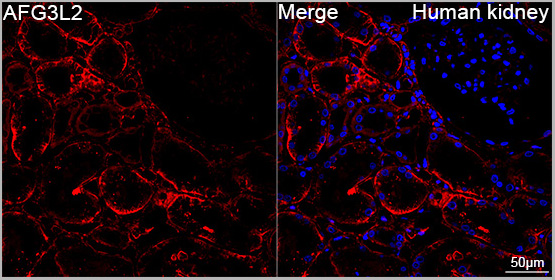
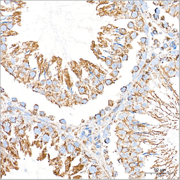
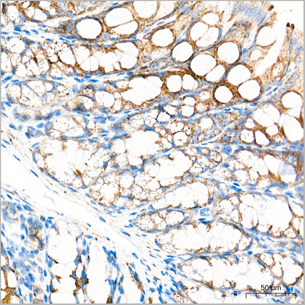
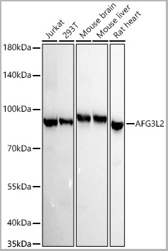
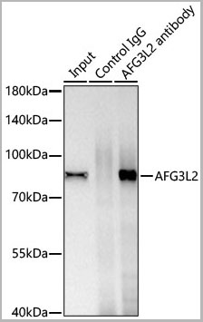
(Immunoprecipitation of AFG3L2 from 600 µg extracts of Mouse liver tissue was performed using 1 µg of AFG3L2 Rabbit PolymAb® (AAA28589). Rabbit IgG isotype control (AC042) was used to precipitate the Control IgG sample. IP samples were eluted with 1X reducing Laemmli Buffer. The Input lane represents 10% of the total input. Western blot analysis of immunoprecipitates was conducted using AFG3L2 Rabbit PolymAb® (AAA28589) at a dilution of 1:10000.)
IP (Immunoprecipitation)
(Immunoprecipitation of AFG3L2 from 600 µg extracts of Mouse liver tissue was performed using 1 µg of AFG3L2 Rabbit PolymAb® (AAA28589). Rabbit IgG isotype control (AC042) was used to precipitate the Control IgG sample. IP samples were eluted with 1X reducing Laemmli Buffer. The Input lane represents 10% of the total input. Western blot analysis of immunoprecipitates was conducted using AFG3L2 Rabbit PolymAb® (AAA28589) at a dilution of 1:10000.)
AFG3L2, Monoclonal Antibody (Cat# AAA28589)
Full Name
AFG3L2 Rabbit PolymAb
Reactivity
Human
Applications
Western Blot, Immunohistochemistry, Immunofluorescence, Immunocytochemistry, Immunoprecipitation
Purity
Affinity purification
Pricing
Cardiac Troponin-I, Recombinant Protein (Cat# AAA10835)
Full Name
Recombinant Human Cardiac Troponin-I
Gene Names
TNNI3; CMH7; RCM1; cTnI; CMD2A; TNNC1; CMD1FF
Purity
Greater than 95.0% as determined by both SDS-PAGE.
Pricing
 SDS-PAGE
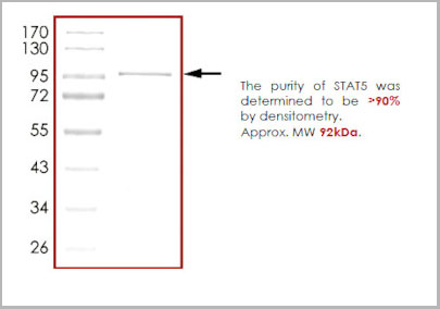
(The purity of STAT5 was determined to be >90% by densitometry. Approx. MW 92kDa.)
SDS-PAGE
(The purity of STAT5 was determined to be >90% by densitometry. Approx. MW 92kDa.)
STAT5, Recombinant Protein (Cat# AAA14248)
Full Name
STAT5 recombinant protein
Gene Names
STAT5A; MGF; STAT5
Purity
>90%
Pricing
 IP (Immunoprecipitation)
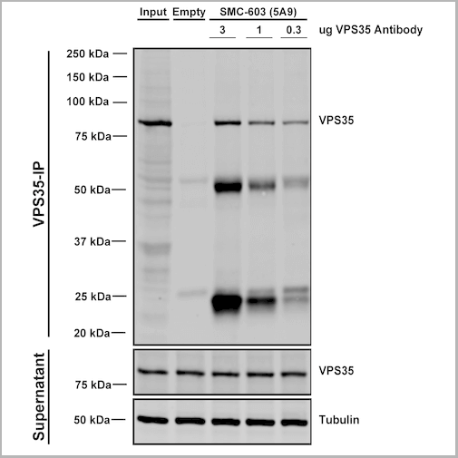
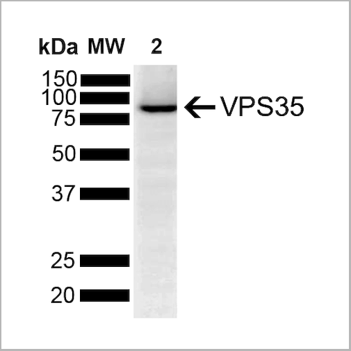
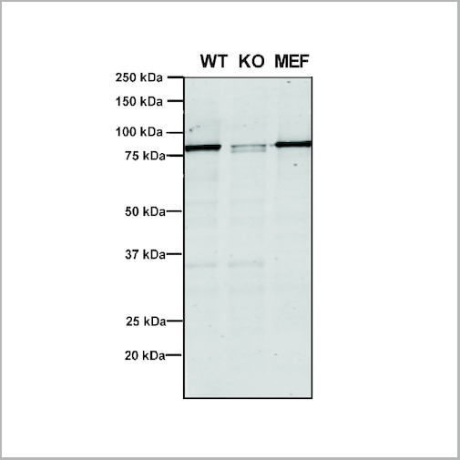
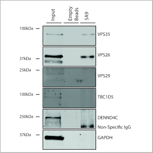
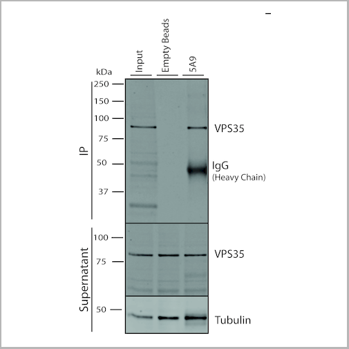
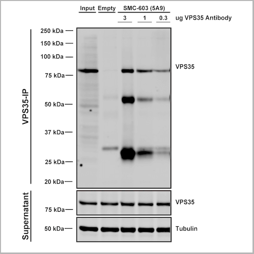
(Immunoprecipitation analysis using Mouse Anti-VPS35 Monoclonal Antibody, Clone 5A9. Tissue: embryonic fibroblast. Species: Mouse. Primary Antibody: Mouse Anti-VPS35 Monoclonal Antibody. Three amounts of (3, 1 and 0.3 ug) were non-covalently coupled to 10uL of A/G sepharose beads for 1 hour at 4 degree C and next incubated with 250ug of MEF lysate for 2 hours at 4 degree C.)
IP (Immunoprecipitation)
(Immunoprecipitation analysis using Mouse Anti-VPS35 Monoclonal Antibody, Clone 5A9. Tissue: embryonic fibroblast. Species: Mouse. Primary Antibody: Mouse Anti-VPS35 Monoclonal Antibody. Three amounts of (3, 1 and 0.3 ug) were non-covalently coupled to 10uL of A/G sepharose beads for 1 hour at 4 degree C and next incubated with 250ug of MEF lysate for 2 hours at 4 degree C.)
VPS35, Monoclonal Antibody (Cat# AAA27674)
Full Name
VPS35 Antibody, Clone 5A9: ATTO 390
Gene Names
VPS35; MEM3; PARK17
Reactivity
Human, Mouse, Rat
Applications
Western Blot, Immunocytochemistry, Immunofluorescence, Immunoprecipitation
Purity
Protein G Purified
Pricing

 Bioactivity
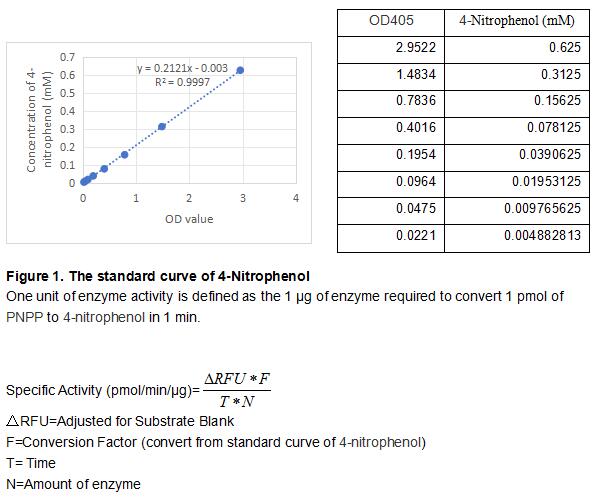
(Acid phosphatase locus 1 (ACP1) is a low molecular weight protein tyrosine phosphatase that has been shown to be an important regulator of insulin receptor signaling. The activity assay of recombinant mouse ACP1 was measured by its ability to cleave a peptide substrate, 4-Nitrophenyl phosphate disodium (PNPP). The reaction was performed in 50 mM NaOAc, pH 5.0 (assay buffer), initiated by addition 50 ul of 3 ug/ml ACP1 (diluted by assay buffer) to 50 uL of 2 mM substrate. Incubated at room temperature for 5 minutes in the dark and add 100 ul of 0.2 M NaOH to stop the reaction. Read at a wavelength of 405 nm (top read), the specific activity of recombinant mouse ACP1 is >32000 pmol/min/ug.)
Bioactivity
(Acid phosphatase locus 1 (ACP1) is a low molecular weight protein tyrosine phosphatase that has been shown to be an important regulator of insulin receptor signaling. The activity assay of recombinant mouse ACP1 was measured by its ability to cleave a peptide substrate, 4-Nitrophenyl phosphate disodium (PNPP). The reaction was performed in 50 mM NaOAc, pH 5.0 (assay buffer), initiated by addition 50 ul of 3 ug/ml ACP1 (diluted by assay buffer) to 50 uL of 2 mM substrate. Incubated at room temperature for 5 minutes in the dark and add 100 ul of 0.2 M NaOH to stop the reaction. Read at a wavelength of 405 nm (top read), the specific activity of recombinant mouse ACP1 is >32000 pmol/min/ug.)
Acid Phosphatase 1 (ACP1), Active Protein (Cat# AAA161821)
Full Name
Active Acid Phosphatase 1 (ACP1)
Gene Names
Acp1; Acp-1; LMW-PTP; AI427468; 4632432E04Rik
Applications
Activity Assay, Cell Culture
Purity
Greater than 90% by SDS-PAGE
Pricing
 Bioactivity
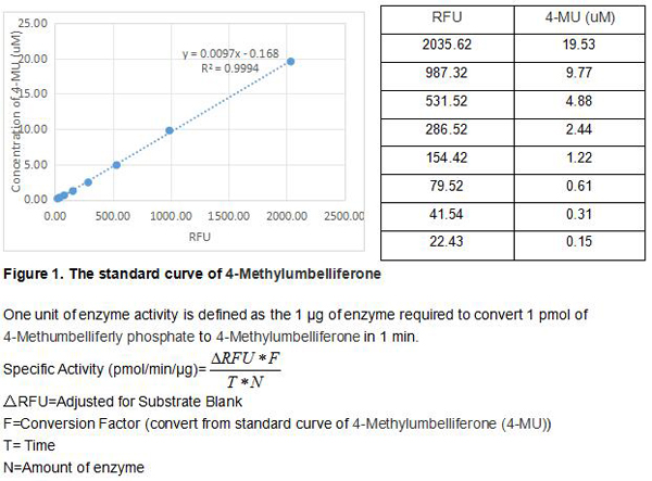
(Four distinct genes encode alkaline phosphatases (APs) in humans. The ALPL gene encodes the liver/bone/kidney isozyme, also known as the tissuenonspecific AP (TNAP). In comparison, ALPI, ALPP and ALPPL2 encode intestinal, placental and placental-like or germ cell APs, respectively. The serum levels of human APs are useful tumor markers. There are many mutations in the ALPL gene, leading to different forms of hypophosphatasia, characterized by poorly mineralized cartilage and bones. The native ALPL is a glycosylated homodimer attached to the membrane through a GPI-anchor. The activity assay of recombinant mouse ALPL was measured by its ability to cleave a peptide substrate, 4-Methumbelliferly phosphate. The reaction was performed in 50 mM Tris, 1 mM MgCl2, pH 9.0 (assay buffer), ainitiated by addition 50 ul of 0.3 ug/ml ALPL (diluted by assay buffer) to 50 uL of 50 uM substrate. Read at excitation and emission wavelengths of 365 nm and 445 nm (top read), respectively, in kinetic mode for 5 minutes. The specific activity of recombinant mouse ALPL is >11000 pmol/min/ug.)
Bioactivity
(Four distinct genes encode alkaline phosphatases (APs) in humans. The ALPL gene encodes the liver/bone/kidney isozyme, also known as the tissuenonspecific AP (TNAP). In comparison, ALPI, ALPP and ALPPL2 encode intestinal, placental and placental-like or germ cell APs, respectively. The serum levels of human APs are useful tumor markers. There are many mutations in the ALPL gene, leading to different forms of hypophosphatasia, characterized by poorly mineralized cartilage and bones. The native ALPL is a glycosylated homodimer attached to the membrane through a GPI-anchor. The activity assay of recombinant mouse ALPL was measured by its ability to cleave a peptide substrate, 4-Methumbelliferly phosphate. The reaction was performed in 50 mM Tris, 1 mM MgCl2, pH 9.0 (assay buffer), ainitiated by addition 50 ul of 0.3 ug/ml ALPL (diluted by assay buffer) to 50 uL of 50 uM substrate. Read at excitation and emission wavelengths of 365 nm and 445 nm (top read), respectively, in kinetic mode for 5 minutes. The specific activity of recombinant mouse ALPL is >11000 pmol/min/ug.)
Alkaline Phosphatase, Tissue-nonspecific (ALPL), Active Protein (Cat# AAA161830)
Full Name
Active Alkaline Phosphatase, Tissue-nonspecific (ALPL)
Gene Names
Alpl; ALP; Akp2; TNAP; Akp-2; APTNAP; TNSALP
Applications
Activity Assay, Cell Culture
Purity
Greater than 90% by SDS-PAGE
Pricing
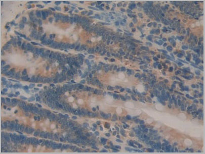
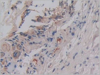
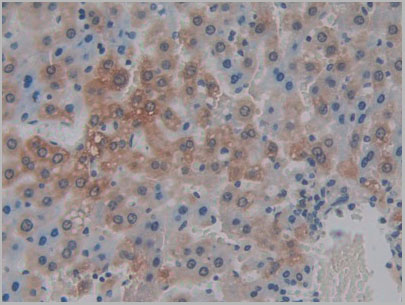 IHC (Immunohistchemistry)
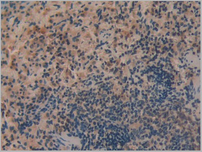
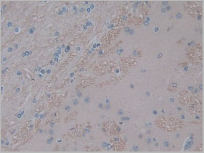
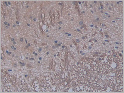
(DAB staining on IHCP;Sample: Rat Liver Tissue; Primary Ab: 30ug/ml Mouse AntiRat IL1a AntibodySecond Ab: 2ug/mL HRPLinked Caprine AntiMouse IgG Polyclonal Antibody(Catalog: SAA544Mu19))
IHC (Immunohistchemistry)
(DAB staining on IHCP;Sample: Rat Liver Tissue; Primary Ab: 30ug/ml Mouse AntiRat IL1a AntibodySecond Ab: 2ug/mL HRPLinked Caprine AntiMouse IgG Polyclonal Antibody(Catalog: SAA544Mu19))
Interleukin 1 Alpha (IL1a), Monoclonal Antibody (Cat# AAA21179)
Full Name
Monoclonal Antibody to Interleukin 1 Alpha (IL1a)
Gene Names
Il1a; IL-1 alpha
Reactivity
Rat
Applications
Western Blot, Immunohistochemistry, Immunocytochemistry, Immunoprecipitation
Purity
Protein A + Protein G affinity chromatography
Pricing
HBcAg, Recombinant Protein (Cat# AAA14357)
Full Name
HBcAg protein
Applications
Western Blot
Purity
>95 % (by SDS-PAGE)
Pricing
 Bioactivity
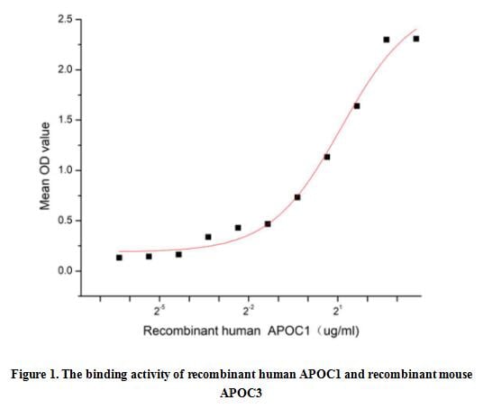
(Apolipoprotein C1 (APOC1), also known as Apo-CI; ApoC-I; apo-CIB; apoC-IB, is the smallest size apolipoprotein of all apolipoprotein C family (Mr = 6.6 kDa) and located at position 19q13.32. APOC1 is primarily expressed in the liver and activated when monocytes differentiate into macrophages. It plays important roles in the innate immune response as effector of glucocorticoid-mediated responses and regulator of the inflammatory process. It has anti-inflammatory activity and also can promote the differentiation of T-cells into Th1 cells and negatively regulates differentiation into Th2 cells. Besides, Apolipoprotein C3 (APOC3) has been identified as an interactor of APOC1, thus a functional binding ELISA assay was conducted to detect the interaction of recombinant human APOC1 and recombinant mouse APOC3. Briefly, APOC1 was diluted serially in PBS with 0.01% BSA (pH 7.4). Duplicate samples of 100 ul were then transferred to APOC3-coated microtiter wells and incubated for 1h at 37 degree C. Wells were washed with PBST and incubated for 1h with anti-APOC1 pAb, then aspirated and washed 3 times. After incubation with HRP labelled secondary antibody for 1h at 37 degree C, wells were aspirated and washed 5 times. With the addition of substrate solution, wells were incubated 15-25 minutes at 37 degree C. Finally, add 50 uL stop solution to the wells and read at 450/630 nm immediately. The binding activity of recombinant human APOC1 and recombinant mouse APOC3 was shown in Figure 1, the EC50 for this effect is 2.19 ug/mL.)
Bioactivity
(Apolipoprotein C1 (APOC1), also known as Apo-CI; ApoC-I; apo-CIB; apoC-IB, is the smallest size apolipoprotein of all apolipoprotein C family (Mr = 6.6 kDa) and located at position 19q13.32. APOC1 is primarily expressed in the liver and activated when monocytes differentiate into macrophages. It plays important roles in the innate immune response as effector of glucocorticoid-mediated responses and regulator of the inflammatory process. It has anti-inflammatory activity and also can promote the differentiation of T-cells into Th1 cells and negatively regulates differentiation into Th2 cells. Besides, Apolipoprotein C3 (APOC3) has been identified as an interactor of APOC1, thus a functional binding ELISA assay was conducted to detect the interaction of recombinant human APOC1 and recombinant mouse APOC3. Briefly, APOC1 was diluted serially in PBS with 0.01% BSA (pH 7.4). Duplicate samples of 100 ul were then transferred to APOC3-coated microtiter wells and incubated for 1h at 37 degree C. Wells were washed with PBST and incubated for 1h with anti-APOC1 pAb, then aspirated and washed 3 times. After incubation with HRP labelled secondary antibody for 1h at 37 degree C, wells were aspirated and washed 5 times. With the addition of substrate solution, wells were incubated 15-25 minutes at 37 degree C. Finally, add 50 uL stop solution to the wells and read at 450/630 nm immediately. The binding activity of recombinant human APOC1 and recombinant mouse APOC3 was shown in Figure 1, the EC50 for this effect is 2.19 ug/mL.)
Apolipoprotein C1 (APOC1), Active Protein (Cat# AAA161719)
Full Name
Active Apolipoprotein C1 (APOC1)
Applications
Activity Assay, Cell Culture
Purity
Greater than 80% by SDS-PAGE
Pricing
 IHC (Immunohistochemisry)
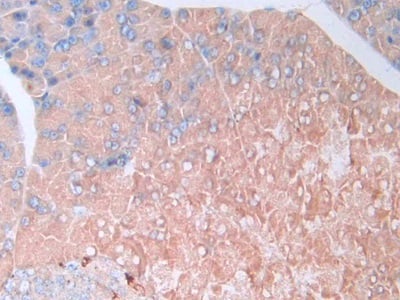
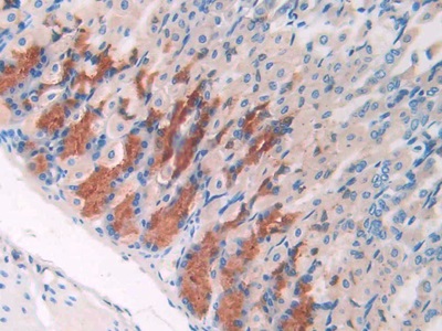
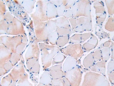
(DAB staining on IHCP;Sample: Mouse Skeletal muscle Tissue; Primary Ab: 40ug/ml Mouse AntiMouse RETN AntibodySecond Ab: 2ug/mL HRPLinked Caprine AntiMouse IgG Polyclonal Antibody(Catalog: SAA544Mu19))
IHC (Immunohistochemisry)
(DAB staining on IHCP;Sample: Mouse Skeletal muscle Tissue; Primary Ab: 40ug/ml Mouse AntiMouse RETN AntibodySecond Ab: 2ug/mL HRPLinked Caprine AntiMouse IgG Polyclonal Antibody(Catalog: SAA544Mu19))
Resistin (RETN), Monoclonal Antibody (Cat# AAA151645)
Full Name
Monoclonal Antibody to Resistin (RETN)
Gene Names
Retn; ADSF; Rstn; Xcp4; Fizz3
Reactivity
Mouse
Applications
Immunoprecipitation, Immunocytochemistry, Immunohistochemistry, Western Blot
Purity
Protein A + Protein G affinity chromatography
Pricing
 FCM (Flow Cytometry)
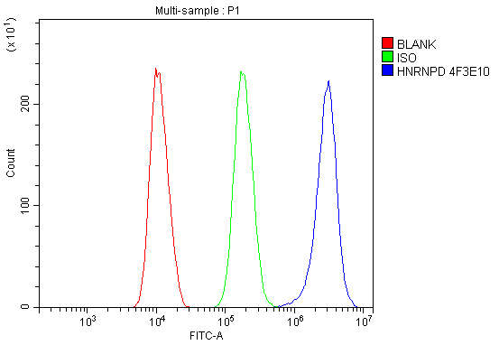
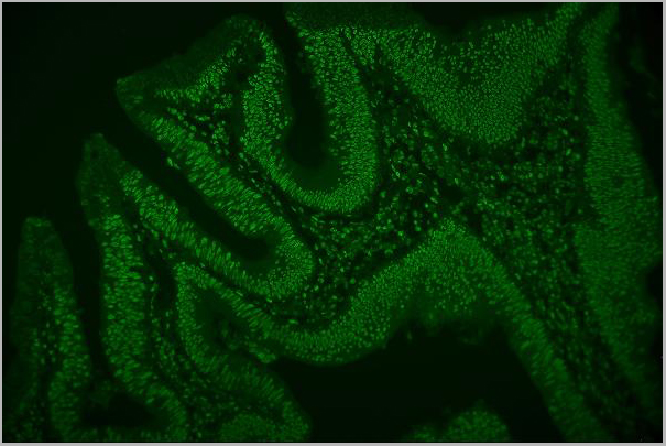
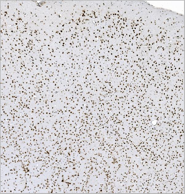
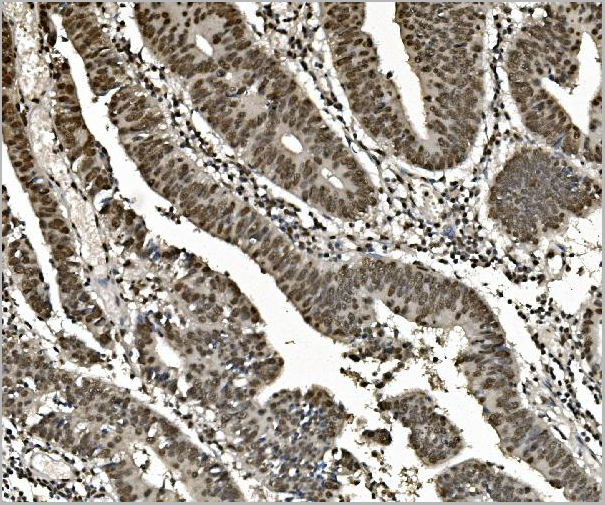
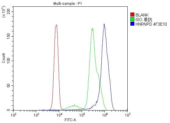
(Figure 6. Flow Cytometry analysis of U937 cells using anti- hnRNP D/AUF1/HNRNPD antibody (AAA19388).Overlay histogram showing U937 cells stained with AAA19388 (Blue line). The cells were blocked with 10% normal goat serum. And then incubated with mouse anti-hnRNP D/AUF1/HNRNPD Antibody (AAA19388, 1μg/1x106 cells) for 30 min at 20 degree C. DyLight®488 conjugated goat anti-mouse IgG (BA1126, 5-10μg/1x106 cells) was used as secondary antibody for 30 minutes at 20 degree C. Isotype control antibody (Green line) was mouse IgG (1μg/1x106) used under the same conditions. Unlabelled sample (Red line) was also used as a control.)
FCM (Flow Cytometry)
(Figure 6. Flow Cytometry analysis of U937 cells using anti- hnRNP D/AUF1/HNRNPD antibody (AAA19388).Overlay histogram showing U937 cells stained with AAA19388 (Blue line). The cells were blocked with 10% normal goat serum. And then incubated with mouse anti-hnRNP D/AUF1/HNRNPD Antibody (AAA19388, 1μg/1x106 cells) for 30 min at 20 degree C. DyLight®488 conjugated goat anti-mouse IgG (BA1126, 5-10μg/1x106 cells) was used as secondary antibody for 30 minutes at 20 degree C. Isotype control antibody (Green line) was mouse IgG (1μg/1x106) used under the same conditions. Unlabelled sample (Red line) was also used as a control.)
hnRNP D/AUF1/HNRNPD, Monoclonal Antibody (Cat# AAA19388)
Full Name
Anti-hnRNP D/AUF1/HNRNPD Antibody (monoclonal, 4F3)
Gene Names
HNRNPD; P37; AUF1; AUF1A; HNRPD; hnRNPD0
Reactivity
Human, Mouse, Rat
Applications
Western Blot, Immunohistochemistry, Immunocytochemistry, Immunofluorescence, Flow Cytometry
Purity
Immunogen affinity purified.
Pricing
 IF (Immunofluorescence)
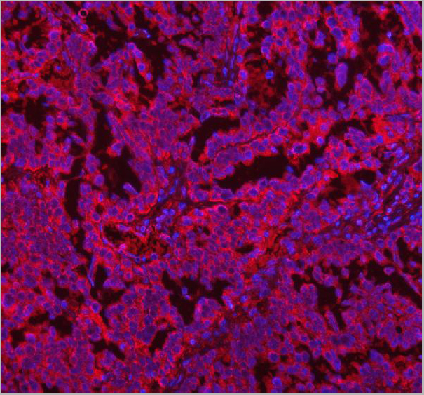
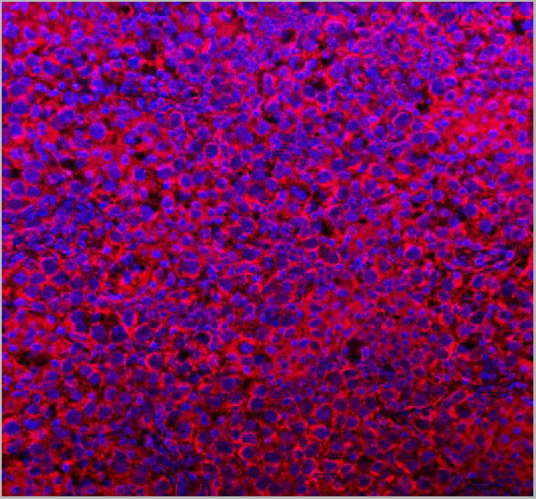
(Figure 14. IF analysis of COQ8B using anti-COQ8B antibody (AAA20014).COQ8B was detected in a paraffin-embedded section of human testis cancer tissue. Heat mediated antigen retrieval was performed in EDTA buffer (pH 8.0, epitope retrieval solution). The tissue section was blocked with 10% goat serum. The tissue section was then incubated with 5ug/mL rabbit anti-COQ8B Antibody (AAA20014) overnight at 4 degree C. DyLight®594 Conjugated Goat Anti-Rabbit IgG (BA1141) was used as secondary antibody at 1:500 dilution and incubated for 30 minutes at 37 degree C. The section was counterstained with DAPI. Visualize using a fluorescence microscope and filter sets appropriate for the label used.)
IF (Immunofluorescence)
(Figure 14. IF analysis of COQ8B using anti-COQ8B antibody (AAA20014).COQ8B was detected in a paraffin-embedded section of human testis cancer tissue. Heat mediated antigen retrieval was performed in EDTA buffer (pH 8.0, epitope retrieval solution). The tissue section was blocked with 10% goat serum. The tissue section was then incubated with 5ug/mL rabbit anti-COQ8B Antibody (AAA20014) overnight at 4 degree C. DyLight®594 Conjugated Goat Anti-Rabbit IgG (BA1141) was used as secondary antibody at 1:500 dilution and incubated for 30 minutes at 37 degree C. The section was counterstained with DAPI. Visualize using a fluorescence microscope and filter sets appropriate for the label used.)
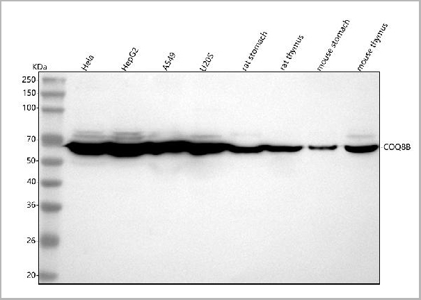
COQ8B, Polyclonal Antibody (Cat# AAA20014)
Full Name
Anti-COQ8B Antibody Picoband
Reactivity
Human, Mouse, Rat
Applications
Western Blot, Immunohistochemistry, Immunocytochemistry, Immunofluorescence
Purity
Immunogen affinity purified.
Pricing
 FCM (Flow Cytometry)
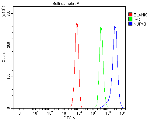
(Figure 23. Flow Cytometry analysis of K562 cells using anti-NUP43 antibody (AAA19968).Overlay histogram showing K562 cells stained with AAA19968 (Blue line). To facilitate intracellular staining, cells were fixed with 4% paraformaldehyde and permeabilized with permeabilization buffer. The cells were blocked with 10% normal goat serum. And then incubated with rabbit anti-NUP43 Antibody (AAA19968, 1ug/1x106 cells) for 30 min at 20 degree C. DyLight®488 conjugated goat anti-rabbit IgG (BA1127, 5-10ug/1x106 cells) was used as secondary antibody for 30 minutes at 20 degree C. Isotype control antibody (Green line) was rabbit IgG (1ug/1x106) used under the same conditions. Unlabelled sample (Red line) was also used as a control.)
FCM (Flow Cytometry)
(Figure 23. Flow Cytometry analysis of K562 cells using anti-NUP43 antibody (AAA19968).Overlay histogram showing K562 cells stained with AAA19968 (Blue line). To facilitate intracellular staining, cells were fixed with 4% paraformaldehyde and permeabilized with permeabilization buffer. The cells were blocked with 10% normal goat serum. And then incubated with rabbit anti-NUP43 Antibody (AAA19968, 1ug/1x106 cells) for 30 min at 20 degree C. DyLight®488 conjugated goat anti-rabbit IgG (BA1127, 5-10ug/1x106 cells) was used as secondary antibody for 30 minutes at 20 degree C. Isotype control antibody (Green line) was rabbit IgG (1ug/1x106) used under the same conditions. Unlabelled sample (Red line) was also used as a control.)
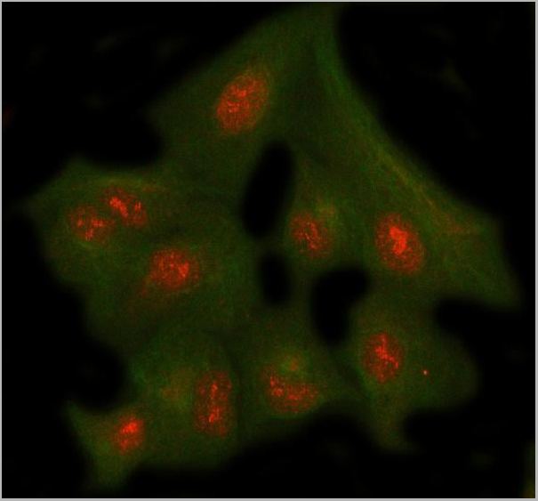
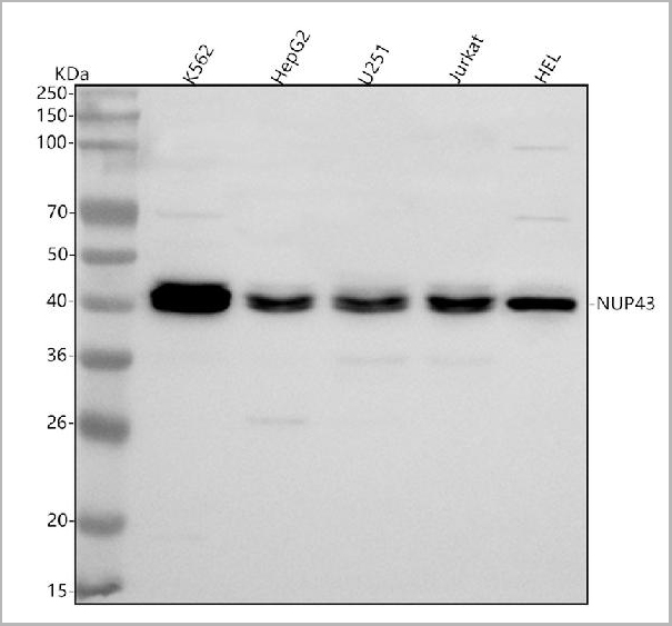
NUP43, Polyclonal Antibody (Cat# AAA19968)
Full Name
Anti-NUP43 Antibody Picoband
Gene Names
NUP43; p42; FLJ13287; bA350J20.1
Reactivity
Human, Mouse, Rat
Applications
Western Blot, Immunohistochemistry, Immunocytochemistry, Immunofluorescence, Flow Cytometry
Purity
Immunogen affinity purified.
Pricing

 Bioactivity
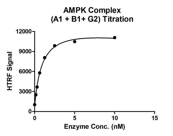
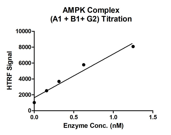
(HTRF assay for AMPK Complex (A1+B1+G2) activity 1 uM STK S1 substrate was incubated with different concentrations of AMPK Complex (A1+B1+G2) protein in a 10 ul reaction system containing 1×Enzymatic Buffer, 5 mM MgCl2, 1 mM DTT, 50 uM AMP and 100 uM ATP for 1 hr. The 10 ul detection reagents containing anti-STK antibody (1:100) and SA-XL665 (1:100) diluted with 1× Detection Buffer were added and incubated with the reactions for 30 min. All the operations and reactions were performed at room temperature. HTRF assay was used for detection.)
Bioactivity
(HTRF assay for AMPK Complex (A1+B1+G2) activity 1 uM STK S1 substrate was incubated with different concentrations of AMPK Complex (A1+B1+G2) protein in a 10 ul reaction system containing 1×Enzymatic Buffer, 5 mM MgCl2, 1 mM DTT, 50 uM AMP and 100 uM ATP for 1 hr. The 10 ul detection reagents containing anti-STK antibody (1:100) and SA-XL665 (1:100) diluted with 1× Detection Buffer were added and incubated with the reactions for 30 min. All the operations and reactions were performed at room temperature. HTRF assay was used for detection.)
AMPK Complex (A1+B1+G2), Active Protein (Cat# AAA60344)
Full Name
Recombinant AMPK Complex (A1+B1+G2) protein
Purity
>=80%
Pricing
 Application Data
TRIM21, Recombinant Protein (Cat# AAA24007)
Full Name
TRIM21 protein (His tag)
Gene Names
Trim21; Ro52; Ssa1
Purity
> 90% pure
Purified using propietary chromatographic techniques.
Purified using propietary chromatographic techniques.
Pricing
 IP (Immunoprecipitation)
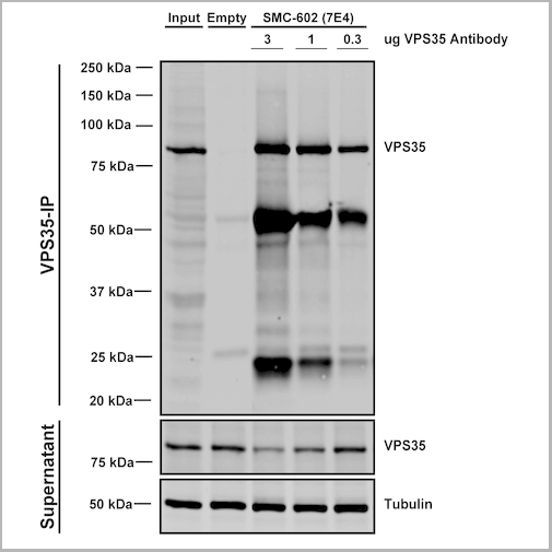
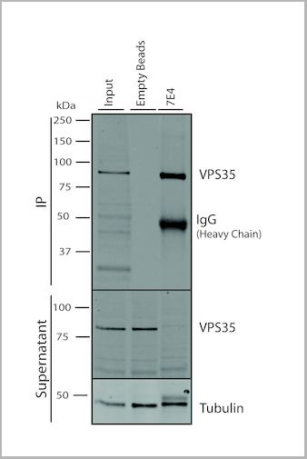
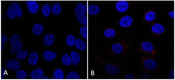
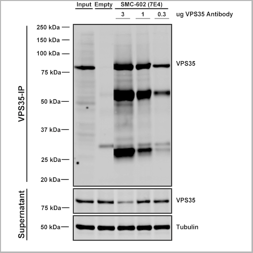
(Immunoprecipitation analysis using Mouse Anti-VPS35 Monoclonal Antibody, Clone 7E4. Tissue: embryonic fibroblast. Species: Mouse. Primary Antibody: Mouse Anti-VPS35 Monoclonal Antibody. Three amounts (3, 1 and 0.3 ug) were non-covalently coupled to 10uL of A/G sepharose beads for 1 hour at 4 degree C and next incubated with 250ug of MEF lysate for 2 hours at 4 degree C.)
IP (Immunoprecipitation)
(Immunoprecipitation analysis using Mouse Anti-VPS35 Monoclonal Antibody, Clone 7E4. Tissue: embryonic fibroblast. Species: Mouse. Primary Antibody: Mouse Anti-VPS35 Monoclonal Antibody. Three amounts (3, 1 and 0.3 ug) were non-covalently coupled to 10uL of A/G sepharose beads for 1 hour at 4 degree C and next incubated with 250ug of MEF lysate for 2 hours at 4 degree C.)
VPS35, Monoclonal Antibody (Cat# AAA27672)
Full Name
VPS35 Antibody, Clone 7E4: RPE
Gene Names
VPS35; MEM3; PARK17
Reactivity
Human, Mouse, Rat
Applications
Western Blot, Immunocytochemistry, Immunofluorescence, Immunoprecipitation
Purity
Protein G Purified
Pricing
 Bioactivity
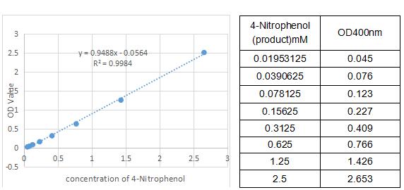
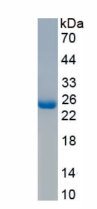
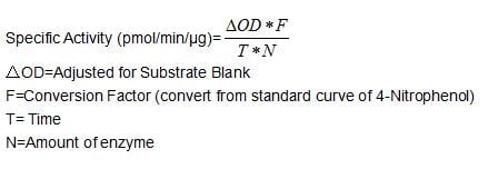
(One unit of enzyme activity is defined as the 1 ug of enzyme required to convert 1 pmol of 4-Nitrophenyl acetate to 4-Nitrophenol in 1min at 37 degree C. The specific activity of recombinant mouse CA4 is > 50 pmol/min/ug.)
Bioactivity
(One unit of enzyme activity is defined as the 1 ug of enzyme required to convert 1 pmol of 4-Nitrophenyl acetate to 4-Nitrophenol in 1min at 37 degree C. The specific activity of recombinant mouse CA4 is > 50 pmol/min/ug.)
Carbonic Anhydrase IV (CA4), Active Protein (Cat# AAA161901)
Full Name
Active Carbonic Anhydrase IV (CA4)
Gene Names
Car4; Ca4; AW456718
Applications
Activity Assay, Cell Culture
Purity
Greater than 90% by SDS-PAGE
Pricing
 Bioactivity
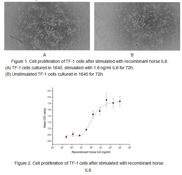
(Interleukin 6 (IL-6) is an interleukin that acts as both a pro-inflammatory cytokine and an anti-inflammatory myokine. Interleukin 6 is secreted by T cells and macrophages to stimulate immune response and also plays a role in fighting infection. It supports the growth of B cells and is antagonistic to regulatory T cells. To test the effect of IL6 on cell proliferation, TF-1 cells were seeded into triplicate wells of 96-well plates at a density of 20,000 cells/well with 10% serum standard 1640 which contains various concentrations of recombinant horse IL6. After incubated for 72h, cells were observed by inverted microscope and cell proliferation was measured by Cell Counting Kit-8 (CCK-8). Briefly, 10 ul of CCK-8 solution was added to each well of the plate, then the absorbance at 450 nm was measured using a microplate reader after incubating the plate for 1-4 hours at 37 degree C. Proliferation of TF-1 cells after incubation with IL6 for 72h observed by inverted microscope was shown in Figure 1. Cell viability was assessed by CCK-8 (Cell Counting Kit-8) assay after incubation with recombinant horse IL6 for 72h. The result was shown in Figure 2. It was obvious that IL6 significantly increased cell viability of TF-1 cells, the EC50 was 1.3 ng/ml.)
Bioactivity
(Interleukin 6 (IL-6) is an interleukin that acts as both a pro-inflammatory cytokine and an anti-inflammatory myokine. Interleukin 6 is secreted by T cells and macrophages to stimulate immune response and also plays a role in fighting infection. It supports the growth of B cells and is antagonistic to regulatory T cells. To test the effect of IL6 on cell proliferation, TF-1 cells were seeded into triplicate wells of 96-well plates at a density of 20,000 cells/well with 10% serum standard 1640 which contains various concentrations of recombinant horse IL6. After incubated for 72h, cells were observed by inverted microscope and cell proliferation was measured by Cell Counting Kit-8 (CCK-8). Briefly, 10 ul of CCK-8 solution was added to each well of the plate, then the absorbance at 450 nm was measured using a microplate reader after incubating the plate for 1-4 hours at 37 degree C. Proliferation of TF-1 cells after incubation with IL6 for 72h observed by inverted microscope was shown in Figure 1. Cell viability was assessed by CCK-8 (Cell Counting Kit-8) assay after incubation with recombinant horse IL6 for 72h. The result was shown in Figure 2. It was obvious that IL6 significantly increased cell viability of TF-1 cells, the EC50 was 1.3 ng/ml.)
Interleukin 6 (IL6), Active Protein (Cat# AAA161679)
Full Name
Active Interleukin 6 (IL6)
Gene Names
IL6; IL-6
Applications
Activity Assay, Cell Culture
Purity
Greater than 90% by SDS-PAGE
Pricing
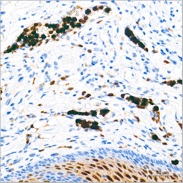
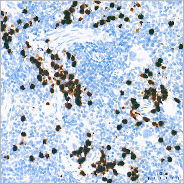
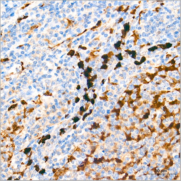
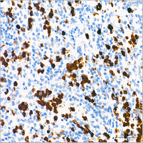
 ICC (Immunocytochemistry)
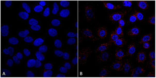
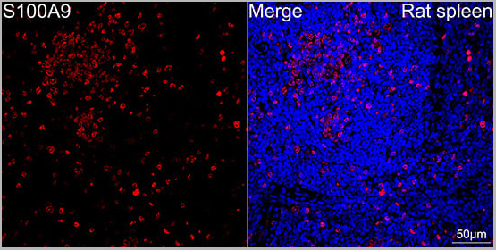
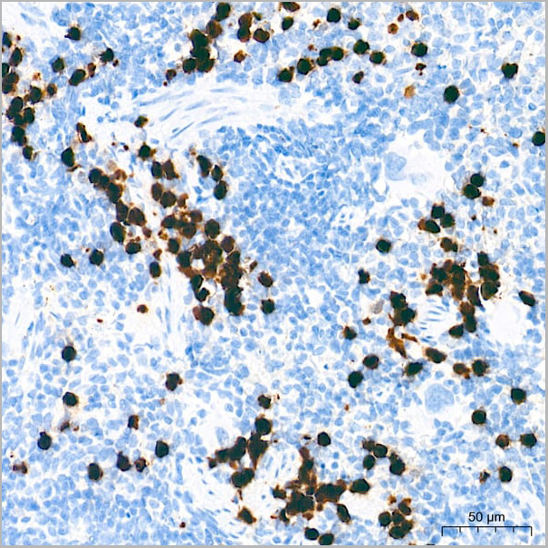
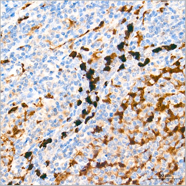
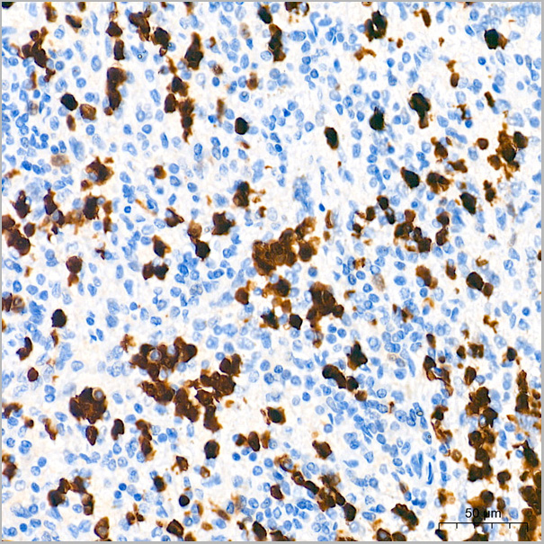
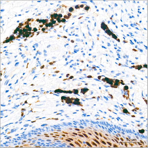
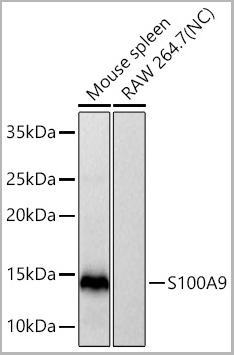
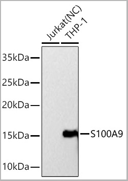
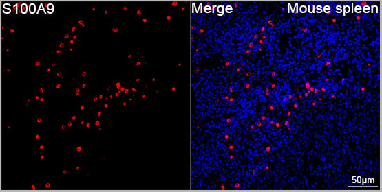
(Confocal imaging of paraffin-embedded Mouse spleen tissue using S100A9 Rabbit PolymAb® (AAA28595, dilution 1:200) followed by a further incubation with Cy3 Goat Anti-Rabbit IgG (H+L) (AS007, dilution 1:500) (Red). DAPI was used for nuclear staining (Blue). High pressure antigen retrieval performed with 0.01M Citrate Buffer (pH 6.0) prior to IF staining. Objective: 40x.)
ICC (Immunocytochemistry)
(Confocal imaging of paraffin-embedded Mouse spleen tissue using S100A9 Rabbit PolymAb® (AAA28595, dilution 1:200) followed by a further incubation with Cy3 Goat Anti-Rabbit IgG (H+L) (AS007, dilution 1:500) (Red). DAPI was used for nuclear staining (Blue). High pressure antigen retrieval performed with 0.01M Citrate Buffer (pH 6.0) prior to IF staining. Objective: 40x.)
S100A9, Monoclonal Antibody (Cat# AAA28595)
Full Name
S100A9 Rabbit PolymAb
Reactivity
Human, Mouse
Applications
Western Blot, Immunohistochemistry, Immunofluorescence, Immunocytochemistry
Purity
Affinity purification
Pricing
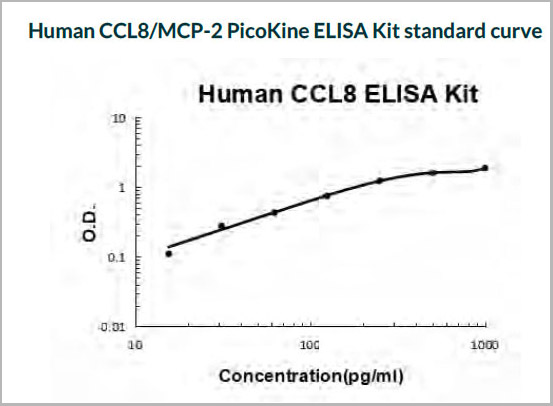 Standard Curve (Sample)
CCL8/MCP-2, ELISA Kit (Cat# AAA11593)
Full Name
Human CCL8/MCP-2 PicoKine ELISA Kit
Gene Names
CCL8; HC14; MCP2; MCP-2; SCYA8; SCYA10
Reactivity
Human
Pricing
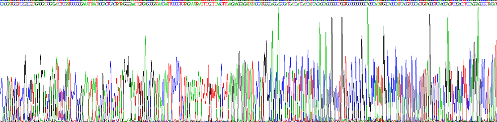
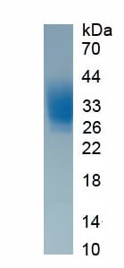 SDS-PAGE
(Figure. SDS-PAGE)
SDS-PAGE
(Figure. SDS-PAGE)
Interleukin 22 (IL22), Protein (Cat# AAA161951)
Full Name
Eukaryotic Interleukin 22 (IL22)
Gene Names
IL22; IL-22
Applications
Western Blot
Purity
Greater than 95% by SDS-PAGE
Pricing
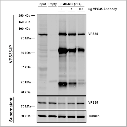
 IP (Immunoprecipitation)
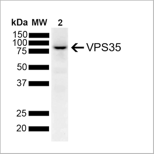
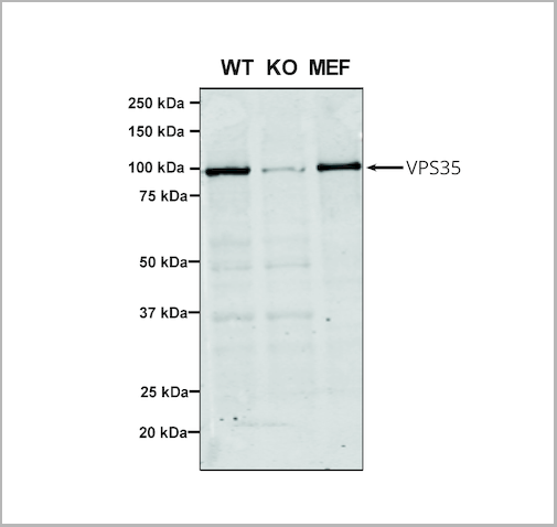
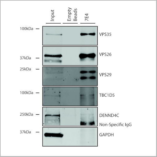
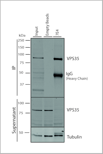
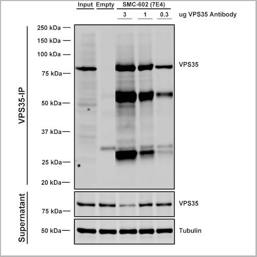
(Immunoprecipitation analysis using Mouse Anti-VPS35 Monoclonal Antibody, Clone 7E4. Tissue: embryonic fibroblast. Species: Mouse. Primary Antibody: Mouse Anti-VPS35 Monoclonal Antibody. Three amounts (3, 1 and 0.3 ug) were non-covalently coupled to 10uL of A/G sepharose beads for 1 hour at 4 degree C and next incubated with 250ug of MEF lysate for 2 hours at 4 degree C.)
IP (Immunoprecipitation)
(Immunoprecipitation analysis using Mouse Anti-VPS35 Monoclonal Antibody, Clone 7E4. Tissue: embryonic fibroblast. Species: Mouse. Primary Antibody: Mouse Anti-VPS35 Monoclonal Antibody. Three amounts (3, 1 and 0.3 ug) were non-covalently coupled to 10uL of A/G sepharose beads for 1 hour at 4 degree C and next incubated with 250ug of MEF lysate for 2 hours at 4 degree C.)
VPS35, Monoclonal Antibody (Cat# AAA27669)
Full Name
VPS35 Antibody, Clone 7E4: FITC
Gene Names
VPS35; MEM3; PARK17
Reactivity
Human, Mouse, Rat
Applications
Western Blot, Immunocytochemistry, Immunofluorescence, Immunoprecipitation
Purity
Protein G Purified
Pricing
 IP (Immunoprecipitation)
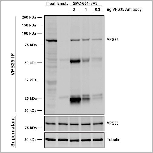
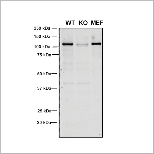
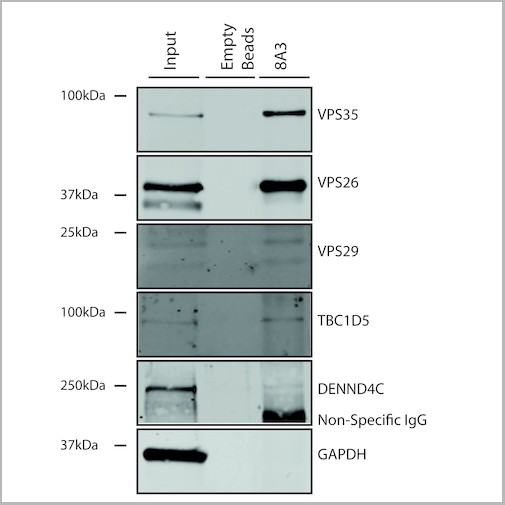
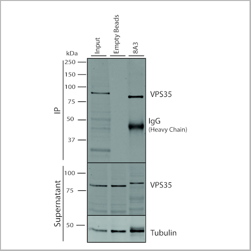
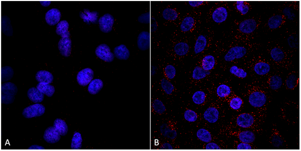
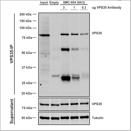
(Immunoprecipitation analysis using Mouse Anti-VPS35 Monoclonal Antibody, Clone 8A3. Tissue: embryonic fibroblast. Species: Mouse. Primary Antibody: Mouse Anti-VPS35 Monoclonal Antibody. Three amounts of (3, 1 and 0.3 ug) were non-covalently coupled to 10uL of A/G sepharose beads for 1 hour at 4 degree C and next incubated with 250ug of MEF lysate for 2 hours at 4 degree C.)
IP (Immunoprecipitation)
(Immunoprecipitation analysis using Mouse Anti-VPS35 Monoclonal Antibody, Clone 8A3. Tissue: embryonic fibroblast. Species: Mouse. Primary Antibody: Mouse Anti-VPS35 Monoclonal Antibody. Three amounts of (3, 1 and 0.3 ug) were non-covalently coupled to 10uL of A/G sepharose beads for 1 hour at 4 degree C and next incubated with 250ug of MEF lysate for 2 hours at 4 degree C.)
VPS35, Monoclonal Antibody (Cat# AAA27684)
Full Name
VPS35 Antibody, Clone 8A3: ATTO 488
Gene Names
VPS35; MEM3; PARK17
Reactivity
Human, Mouse, Rat
Applications
Western Blot, Immunocytochemistry, Immunofluorescence, Immunoprecipitation
Purity
Protein G Purified
Pricing

 WB (Western Blot)
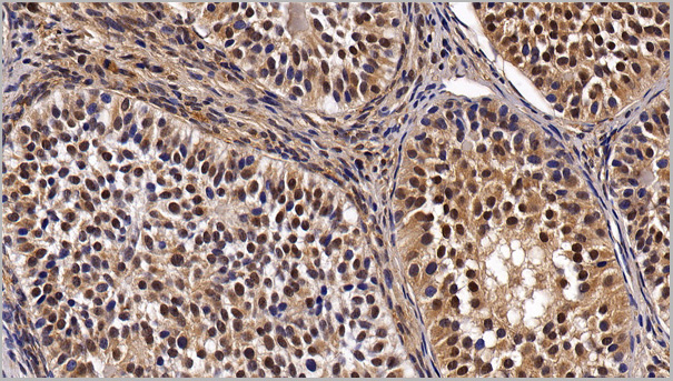
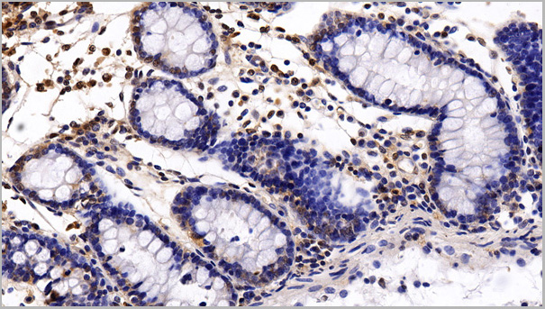
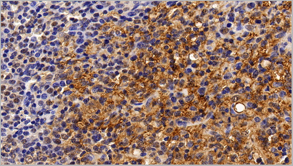
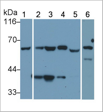
(Western Blot; Sample: Lane1: Human Lung lysate; Lane2: A549 cell lysate; Lane3: HepG2 cell lysate; Lane4: Hela cell lysate; Lane5: Bovine Testis lysate; Lane6: Rat Testis lysatePrimary Ab: 0.2ug/ml Mouse AntiHuman HDAC1 AntibodySecond Ab: 0.2ug/mL HRPLinked Caprine AntiMouse IgG Polyclonal Antibody(Catalog: SAA544Mu19))
WB (Western Blot)
(Western Blot; Sample: Lane1: Human Lung lysate; Lane2: A549 cell lysate; Lane3: HepG2 cell lysate; Lane4: Hela cell lysate; Lane5: Bovine Testis lysate; Lane6: Rat Testis lysatePrimary Ab: 0.2ug/ml Mouse AntiHuman HDAC1 AntibodySecond Ab: 0.2ug/mL HRPLinked Caprine AntiMouse IgG Polyclonal Antibody(Catalog: SAA544Mu19))
Histone Deacetylase 1 (HDAC1), Monoclonal Antibody (Cat# AAA21204)
Full Name
Monoclonal Antibody to Histone Deacetylase 1 (HDAC1)
Gene Names
HDAC1; HD1; RPD3; GON-10; RPD3L1
Reactivity
Human, Rat, Pig, Bovine
Applications
Western Blot, Immunohistochemistry, Immunocytochemistry, Immunoprecipitation
Purity
Protein A + Protein G affinity chromatography
Pricing
 WB (Western Blot)
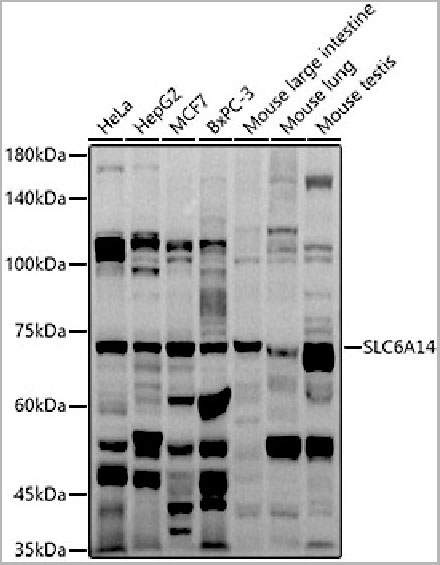
(Western blot analysis of extracts of various cell lines, using SLC6A14 antibody (AAA28230) at 1:1000 dilution.Secondary antibody: HRP Goat Anti-Rabbit IgG (H+L) at 1:10000 dilution. Lysates/proteins: 25ug per lane. Blocking buffer: 3% nonfat dry milk in TBST. Detection: ECL Basic Kit. Exposure time: 90s)
WB (Western Blot)
(Western blot analysis of extracts of various cell lines, using SLC6A14 antibody (AAA28230) at 1:1000 dilution.Secondary antibody: HRP Goat Anti-Rabbit IgG (H+L) at 1:10000 dilution. Lysates/proteins: 25ug per lane. Blocking buffer: 3% nonfat dry milk in TBST. Detection: ECL Basic Kit. Exposure time: 90s)
SLC6A14, Polyclonal Antibody (Cat# AAA28230)
Full Name
SLC6A14 Polyclonal Antibody
Gene Names
SLC6A14; BMIQ11
Reactivity
Human, Mouse
Applications
Western Blot
Purity
Affinity Purification
Pricing
 Application Data
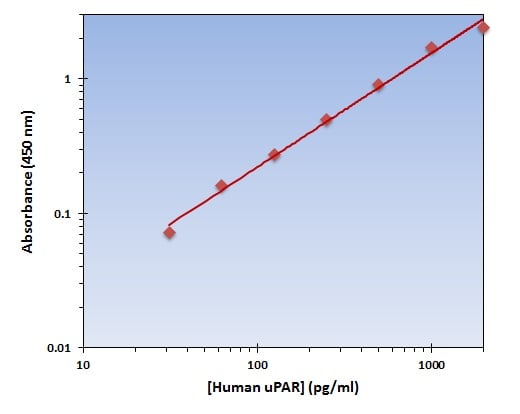
(The OmniKine Human uPAR ELISA Kit allows for the detection and quantification of endogenous levels of natural and/or recombinant Human uPAR proteins within the range of 32-2000 pg/ml.)
Application Data
(The OmniKine Human uPAR ELISA Kit allows for the detection and quantification of endogenous levels of natural and/or recombinant Human uPAR proteins within the range of 32-2000 pg/ml.)
uPAR, ELISA Kit (Cat# AAA315842)
Full Name
uPAR (Human) OmniKine ELISA Kit
Gene Names
PLAUR; CD87; UPAR; URKR; U-PAR
Reactivity
Human
Pricing
 Amino Acid Info
Triggering Receptor Expressed On Myeloid Cells 2 (TREM2), Recombinant Protein (Cat# AAA153655)
Full Name
Recombinant Triggering Receptor Expressed On Myeloid Cells 2 (TREM2)
Applications
Western Blot
Purity
Greater than 80% by SDS-PAGE
Pricing
 IF (Immunofluorescence)
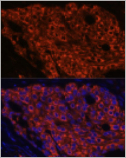
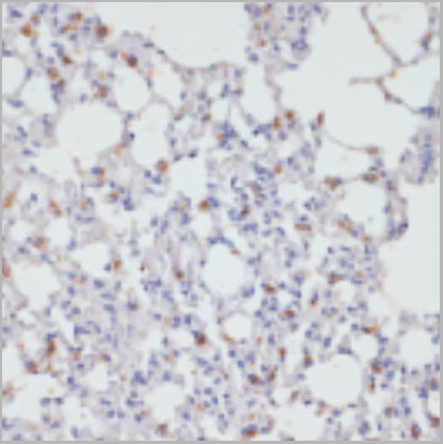
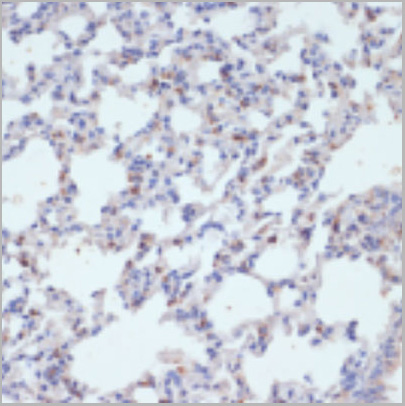
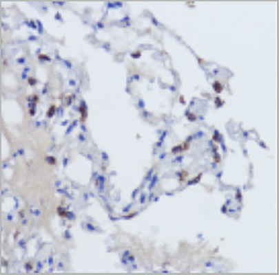
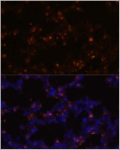
(Immunofluorescence analysis of mouse lung using SFTPC Polyclonal Antibody at dilution of 1:100 (40x lens). Blue: DAPI for nuclear staining.)
IF (Immunofluorescence)
(Immunofluorescence analysis of mouse lung using SFTPC Polyclonal Antibody at dilution of 1:100 (40x lens). Blue: DAPI for nuclear staining.)
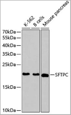
SFTPC, Polyclonal Antibody (Cat# AAA29679)
Full Name
SFTPC Antibody
Gene Names
SFTPC; SP-C; PSP-C; SFTP2; SMDP2; BRICD6
Reactivity
Human, Mouse, Rat
Applications
Western Blot, Immunohistochemistry, Immunofluorescence
Purity
Antibodies were purified by affinity purification using immunogen.
Pricing
 IP (Immunoprecipitation)
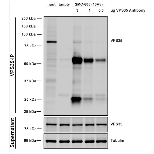
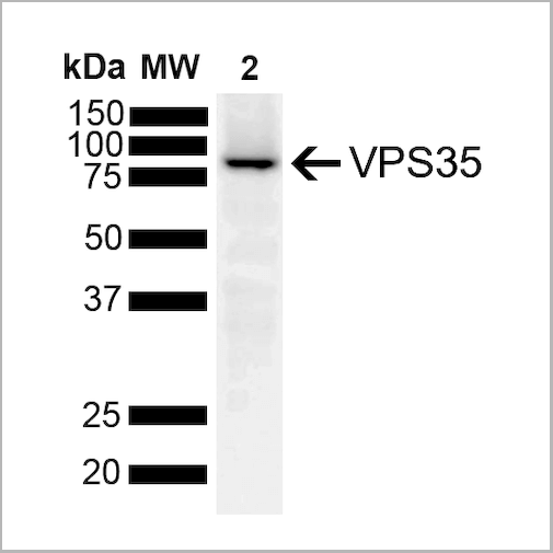
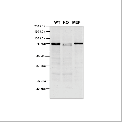
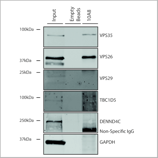
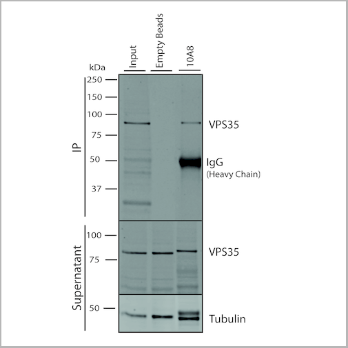
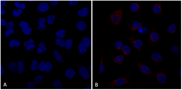
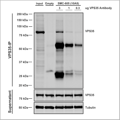
(Immunoprecipitation analysis using Mouse Anti-VPS35 Monoclonal Antibody, Clone 10A8. Tissue: embryonic fibroblast. Species: Mouse. Primary Antibody: Mouse Anti-VPS35 Monoclonal Antibody. Three amounts of (3, 1 and 0.3 ug) were non-covalently coupled to 10uL of A/G sepharose beads for 1 hour at 4 degree C and next incubated with 250ug of MEF lysate for 2 hours at 4 degree C.)
IP (Immunoprecipitation)
(Immunoprecipitation analysis using Mouse Anti-VPS35 Monoclonal Antibody, Clone 10A8. Tissue: embryonic fibroblast. Species: Mouse. Primary Antibody: Mouse Anti-VPS35 Monoclonal Antibody. Three amounts of (3, 1 and 0.3 ug) were non-covalently coupled to 10uL of A/G sepharose beads for 1 hour at 4 degree C and next incubated with 250ug of MEF lysate for 2 hours at 4 degree C.)
VPS35, Monoclonal Antibody (Cat# AAA27697)
Full Name
VPS35 Antibody, Clone 10A8: HRP
Gene Names
VPS35; MEM3; PARK17
Reactivity
Human, Mouse, Rat
Applications
Western Blot, Immunocytochemistry, Immunofluorescence, Immunoprecipitation
Purity
Protein G Purified
Pricing
 IP (Immunoprecipitation)
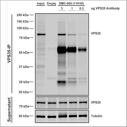
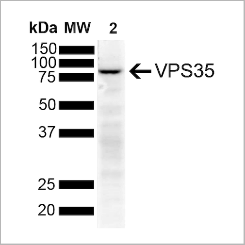
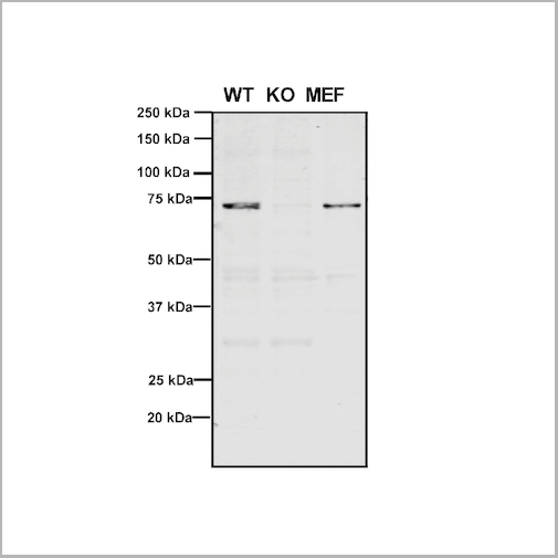
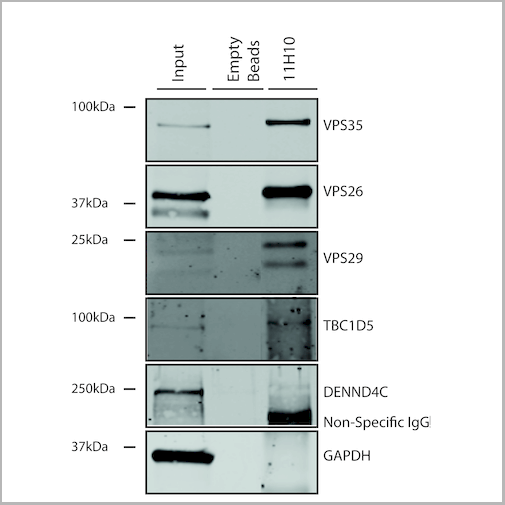
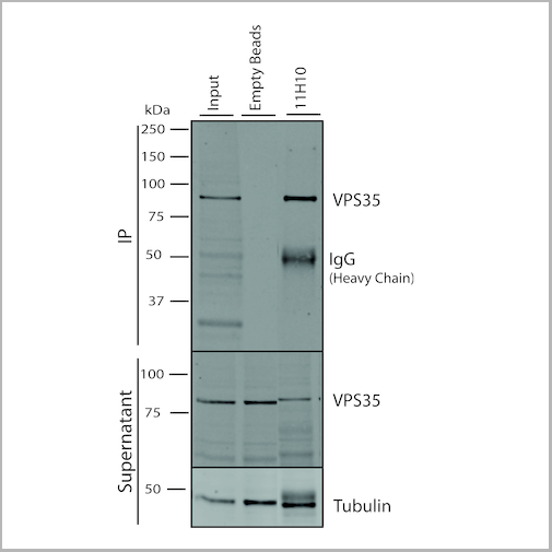
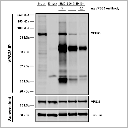
(Immunoprecipitation analysis using Mouse Anti-VPS35 Monoclonal Antibody, Clone 11H10. Tissue: embryonic fibroblast. Species: Mouse. Primary Antibody: Mouse Anti-VPS35 Monoclonal Antibody. Three amounts of (3, 1 and 0.3 ug) were non-covalently coupled to 10uL of A/G sepharose beads for 1 hour at 4 degree C and next incubated with 250ug of MEF lysate for 2 hours at 4 degree C.)
IP (Immunoprecipitation)
(Immunoprecipitation analysis using Mouse Anti-VPS35 Monoclonal Antibody, Clone 11H10. Tissue: embryonic fibroblast. Species: Mouse. Primary Antibody: Mouse Anti-VPS35 Monoclonal Antibody. Three amounts of (3, 1 and 0.3 ug) were non-covalently coupled to 10uL of A/G sepharose beads for 1 hour at 4 degree C and next incubated with 250ug of MEF lysate for 2 hours at 4 degree C.)
VPS35, Monoclonal Antibody (Cat# AAA27706)
Full Name
VPS35 Antibody, Clone 11H10: HRP
Gene Names
VPS35; MEM3; PARK17
Reactivity
Human, Mouse, Rat
Applications
Western Blot, Immunoprecipitation
Purity
Protein G Purified
Pricing

 Bioactivity
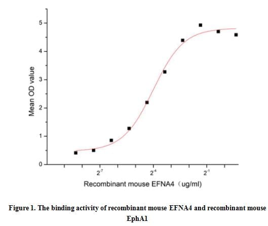
(Ephrin-A4, also known as EFNA4 and EFL-4, is a member of the ligand of the EPH family. It is mainly expressed in the spleen, lymph nodes, ovary, small intestine, and colon of adults, as well as in the heart, lungs, liver, and kidneys of the fetus. It is involved in the development of neurons, blood vessels, and epithelium by regulating cell migration, rejection, and adhesion. Ephrin-A4 has been shown to bind EphA1,EphA2, EphA3, EphA4, EphA5, EphA6, EphA7 and EphB1. Thus a functional binding ELISA assay was conducted to detect the interaction of recombinant mouse EFNA4 and recombinant mouse EPHA1. Briefly, EFNA4 was diluted serially in PBS with 0.01% BSA (pH 7.4). Duplicate samples of 100 ul were then transferred to EphA1-coated microtiter wells and incubated for 1h at 37 degree C. Wells were washed with PBST and incubated for 1h with anti-EFNA4 pAb, then aspirated and washed 3 times. After incubation with HRP labelled secondary antibody for 1h at 37 degree C, wells were aspirated and washed 5 times. With the addition of substrate solution, wells were incubated 15-25 minutes at 37 degree C. Finally, add 50 uL stop solution to the wells and read at 450/630 nm immediately. The binding activity of recombinant mouse EFNA4 and recombinant mouse EphA1 was shown in Figure 1, the EC50 for this effect is 0.06 ug/mL.)
Bioactivity
(Ephrin-A4, also known as EFNA4 and EFL-4, is a member of the ligand of the EPH family. It is mainly expressed in the spleen, lymph nodes, ovary, small intestine, and colon of adults, as well as in the heart, lungs, liver, and kidneys of the fetus. It is involved in the development of neurons, blood vessels, and epithelium by regulating cell migration, rejection, and adhesion. Ephrin-A4 has been shown to bind EphA1,EphA2, EphA3, EphA4, EphA5, EphA6, EphA7 and EphB1. Thus a functional binding ELISA assay was conducted to detect the interaction of recombinant mouse EFNA4 and recombinant mouse EPHA1. Briefly, EFNA4 was diluted serially in PBS with 0.01% BSA (pH 7.4). Duplicate samples of 100 ul were then transferred to EphA1-coated microtiter wells and incubated for 1h at 37 degree C. Wells were washed with PBST and incubated for 1h with anti-EFNA4 pAb, then aspirated and washed 3 times. After incubation with HRP labelled secondary antibody for 1h at 37 degree C, wells were aspirated and washed 5 times. With the addition of substrate solution, wells were incubated 15-25 minutes at 37 degree C. Finally, add 50 uL stop solution to the wells and read at 450/630 nm immediately. The binding activity of recombinant mouse EFNA4 and recombinant mouse EphA1 was shown in Figure 1, the EC50 for this effect is 0.06 ug/mL.)
Ephrin A4 (EFNA4), Active Protein (Cat# AAA161917)
Full Name
Active Ephrin A4 (EFNA4)
Gene Names
Efna4; Epl4; EFL-4; LERK-4
Applications
Activity Assay, Cell Culture
Purity
Greater than 90% by SDS-PAGE
Pricing
 Bioactivity
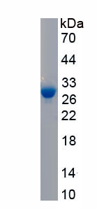
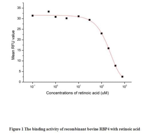
(Retinol-binding protein 4 (RBP4) is the specific carrier for retinol (also known as vitamin A), and is responsible for the conversion of unstable and insoluble retinol in aqueous solution into stable and soluble complex in plasma through their tight interaction. As a member of the lipocalin superfamily, RBP4 containing a beta-barrel structure with a well-defined cavity is secreted from the liver, and in turn delivers retinol from the liver stores to the peripheral tissues. In plasma, the RBP4-retinol complex interacts with transthyretin (TTR), and this binding is crucial for preventing RBP4 excretion through the kidney glomeruli. RBP4 expressed from an ectopic source efficiently delivers retinol to the eyes, and its deficiency affects night vision largely. Recently, RBP4 as an adipokine, is found to be expressed in adipose tissue and correlated with obesity, insulin resistance (IR) and type 2 diabetes (T2DM). The activity of recombinant bovine RBP4 was measured by its ability to bind all-trans retinoic acid. The binding of retinoic acid results in the quenching of Trp fluorescence in RBP4. RBP4 was diluted to 50 ug/ml in 50 mM Tris, 10 mM CaCl2, 150 mM NaCl, pH 7.5 (assay buffer) and the retinoic acid was diluted to 800, 400, 200, 100, 30, 10, 3, 1, 0.5 and 0.1 uM in 95% ethanol. Mixing 112.5 uL of 50 ug/mL rbRBP4 and 12.5 uL of retinoic acid serial dilutions in microtubes and a blank containing 112.5 uL of 50 ug/mL rbRBP4 and 12.5 uL of 95% ethanol, then incubate at room temperature for 30 minutes. Loading 100 ul of the reaction mixtures and blank and read at excitation and emission wavelengths of 280 nm and 340 nm (top read), respectively, in endpoint mode. The result was shown in figure 1, the 50% binding concentration (BC50) is >190 uM.)
Bioactivity
(Retinol-binding protein 4 (RBP4) is the specific carrier for retinol (also known as vitamin A), and is responsible for the conversion of unstable and insoluble retinol in aqueous solution into stable and soluble complex in plasma through their tight interaction. As a member of the lipocalin superfamily, RBP4 containing a beta-barrel structure with a well-defined cavity is secreted from the liver, and in turn delivers retinol from the liver stores to the peripheral tissues. In plasma, the RBP4-retinol complex interacts with transthyretin (TTR), and this binding is crucial for preventing RBP4 excretion through the kidney glomeruli. RBP4 expressed from an ectopic source efficiently delivers retinol to the eyes, and its deficiency affects night vision largely. Recently, RBP4 as an adipokine, is found to be expressed in adipose tissue and correlated with obesity, insulin resistance (IR) and type 2 diabetes (T2DM). The activity of recombinant bovine RBP4 was measured by its ability to bind all-trans retinoic acid. The binding of retinoic acid results in the quenching of Trp fluorescence in RBP4. RBP4 was diluted to 50 ug/ml in 50 mM Tris, 10 mM CaCl2, 150 mM NaCl, pH 7.5 (assay buffer) and the retinoic acid was diluted to 800, 400, 200, 100, 30, 10, 3, 1, 0.5 and 0.1 uM in 95% ethanol. Mixing 112.5 uL of 50 ug/mL rbRBP4 and 12.5 uL of retinoic acid serial dilutions in microtubes and a blank containing 112.5 uL of 50 ug/mL rbRBP4 and 12.5 uL of 95% ethanol, then incubate at room temperature for 30 minutes. Loading 100 ul of the reaction mixtures and blank and read at excitation and emission wavelengths of 280 nm and 340 nm (top read), respectively, in endpoint mode. The result was shown in figure 1, the 50% binding concentration (BC50) is >190 uM.)
Retinol Binding Protein 4 (RBP4), Active Protein (Cat# AAA161803)
Full Name
Active Retinol Binding Protein 4 (RBP4)
Applications
Activity Assay, Cell Culture
Purity
Greater than 95% by SDS-PAGE
Pricing
 SDS-PAGE
DsbG, Recombinant Protein (Cat# AAA48495)
Full Name
DsbG, 18-248aa, E Coli, E Coli
Gene Names
dsbG; ECK0598; JW0597; ybdP
Applications
SDS-PAGE
Purity
> 95% by SDS-PAGE
Pricing
 Bioactivity
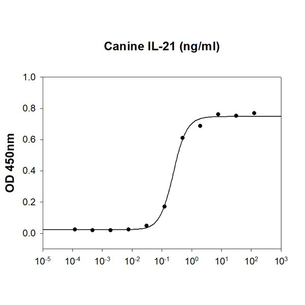
(Canine IL-21 stimulates IFN-g secretion of the NK-92 human natural killer cells. The ED50 range )
Bioactivity
(Canine IL-21 stimulates IFN-g secretion of the NK-92 human natural killer cells. The ED50 range )
IL-21, Active Protein (Cat# AAA48379)
Full Name
Recombinant canine IL-21 protein
Gene Names
IL21; IL-21
Applications
Bioactivity, SDS-PAGE
Purity
>95% by SDS-PAGE
Pricing

 SDS-PAGE
(Figure. SDS-PAGE)
SDS-PAGE
(Figure. SDS-PAGE)
Myelin Basic Protein (MBP), Protein (Cat# AAA161946)
Full Name
Eukaryotic Myelin Basic Protein (MBP)
Gene Names
Mbp; jve; mld; shi; Hmbpr; C76307; R75289; golli-mbp
Applications
Western Blot
Purity
Greater than 90% by SDS-PAGE
Pricing
 IP (Immunoprecipitation)
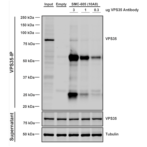
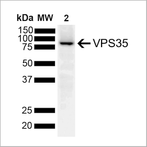
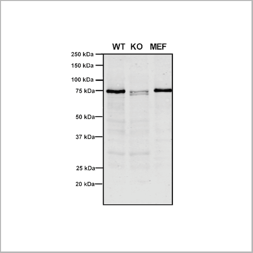
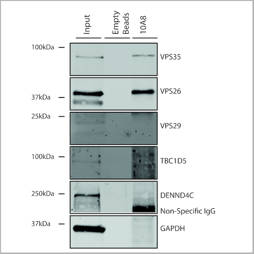
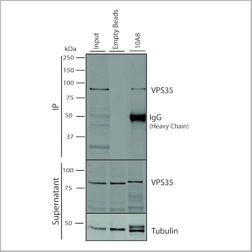
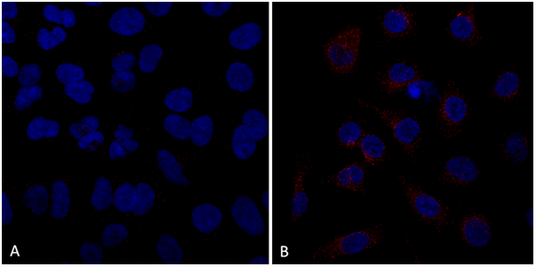
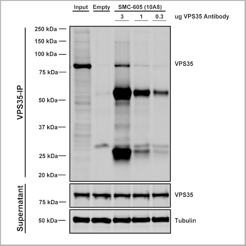
(Immunoprecipitation analysis using Mouse Anti-VPS35 Monoclonal Antibody, Clone 10A8. Tissue: embryonic fibroblast. Species: Mouse. Primary Antibody: Mouse Anti-VPS35 Monoclonal Antibody. Three amounts of (3, 1 and 0.3 ug) were non-covalently coupled to 10uL of A/G sepharose beads for 1 hour at 4 degree C and next incubated with 250ug of MEF lysate for 2 hours at 4 degree C.)
IP (Immunoprecipitation)
(Immunoprecipitation analysis using Mouse Anti-VPS35 Monoclonal Antibody, Clone 10A8. Tissue: embryonic fibroblast. Species: Mouse. Primary Antibody: Mouse Anti-VPS35 Monoclonal Antibody. Three amounts of (3, 1 and 0.3 ug) were non-covalently coupled to 10uL of A/G sepharose beads for 1 hour at 4 degree C and next incubated with 250ug of MEF lysate for 2 hours at 4 degree C.)
VPS35, Monoclonal Antibody (Cat# AAA27699)
Full Name
VPS35 Antibody, Clone 10A8: RPE
Gene Names
VPS35; MEM3; PARK17
Reactivity
Human, Mouse, Rat
Applications
Western Blot, Immunocytochemistry, Immunofluorescence, Immunoprecipitation
Purity
Protein G Purified
Pricing
 Bioactivity
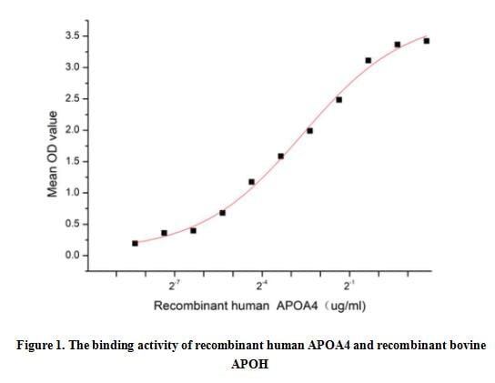
(Apolipoprotein A4 (APOA4) is a lipid-binding protein, which is primarily synthesized in the small intestine, packaged into chylomicrons, and secreted into intestinal lymph during fat absorption. In the circulation, apoA-IV is present on chylomicron remnants, high-density lipoproteins, and also in lipid-free form. ApoA-IV is involved in a myriad of physiological processes such as lipid absorption and metabolism, anti-atherosclerosis, platelet aggregation and thrombosis, glucose homeostasis and food intake. Besides, Apolipoprotein H (APOH) has been identified as an interactor of APOA4, thus a functional binding ELISA assay was conducted to detect the interaction of recombinant human APOA4 and recombinant bovine APOH. Briefly, APOA4 was diluted serially in PBS with 0.01% BSA (pH 7.4). Duplicate samples of 100 ul were then transferred to APOH-coated microtiter wells and incubated for 1h at 37 degree C. Wells were washed with PBST and incubated for 1h with anti-APOA4 pAb, then aspirated and washed 3 times. After incubation with HRP labelled secondary antibody for 1h at 37 degree C, wells were aspirated and washed 5 times. With the addition of substrate solution, wells were incubated 15-25 minutes at 37 degree C. Finally, add 50 uL stop solution to the wells and read at 450/630 nm immediately. The binding activity of recombinant human APOA4 and recombinant bovine APOH was shown in Figure 1, the EC50 for this effect is 0.16 ug/mL.)
Bioactivity
(Apolipoprotein A4 (APOA4) is a lipid-binding protein, which is primarily synthesized in the small intestine, packaged into chylomicrons, and secreted into intestinal lymph during fat absorption. In the circulation, apoA-IV is present on chylomicron remnants, high-density lipoproteins, and also in lipid-free form. ApoA-IV is involved in a myriad of physiological processes such as lipid absorption and metabolism, anti-atherosclerosis, platelet aggregation and thrombosis, glucose homeostasis and food intake. Besides, Apolipoprotein H (APOH) has been identified as an interactor of APOA4, thus a functional binding ELISA assay was conducted to detect the interaction of recombinant human APOA4 and recombinant bovine APOH. Briefly, APOA4 was diluted serially in PBS with 0.01% BSA (pH 7.4). Duplicate samples of 100 ul were then transferred to APOH-coated microtiter wells and incubated for 1h at 37 degree C. Wells were washed with PBST and incubated for 1h with anti-APOA4 pAb, then aspirated and washed 3 times. After incubation with HRP labelled secondary antibody for 1h at 37 degree C, wells were aspirated and washed 5 times. With the addition of substrate solution, wells were incubated 15-25 minutes at 37 degree C. Finally, add 50 uL stop solution to the wells and read at 450/630 nm immediately. The binding activity of recombinant human APOA4 and recombinant bovine APOH was shown in Figure 1, the EC50 for this effect is 0.16 ug/mL.)
Apolipoprotein A4 (APOA4), Active Protein (Cat# AAA161870)
Full Name
Active Apolipoprotein A4 (APOA4)
Applications
Activity Assay, Cell Culture
Purity
Greater than 80% by SDS-PAGE
Pricing

 ELISA
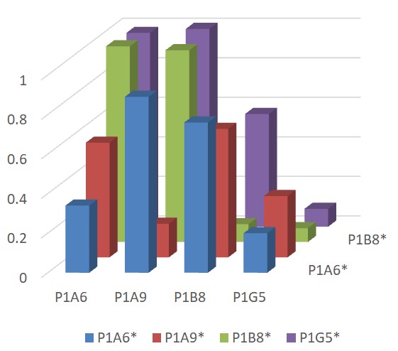
(Figure 2 Epitope Mapping of four Spike S2 Antibodies by Direct Competitive ELISA A direct competitive ELISA was developed for the epitope mapping using Spike trimer proteins of SARS-CoV-2 Delta variant at 1ug/ml as the coating antigen and biotinylated Spike S2 single domain antibodies (sdAbs) as the competitive antibodies. The sdAb mixtures (biotinylated and unconjugated sdAbs) were added to ELISA plate and streptavidin-HRP was used to detect the captured biotinylated sdAbs. The assay suggests the antibodies bind to different epitopes of the Spike trimer.)
ELISA
(Figure 2 Epitope Mapping of four Spike S2 Antibodies by Direct Competitive ELISA A direct competitive ELISA was developed for the epitope mapping using Spike trimer proteins of SARS-CoV-2 Delta variant at 1ug/ml as the coating antigen and biotinylated Spike S2 single domain antibodies (sdAbs) as the competitive antibodies. The sdAb mixtures (biotinylated and unconjugated sdAbs) were added to ELISA plate and streptavidin-HRP was used to detect the captured biotinylated sdAbs. The assay suggests the antibodies bind to different epitopes of the Spike trimer.)
SARS-CoV-2 (COVID-19) Spike S2, Monoclonal Recombinant Antibody (Cat# AAA40984)
Full Name
SARS-CoV-2 (COVID-19) Spike S2 Single Domain Antibody [P1A6]
Reactivity
Virus
Applications
ELISA
Purity
SARS-CoV-2 (COVID-19) Spike S2 Antibody is affinity chromatography purified via Nickel column. Antibody is supplied as a His-tagged purified protein. It also contains a myc-tag for detection.
Pricing
 Bioactivity
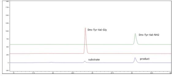
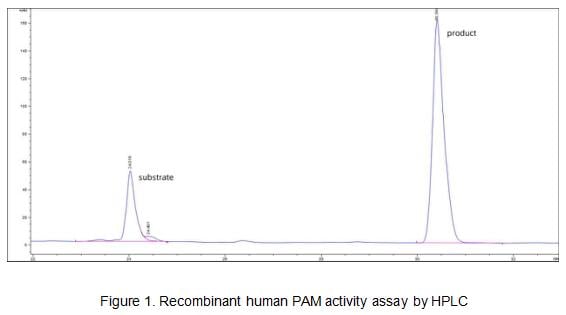
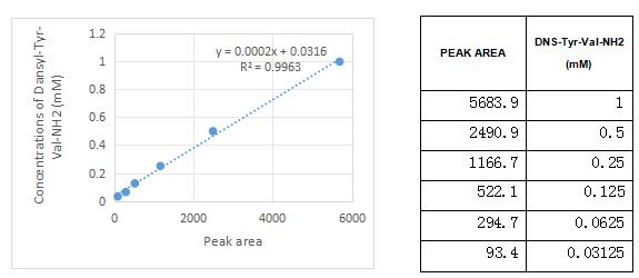
(Figure 3. The sandard curve of Dns-Tyr-Val-NH2)
Bioactivity
(Figure 3. The sandard curve of Dns-Tyr-Val-NH2)
Peptidylglycine Alpha Amidating Monooxygenase (PAM), Active Protein (Cat# AAA161894)
Full Name
Active Peptidylglycine Alpha Amidating Monooxygenase (PAM)
Gene Names
PAM; PAL; PHM
Applications
Activity Assay, Cell Culture
Purity
Greater than 90% by SDS-PAGE
Pricing
 WB (Western Blot)
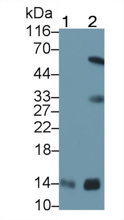
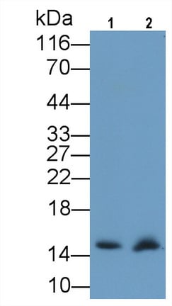
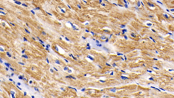
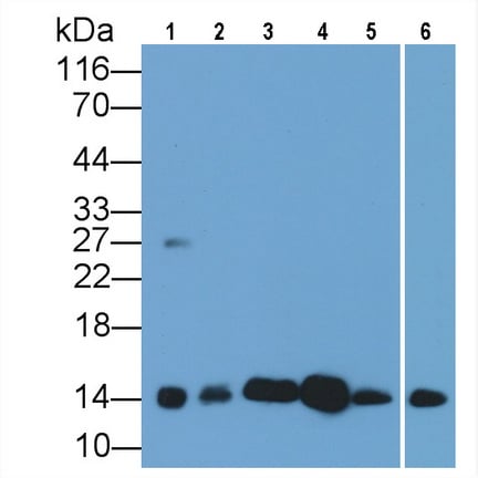
(Western Blot; Sample: Lane1: Gallus Heart lysate; Lane2: Canine Heart lysate; Lane3: Caprine Heart lysate; Lane4: Cavia Heart lysate; Lane5: Rabbit Heart lysate; Lane6: Equine Heart lysatePrimary Ab: 0.2ug/ml Mouse AntiHuman CYCS AntibodySecond Ab: 0.2ug/mL HRPLinked Caprine AntiMouse IgG Polyclonal Antibody(Catalog: SAA544Mu19))
WB (Western Blot)
(Western Blot; Sample: Lane1: Gallus Heart lysate; Lane2: Canine Heart lysate; Lane3: Caprine Heart lysate; Lane4: Cavia Heart lysate; Lane5: Rabbit Heart lysate; Lane6: Equine Heart lysatePrimary Ab: 0.2ug/ml Mouse AntiHuman CYCS AntibodySecond Ab: 0.2ug/mL HRPLinked Caprine AntiMouse IgG Polyclonal Antibody(Catalog: SAA544Mu19))
Cytochrome C (CYCS), Monoclonal Antibody (Cat# AAA151581)
Full Name
Monoclonal Antibody to Cytochrome C (CYCS)
Gene Names
CYCS; CYC; HCS; THC4
Reactivity
Human, Mouse, Rat, Guinea pig, Rabbit, Dog, Pig, Goat, Sheep, Horse, Chicken
Applications
Immunoprecipitation, Immunocytochemistry, Immunohistochemistry, Western Blot
Purity
Protein A + Protein G affinity chromatography
Pricing
 SDS-PAGE
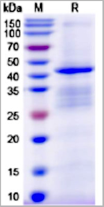
SLC34A2, Recombinant Protein (Cat# AAA11076)
Full Name
Recombinant Human SLC34A2 protein, N- GST Tag
Gene Names
SLC34A2; NPTIIb; NAPI-3B; NAPI-IIb
Purity
>90% as determined by SDS-PAGE
Pricing
 WB (Western Blot)
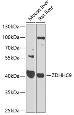
(Western blot analysis of various lysates using ZDHHC9 Rabbit pAb (AAA28212) at 1:1000 dilution.Secondary antibody: HRP Goat Anti-Rabbit IgG (H+L) at 1:10000 dilution.Lysates/proteins: 25ug per lane.Blocking buffer: 3% nonfat dry milk in TBST.Detection: ECL Enhanced Kit.Exposure time: 90s.)
WB (Western Blot)
(Western blot analysis of various lysates using ZDHHC9 Rabbit pAb (AAA28212) at 1:1000 dilution.Secondary antibody: HRP Goat Anti-Rabbit IgG (H+L) at 1:10000 dilution.Lysates/proteins: 25ug per lane.Blocking buffer: 3% nonfat dry milk in TBST.Detection: ECL Enhanced Kit.Exposure time: 90s.)
ZDHHC9, Polyclonal Antibody (Cat# AAA28212)
Full Name
ZDHHC9 Polyclonal Antibody
Gene Names
ZDHHC9; CGI89; DHHC9; MMSA1; MRXSZ; ZNF379; ZNF380; CXorf11; ZDHHC10
Applications
Western Blot
Purity
Affinity Purification
Pricing
 Bioactivity
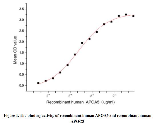
(Apolipoprotein A5 (APOA5) is a small protein, expressed predominantly in the liver. It has been identified to play an important role in lipid metabolism, specifically in triglyceride (TG) and TG-rich lipoproteins (TRLs) metabolism. Apolipoprotein C3 (APOC3) has been proved to be one of the ligands of APOA5. Thus a functional binding ELISA assay was conducted to detect the interaction of recombinant human APOA5 and recombinant human APOC3. Briefly, APOA5 was diluted serially in PBS with 0.01% BSA (pH 7.4). Duplicate samples of 100 ul were then transferred to APOC3-coated microtiter wells and incubated for 1h at 37 degree C. Wells were washed with PBST and incubated for 1h with anti-APOA5 pAb, then aspirated and washed 3 times. After incubation with HRP labelled secondary antibody for 1h at 37 degree C, wells were aspirated and washed 5 times. With the addition of substrate solution, wells were incubated 15-25 minutes at 37 degree C. Finally, add 50 uL stop solution to the wells and read at 450/630 nm immediately. The binding activity of recombinant human APOA5 and recombinant human APOC3 was shown in Figure 1, the EC50 for this effect is 0.14 ug/mL.)
Bioactivity
(Apolipoprotein A5 (APOA5) is a small protein, expressed predominantly in the liver. It has been identified to play an important role in lipid metabolism, specifically in triglyceride (TG) and TG-rich lipoproteins (TRLs) metabolism. Apolipoprotein C3 (APOC3) has been proved to be one of the ligands of APOA5. Thus a functional binding ELISA assay was conducted to detect the interaction of recombinant human APOA5 and recombinant human APOC3. Briefly, APOA5 was diluted serially in PBS with 0.01% BSA (pH 7.4). Duplicate samples of 100 ul were then transferred to APOC3-coated microtiter wells and incubated for 1h at 37 degree C. Wells were washed with PBST and incubated for 1h with anti-APOA5 pAb, then aspirated and washed 3 times. After incubation with HRP labelled secondary antibody for 1h at 37 degree C, wells were aspirated and washed 5 times. With the addition of substrate solution, wells were incubated 15-25 minutes at 37 degree C. Finally, add 50 uL stop solution to the wells and read at 450/630 nm immediately. The binding activity of recombinant human APOA5 and recombinant human APOC3 was shown in Figure 1, the EC50 for this effect is 0.14 ug/mL.)
Apolipoprotein A5 (APOA5), Active Protein (Cat# AAA161872)
Full Name
Active Apolipoprotein A5 (APOA5)
Gene Names
APOA5; RAP3; APOAV
Applications
Activity Assay, Cell Culture
Purity
Greater than 90% by SDS-PAGE
Pricing
 IP (Immunoprecipitation)
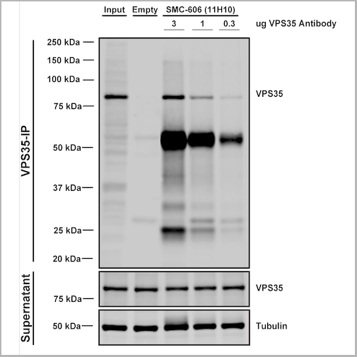
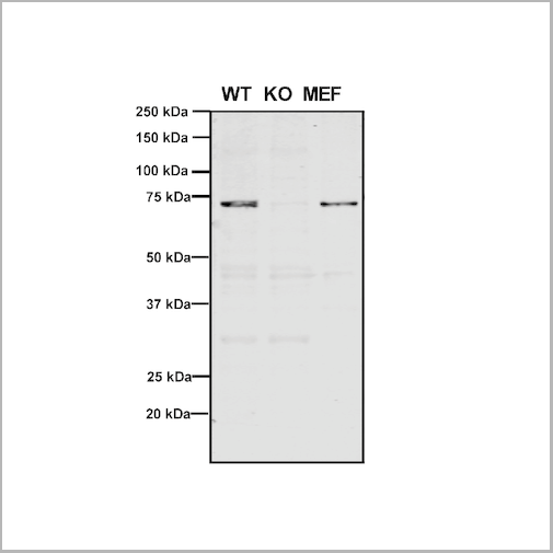
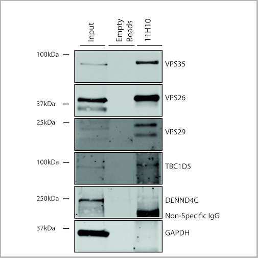
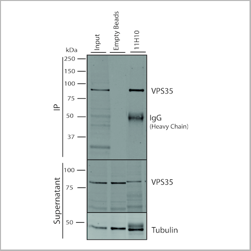
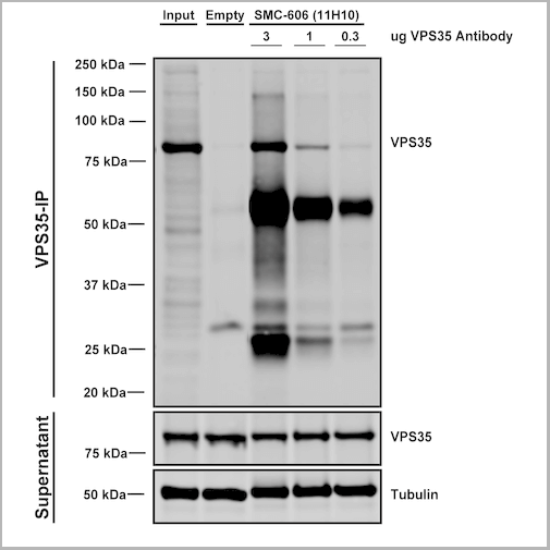
(Immunoprecipitation analysis using Mouse Anti-VPS35 Monoclonal Antibody, Clone 11H10. Tissue: embryonic fibroblast. Species: Mouse. Primary Antibody: Mouse Anti-VPS35 Monoclonal Antibody. Three amounts of (3, 1 and 0.3 ug) were non-covalently coupled to 10uL of A/G sepharose beads for 1 hour at 4 degree C and next incubated with 250ug of MEF lysate for 2 hours at 4 degree C.)
IP (Immunoprecipitation)
(Immunoprecipitation analysis using Mouse Anti-VPS35 Monoclonal Antibody, Clone 11H10. Tissue: embryonic fibroblast. Species: Mouse. Primary Antibody: Mouse Anti-VPS35 Monoclonal Antibody. Three amounts of (3, 1 and 0.3 ug) were non-covalently coupled to 10uL of A/G sepharose beads for 1 hour at 4 degree C and next incubated with 250ug of MEF lysate for 2 hours at 4 degree C.)
VPS35, Monoclonal Antibody (Cat# AAA27701)
Full Name
VPS35 Antibody, Clone 11H10: ATTO 390
Gene Names
VPS35; MEM3; PARK17
Reactivity
Human, Mouse, Rat
Applications
Western Blot, Immunoprecipitation
Purity
Protein G Purified
Pricing