
Filters
Clonality
Type
Reactivity
Gene Name
Isotype
Host
Application
Clone
10000 results for "Recombinant Proteins" - showing 2200-2250
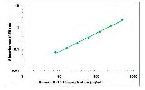 Standard Curve (Sample)
IL-15, ELISA Kit (Cat# AAA105855)
Full Name
Human IL-15 ELISA Kit
Gene Names
IL15; IL-15
Reactivity
Human
Applications
Sandwich ELISA
Pricing
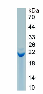
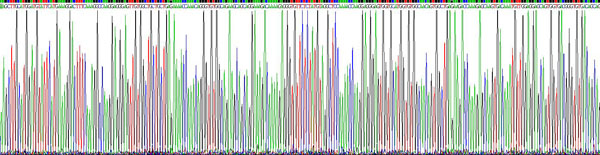
 Amino Acid Info
Synuclein Alpha (SNCa), Recombinant Protein (Cat# AAA153436)
Full Name
Recombinant Synuclein Alpha (SNCa)
Gene Names
Snca; NACP; alphaSYN
Applications
Western Blot
Purity
Greater than 90% by SDS-PAGE
Pricing
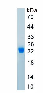
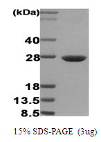 SDS-PAGE
Hsp27, Recombinant Protein (Cat# AAA48442)
Full Name
Hsp27, 1-205aa, Human, E Coli
Gene Names
HSPB1; CMT2F; HMN2B; HSP27; HSP28; Hsp25; SRP27; HS.76067; HEL-S-102
Applications
SDS-PAGE
Purity
> 95% by SDS-PAGE
Pricing
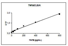 Standard Curve (Sample)
TNF alpha, ELISA Kit (Cat# AAA58614)
Full Name
Human TNF alpha ELISA Kit
Gene Names
TNF; DIF; TNFA; TNFSF2; TNF-alpha
Reactivity
Human. No cross reactivity with other human cytokines. Cross reactivity with macac TNF-a. No interference with soluble TNFR I and TNFR II.
Pricing
 Bioactivity
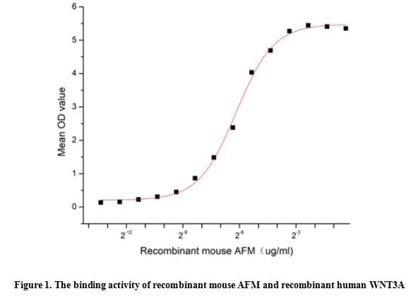
(Afamin (AFM, also known as Alpha -Albumin), a bioactive protein, is expressed in the liver and secreted into the bloodstream. Human afamin is a 87 kDa protein which is encoded by the fourth member of the human albumin gene family. Mouse AFM shares 66% aa sequence identity with Full-length human AFM. This protein can bind and transport vitamin E across the blood-brain barrier in body fluids under conditions where the lipoprotein system is not sufficient, it also can act as a carrier for hydrophobic molecules in body fluids. Essential for the solubility and activity of lipidated Wnt family members. Thus a functional binding ELISA assay was conducted to detect the interaction of recombinant mouse AFM and recombinant human WNT3A. Briefly, AFM was diluted serially in PBS with 0.01% BSA (pH 7.4). Duplicate samples of 100 ul were then transferred to WNT3A-coated microtiter wells and incubated for 1h at 37 degree C. Wells were washed with PBST and incubated for 1h with anti-AFM pAb, then aspirated and washed 3 times. After incubation with HRP labelled secondary antibody for 1h at 37 degree C, wells were aspirated and washed 5 times. With the addition of substrate solution, wells were incubated 15-25 minutes at 37 degree C. Finally, add 50 uL stop solution to the wells and read at 450/630 nm immediately. The binding activity of recombinant mouse AFM and recombinant human WNT3A was shown in Figure 1, the EC50 for this effect is 0.01 ug/mL.)
Bioactivity
(Afamin (AFM, also known as Alpha -Albumin), a bioactive protein, is expressed in the liver and secreted into the bloodstream. Human afamin is a 87 kDa protein which is encoded by the fourth member of the human albumin gene family. Mouse AFM shares 66% aa sequence identity with Full-length human AFM. This protein can bind and transport vitamin E across the blood-brain barrier in body fluids under conditions where the lipoprotein system is not sufficient, it also can act as a carrier for hydrophobic molecules in body fluids. Essential for the solubility and activity of lipidated Wnt family members. Thus a functional binding ELISA assay was conducted to detect the interaction of recombinant mouse AFM and recombinant human WNT3A. Briefly, AFM was diluted serially in PBS with 0.01% BSA (pH 7.4). Duplicate samples of 100 ul were then transferred to WNT3A-coated microtiter wells and incubated for 1h at 37 degree C. Wells were washed with PBST and incubated for 1h with anti-AFM pAb, then aspirated and washed 3 times. After incubation with HRP labelled secondary antibody for 1h at 37 degree C, wells were aspirated and washed 5 times. With the addition of substrate solution, wells were incubated 15-25 minutes at 37 degree C. Finally, add 50 uL stop solution to the wells and read at 450/630 nm immediately. The binding activity of recombinant mouse AFM and recombinant human WNT3A was shown in Figure 1, the EC50 for this effect is 0.01 ug/mL.)
Afamin (AFM), Active Protein (Cat# AAA161881)
Full Name
Active Afamin (AFM)
Gene Names
Afm; Alf; alpha-Alb
Applications
Activity Assay, Cell Culture
Purity
Greater than 90% by SDS-PAGE
Pricing
 IP (Immunoprecipitation)
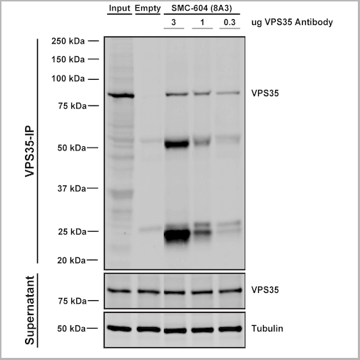
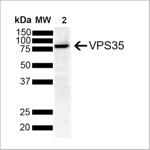
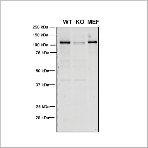
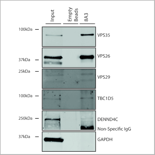
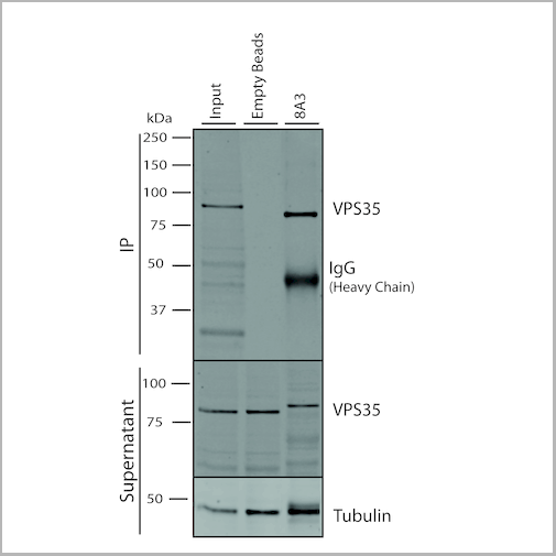
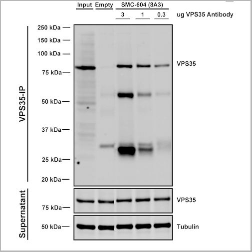
(Immunoprecipitation analysis using Mouse Anti-VPS35 Monoclonal Antibody, Clone 8A3. Tissue: embryonic fibroblast. Species: Mouse. Primary Antibody: Mouse Anti-VPS35 Monoclonal Antibody. Three amounts of (3, 1 and 0.3 ug) were non-covalently coupled to 10uL of A/G sepharose beads for 1 hour at 4 degree C and next incubated with 250ug of MEF lysate for 2 hours at 4 degree C.)
IP (Immunoprecipitation)
(Immunoprecipitation analysis using Mouse Anti-VPS35 Monoclonal Antibody, Clone 8A3. Tissue: embryonic fibroblast. Species: Mouse. Primary Antibody: Mouse Anti-VPS35 Monoclonal Antibody. Three amounts of (3, 1 and 0.3 ug) were non-covalently coupled to 10uL of A/G sepharose beads for 1 hour at 4 degree C and next incubated with 250ug of MEF lysate for 2 hours at 4 degree C.)
VPS35, Monoclonal Antibody (Cat# AAA27686)
Full Name
VPS35 Antibody, Clone 8A3: Biotin
Gene Names
VPS35; MEM3; PARK17
Reactivity
Human, Mouse, Rat
Applications
Western Blot, Immunocytochemistry, Immunofluorescence, Immunoprecipitation
Purity
Protein G Purified
Pricing
 DB (Dot Blot)
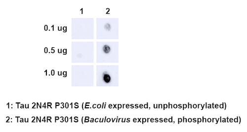
(Dot Blot analysis using Rabbit Anti-Tau Monoclonal Antibody, Clone AH36. Species: E. Coli, Baculovirus. Primary Antibody: Rabbit Anti-Tau Monoclonal Antibody at 1:500. Secondary Antibody: Goat anti-rabbit IgG:HRP.)
DB (Dot Blot)
(Dot Blot analysis using Rabbit Anti-Tau Monoclonal Antibody, Clone AH36. Species: E. Coli, Baculovirus. Primary Antibody: Rabbit Anti-Tau Monoclonal Antibody at 1:500. Secondary Antibody: Goat anti-rabbit IgG:HRP.)
Tau, Monoclonal Antibody (Cat# AAA253978)
Full Name
Tau Antibody (pSer202/ pThr205), Clone AH36
Gene Names
MAPT; TAU; MSTD; PPND; DDPAC; MAPTL; MTBT1; MTBT2; FTDP-17; PPP1R103
Reactivity
Human, Mouse
Applications
Immunohistochemistry, Immunofluorescence, Immunocytochemistry, Dot Blot, Western Blot
Purity
Affinity Purified
Pricing
 Standard Curve (Sample)
TNF-alpha, CLIA Kit (Cat# AAA318562)
Full Name
TNF-alpha (Human) LumiAb ELISA Kit
Gene Names
TNF; DIF; TNFA; TNFSF2; TNLG1F; TNF-alpha
Reactivity
Human
Pricing
 Bioactivity
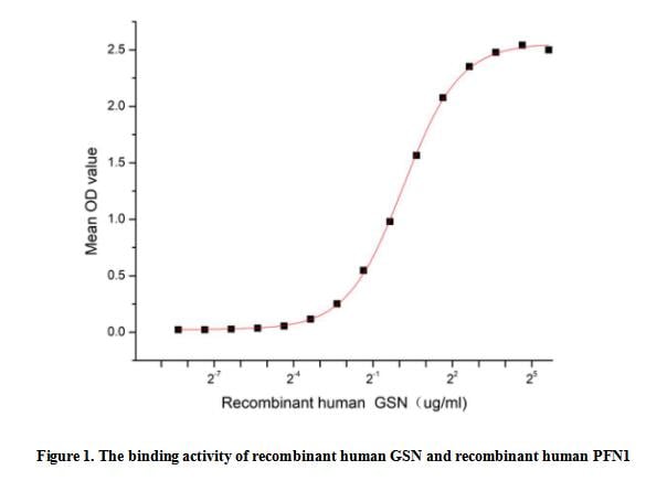
(Gelsolin (GSN), one of the most abundant actin-binding proteins, is a multifunctional regulator of physiological and pathological cellular process and regulate cell migration, cell morphology, proliferation and apoptosis. Widely expressed, Gelsolin binds to actin and fibronectin, and is found both secreted in plasma and in cytoplasm and a previous study revealed that the levels of GSN are decreased in various cancer. Profilin-1 (PFN1) can also bind to actin and affects the structure of the cytoskeleton. PFN1 is one of the ligands of GSN, thus a functional binding ELISA assay was conducted to detect the interaction of recombinant human GSN and recombinant human PFN1. Briefly, GSN was diluted serially in PBS with 0.01% BSA (pH 7.4). Duplicate samples of 100 ul were then transferred to PFN1-coated microtiter wells and incubated for 1h at 37 degree C. Wells were washed with PBST and incubated for 1h with anti-GSN pAb, then aspirated and washed 3 times. After incubation with HRP labelled secondary antibody for 1h at 37 degree C, wells were aspirated and washed 5 times. With the addition of substrate solution, wells were incubated 15-25 minutes at 37 degree C. Finally, add 50 uL stop solution to the wells and read at 450/630 nm immediately. The binding activity of recombinant human GSN and recombinant human PFN1 was shown in Figure 1, the EC50 for this effect is 1.11 ug/mL.)
Bioactivity
(Gelsolin (GSN), one of the most abundant actin-binding proteins, is a multifunctional regulator of physiological and pathological cellular process and regulate cell migration, cell morphology, proliferation and apoptosis. Widely expressed, Gelsolin binds to actin and fibronectin, and is found both secreted in plasma and in cytoplasm and a previous study revealed that the levels of GSN are decreased in various cancer. Profilin-1 (PFN1) can also bind to actin and affects the structure of the cytoskeleton. PFN1 is one of the ligands of GSN, thus a functional binding ELISA assay was conducted to detect the interaction of recombinant human GSN and recombinant human PFN1. Briefly, GSN was diluted serially in PBS with 0.01% BSA (pH 7.4). Duplicate samples of 100 ul were then transferred to PFN1-coated microtiter wells and incubated for 1h at 37 degree C. Wells were washed with PBST and incubated for 1h with anti-GSN pAb, then aspirated and washed 3 times. After incubation with HRP labelled secondary antibody for 1h at 37 degree C, wells were aspirated and washed 5 times. With the addition of substrate solution, wells were incubated 15-25 minutes at 37 degree C. Finally, add 50 uL stop solution to the wells and read at 450/630 nm immediately. The binding activity of recombinant human GSN and recombinant human PFN1 was shown in Figure 1, the EC50 for this effect is 1.11 ug/mL.)
Gelsolin (GSN), Active Protein (Cat# AAA161725)
Full Name
Active Gelsolin (GSN)
Gene Names
GSN; ADF; AGEL
Applications
Activity Assay, Cell Culture
Purity
Greater than 90% by SDS-PAGE
Pricing
 SDS-PAGE
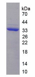
Procollagen III N-Terminal Propeptide (PIIINP), Recombinant Protein (Cat# AAA151022)
Full Name
Recombinant Procollagen III N-Terminal Propeptide (PIIINP)
Gene Names
COL3A1; EDS4A
Reactivity
Homo sapiens (Human)
Applications
Western Blot
Purity
> 90%
Pricing
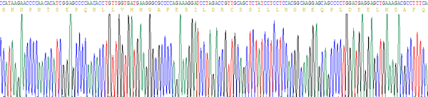
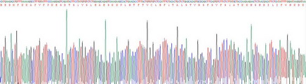 Gene Sequence
(Gene Sequencing (extract))
Gene Sequence
(Gene Sequencing (extract))
C Reactive Protein (CRP), Recombinant Protein (Cat# AAA133994)
Full Name
Recombinant C Reactive Protein (CRP)
Gene Names
Crp; AI255847
Applications
Western Blot
Purity
> 90%
Pricing
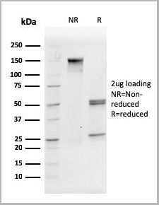
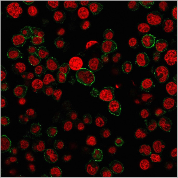
 Application Data
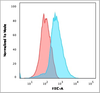
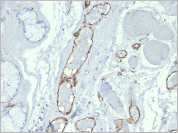
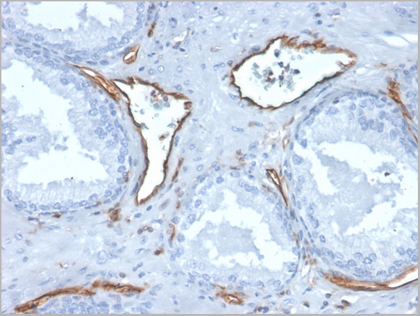
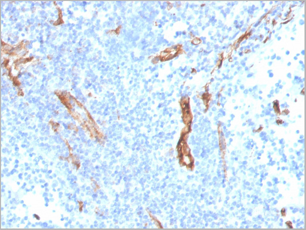
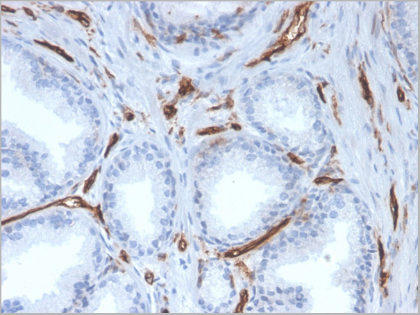
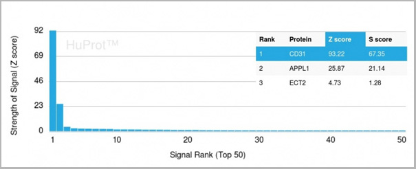
(Analysis of Protein Array containing more than 19,000 full-length human proteins using CD31 Mouse Monoclonal Antibody (PECAM1/3527) Z- and S- Score: The Z-score represents the strength of a signal that a monoclonal antibody (MAb) (in combination with a fluorescently-tagged anti-IgG secondary antibody) produces when binding to a particular protein on the HuProtTM array. Z-scores are described in units of standard deviations (SD’s) above the mean value of all signals generated on that array. If targets on HuProtTM are arranged in descending order of the Z-score, the S-score is the difference (also in units of SD’s) between the Z-score. S-score therefore represents the relative target specificity of a MAb to its intended target. A MAb is considered to specific to its intended target, if the MAb has an S-score of at least 2.5. For example, if a MAb binds to protein X with a Z-score of 43 and to protein Y with a Z-score of 14, then the S-score for the binding of that MAb to protein X is equal to 29.)
Application Data
(Analysis of Protein Array containing more than 19,000 full-length human proteins using CD31 Mouse Monoclonal Antibody (PECAM1/3527) Z- and S- Score: The Z-score represents the strength of a signal that a monoclonal antibody (MAb) (in combination with a fluorescently-tagged anti-IgG secondary antibody) produces when binding to a particular protein on the HuProtTM array. Z-scores are described in units of standard deviations (SD’s) above the mean value of all signals generated on that array. If targets on HuProtTM are arranged in descending order of the Z-score, the S-score is the difference (also in units of SD’s) between the Z-score. S-score therefore represents the relative target specificity of a MAb to its intended target. A MAb is considered to specific to its intended target, if the MAb has an S-score of at least 2.5. For example, if a MAb binds to protein X with a Z-score of 43 and to protein Y with a Z-score of 14, then the S-score for the binding of that MAb to protein X is equal to 29.)
CD31/PECAM-1, Monoclonal Antibody (Cat# AAA23931)
Full Name
CD31/PECAM-1 (Endothelial Cell Marker)
Gene Names
PECAM1; CD31; PECA1; GPIIA'; PECAM-1; endoCAM; CD31/EndoCAM
Reactivity
Human
Applications
Flow Cytometry, Immunofluorescence, Immunohistochemistry
Purity
Purified Ab with BSA and Azide at 200ug/ml OR Purified Ab WITHOUT BSA and Azide at 1.0mg/ml
Pricing
 Bioactivity
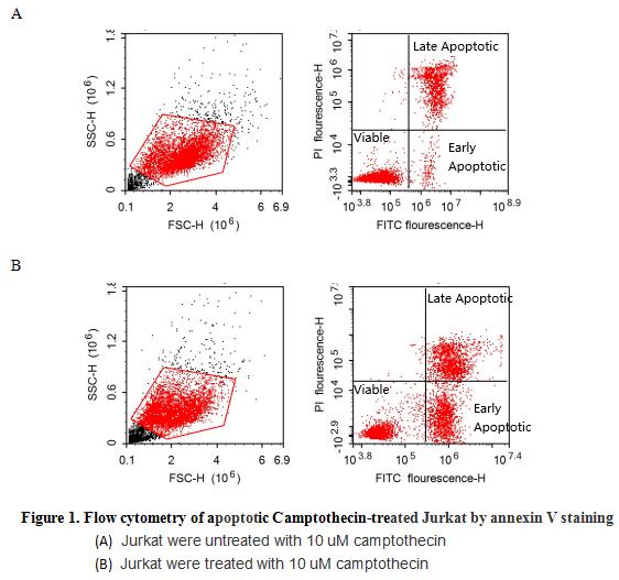
(Annexin V (ANXA5) is a multifunctional protein that is highly expressed on the apical surfaces of syncytiotrophoblasts, and plays an important role in haemostatic regulations, maintaining blood fluidity of the placenta. Lower ANXA5 levels have been observed in M2/ANXA5 haplotype carrying chorion. The association found between the maternal carriage of the M2/ANXA5 haplotype and an elevated risk of IUGR and/or PE supports the hypothesis that carrier status of this haplotype and the consequently reduced placental ANXA5 expression might be responsible, at least partially, for the onset of these gestational vascular complications. Annexin V is a calcium-dependent phospholipid binding protein that can be used to bind Phosphatidylserine (PS) during an early apoptosis event where the PS becomes exposed at the cell surface. Jurkat cells were treated with 10 uM camptothecin for 4h, 2*105 cells which were resuspended in binding buffer were stained with 5 ug recombinant human Annexin V-FITC and 10 ul Propidium iodide (PI) for 20min in dark room temperature. The flow cytometry was used to detect the early apoptotic and late apoptotic of camptothecin-treated Jurkat cells (Figure 1), the combination of Annexin V-FITC and propidium iodide allows for the distinction between early apoptotic cells (Annexin V-FITC positive and propidium iodide negative), late apoptotic and/or necrotic cells (Annexin V-FITC and propidium iodide positive), and viable cells (unstained). Thus, the recombinant human Annexin V-FITC can bind Phosphatidylserine (PS) at early apoptosis of Jurkat.)
Bioactivity
(Annexin V (ANXA5) is a multifunctional protein that is highly expressed on the apical surfaces of syncytiotrophoblasts, and plays an important role in haemostatic regulations, maintaining blood fluidity of the placenta. Lower ANXA5 levels have been observed in M2/ANXA5 haplotype carrying chorion. The association found between the maternal carriage of the M2/ANXA5 haplotype and an elevated risk of IUGR and/or PE supports the hypothesis that carrier status of this haplotype and the consequently reduced placental ANXA5 expression might be responsible, at least partially, for the onset of these gestational vascular complications. Annexin V is a calcium-dependent phospholipid binding protein that can be used to bind Phosphatidylserine (PS) during an early apoptosis event where the PS becomes exposed at the cell surface. Jurkat cells were treated with 10 uM camptothecin for 4h, 2*105 cells which were resuspended in binding buffer were stained with 5 ug recombinant human Annexin V-FITC and 10 ul Propidium iodide (PI) for 20min in dark room temperature. The flow cytometry was used to detect the early apoptotic and late apoptotic of camptothecin-treated Jurkat cells (Figure 1), the combination of Annexin V-FITC and propidium iodide allows for the distinction between early apoptotic cells (Annexin V-FITC positive and propidium iodide negative), late apoptotic and/or necrotic cells (Annexin V-FITC and propidium iodide positive), and viable cells (unstained). Thus, the recombinant human Annexin V-FITC can bind Phosphatidylserine (PS) at early apoptosis of Jurkat.)
Annexin V (ANXA5), Active Protein (Cat# AAA161722)
Full Name
Active Annexin V (ANXA5)
Gene Names
ANXA5; PP4; ANX5; ENX2; RPRGL3; HEL-S-7
Applications
Activity Assay, Cell Culture
Purity
Greater than 95% by SDS-PAGE
Pricing
Melanoma Antigen Family A, Recombinant Protein (Cat# AAA10890)
Full Name
Recombinant Human Melanoma Antigen Family A, 3
Gene Names
MAGEA3; HIP8; HYPD; CT1.3; MAGE3; MAGEA6
Purity
Greater than 90% as determined by SDS-PAGE.
Pricing
Rhinovirus outer capsid protein 3, Monoclonal Antibody (Cat# AAA13449)
Full Name
Rhinovirus outer capsid protein 3 (VP3)
Applications
Immunofluorescence
Purity
> 90% pure. Protein A Chromatography
Pricing
 SDS-PAGE
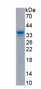
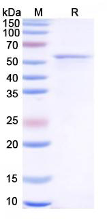
(SDS PAGE for Recombinant Human BP180/NC16a Protein)
SDS-PAGE
(SDS PAGE for Recombinant Human BP180/NC16a Protein)
BP180/NC16a, Recombinant Protein (Cat# AAA120534)
Full Name
Recombinant Human BP180/NC16a Protein
Gene Names
COL17A1; BP180; BPA-2; BPAG2; LAD-1; BA16H23.2
Purity
>95% as determined by SDS-PAGE
Pricing
 Bioactivity
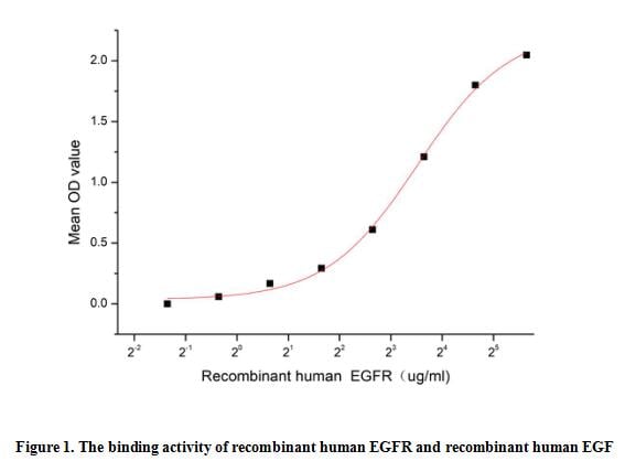
(The epidermal growth factor receptor (EGFR) is a growth factor receptor that induces cell differentiation and proliferation upon activation through the binding of one of its ligands. The receptor is located at the cell surface, where the binding of a ligand activates a tyrosine kinase in the intracellular region of the receptor. This tyrosine kinase phosphorylates a number of intracellular substrates that activates pathways leading to cell growth, DNA synthesis and the expression of oncogenes such as fos and jun. Epidermal growth factor (EGF) family peptides are ligands for the EGF receptor (EGFR), thus a functional binding ELISA assay was conducted to detect the interaction of recombinant human EGFR and recombinant human EGF. Briefly, biotin-linked EGFR were diluted serially in PBS, with 0.01% BSA (pH 7.4). Duplicate samples of 100 ul were then transferred to EGF-coated microtiter wells and incubated for 1h at 37 degree C. Wells were washed with PBST 3 times and incubation with Streptavidin-HRP for 30min, then wells were aspirated and washed 5 times. With the addition of substrate solution, wells were incubated 15-25 minutes at 37 degree C. Finally, add 50 ul stop solution to the wells and read at 450 nm immediately. The binding activity of recombinant human EGFR and recombinant human EGF was shown in Figure 1, the EC50 for this effect is 11.46 ug/mL.)
Bioactivity
(The epidermal growth factor receptor (EGFR) is a growth factor receptor that induces cell differentiation and proliferation upon activation through the binding of one of its ligands. The receptor is located at the cell surface, where the binding of a ligand activates a tyrosine kinase in the intracellular region of the receptor. This tyrosine kinase phosphorylates a number of intracellular substrates that activates pathways leading to cell growth, DNA synthesis and the expression of oncogenes such as fos and jun. Epidermal growth factor (EGF) family peptides are ligands for the EGF receptor (EGFR), thus a functional binding ELISA assay was conducted to detect the interaction of recombinant human EGFR and recombinant human EGF. Briefly, biotin-linked EGFR were diluted serially in PBS, with 0.01% BSA (pH 7.4). Duplicate samples of 100 ul were then transferred to EGF-coated microtiter wells and incubated for 1h at 37 degree C. Wells were washed with PBST 3 times and incubation with Streptavidin-HRP for 30min, then wells were aspirated and washed 5 times. With the addition of substrate solution, wells were incubated 15-25 minutes at 37 degree C. Finally, add 50 ul stop solution to the wells and read at 450 nm immediately. The binding activity of recombinant human EGFR and recombinant human EGF was shown in Figure 1, the EC50 for this effect is 11.46 ug/mL.)
Epidermal Growth Factor Receptor (EGFR), Active Protein (Cat# AAA161774)
Full Name
Active Epidermal Growth Factor Receptor (EGFR)
Gene Names
EGFR; ERBB; HER1; mENA; ERBB1; PIG61
Applications
Activity Assay, Cell Culture
Purity
Greater than 90% by SDS-PAGE
Pricing
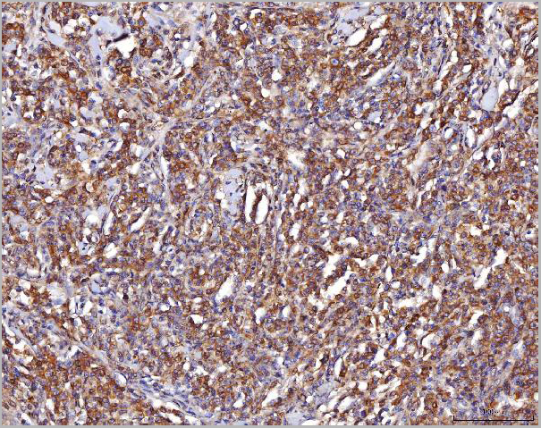
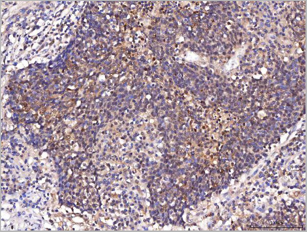
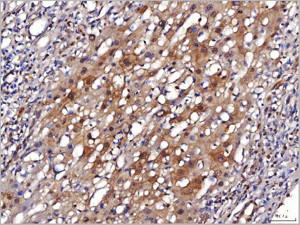
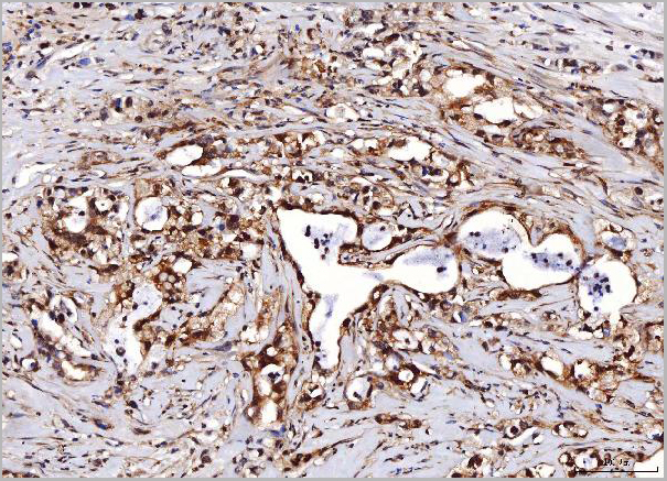
 FCM (Flow Cytometry)
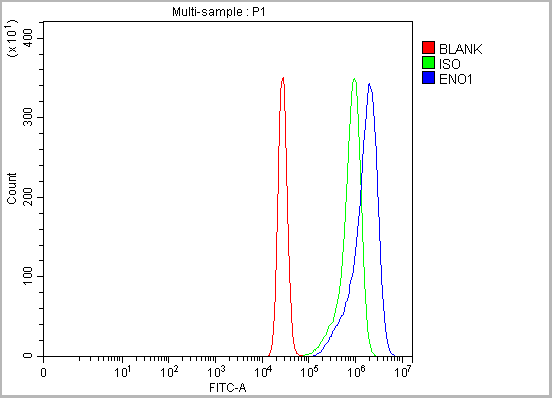
(Figure 10. Flow Cytometry analysis of K562 cells using anti-ENO1 antibody (AAA19420).Overlay histogram showing K562 cells stained with AAA19420 (Blue line). The cells were blocked with 10% normal goat serum. And then incubated with rabbit anti-ENO1 Antibody (AAA19420, 1 ug/1x10^6 cells) for 30 min at 20 degree C. DyLight488 conjugated goat anti-rabbit IgG was used as secondary antibody for 30 minutes at 20 degree C. Isotype control antibody (Green line) was rabbit IgG (1 ug/1x10^6) used under the same conditions. Unlabelled sample (Red line) was also used as a control.)
FCM (Flow Cytometry)
(Figure 10. Flow Cytometry analysis of K562 cells using anti-ENO1 antibody (AAA19420).Overlay histogram showing K562 cells stained with AAA19420 (Blue line). The cells were blocked with 10% normal goat serum. And then incubated with rabbit anti-ENO1 Antibody (AAA19420, 1 ug/1x10^6 cells) for 30 min at 20 degree C. DyLight488 conjugated goat anti-rabbit IgG was used as secondary antibody for 30 minutes at 20 degree C. Isotype control antibody (Green line) was rabbit IgG (1 ug/1x10^6) used under the same conditions. Unlabelled sample (Red line) was also used as a control.)
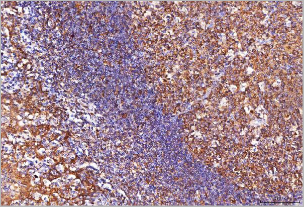
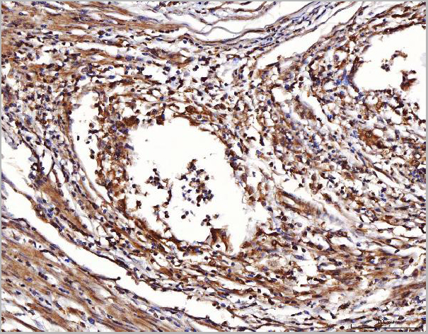
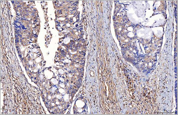
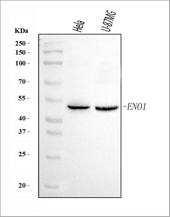
ENO1, Polyclonal Antibody (Cat# AAA19420)
Full Name
Anti-ENO1 Antibody Picoband
Gene Names
ENO1; NNE; PPH; MPB1; ENO1L1
Reactivity
Human
Applications
Flow Cytometry, Immunohistochemistry, Western Blot
Purity
Immunogen affinity purified.
Pricing
 SDS-PAGE
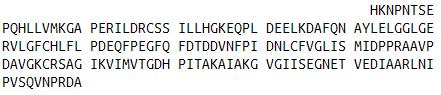
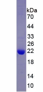
(SDS-PAGE)
SDS-PAGE
(SDS-PAGE)
ATPase, Na+/K+ Transporting Alpha 1 Polypeptide (ATP1a1), Recombinant Protein (Cat# AAA152316)
Full Name
Recombinant ATPase, Na+/K+ Transporting Alpha 1 Polypeptide (ATP1a1)
Applications
Western Blot
Purity
Greater than 90% by SDS-PAGE
Pricing
 Amino Acid Info
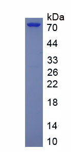
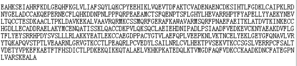
Eukaryotic Albumin (ALB), Protein (Cat# AAA153194)
Full Name
Eukaryotic Albumin (ALB)
Gene Names
Alb; Alb1; Albza
Applications
Western Blot
Purity
Greater than 95% by SDS-PAGE
Pricing
 Bioactivity
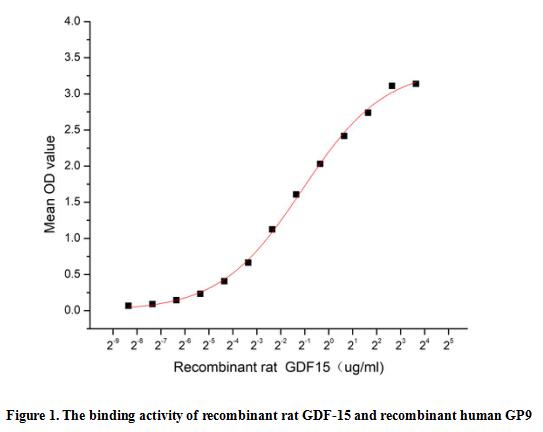
(Growth Differentiation Factor 15 (GDF-15), also called Macrophage Inhibitory Cytokine 1 (MIC-1), is a divergent member of the Transforming Growth Factor beta (TGF-beta) superfamily. Human GDF-15 shares 66% and 68% amino acid sequence identity with the rat and mouse proteins, respectively. GDF-15 is highly expressed in placenta and brain, and it is expressed at lower levels in kidney, pancreas, prostate, and colon. Similar to other TGF-beta family proteins, GDF-15 is synthesized as a large precursor protein that is cleaved at a dibasic cleavage site (RxxR) to release the mature protein. Biologically active GDF-15 is a disulfide-linked homodimer of the mature protein. GDF15 has been shown to have various functions, including inhibition of Tumor Necrosis Factor alpha (TNF-alpha) production from lipopolysaccharide-stimulated macrophages and the induction of cartilage formation. A functional binding ELISA assay was conducted to detect the interaction of recombinant rat GDF-15 and recombinant human GP9. Briefly, GDF-15 was diluted serially in PBS with 0.01% BSA (pH 7.4). Duplicate samples of 100 ul were then transferred to GP9-coated microtiter wells and incubated for 1h at 37 degree C. Wells were washed with PBST and incubated for 1h with anti-GDF-15 pAb, then aspirated and washed 3 times. After incubation with HRP labelled secondary antibody for 1h at 37 degree C, wells were aspirated and washed 5 times. With the addition of substrate solution, wells were incubated 15-25 minutes at 37 degree C. Finally, add 50 uL stop solution to the wells and read at 450/630 nm immediately. The binding activity of GDF-15 and GP9 was shown in Figure 1, the EC50 for this effect is 0.47 ug/mL.)
Bioactivity
(Growth Differentiation Factor 15 (GDF-15), also called Macrophage Inhibitory Cytokine 1 (MIC-1), is a divergent member of the Transforming Growth Factor beta (TGF-beta) superfamily. Human GDF-15 shares 66% and 68% amino acid sequence identity with the rat and mouse proteins, respectively. GDF-15 is highly expressed in placenta and brain, and it is expressed at lower levels in kidney, pancreas, prostate, and colon. Similar to other TGF-beta family proteins, GDF-15 is synthesized as a large precursor protein that is cleaved at a dibasic cleavage site (RxxR) to release the mature protein. Biologically active GDF-15 is a disulfide-linked homodimer of the mature protein. GDF15 has been shown to have various functions, including inhibition of Tumor Necrosis Factor alpha (TNF-alpha) production from lipopolysaccharide-stimulated macrophages and the induction of cartilage formation. A functional binding ELISA assay was conducted to detect the interaction of recombinant rat GDF-15 and recombinant human GP9. Briefly, GDF-15 was diluted serially in PBS with 0.01% BSA (pH 7.4). Duplicate samples of 100 ul were then transferred to GP9-coated microtiter wells and incubated for 1h at 37 degree C. Wells were washed with PBST and incubated for 1h with anti-GDF-15 pAb, then aspirated and washed 3 times. After incubation with HRP labelled secondary antibody for 1h at 37 degree C, wells were aspirated and washed 5 times. With the addition of substrate solution, wells were incubated 15-25 minutes at 37 degree C. Finally, add 50 uL stop solution to the wells and read at 450/630 nm immediately. The binding activity of GDF-15 and GP9 was shown in Figure 1, the EC50 for this effect is 0.47 ug/mL.)
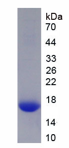
Growth Differentiation Factor 15 (GDF15), Active Protein (Cat# AAA161875)
Full Name
Active Growth Differentiation Factor 15 (GDF15)
Applications
Activity Assay, Cell Culture
Purity
Greater than 97% by SDS-PAGE
Pricing

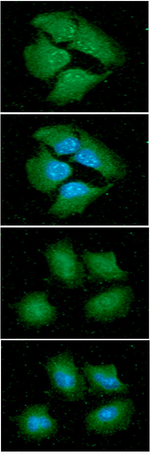 IF (Immunofluorescence)
(ICC/IF analysis of 14-3-3 epsilon in HeLa cells line, stained with DAPI (Blue) for nucleus staining and monoclonal anti-human 14-3-3 epsilon antibody (1:100) with goat anti-mouse IgG-Alexa fluor 488 conjugate (Green). ICC/IF analysis of 14-3-3 epsilon in A549 cells line, stained with DAPI (Blue) for nucleus staining and monoclonal anti-human 14-3-3 epsilon antibody (1:100) with goat anti-mouse IgG-Alexa fluor 488 conjugate (Green).)
IF (Immunofluorescence)
(ICC/IF analysis of 14-3-3 epsilon in HeLa cells line, stained with DAPI (Blue) for nucleus staining and monoclonal anti-human 14-3-3 epsilon antibody (1:100) with goat anti-mouse IgG-Alexa fluor 488 conjugate (Green). ICC/IF analysis of 14-3-3 epsilon in A549 cells line, stained with DAPI (Blue) for nucleus staining and monoclonal anti-human 14-3-3 epsilon antibody (1:100) with goat anti-mouse IgG-Alexa fluor 488 conjugate (Green).)
14-3-3 epsilon, Monoclonal Antibody (Cat# AAA48240)
Full Name
14-3-3 epsilon antibody
Gene Names
YWHAE; MDS; HEL2; MDCR; KCIP-1; 14-3-3E
Reactivity
Human
Applications
Immunofluorescence, Immunocytochemistry, Flow Cytometry, Western Blot
Purity
By protein-A affinity chromatography
Pricing
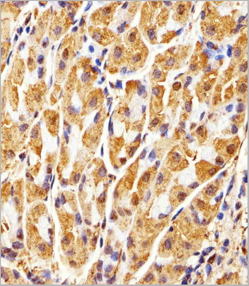
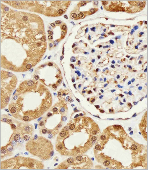
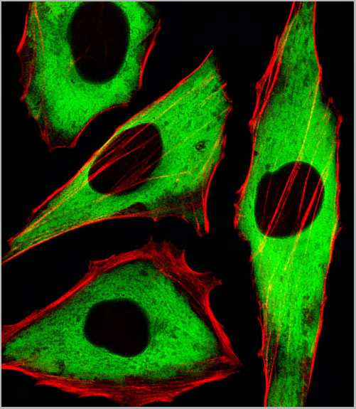
 WB (Western Blot)
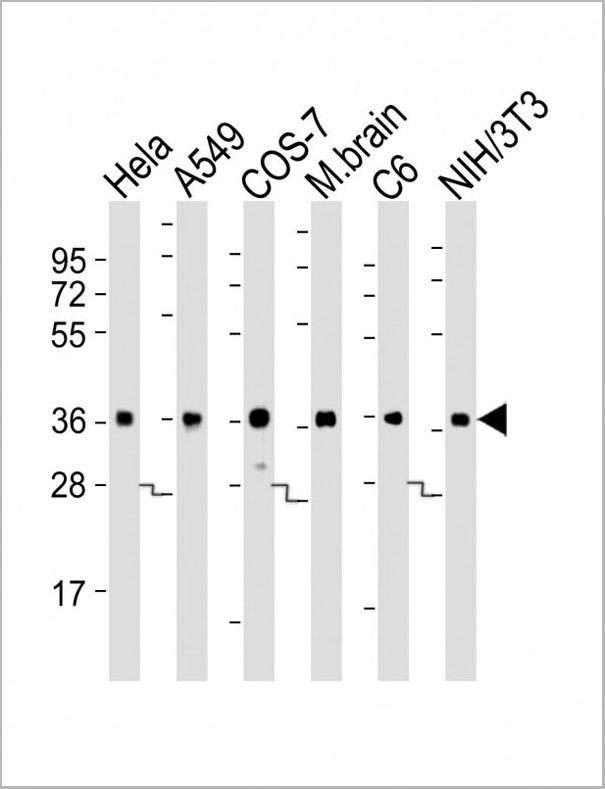
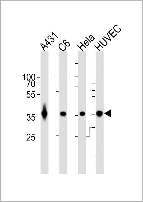
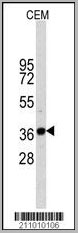
(Western blot analysis of anti-GAPDH Monoclonal Antibody in CEM cell line lysates (35ug/lane). GAPDH(arrow) was detected using the purified Mab.)
WB (Western Blot)
(Western blot analysis of anti-GAPDH Monoclonal Antibody in CEM cell line lysates (35ug/lane). GAPDH(arrow) was detected using the purified Mab.)
GAPDH, Monoclonal Antibody (Cat# AAA28634)
Full Name
GAPDH Antibody
Gene Names
GAPDH; G3PD; GAPD; HEL-S-162eP
Reactivity
Human, mouse, rat
Applications
Western Blot, Immunofluorescence, Immunohistochemistry
Purity
Purified Mouse Monoclonal Antibody (Mab)
Pricing
 Bioactivity
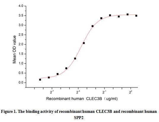
(CLEC3B (Ctype lectin domain family 3 member B), also known as Tetranectin (TN), is a plasminogen-kringle-4-binding protein that is detected in various endocrine tissues and in epithelial and mesenchymal cells, including fibroblasts, monocytes, and neutrophils. Studies have shown that abnormal CLEC3B expression plays an important role in tumourigenesis and metastasis in gastric adenocarcinoma, breast cancer, cervical cancer, ovarian cancer, carcinoma of the urinary bladder, and melanoma. Besides, Secreted Phosphoprotein 2 (SPP2) has been identified as an interactor of CLEC3B, thus a functional binding ELISA assay was conducted to detect the interaction of recombinant human SPP2 and recombinant human CLEC3B. Briefly, CLEC3B was diluted serially in PBS with 0.01% BSA (pH 7.4). Duplicate samples of 100 ul were then transferred to SPP2-coated microtiter wells and incubated for 1h at 37 degree C. Wells were washed with PBST and incubated for 1h with anti-CLEC3B pAb, then aspirated and washed 3 times. After incubation with HRP labelled secondary antibody for 1h at 37 degree C, wells were aspirated and washed 5 times. With the addition of substrate solution, wells were incubated 15-25 minutes at 37 degree C. Finally, add 50 uL stop solution to the wells and read at 450/630 nm immediately. The binding activity of recombinant human CLEC3B and recombinant human SPP2 was shown in Figure 1, the EC50 for this effect is 0.08 ug/mL.)
Bioactivity
(CLEC3B (Ctype lectin domain family 3 member B), also known as Tetranectin (TN), is a plasminogen-kringle-4-binding protein that is detected in various endocrine tissues and in epithelial and mesenchymal cells, including fibroblasts, monocytes, and neutrophils. Studies have shown that abnormal CLEC3B expression plays an important role in tumourigenesis and metastasis in gastric adenocarcinoma, breast cancer, cervical cancer, ovarian cancer, carcinoma of the urinary bladder, and melanoma. Besides, Secreted Phosphoprotein 2 (SPP2) has been identified as an interactor of CLEC3B, thus a functional binding ELISA assay was conducted to detect the interaction of recombinant human SPP2 and recombinant human CLEC3B. Briefly, CLEC3B was diluted serially in PBS with 0.01% BSA (pH 7.4). Duplicate samples of 100 ul were then transferred to SPP2-coated microtiter wells and incubated for 1h at 37 degree C. Wells were washed with PBST and incubated for 1h with anti-CLEC3B pAb, then aspirated and washed 3 times. After incubation with HRP labelled secondary antibody for 1h at 37 degree C, wells were aspirated and washed 5 times. With the addition of substrate solution, wells were incubated 15-25 minutes at 37 degree C. Finally, add 50 uL stop solution to the wells and read at 450/630 nm immediately. The binding activity of recombinant human CLEC3B and recombinant human SPP2 was shown in Figure 1, the EC50 for this effect is 0.08 ug/mL.)
C-Type Lectin Domain Family 3, Member B (CLEC3B), Active Protein (Cat# AAA161934)
Full Name
Active C-Type Lectin Domain Family 3, Member B (CLEC3B)
Gene Names
CLEC3B; TN; TNA
Applications
Activity Assay, Cell Culture
Purity
Greater than 90% by SDS-PAGE
Pricing
 IHC (Immunohistochemistry)
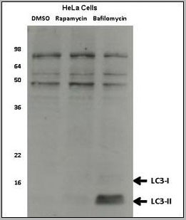
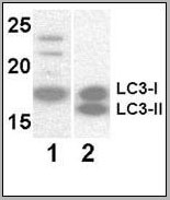
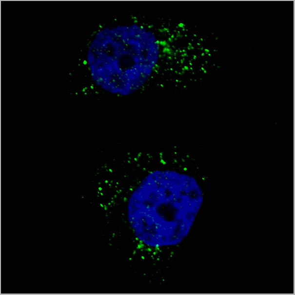
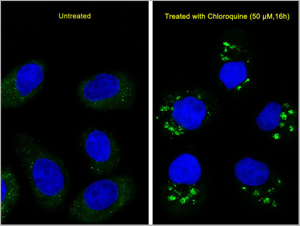
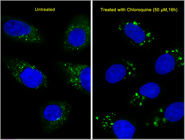
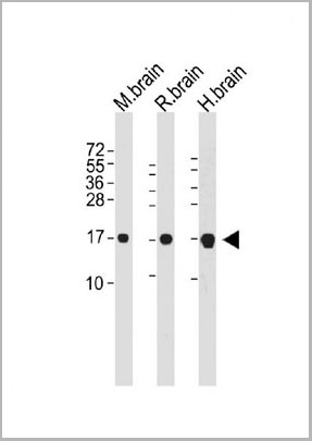
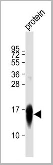
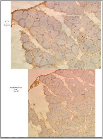
(10X(lower panel) and 20X (upper panel) immunohistochemistry images from muscle tissue of a diseased mouse off Dox after 5 weeks on regular food. Several fibers that have autophagic vesicles throughout are visible. Primary antibody used is. Data courtesy of Dr. Christy Caudill, Cincinnati Children's Hospital Medical Center.)
IHC (Immunohistochemistry)
(10X(lower panel) and 20X (upper panel) immunohistochemistry images from muscle tissue of a diseased mouse off Dox after 5 weeks on regular food. Several fibers that have autophagic vesicles throughout are visible. Primary antibody used is. Data courtesy of Dr. Christy Caudill, Cincinnati Children's Hospital Medical Center.)
LC3, Monoclonal Antibody (Cat# AAA28636)
Full Name
LC3 Antibody (APG8)
Gene Names
MAP1LC3A; LC3; LC3A; ATG8E; MAP1ALC3; MAP1BLC3
Reactivity
Human, mouse, rat
Applications
Western Blot
Purity
This antibody is purified through a protein G column, followed by dialysis against PBS.
Pricing
 FCM (Flow Cytometry)
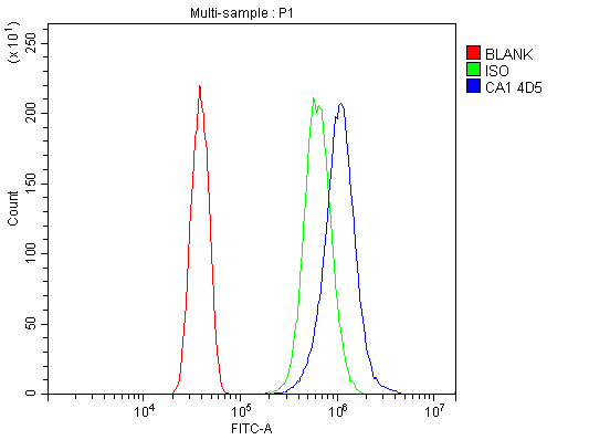
(Figure 9. Flow Cytometry analysis of SiHa cells using anti-Carbonic Anhydrase I/CA1 antibody (AAA19347).Overlay histogram showing SiHa cells stained with AAA19347 (Blue line). The cells were blocked with 10% normal goat serum. And then incubated with mouse anti- Carbonic Anhydrase I/CA1 Antibody (AAA19347, 1μg/1x106 cells) for 30 min at 20 degree C. DyLight®488 conjugated goat anti-mouse IgG (BA1126, 5-10μg/1x106 cells) was used as secondary antibody for 30 minutes at 20 degree C. Isotype control antibody (Green line) was mouse IgG (1μg/1x106) used under the same conditions. Unlabelled sample (Red line) was also used as a control.)
FCM (Flow Cytometry)
(Figure 9. Flow Cytometry analysis of SiHa cells using anti-Carbonic Anhydrase I/CA1 antibody (AAA19347).Overlay histogram showing SiHa cells stained with AAA19347 (Blue line). The cells were blocked with 10% normal goat serum. And then incubated with mouse anti- Carbonic Anhydrase I/CA1 Antibody (AAA19347, 1μg/1x106 cells) for 30 min at 20 degree C. DyLight®488 conjugated goat anti-mouse IgG (BA1126, 5-10μg/1x106 cells) was used as secondary antibody for 30 minutes at 20 degree C. Isotype control antibody (Green line) was mouse IgG (1μg/1x106) used under the same conditions. Unlabelled sample (Red line) was also used as a control.)
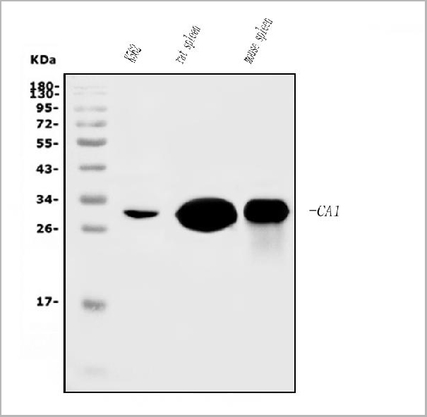
Carbonic Anhydrase I/CA1, Monoclonal Antibody (Cat# AAA19347)
Full Name
Anti-Carbonic Anhydrase I/CA1 Antibody (monoclonal, 4D5)
Gene Names
CA1; CAB; CA-I; Car1
Reactivity
Human, Mouse, Rat
Applications
Western Blot, Immunohistochemistry, Flow Cytometry
Purity
Immunogen affinity purified.
Pricing

 Bioactivity
(Fas ligand (FasL) is a 40 kDa type II membrane protein belonging to the TNF family. In the new TNF super family nomenclature, FasL is referred to as TNFSF6. The specific receptor for FasL is Fas (CD95, Apo-1), a 45 kDa type I transmembrane protein that is a member of the TNF receptor family. FasL is predominantly expressed on activated T cells and NK cells, while Fas is expressed on various types of cells. The Fas/FasL system plays a crucial role in modulating immune response by inducing cell apoptosis to maintain homeostasis, self-tolerance of lymphocytes, and immune privilege. FasL was reported to be a potent chemoattractant for neutrophils, suggesting a novel proinflammatory function of this molecule. A functional ELISA assay was conducted to detect the interaction of recombinant human FASL and recombinant human FAS. Briefly, FASL was diluted serially in PBS with 0.01% BSA (pH 7.4). Duplicate samples of 100 ul were then transferred to FAS-coated microtiter wells and incubated for 1h at 37 degree C. Wells were washed with PBST and incubated for 1h with anti-FASL pAb, then aspirated and washed 3 times. After incubation with HRP labelled secondary antibody for 1h at 37 degree C, wells were aspirated and washed 5 times. With the addition of substrate solution, wells were incubated 15-25 minutes at 37 degree C. Finally, add 50 uL stop solution to the wells and read at 450/630nm immediately. The binding activity of recombinant human FASL and recombinant human FAS was shown in Figure 1, the EC50 for this effect is 0.044 ug/mL.)
Bioactivity
(Fas ligand (FasL) is a 40 kDa type II membrane protein belonging to the TNF family. In the new TNF super family nomenclature, FasL is referred to as TNFSF6. The specific receptor for FasL is Fas (CD95, Apo-1), a 45 kDa type I transmembrane protein that is a member of the TNF receptor family. FasL is predominantly expressed on activated T cells and NK cells, while Fas is expressed on various types of cells. The Fas/FasL system plays a crucial role in modulating immune response by inducing cell apoptosis to maintain homeostasis, self-tolerance of lymphocytes, and immune privilege. FasL was reported to be a potent chemoattractant for neutrophils, suggesting a novel proinflammatory function of this molecule. A functional ELISA assay was conducted to detect the interaction of recombinant human FASL and recombinant human FAS. Briefly, FASL was diluted serially in PBS with 0.01% BSA (pH 7.4). Duplicate samples of 100 ul were then transferred to FAS-coated microtiter wells and incubated for 1h at 37 degree C. Wells were washed with PBST and incubated for 1h with anti-FASL pAb, then aspirated and washed 3 times. After incubation with HRP labelled secondary antibody for 1h at 37 degree C, wells were aspirated and washed 5 times. With the addition of substrate solution, wells were incubated 15-25 minutes at 37 degree C. Finally, add 50 uL stop solution to the wells and read at 450/630nm immediately. The binding activity of recombinant human FASL and recombinant human FAS was shown in Figure 1, the EC50 for this effect is 0.044 ug/mL.)
Factor Related Apoptosis Ligand (FASL), Active Protein (Cat# AAA161660)
Full Name
Active Factor Related Apoptosis Ligand (FASL)
Gene Names
FASLG; APTL; FASL; CD178; CD95L; ALPS1B; CD95-L; TNFSF6; APT1LG1
Applications
Activity Assay, Cell Culture
Purity
Greater than 80% by SDS-PAGE
Pricing
 Bioactivity
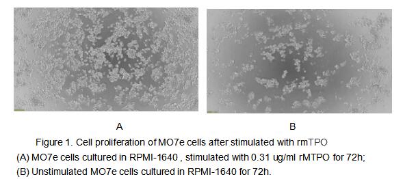
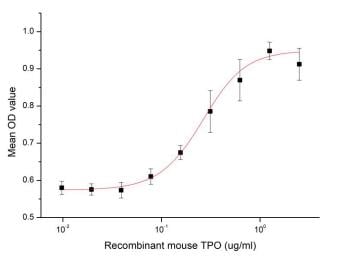
(Figure 2. The dose-effect curve of rmTPO on MO7e cells)
Bioactivity
(Figure 2. The dose-effect curve of rmTPO on MO7e cells)
Thrombopoietin (TPO), Active Protein (Cat# AAA161704)
Full Name
Active Thrombopoietin (TPO)
Gene Names
Thpo; Ml; Tpo; Mgdf; Tpo1; Tpo2; Tpo3; Tpo4; Mpllg
Applications
Activity Assay, Cell Culture
Purity
Greater than 90% by SDS-PAGE
Pricing
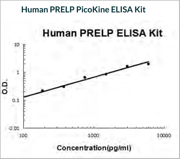 Standard Curve (Sample)
PRELP, ELISA Kit (Cat# AAA19191)
Full Name
Human PRELP ELISA Kit
Gene Names
PRELP; MST161; SLRR2A; MSTP161
Reactivity
Human
There is no detectable cross-reactivity with other relevant proteins.
There is no detectable cross-reactivity with other relevant proteins.
Pricing

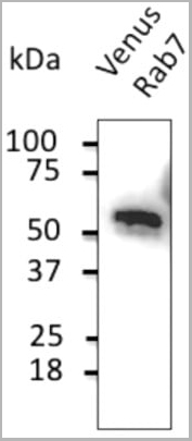 WB (Western Blot)
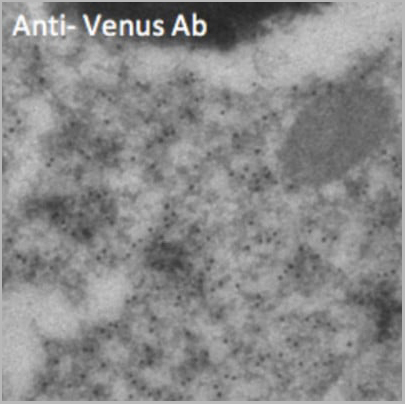
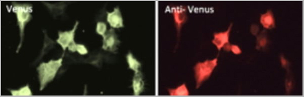
(Anti-Venus Ab at 1/2,000 dilution; 293HEK cells transduced with Venus-Rab7 Ad; lysates at 50 µg per lane; rabbit polyclonal to goat IgG (HRP) at 1/10,000 dilution;)
WB (Western Blot)
(Anti-Venus Ab at 1/2,000 dilution; 293HEK cells transduced with Venus-Rab7 Ad; lysates at 50 µg per lane; rabbit polyclonal to goat IgG (HRP) at 1/10,000 dilution;)
Venus, Polyclonal Antibody (Cat# AAA13879)
Full Name
Venus Polyclonal Antibody
Reactivity
Transfected cells proteins
Applications
Western Blot, Immunofluorescence, Immunohistochemistry, Immunohistochemistry
Pricing
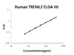 Standard Curve (Sample)
TREML2, ELISA Kit (Cat# AAA125109)
Full Name
Human TREML2 PicoKine ELISA Kit
Gene Names
TREML2; TLT2; TLT-2; C6orf76; dJ238O23.1
Reactivity
Human
There is no detectable cross-reactivity with other relevant proteins.
There is no detectable cross-reactivity with other relevant proteins.
Pricing
Alpha-1 Antitrypsin, Active Protein (Cat# AAA38953)
Full Name
Recombinant Human Alpha-1 Antitrypsin, Active
Gene Names
SERPINA1; PI; A1A; AAT; PI1; A1AT; PRO2275; alpha1AT
Purity
Greater than 95% as determined by SDS-PAGE.
Pricing
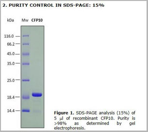 SDS-PAGE
CFP10, Recombinant Protein (Cat# AAA14239)
Full Name
Recombinant antigen CFP10 for Mycobacterium tuberculosis (Koch's bacillus)
Pricing
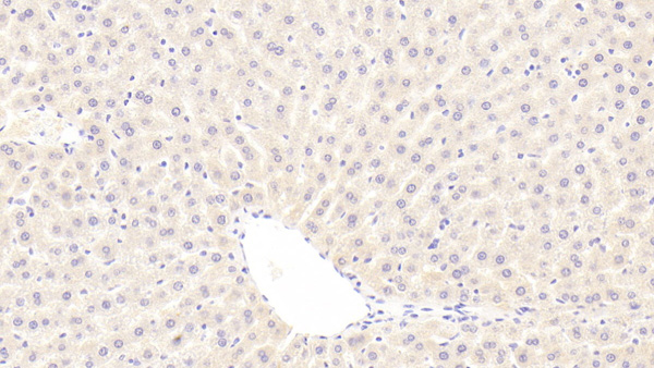
 IHC (Immunohiostchemistry)
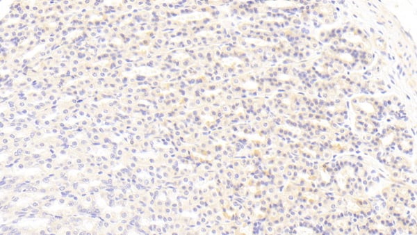
(DAB staining on IHCP;Sample: Rat Stomach Tissue; Primary Ab: 30ug/ml Mouse AntiRat BMP3 AntibodySecond Ab: 2ug/mL HRPLinked Caprine AntiMouse IgG Polyclonal Antibody(Catalog: SAA544Mu19))
IHC (Immunohiostchemistry)
(DAB staining on IHCP;Sample: Rat Stomach Tissue; Primary Ab: 30ug/ml Mouse AntiRat BMP3 AntibodySecond Ab: 2ug/mL HRPLinked Caprine AntiMouse IgG Polyclonal Antibody(Catalog: SAA544Mu19))
Bone Morphogenetic Protein 3 (BMP3), Monoclonal Antibody (Cat# AAA151790)
Full Name
Monoclonal Antibody to Bone Morphogenetic Protein 3 (BMP3)
Gene Names
Bmp3; PBMP3
Reactivity
Rat
Applications
Immunoprecipitation, Immunocytochemistry, Immunohistochemistry, Western Blot
Purity
Protein A + Protein G affinity chromatography
Pricing

 WB (Western Blot)
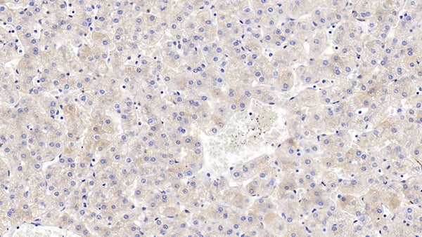
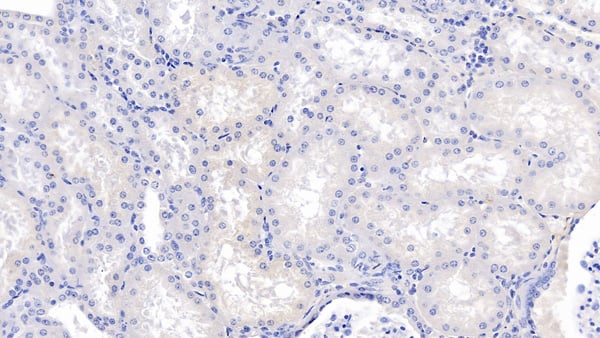
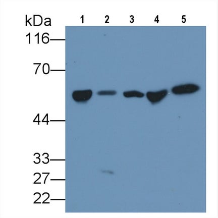
(Western Blot; Sample: Lane1: Human Serum; Lane2: Human Lung lysate; Lane3: Human Placenta lysate; Lane4: Human Plasma; Lane5: Rat PlasmaPrimary Ab: 3ug/ml Mouse AntiHuman DBP AntibodySecond Ab: 0.2ug/mL HRPLinked Caprine AntiMouse IgG Polyclonal Antibody(Catalog: SAA544Mu19))
WB (Western Blot)
(Western Blot; Sample: Lane1: Human Serum; Lane2: Human Lung lysate; Lane3: Human Placenta lysate; Lane4: Human Plasma; Lane5: Rat PlasmaPrimary Ab: 3ug/ml Mouse AntiHuman DBP AntibodySecond Ab: 0.2ug/mL HRPLinked Caprine AntiMouse IgG Polyclonal Antibody(Catalog: SAA544Mu19))
Vitamin D Binding Protein (DBP), Monoclonal Antibody (Cat# AAA151750)
Full Name
Monoclonal Antibody to Vitamin D Binding Protein (DBP)
Gene Names
GC; DBP; GRD3; VDBG; VDBP; DBP/GC
Reactivity
Human, Rat, Pig
Applications
Immunoprecipitation, Immunocytochemistry, Immunohistochemistry, Western Blot
Purity
Protein A + Protein G affinity chromatography
Pricing
 Bioactivity
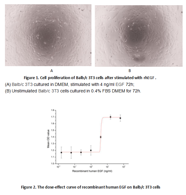
(Epidermal growth factor (EGF) is a growth factor that stimulates cell growth, proliferation, and differentiation by binding to its receptor EGFR. To test the effect of EGF on cell proliferation of 3T3 fibroblasts, Balb/c 3T3 cells were seeded into triplicate wells of 96-well plates and allowed to attach overnight, then the medium was replaced with various concentrations of recombinant human EGF. After incubated for 48h, cells were observed by inverted microscope and cell proliferation was measured by Cell Counting Kit-8 (CCK-8). Briefly, 10 ul of CCK-8 solution was added to each well of the plate, then measure the absorbance at 450 nm using a microplate reader after incubating the plate for 1-4 hours at 37 degree C. Cell proliferation of Balb/c 3T3 cells after incubation with rhEGF for 48h observed by inverted microscope was shown in Figure 1. The dose-effect curve of rhEGF was shown in Figure2. It was obvious that rhEGF significantly promoted cell proliferation of 3T3 cells. The ED50 for this effect is typically 3.9 ng/ml.)
Bioactivity
(Epidermal growth factor (EGF) is a growth factor that stimulates cell growth, proliferation, and differentiation by binding to its receptor EGFR. To test the effect of EGF on cell proliferation of 3T3 fibroblasts, Balb/c 3T3 cells were seeded into triplicate wells of 96-well plates and allowed to attach overnight, then the medium was replaced with various concentrations of recombinant human EGF. After incubated for 48h, cells were observed by inverted microscope and cell proliferation was measured by Cell Counting Kit-8 (CCK-8). Briefly, 10 ul of CCK-8 solution was added to each well of the plate, then measure the absorbance at 450 nm using a microplate reader after incubating the plate for 1-4 hours at 37 degree C. Cell proliferation of Balb/c 3T3 cells after incubation with rhEGF for 48h observed by inverted microscope was shown in Figure 1. The dose-effect curve of rhEGF was shown in Figure2. It was obvious that rhEGF significantly promoted cell proliferation of 3T3 cells. The ED50 for this effect is typically 3.9 ng/ml.)
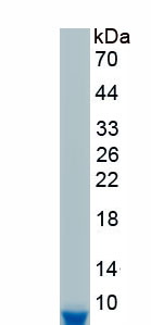
Epidermal Growth Factor (EGF), Active Protein (Cat# AAA161747)
Full Name
Active Epidermal Growth Factor (EGF)
Gene Names
EGF; URG; HOMG4
Applications
Activity Assay, Cell Culture
Purity
Greater than 90% by SDS-PAGE
Pricing
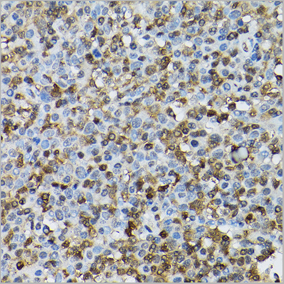
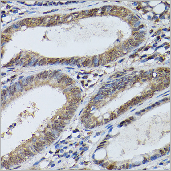
 IP (Immunoprecipitation)
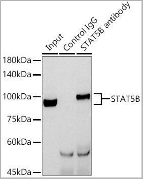
(Immunoprecipitation analysis of 600ug extracts of Mouse thymus cells using 3ug STAT5B antibody. Western blot was performed from the immunoprecipitate using STAT5B antibody at a dilution of 1:1000.)
IP (Immunoprecipitation)
(Immunoprecipitation analysis of 600ug extracts of Mouse thymus cells using 3ug STAT5B antibody. Western blot was performed from the immunoprecipitate using STAT5B antibody at a dilution of 1:1000.)
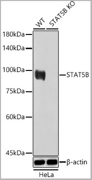
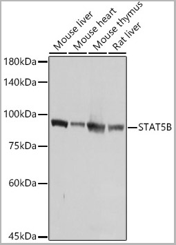
STAT5B, Polyclonal Antibody (Cat# AAA28416)
Full Name
[KO Validated] STAT5B Rabbit pAb
Reactivity
Human, Mouse, Rat
Applications
Western Blot, Immunohistochemistry, Immunoprecipitation, Immunofluorescence, Immunocytochemistry
Purity
Affinity purification
Pricing
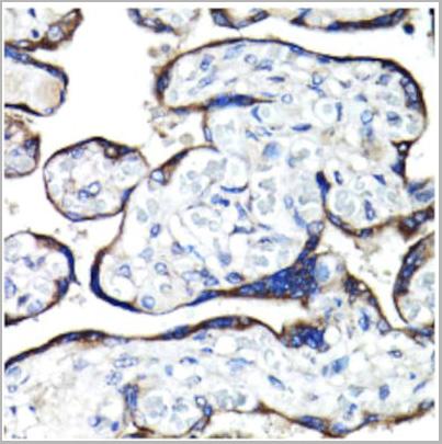
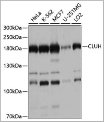
 IF (Immunofluorescence)
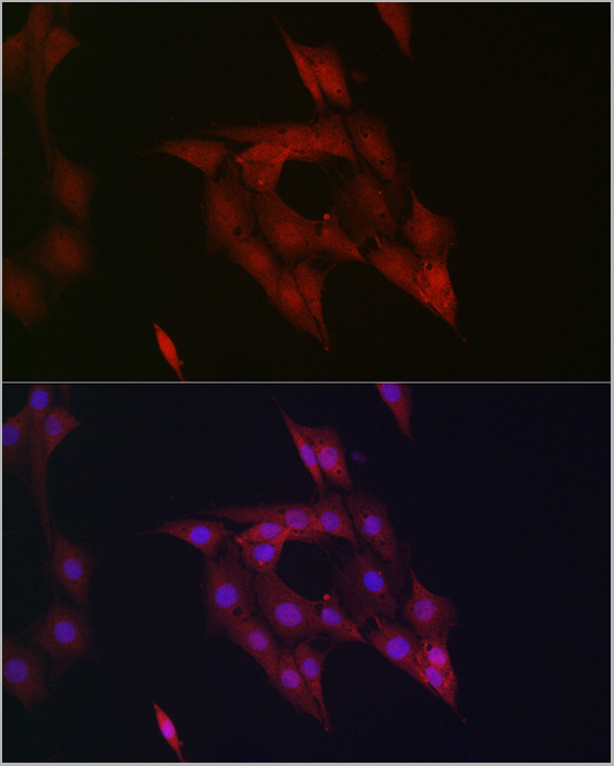
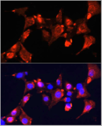
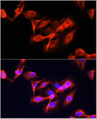
(Immunofluorescence analysis of U-2 OS cells using CLUH Polyclonal Antibody (AAA28214) at dilution of 1:100 (40x lens). Blue: DAPI for nuclear staining.)
IF (Immunofluorescence)
(Immunofluorescence analysis of U-2 OS cells using CLUH Polyclonal Antibody (AAA28214) at dilution of 1:100 (40x lens). Blue: DAPI for nuclear staining.)
CLUH, Polyclonal Antibody (Cat# AAA28214)
Full Name
CLUH Polyclonal Antibody
Gene Names
CLUH; CLU1; KIAA0664
Reactivity
Human, Mouse
Applications
Western Blot, Immunohistochemistry, Immunofluorescence, Immunocytochemistry
Purity
Affinity purification
Pricing
Rhinovirus outer capsid Protein 3, Monoclonal Antibody (Cat# AAA13450)
Full Name
Rhinovirus outer capsid Protein 3 (VP3)
Applications
Immunofluorescence
Purity
> 90% pure. Protein A Chromatography.
Pricing
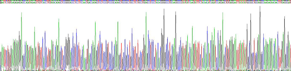
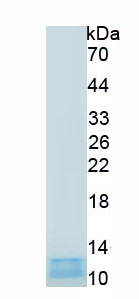 SDS-PAGE
(Figure. SDS-PAGE)
SDS-PAGE
(Figure. SDS-PAGE)
Aprotinin (AP), Protein (Cat# AAA161949)
Full Name
Eukaryotic Aprotinin (AP)
Applications
Western Blot
Purity
Greater than 90% by SDS-PAGE
Pricing

 Bioactivity
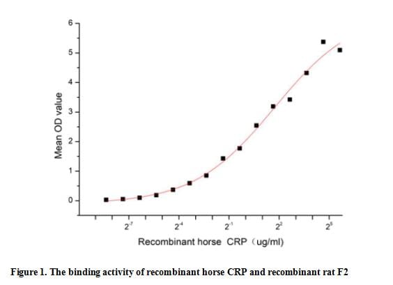
(C reactive protein (CRP) is an annular (ring-shaped), pentameric protein found in blood plasma, whose levels rise in response to inflammation. It is an acute-phase protein of hepatic origin that increases following interleukin-6 secretion by macrophages and T cells. Its physiological role is to bind to lysophosphatidylcholine expressed on the surface of dead or dying cells (and some types of bacteria) in order to activate the complement system via C1q. Besides, Coagulation Factor II (F2) has been identified as an interactor of CRP, thus a functional binding ELISA assay was conducted to detect the interaction of recombinant horse CRP and recombinant rat F2. Briefly, CRP was diluted serially in PBS with 0.01% BSA (pH 7.4). Duplicate samples of 100 ul were then transferred to F2-coated microtiter wells and incubated for 1h at 37 degree C. Wells were washed with PBST and incubated for 1h with anti-CRP pAb, then aspirated and washed 3 times. After incubation with HRP labelled secondary antibody for 1h at 37 degree C, wells were aspirated and washed 5 times. With the addition of substrate solution, wells were incubated 15-25 minutes at 37 degree C. Finally, add 50 uL stop solution to the wells and read at 450/630 nm immediately. The binding activity of recombinant horse CRP and recombinant rat F2 was shown in Figure 1, the EC50 for this effect is 3.29 ug/mL.)
Bioactivity
(C reactive protein (CRP) is an annular (ring-shaped), pentameric protein found in blood plasma, whose levels rise in response to inflammation. It is an acute-phase protein of hepatic origin that increases following interleukin-6 secretion by macrophages and T cells. Its physiological role is to bind to lysophosphatidylcholine expressed on the surface of dead or dying cells (and some types of bacteria) in order to activate the complement system via C1q. Besides, Coagulation Factor II (F2) has been identified as an interactor of CRP, thus a functional binding ELISA assay was conducted to detect the interaction of recombinant horse CRP and recombinant rat F2. Briefly, CRP was diluted serially in PBS with 0.01% BSA (pH 7.4). Duplicate samples of 100 ul were then transferred to F2-coated microtiter wells and incubated for 1h at 37 degree C. Wells were washed with PBST and incubated for 1h with anti-CRP pAb, then aspirated and washed 3 times. After incubation with HRP labelled secondary antibody for 1h at 37 degree C, wells were aspirated and washed 5 times. With the addition of substrate solution, wells were incubated 15-25 minutes at 37 degree C. Finally, add 50 uL stop solution to the wells and read at 450/630 nm immediately. The binding activity of recombinant horse CRP and recombinant rat F2 was shown in Figure 1, the EC50 for this effect is 3.29 ug/mL.)
C ReProtein (CRP), Active Protein (Cat# AAA161779)
Full Name
Active C Reactive Protein (CRP)
Applications
Activity Assay, Cell Culture
Purity
Greater than 90% by SDS-PAGE
Pricing
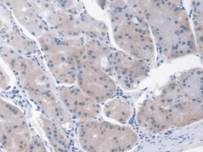 IHC (Immunohistochemisry)
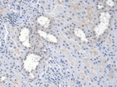
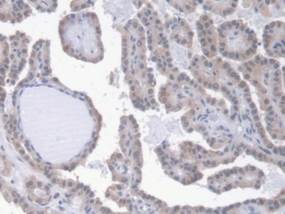
(DAB staining on IHCP;Sample: Human Stomach Tissue; Primary Ab: 30ug/ml Mouse AntiHuman EPO AntibodySecond Ab: 2ug/mL HRPLinked Caprine AntiMouse IgG Polyclonal Antibody(Catalog: SAA544Mu19))
IHC (Immunohistochemisry)
(DAB staining on IHCP;Sample: Human Stomach Tissue; Primary Ab: 30ug/ml Mouse AntiHuman EPO AntibodySecond Ab: 2ug/mL HRPLinked Caprine AntiMouse IgG Polyclonal Antibody(Catalog: SAA544Mu19))
Erythropoietin (EPO), Monoclonal Antibody (Cat# AAA151434)
Full Name
Monoclonal Antibody to Erythropoietin (EPO)
Gene Names
EPO; EP; MVCD2
Reactivity
Human
Applications
Immunoprecipitation, Immunocytochemistry, Immunohistochemistry, Western Blot
Purity
Protein A + Protein G affinity chromatography
Pricing
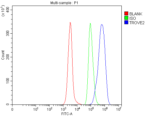 FCM/FACS (Flow Cytometry)
(Figure 2. Flow Cytometry analysis of Daudi cells using anti-TROVE2/SS-A/RO60 antibody (AAA126305).Overlay histogram showing Daudi cells stained with AAA126305 (Blue line). The cells were blocked with 10% normal goat serum. And then incubated with rabbit anti-TROVE2/SS-A/RO60 Antibody (AAA126305, 1 ug/1x10^6 cells) for 30 min at 20 degree C. DyLight488 conjugated goat anti-rabbit IgG was used as secondary antibody for 30 minutes at 20 degree C. Isotype control antibody (Green line) was rabbit IgG (1 ug/1x10^6) used under the same conditions. Unlabelled sample (Red line) was also used as a control.)
FCM/FACS (Flow Cytometry)
(Figure 2. Flow Cytometry analysis of Daudi cells using anti-TROVE2/SS-A/RO60 antibody (AAA126305).Overlay histogram showing Daudi cells stained with AAA126305 (Blue line). The cells were blocked with 10% normal goat serum. And then incubated with rabbit anti-TROVE2/SS-A/RO60 Antibody (AAA126305, 1 ug/1x10^6 cells) for 30 min at 20 degree C. DyLight488 conjugated goat anti-rabbit IgG was used as secondary antibody for 30 minutes at 20 degree C. Isotype control antibody (Green line) was rabbit IgG (1 ug/1x10^6) used under the same conditions. Unlabelled sample (Red line) was also used as a control.)
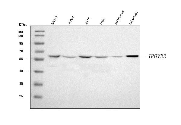
TROVE2/SS-A/RO60, Polyclonal Antibody (Cat# AAA126305)
Full Name
Anti-TROVE2/SS-A/RO60 Antibody Picoband
Gene Names
TROVE2; RO60; SSA2; RORNP
Reactivity
Human, Rat
Applications
Flow Cytometry, Western Blot
Purity
Immunogen affinity purified.
Pricing
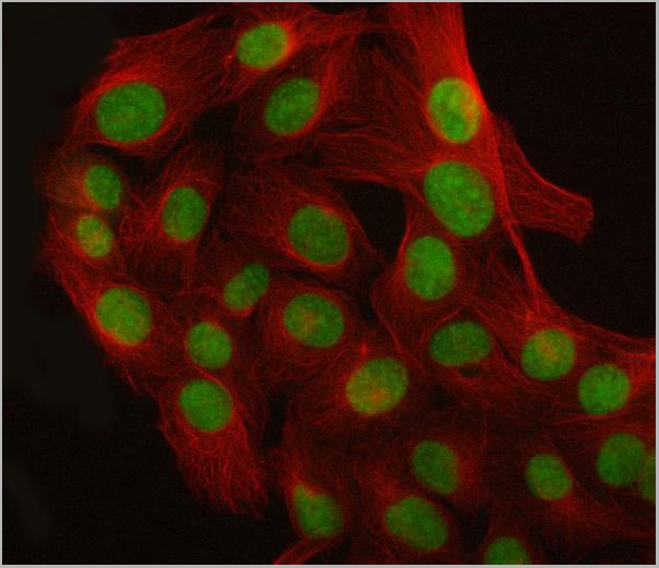
 FCM (Flow Cytometry)
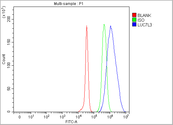
(Figure 13. Flow Cytometry analysis of JK cells using anti-CROP/LUC7L3 antibody (AAA19906).Overlay histogram showing JK cells stained with AAA19906 (Blue line). To facilitate intracellular staining, cells were fixed with 4% paraformaldehyde and permeabilized with permeabilization buffer. The cells were blocked with 10% normal goat serum. And then incubated with rabbit anti-CROP/LUC7L3 Antibody (AAA19906, 1ug/1x106 cells) for 30 min at 20 degree C. DyLight®488 conjugated goat anti-rabbit IgG (BA1127, 5-10ug/1x106 cells) was used as secondary antibody for 30 minutes at 20 degree C. Isotype control antibody (Green line) was rabbit IgG (1ug/1x106) used under the same conditions. Unlabelled sample (Red line) was also used as a control.)
FCM (Flow Cytometry)
(Figure 13. Flow Cytometry analysis of JK cells using anti-CROP/LUC7L3 antibody (AAA19906).Overlay histogram showing JK cells stained with AAA19906 (Blue line). To facilitate intracellular staining, cells were fixed with 4% paraformaldehyde and permeabilized with permeabilization buffer. The cells were blocked with 10% normal goat serum. And then incubated with rabbit anti-CROP/LUC7L3 Antibody (AAA19906, 1ug/1x106 cells) for 30 min at 20 degree C. DyLight®488 conjugated goat anti-rabbit IgG (BA1127, 5-10ug/1x106 cells) was used as secondary antibody for 30 minutes at 20 degree C. Isotype control antibody (Green line) was rabbit IgG (1ug/1x106) used under the same conditions. Unlabelled sample (Red line) was also used as a control.)
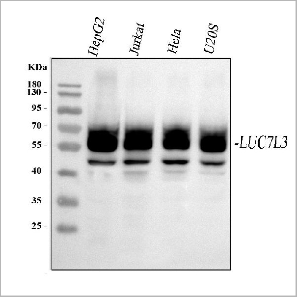
CROP/LUC7L3, Polyclonal Antibody (Cat# AAA19906)
Full Name
Anti-CROP/LUC7L3 Antibody Picoband
Gene Names
LUC7L3; CRA; CROP; LUC7A; hLuc7A; CREAP-1; OA48-18
Reactivity
Human, Mouse, Rat
Applications
Western Blot, Immunohistochemistry, Immunocytochemistry, Immunofluorescence, Flow Cytometry
Purity
Immunogen affinity purified.
Pricing

 Bioactivity
 IF (Immunofluorescence)
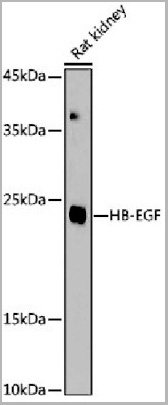
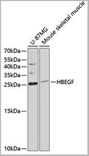
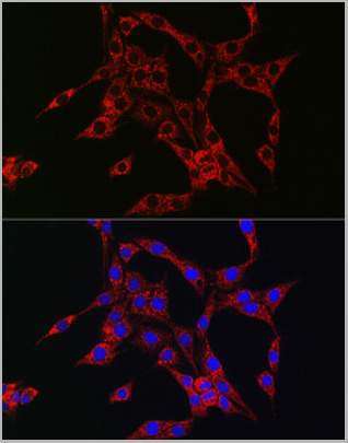
(Immunofluorescence analysis of PC-12 cells using HB-EGF Rabbit pAb (AAA28122) at dilution of 1:150 (40x lens). Blue: DAPI for nuclear staining.)
IF (Immunofluorescence)
(Immunofluorescence analysis of PC-12 cells using HB-EGF Rabbit pAb (AAA28122) at dilution of 1:150 (40x lens). Blue: DAPI for nuclear staining.)
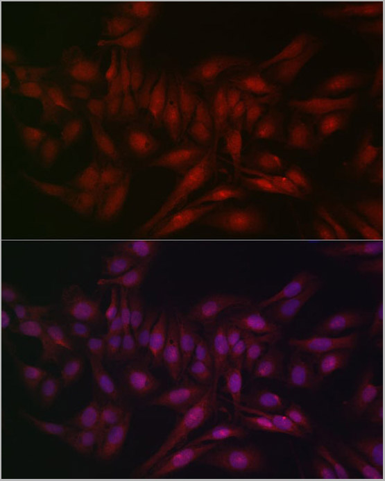
HBEGF, Polyclonal Antibody (Cat# AAA28122)
Full Name
HBEGF Polyclonal Antibody
Gene Names
HBEGF; DTR; DTS; DTSF; HEGFL
Applications
Western Blot, Immunofluorescence, Immunocytochemistry
Purity
Affinity purification
Pricing
 Application Data
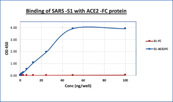
(Figure 2: Binding assay of recombinant SARS CoV-2 Spike S1 C-Term His Tag Protein with recombinant Human ACE2 Fc Protein. An ELISA plate was coated with 1ug/ml of recombinant SARS CoV-2 Spike S1 C-Term His Tag Protein, and the plate was incubated overnight at 4 degree C. Different concentrations (100 ng/well to 0 ng/well) of recombinant Human ACE2 Fc Protein was added to the wells. HRP conjugated goat anti-human Fc was used as secondary antibody.)
Application Data
(Figure 2: Binding assay of recombinant SARS CoV-2 Spike S1 C-Term His Tag Protein with recombinant Human ACE2 Fc Protein. An ELISA plate was coated with 1ug/ml of recombinant SARS CoV-2 Spike S1 C-Term His Tag Protein, and the plate was incubated overnight at 4 degree C. Different concentrations (100 ng/well to 0 ng/well) of recombinant Human ACE2 Fc Protein was added to the wells. HRP conjugated goat anti-human Fc was used as secondary antibody.)
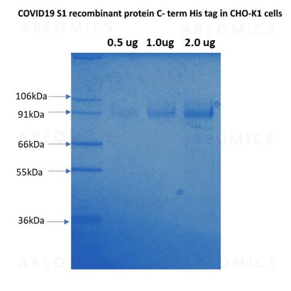
COVID 19 Spike S1 Coronavirus, Recombinant Protein (Cat# AAA78555)
Full Name
SARS CoV2 Spike S1 C-Term His Tag Protein
Applications
Functional Assay
Purity
>95% by SDS-PAGE
Pricing
 Bioactivity
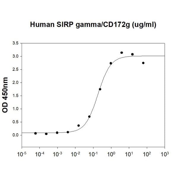
(Human CD47 is coated at 1 ug/ml (100 ul/well) can bind Human SIRP gamma/CD172g (CAT# ATGP4030) in a Functional ELISA assay.)
Bioactivity
(Human CD47 is coated at 1 ug/ml (100 ul/well) can bind Human SIRP gamma/CD172g (CAT# ATGP4030) in a Functional ELISA assay.)
CD47, Active Protein (Cat# AAA48392)
Full Name
Recombinant human CD47 protein
Gene Names
CD47; IAP; OA3; MER6
Applications
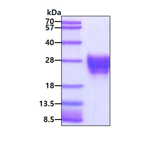
Bioactivity, SDS-PAGE
Purity
>95% by SDS-PAGE
Pricing
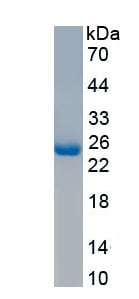 SDS-PAGE
(Figure. SDS-PAGE)
SDS-PAGE
(Figure. SDS-PAGE)
Ras Related C3 Botulinum Toxin Substrate 1 (Rac1), Recombinant Protein (Cat# AAA162023)
Full Name
Recombinant Ras Related C3 Botulinum Toxin Substrate 1 (Rac1)
Gene Names
Rac1; AL023026; D5Ertd559e
Applications
Western Blot
Purity
Greater than 95% by SDS-PAGE
Pricing
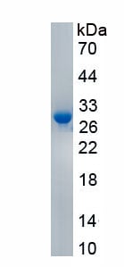
 Bioactivity
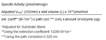
(Granzyme B is a member of the granzyme family of the serine proteases found specifically in the cytotoxic granules of cytotoxic T lymphocytes (CTL) and natural killer (NK) cells. Granzyme B plays an essential role in granule-mediated apoptosis and may have additional roles in rheumatoid arthritis and in bacterial and viral infections. It activates various caspases and cleaves proteins such as aggrecan. The activity of recombinant pig Granzyme B was measured by its ability to cleaves a thioester substrate Z-Lys-SBzl•HCl. The reaction was performed in 0.05 M Tris, 0.15 M NaCl, 0.01% Triton X-100, pH 8.0 (Assay Buffer), initiated by addition 50 uL of various concentrations of GZMB (diluted by Assay Buffer) to 50 uL of 1.2 mM Substrate and DTNB mixture. The final well serves as a negative control with no GZMB, replaced with 50 uL assay buffer. Incubated at 25 degree C for 5min, then read at a wavelength of 405 nm. The specific activity of recombinant pig Granzyme B is >440 pmol/min/ug.)
Bioactivity
(Granzyme B is a member of the granzyme family of the serine proteases found specifically in the cytotoxic granules of cytotoxic T lymphocytes (CTL) and natural killer (NK) cells. Granzyme B plays an essential role in granule-mediated apoptosis and may have additional roles in rheumatoid arthritis and in bacterial and viral infections. It activates various caspases and cleaves proteins such as aggrecan. The activity of recombinant pig Granzyme B was measured by its ability to cleaves a thioester substrate Z-Lys-SBzl•HCl. The reaction was performed in 0.05 M Tris, 0.15 M NaCl, 0.01% Triton X-100, pH 8.0 (Assay Buffer), initiated by addition 50 uL of various concentrations of GZMB (diluted by Assay Buffer) to 50 uL of 1.2 mM Substrate and DTNB mixture. The final well serves as a negative control with no GZMB, replaced with 50 uL assay buffer. Incubated at 25 degree C for 5min, then read at a wavelength of 405 nm. The specific activity of recombinant pig Granzyme B is >440 pmol/min/ug.)
Granzyme B (GZMB), Active Protein (Cat# AAA161751)
Full Name
Active Granzyme B (GZMB)
Applications
Activity Assay, Cell Culture
Purity
Greater than 90% by SDS-PAGE
Pricing