
Filters
Clonality
Type
Reactivity
Gene Name
Isotype
Host
Application
Clone
10000 results for "Recombinant Proteins" - showing 2250-2300
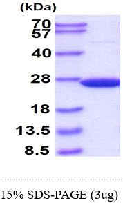 SDS-PAGE
SOD2, Active Protein (Cat# AAA48636)
Full Name
SOD2, 25-222aa, Human, His tag, E Coli (Bioactivity Validated)
Gene Names
SOD2; IPOB; IPO-B; MNSOD; MVCD6; Mn-SOD
Applications
SDS-PAGE
Purity
> 95% by SDS-PAGE
Pricing
 Bioactivity
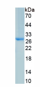
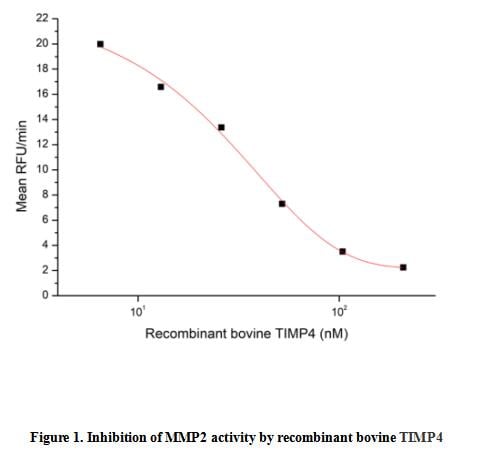
(Tissue Inhibitors of Metalloproteinase 4 (TIMP4) is an enzyme that in humans is encoded by the TIMP4 gene. This gene belongs to the tissue inhibitor of metalloproteinases gene family. The proteins encoded by this gene family are inhibitors of the matrix metalloproteinases, a group of peptidases involved in degradation of the extracellular matrix. The secreted, netrin domain-containing protein encoded by this gene is involved in regulation of platelet aggregation and recruitment and may play role in hormonal regulation and endometrial tissue remodeling. The activity of recombinant bovine TIMP4 was measured by its ability to inhibit rhMMP2 cleavage of a fluorogenic peptide substrate MCA-Pro-Leu-Gly-Leu-DPA-Ala-Arg-NH2 in the assay buffer 50 mM Tris, 10 mM CaCl2, 150 mM NaCl, 0.05% (w/v) Brij-35, pH 7.5. rhMMP2 was diluted to 100 ug/ml and activated with 1 mM APMA at 37 degree C for 1 hour and rbTIMP4 (MW: 24.05 KD) was diluted to different concentrations with the assay buffer. Mix 8 ul of rbTIMP4 curve dilutions, 12.8 ul of activated rhMMP-2, and 59.2 ul of assay buffer, including a control containing assay buffer and the diluted rhMMP-2 and incubate the reactions for 2 hours at 37 degree C. Loading 50 ul of the incubated mixtures which were diluted five-fold in assay buffer into empty wells of a plate, and start the reaction by adding 50 ul of 20 uM substrate. Include a substrate blank containing 50 ul of assay buffer and 50 ul of 20 uM substrate. Then read at excitiation and emission wavelengths of 320 nm and 405 nm, respectively, in kinetic mode for 5 minutes. The result was shown in Figure 1 and it was obvious that recombinant bovine TIMP4 significantly decreased rhMMP2 activity. The inhibition IC50 was )
Bioactivity
(Tissue Inhibitors of Metalloproteinase 4 (TIMP4) is an enzyme that in humans is encoded by the TIMP4 gene. This gene belongs to the tissue inhibitor of metalloproteinases gene family. The proteins encoded by this gene family are inhibitors of the matrix metalloproteinases, a group of peptidases involved in degradation of the extracellular matrix. The secreted, netrin domain-containing protein encoded by this gene is involved in regulation of platelet aggregation and recruitment and may play role in hormonal regulation and endometrial tissue remodeling. The activity of recombinant bovine TIMP4 was measured by its ability to inhibit rhMMP2 cleavage of a fluorogenic peptide substrate MCA-Pro-Leu-Gly-Leu-DPA-Ala-Arg-NH2 in the assay buffer 50 mM Tris, 10 mM CaCl2, 150 mM NaCl, 0.05% (w/v) Brij-35, pH 7.5. rhMMP2 was diluted to 100 ug/ml and activated with 1 mM APMA at 37 degree C for 1 hour and rbTIMP4 (MW: 24.05 KD) was diluted to different concentrations with the assay buffer. Mix 8 ul of rbTIMP4 curve dilutions, 12.8 ul of activated rhMMP-2, and 59.2 ul of assay buffer, including a control containing assay buffer and the diluted rhMMP-2 and incubate the reactions for 2 hours at 37 degree C. Loading 50 ul of the incubated mixtures which were diluted five-fold in assay buffer into empty wells of a plate, and start the reaction by adding 50 ul of 20 uM substrate. Include a substrate blank containing 50 ul of assay buffer and 50 ul of 20 uM substrate. Then read at excitiation and emission wavelengths of 320 nm and 405 nm, respectively, in kinetic mode for 5 minutes. The result was shown in Figure 1 and it was obvious that recombinant bovine TIMP4 significantly decreased rhMMP2 activity. The inhibition IC50 was )
Tissue Inhibitors Of Metalloproteinase 4 (TIMP4), Active Protein (Cat# AAA161701)
Full Name
Active Tissue Inhibitors Of Metalloproteinase 4 (TIMP4)
Applications
Activity Assay, Cell Culture
Purity
Greater than 95% by SDS-PAGE
Pricing
 Bioactivity
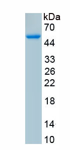
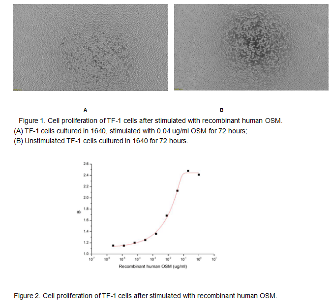
(Oncostatin M (OSM) is an approximately 30 KDa secreted cytokine belonging to the Interleukin-6 family. Like other members of the IL-6 family such as IL-11, CNTF, and Cardiotrophin-1, OSM plays crucial roles in inflammation, neuroprotection, hematopoiesis, metabolism and development. To test the effect of OSM on cell proliferation, TF-1 cells were seeded into triplicate wells of 96-well plates with various concentrations of recombinant human OSM. After incubated for 72 hours, cells were observed by inverted microscope and cell proliferation was measured by Cell Counting Kit-8 (CCK-8). Briefly, 10 ul of CCK-8 solution was added to each well of the plate, then the absorbance at 450 nm was measured using a microplate reader after incubating the plate for 1-4 hours at 37 degree C. Proliferation of TF-1 cells after incubation with OSM for 72 hours observed by inverted microscope was shown in Figure 1. Cell viability was assessed by CCK-8 (Cell Counting Kit-8) assay after incubation with recombinant human OSM for 72 hours. The result was shown in Figure 2. It was obvious that recombinant human OSM significantly increased cell viability of TF-1 cells, the EC50 was 0.014 ug/ml.)
Bioactivity
(Oncostatin M (OSM) is an approximately 30 KDa secreted cytokine belonging to the Interleukin-6 family. Like other members of the IL-6 family such as IL-11, CNTF, and Cardiotrophin-1, OSM plays crucial roles in inflammation, neuroprotection, hematopoiesis, metabolism and development. To test the effect of OSM on cell proliferation, TF-1 cells were seeded into triplicate wells of 96-well plates with various concentrations of recombinant human OSM. After incubated for 72 hours, cells were observed by inverted microscope and cell proliferation was measured by Cell Counting Kit-8 (CCK-8). Briefly, 10 ul of CCK-8 solution was added to each well of the plate, then the absorbance at 450 nm was measured using a microplate reader after incubating the plate for 1-4 hours at 37 degree C. Proliferation of TF-1 cells after incubation with OSM for 72 hours observed by inverted microscope was shown in Figure 1. Cell viability was assessed by CCK-8 (Cell Counting Kit-8) assay after incubation with recombinant human OSM for 72 hours. The result was shown in Figure 2. It was obvious that recombinant human OSM significantly increased cell viability of TF-1 cells, the EC50 was 0.014 ug/ml.)
Oncostatin M (OSM), Active Protein (Cat# AAA161689)
Full Name
Active Oncostatin M (OSM)
Applications
Activity Assay, Cell Culture
Purity
Greater than 90% by SDS-PAGE
Pricing
 IHC (Immunohiostchemistry)
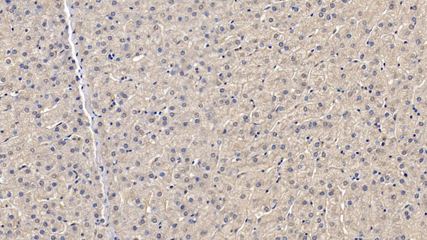
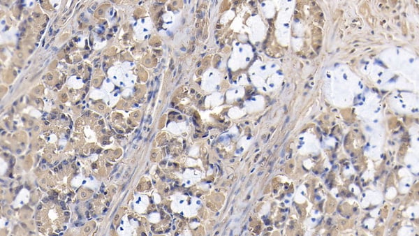
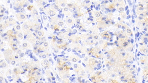
(DAB staining on IHCP;Sample: Human Stomach Tissue; Primary Ab: 20ug/ml Mouse AntiHuman HMG1 AntibodySecond Ab: 2ug/mL HRPLinked Caprine AntiMouse IgG Polyclonal Antibody(Catalog: SAA544Mu19))
IHC (Immunohiostchemistry)
(DAB staining on IHCP;Sample: Human Stomach Tissue; Primary Ab: 20ug/ml Mouse AntiHuman HMG1 AntibodySecond Ab: 2ug/mL HRPLinked Caprine AntiMouse IgG Polyclonal Antibody(Catalog: SAA544Mu19))
High Mobility Group Protein 1 (HMGB1), Monoclonal Antibody (Cat# AAA151531)
Full Name
Monoclonal Antibody to High Mobility Group Protein 1 (HMGB1)
Gene Names
HMGB1; HMG1; HMG3; SBP-1
Reactivity
Human, Pig
Applications
Immunoprecipitation, Immunocytochemistry, Immunohistochemistry, Western Blot
Purity
Protein A + Protein G affinity chromatography
Pricing
 Bioactivity
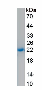
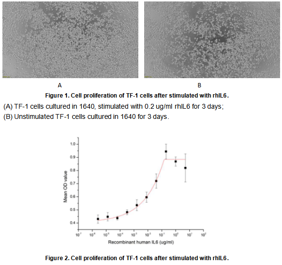
(Interleukin 6 (IL-6) is an interleukin that acts as both a pro-inflammatory cytokine and an anti-inflammatory myokine. Interleukin 6 is secreted by T cells and macrophages to stimulate immune response and also plays a role in fighting infection. It supports the growth of B cells and is antagonistic to regulatory T cells. To test the effect of IL6 on cell proliferation, TF-1 cells were seeded into triplicate wells of 96-well plates at a density of 20,000 cells/well with 10% serum standard 1640 which contains various concentrations of recombinant human IL6. After incubated for 3 days, cells were observed by inverted microscope and cell proliferation was measured by Cell Counting Kit-8 (CCK-8). Briefly, 10 ul of CCK-8 solution was added to each well of the plate, then the absorbance at 450 nm was measured using a microplate reader after incubating the plate for 1-4 hours at 37 degree C. Proliferation of TF-1 cells after incubation with rhIL6 for 3 days observed by inverted microscope was shown in Figure 1. Cell viability was assessed by CCK-8 (Cell Counting Kit-8) assay after incubation with recombinant human IL6 for 3 days. The result was shown in Figure 2. It was obvious that recombinant human IL6 significantly increased cell viability of TF-1 cells, the EC50 was 0.016 ug/ml.)
Bioactivity
(Interleukin 6 (IL-6) is an interleukin that acts as both a pro-inflammatory cytokine and an anti-inflammatory myokine. Interleukin 6 is secreted by T cells and macrophages to stimulate immune response and also plays a role in fighting infection. It supports the growth of B cells and is antagonistic to regulatory T cells. To test the effect of IL6 on cell proliferation, TF-1 cells were seeded into triplicate wells of 96-well plates at a density of 20,000 cells/well with 10% serum standard 1640 which contains various concentrations of recombinant human IL6. After incubated for 3 days, cells were observed by inverted microscope and cell proliferation was measured by Cell Counting Kit-8 (CCK-8). Briefly, 10 ul of CCK-8 solution was added to each well of the plate, then the absorbance at 450 nm was measured using a microplate reader after incubating the plate for 1-4 hours at 37 degree C. Proliferation of TF-1 cells after incubation with rhIL6 for 3 days observed by inverted microscope was shown in Figure 1. Cell viability was assessed by CCK-8 (Cell Counting Kit-8) assay after incubation with recombinant human IL6 for 3 days. The result was shown in Figure 2. It was obvious that recombinant human IL6 significantly increased cell viability of TF-1 cells, the EC50 was 0.016 ug/ml.)
Interleukin 6 (IL6), Active Protein (Cat# AAA161680)
Full Name
Active Interleukin 6 (IL6)
Gene Names
IL6; HGF; HSF; BSF2; IL-6; IFNB2
Applications
Activity Assay, Cell Culture
Purity
Greater than 90% by SDS-PAGE
Pricing
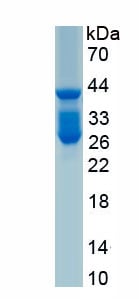 SDS-PAGE
(Figure. SDS-PAGE)
SDS-PAGE
(Figure. SDS-PAGE)
Rhodopsin (RHO), Recombinant Protein (Cat# AAA161996)
Full Name
Recombinant Rhodopsin (RHO)
Gene Names
Rho; Ops; RP4; Opn2; Noerg1
Applications
Western Blot
Purity
Greater than 80% by SDS-PAGE
Pricing

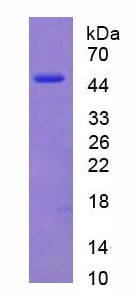 SDS-PAGE
(Figure. SDS-PAGE)
SDS-PAGE
(Figure. SDS-PAGE)
Vimentin (VIM), Recombinant Protein (Cat# AAA150685)
Full Name
Recombinant Vimentin (VIM)
Applications
Western Blot
Purity
>97%
Pricing
 Application Data
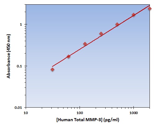
(The OmniKine Human Total MMP-3 ELISA Kit allows for the detection and quantification of endogenous levels of natural and/or recombinant Human Total MMP-3 proteins within the range of 32-2000 pg/ml.)
Application Data
(The OmniKine Human Total MMP-3 ELISA Kit allows for the detection and quantification of endogenous levels of natural and/or recombinant Human Total MMP-3 proteins within the range of 32-2000 pg/ml.)
Total MMP-3, ELISA Kit (Cat# AAA315759)
Full Name
Total MMP-3 (Human) OmniKine ELISA Kit
Gene Names
MMP3; SL-1; STMY; STR1; CHDS6; MMP-3; STMY1
Reactivity
Human
Pricing
 IF (Immunofluorescence)
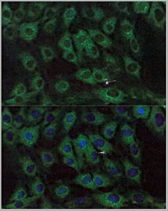
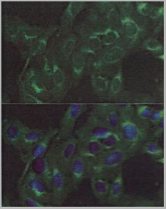
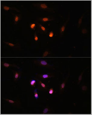
(Immunofluorescence analysis of U2OS cells using FRMD6 antibody (AAA28220) at dilution of 1:100. Blue: DAPI for nuclear staining.)
IF (Immunofluorescence)
(Immunofluorescence analysis of U2OS cells using FRMD6 antibody (AAA28220) at dilution of 1:100. Blue: DAPI for nuclear staining.)
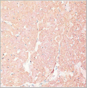
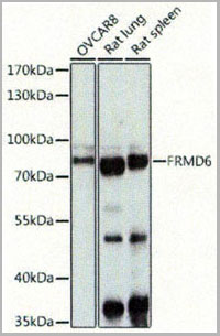
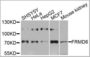
FRMD6, Polyclonal Antibody (Cat# AAA28220)
Full Name
FRMD6 Polyclonal Antibody
Gene Names
FRMD6; EX1; Willin; C14orf31; c14_5320
Reactivity
Human, Mouse, Rat
Applications
Western Blot, Immunohistochemistry, Immunofluorescence
Purity
Affinity purification
Pricing
 IF (Immunofluorescence)
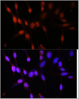
(Immunofluorescence analysis of NIH/3T3 cells using KAT6A antibody at dilution of 1:100. Blue: DAPI for nuclear staining.)
IF (Immunofluorescence)
(Immunofluorescence analysis of NIH/3T3 cells using KAT6A antibody at dilution of 1:100. Blue: DAPI for nuclear staining.)
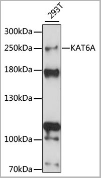
KAT6A, Antibody (Cat# AAA28281)
Full Name
KAT6A Polyclonal Antibody
Gene Names
KAT6A; MOZ; MRD32; MYST3; MYST-3; ZNF220; RUNXBP2; ZC2HC6A
Applications
Western Blot, Immunohistochemistry, Immunofluorescence
Purity
Affinity Purification
Pricing

 FCM/FACS (Flow Cytometry)
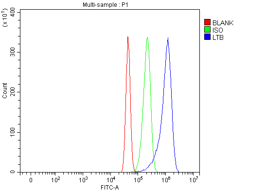
(Figure 2. Flow Cytometry analysis of RAW264.7 cells using anti-Ltb antibody (AAA127129).Overlay histogram showing RAW264.7 cells stained with AAA127129 (Blue line). The cells were fixed with 4% paraformaldehyde and blocked with 10% normal goat serum. And then incubated with rabbit anti-Ltb Antibody (AAA127129, 1ug/1x106 cells) for 30 min at 20 degree C. DyLight488 conjugated goat anti-rabbit IgG was used as secondary antibody for 30 minutes at 20 degree C. Isotype control antibody (Green line) was rabbit IgG (1ug/1x106) used under the same conditions. Unlabelled sample (Red line) was also used as a control.)
FCM/FACS (Flow Cytometry)
(Figure 2. Flow Cytometry analysis of RAW264.7 cells using anti-Ltb antibody (AAA127129).Overlay histogram showing RAW264.7 cells stained with AAA127129 (Blue line). The cells were fixed with 4% paraformaldehyde and blocked with 10% normal goat serum. And then incubated with rabbit anti-Ltb Antibody (AAA127129, 1ug/1x106 cells) for 30 min at 20 degree C. DyLight488 conjugated goat anti-rabbit IgG was used as secondary antibody for 30 minutes at 20 degree C. Isotype control antibody (Green line) was rabbit IgG (1ug/1x106) used under the same conditions. Unlabelled sample (Red line) was also used as a control.)
Ltb, Polyclonal Antibody (Cat# AAA127129)
Full Name
Anti-Ltb Antibody Picoband
Gene Names
Ltb; p33; Tnfc; LTbeta; Tnfsf3; Tnlg1c; AI662801
Reactivity
Mouse, Rat
Applications
Flow Cytometry, Western Blot
Purity
Immunogen affinity purified.
Pricing
 Bioactivity
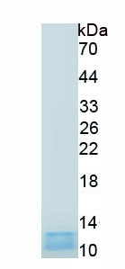
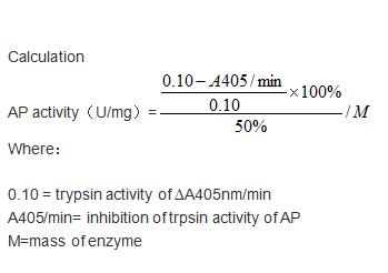
(Aprotinin (AP) is a competitive serine protease inhibitor. Reversibly binds to and blocks the enzymatic active site. Inhibits a range of serine proteases including trypsin, chymotrypsin, kallikrein and plasmin. Inhibits cytopathogenic effect of SARS-CoV-2 and double-stranded RNA formation in SARS-CoV-2-infected cells. The activity of recombinant pig AP was measured by its ability to inhibit trypsin cleavage of a peptide substrate BAPNA in the assay buffer 200 mM Triethanolamine hydrochloride, 20 mM CaCl2, pH 7.8. The reaction was performed in adding 20 ul 4 mg/mL trypsin diluted by 1mM HCl to 160 ul assay buffer and 20 ul 0.85% (w/v) NaCl and start the reaction by adding 100 ul of 1mg/ml BAPNA. Include a substrate blank containing 160 ul assay buffer, 20 ul 1mM HCl, 20 ul 0.85% (w/v) NaCl and 100 uL of 1mg/ml substrate. Rapidly mixing at 25 degree C, then read at 405 nm in kinetic mode for 5 minutes using a microplate reader controlling the ?A405nm/min=0.08-0.12. The 20 ul different concentrations of recombinant pig AP was incubated with 20 ul 4 mg/mL trypsin in 160 ul assay buffer at 25 degree C for 10 minutes followed by adding 100 ul substrate, then read at 405 nm in kinetic mode for 5 minutes using a microplate reader. Under these conditions, the enzyme amount of 50% inhibition of trypsin activity per minute is defined as a unit. The specific activity of recombinant pig AP is >9000 U/mg.)
Bioactivity
(Aprotinin (AP) is a competitive serine protease inhibitor. Reversibly binds to and blocks the enzymatic active site. Inhibits a range of serine proteases including trypsin, chymotrypsin, kallikrein and plasmin. Inhibits cytopathogenic effect of SARS-CoV-2 and double-stranded RNA formation in SARS-CoV-2-infected cells. The activity of recombinant pig AP was measured by its ability to inhibit trypsin cleavage of a peptide substrate BAPNA in the assay buffer 200 mM Triethanolamine hydrochloride, 20 mM CaCl2, pH 7.8. The reaction was performed in adding 20 ul 4 mg/mL trypsin diluted by 1mM HCl to 160 ul assay buffer and 20 ul 0.85% (w/v) NaCl and start the reaction by adding 100 ul of 1mg/ml BAPNA. Include a substrate blank containing 160 ul assay buffer, 20 ul 1mM HCl, 20 ul 0.85% (w/v) NaCl and 100 uL of 1mg/ml substrate. Rapidly mixing at 25 degree C, then read at 405 nm in kinetic mode for 5 minutes using a microplate reader controlling the ?A405nm/min=0.08-0.12. The 20 ul different concentrations of recombinant pig AP was incubated with 20 ul 4 mg/mL trypsin in 160 ul assay buffer at 25 degree C for 10 minutes followed by adding 100 ul substrate, then read at 405 nm in kinetic mode for 5 minutes using a microplate reader. Under these conditions, the enzyme amount of 50% inhibition of trypsin activity per minute is defined as a unit. The specific activity of recombinant pig AP is >9000 U/mg.)
Aprotinin (AP), Active Protein (Cat# AAA161812)
Full Name
Active Aprotinin (AP)
Applications
Activity Assay, Cell Culture
Purity
Greater than 90% by SDS-PAGE
Pricing
 Bioactivity
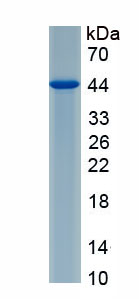
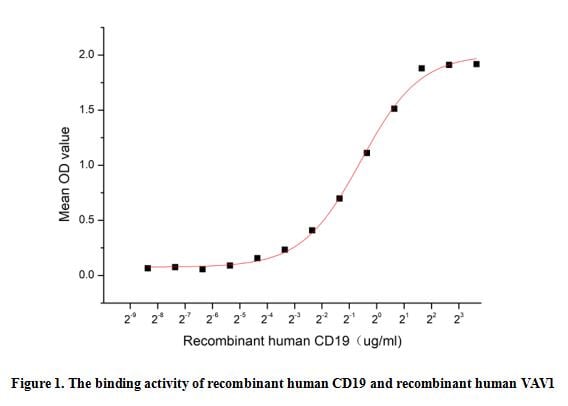
(Cluster Of Differentiation 19 (CD19) is a type I transmembrane glycoprotein belonging to the immunoglobulin superfamily. It is expressed in normal and neoplastic B cells, and it modulates the threshold of B cell activation for amplifying B cell receptor signaling. This protein has recently emerged as a very attractive biomarker and therapeutic target for B-cell malignancies. The development of safe and effective ligands for CD19 has become an important need for the development of targeted conventional and immunotherapies. As far as we know, signaling through CD19 activates Vav/mitogen-activated protein kinase pathway and induces formation of a CD19/Vav/phosphatidylinositol 3-kinase complex in human B cell precursors. Thus a functional binding ELISA assay was conducted to detect the interaction of recombinant human CD19 and recombinant human VAV1. Briefly, biotin-linked CD19 were diluted serially in PBS, with 0.01% BSA (pH 7.4). Duplicate samples of 100 ul were then transferred to VAV1-coated microtiter wells and incubated for 1h at 37 degree C. Wells were washed with PBST 3 times and incubation with Streptavidin-HRP for 30min, then wells were aspirated and washed 5 times. With the addition of substrate solution, wells were incubated 15-25 minutes at 37 degree C. Finally, add 50 ul stop solution to the wells and read at 450 nm immediately. The binding activity of recombinant human CD19 and recombinant human VAV1 was shown in Figure 1, the EC50 for this effect is 0.67 ug/mL.)
Bioactivity
(Cluster Of Differentiation 19 (CD19) is a type I transmembrane glycoprotein belonging to the immunoglobulin superfamily. It is expressed in normal and neoplastic B cells, and it modulates the threshold of B cell activation for amplifying B cell receptor signaling. This protein has recently emerged as a very attractive biomarker and therapeutic target for B-cell malignancies. The development of safe and effective ligands for CD19 has become an important need for the development of targeted conventional and immunotherapies. As far as we know, signaling through CD19 activates Vav/mitogen-activated protein kinase pathway and induces formation of a CD19/Vav/phosphatidylinositol 3-kinase complex in human B cell precursors. Thus a functional binding ELISA assay was conducted to detect the interaction of recombinant human CD19 and recombinant human VAV1. Briefly, biotin-linked CD19 were diluted serially in PBS, with 0.01% BSA (pH 7.4). Duplicate samples of 100 ul were then transferred to VAV1-coated microtiter wells and incubated for 1h at 37 degree C. Wells were washed with PBST 3 times and incubation with Streptavidin-HRP for 30min, then wells were aspirated and washed 5 times. With the addition of substrate solution, wells were incubated 15-25 minutes at 37 degree C. Finally, add 50 ul stop solution to the wells and read at 450 nm immediately. The binding activity of recombinant human CD19 and recombinant human VAV1 was shown in Figure 1, the EC50 for this effect is 0.67 ug/mL.)
Cluster Of Differentiation 19 (CD19), Active Protein (Cat# AAA161866)
Full Name
Active Cluster Of Differentiation 19 (CD19)
Gene Names
CD19; B4; CVID3
Applications
Activity Assay, Cell Culture
Purity
Greater than 90% by SDS-PAGE
Pricing
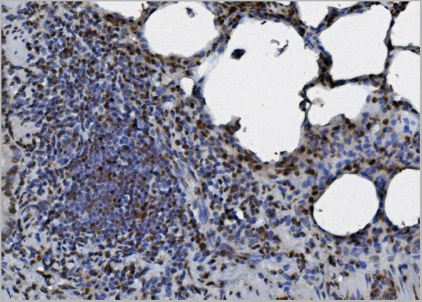
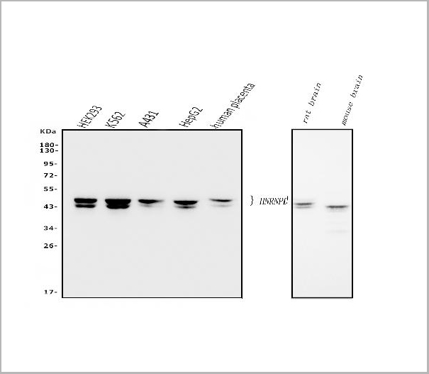
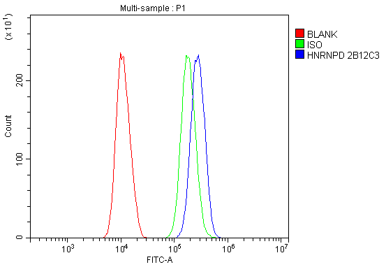
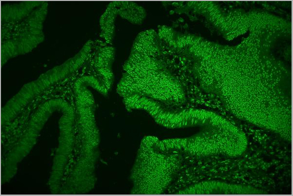
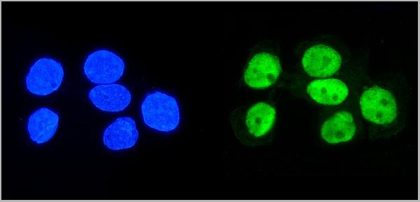
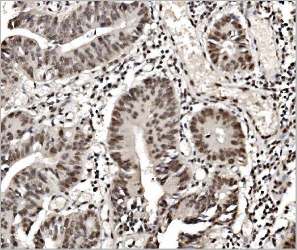
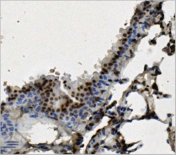 IHC (Immunohistochemistry)
(Figure 7. IHC analysis of hnRNP D/AUF1/HNRNPD using anti-hnRNP D/AUF1/HNRNPD antibody (AAA19387).hnRNP D/AUF1/HNRNPD was detected in paraffin-embedded section of mouse lung tissue. Heat mediated antigen retrieval was performed in EDTA buffer (pH8. 0, epitope retrieval solution). The tissue section was blocked with 10% goat serum. The tissue section was then incubated with 1μg/ml mouse anti-hnRNP D/AUF1/HNRNPD Antibody (AAA19387) overnight at 4 degree C. Biotinylated goat anti-mouse IgG was used as secondary antibody and incubated for 30 minutes at 37 degree C. The tissue section was developed using Strepavidin-Biotin-Complex (SABC) (Catalog # with DAB as the chromogen.)
IHC (Immunohistochemistry)
(Figure 7. IHC analysis of hnRNP D/AUF1/HNRNPD using anti-hnRNP D/AUF1/HNRNPD antibody (AAA19387).hnRNP D/AUF1/HNRNPD was detected in paraffin-embedded section of mouse lung tissue. Heat mediated antigen retrieval was performed in EDTA buffer (pH8. 0, epitope retrieval solution). The tissue section was blocked with 10% goat serum. The tissue section was then incubated with 1μg/ml mouse anti-hnRNP D/AUF1/HNRNPD Antibody (AAA19387) overnight at 4 degree C. Biotinylated goat anti-mouse IgG was used as secondary antibody and incubated for 30 minutes at 37 degree C. The tissue section was developed using Strepavidin-Biotin-Complex (SABC) (Catalog # with DAB as the chromogen.)
hnRNP D/AUF1/HNRNPD, Monoclonal Antibody (Cat# AAA19387)
Full Name
Anti-hnRNP D/AUF1/HNRNPD Antibody (monoclonal, 2B12)
Gene Names
HNRNPD; P37; AUF1; AUF1A; HNRPD; hnRNPD0
Reactivity
Human, Mouse, Rat
Applications
Western Blot, Immunohistochemistry, Immunocytochemistry, Immunofluorescence, Flow Cytometry
Purity
Immunogen affinity purified.
Pricing
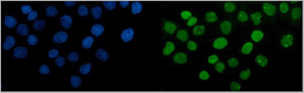
 FCM (Flow Cytometry)
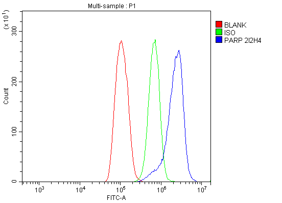
(Figure 10. Flow Cytometry analysis of THP-1 cells using anti-PARP antibody (AAA19345).Overlay histogram showing THP-1 cells stained with AAA19345 (Blue line). The cells were blocked with 10% normal goat serum. And then incubated with mouse anti- PARP Antibody (AAA19345, 1μg/1x106 cells) for 30 min at 20 degree C. DyLight®488 conjugated goat anti-mouse IgG (BA1126, 5-10μg/1x106 cells) was used as secondary antibody for 30 minutes at 20 degree C. Isotype control antibody (Green line) was mouse IgG (1μg/1x106) used under the same conditions. Unlabelled sample (Red line) was also used as a control.)
FCM (Flow Cytometry)
(Figure 10. Flow Cytometry analysis of THP-1 cells using anti-PARP antibody (AAA19345).Overlay histogram showing THP-1 cells stained with AAA19345 (Blue line). The cells were blocked with 10% normal goat serum. And then incubated with mouse anti- PARP Antibody (AAA19345, 1μg/1x106 cells) for 30 min at 20 degree C. DyLight®488 conjugated goat anti-mouse IgG (BA1126, 5-10μg/1x106 cells) was used as secondary antibody for 30 minutes at 20 degree C. Isotype control antibody (Green line) was mouse IgG (1μg/1x106) used under the same conditions. Unlabelled sample (Red line) was also used as a control.)
PARP, Monoclonal Antibody (Cat# AAA19345)
Full Name
Anti-PARP Antibody (monoclonal, 2I2H4)
Gene Names
PARP1; PARP; PPOL; ADPRT; ARTD1; ADPRT1; PARP-1; ADPRT 1; pADPRT-1
Reactivity
Human, Mouse, Rat
Applications
Western Blot, Immunohistochemistry, Immunocytochemistry, Immunofluorescence, Flow Cytometry
Purity
Immunogen affinity purified.
Pricing
 Bioactivity
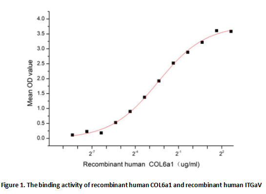
(Collagen alpha-1(VI) chain (COL6a1) is a protein that in humans, the collagens are a superfamily of proteins that play a role in maintaining the integrity of various tissues. Collagens are extracellular matrix proteins and have a triple-helical domain as their common structural element. Besides, Integrin Alpha V (ITGaV) has been identified as an interactor of COL6A1, thus a functional binding ELISA assay was conducted to detect the interaction of recombinant human COL6a1 and recombinant human ITGaV. Briefly, COL6a1 were diluted serially in PBS, with 0.01% BSA (pH 7.4). Duplicate samples of 100 ul were then transferred to ITGaV -coated microtiter wells and incubated for 2h at 37 degree C. Wells were washed with PBST and incubated for 1h with anti-COL6a1 pAb, then aspirated and washed 3 times. After incubation with HRP labelled secondary antibody, wells were aspirated and washed 5 times. With the addition of substrate solution, wells were incubated 15-25 minutes at 37 degree C. Finally, add 50 uL stop solution to the wells and read at 450/630 nm immediately. The binding activity of recombinant human COL6a1 and recombinant human ITGaV was shown in Figure 1, the EC50 for this effect is 0.19 ug/mL.)
Bioactivity
(Collagen alpha-1(VI) chain (COL6a1) is a protein that in humans, the collagens are a superfamily of proteins that play a role in maintaining the integrity of various tissues. Collagens are extracellular matrix proteins and have a triple-helical domain as their common structural element. Besides, Integrin Alpha V (ITGaV) has been identified as an interactor of COL6A1, thus a functional binding ELISA assay was conducted to detect the interaction of recombinant human COL6a1 and recombinant human ITGaV. Briefly, COL6a1 were diluted serially in PBS, with 0.01% BSA (pH 7.4). Duplicate samples of 100 ul were then transferred to ITGaV -coated microtiter wells and incubated for 2h at 37 degree C. Wells were washed with PBST and incubated for 1h with anti-COL6a1 pAb, then aspirated and washed 3 times. After incubation with HRP labelled secondary antibody, wells were aspirated and washed 5 times. With the addition of substrate solution, wells were incubated 15-25 minutes at 37 degree C. Finally, add 50 uL stop solution to the wells and read at 450/630 nm immediately. The binding activity of recombinant human COL6a1 and recombinant human ITGaV was shown in Figure 1, the EC50 for this effect is 0.19 ug/mL.)
Collagen Type VI Alpha 1 (COL6a1), Active Protein (Cat# AAA161878)
Full Name
Active Collagen Type VI Alpha 1 (COL6a1)
Gene Names
COL6A1; OPLL
Applications
Activity Assay, Cell Culture
Purity
Greater than 90% by SDS-PAGE
Pricing
 Bioactivity
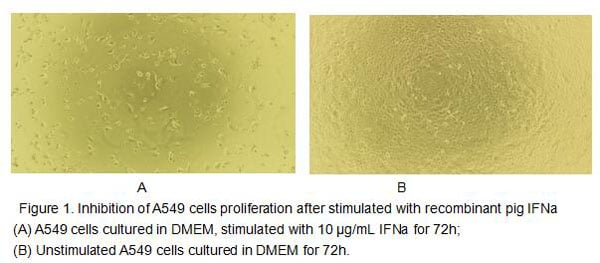
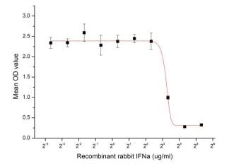
(Figure 2. Inhibition of A549 cells proliferation after stimulated with recombinant rabbit IFN-alpha.)
Bioactivity
(Figure 2. Inhibition of A549 cells proliferation after stimulated with recombinant rabbit IFN-alpha.)
Interferon Alpha (IFNa), Active Protein (Cat# AAA152956)
Full Name
Active Interferon Alpha (IFNa)
Applications
Activity Assay, Cell Culture
Purity
Greater than 80% by SDS-PAGE
Pricing
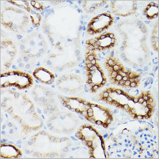
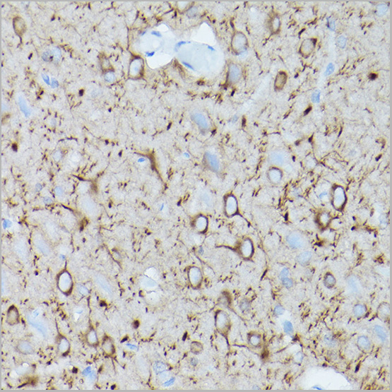
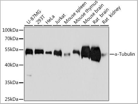
 IF (Immunofluorescence)
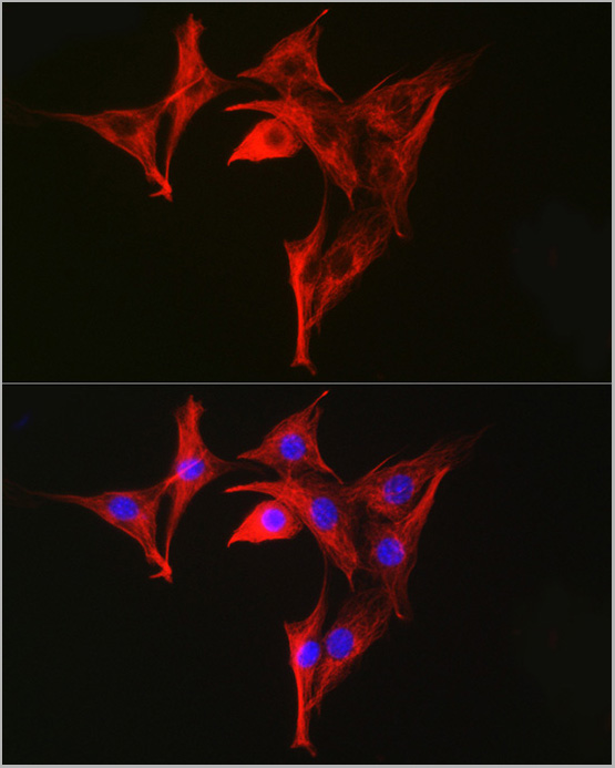
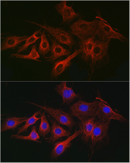
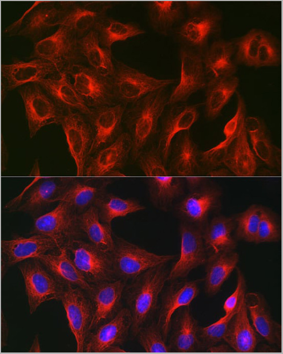
(Immunofluorescence analysis of U-2 OS cells using α-Tubulin Mouse mAb at dilution of 1:100 (40x lens). Blue: DAPI for nuclear staining.)
IF (Immunofluorescence)
(Immunofluorescence analysis of U-2 OS cells using α-Tubulin Mouse mAb at dilution of 1:100 (40x lens). Blue: DAPI for nuclear staining.)
alpha-Tubulin, Monoclonal Antibody (Cat# AAA28393)
Full Name
alpha-Tubulin Mouse mAb
Gene Names
TUBA4A; TUBA1; H2-ALPHA
Reactivity
Human, Mouse, Rat
Applications
Western Blot, Immunohistochemistry, Immunofluorescence, Immunoprecipitation
Purity
Affinity purification
Pricing

 WB (Western Blot)
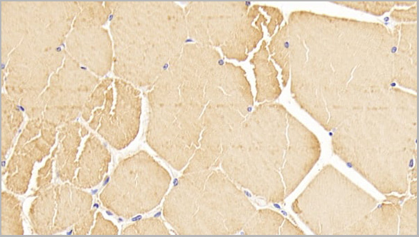
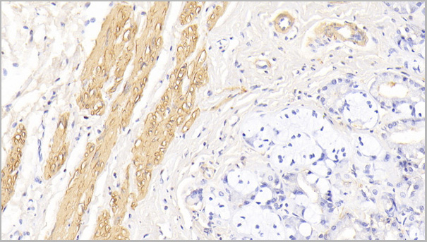
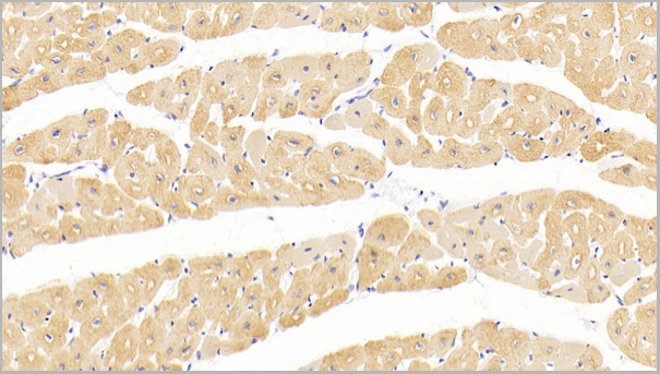
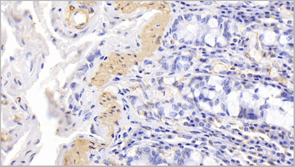
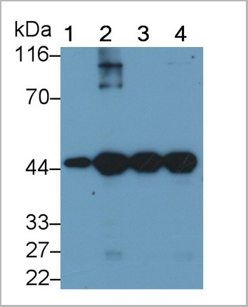
(Western Blot; Sample: Lane1: Rat Heart lysate; Lane2: Rat Skeletal muscle lysate; Lane3: Porcine Heart lysate; Lane4: Porcine Skeletal muscle lysatePrimary Ab: 0.05ug/ml Mouse AntiHuman ACTC1 AntibodySecond Ab: 0.2ug/mL HRPLinked Caprine AntiMouse IgG Polyclonal Antibody(Catalog: SAA544Mu19))
WB (Western Blot)
(Western Blot; Sample: Lane1: Rat Heart lysate; Lane2: Rat Skeletal muscle lysate; Lane3: Porcine Heart lysate; Lane4: Porcine Skeletal muscle lysatePrimary Ab: 0.05ug/ml Mouse AntiHuman ACTC1 AntibodySecond Ab: 0.2ug/mL HRPLinked Caprine AntiMouse IgG Polyclonal Antibody(Catalog: SAA544Mu19))
Actin Alpha 1, Cardiac Muscle (ACTC1), Monoclonal Antibody (Cat# AAA21193)
Full Name
Monoclonal Antibody to Actin Alpha 1, Cardiac Muscle (ACTC1)
Gene Names
ACTC1; ACTC; ASD5; CMD1R; CMH11; LVNC4
Reactivity
Human, Rat, Pig
Applications
Western Blot, Immunohistochemistry, Immunocytochemistry, Immunoprecipitation
Purity
Protein A + Protein G affinity chromatography
Pricing
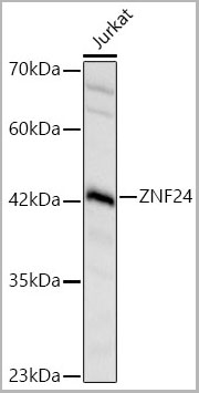
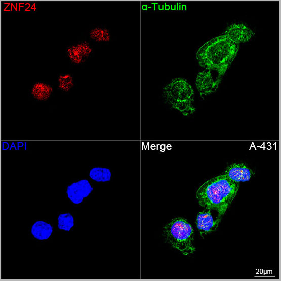 ICC (Immunocytochemistry)
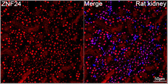
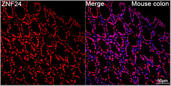
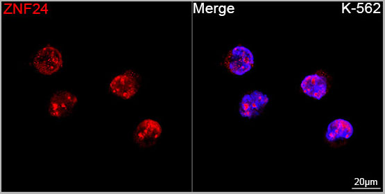
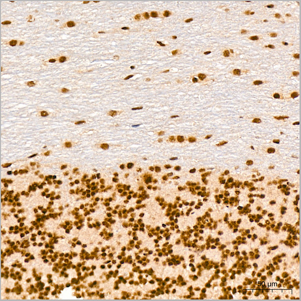
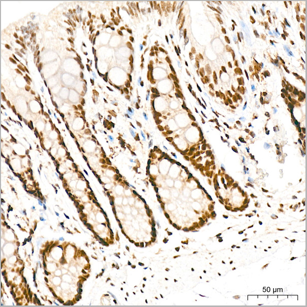
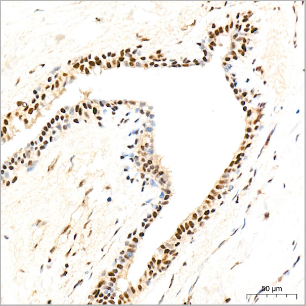
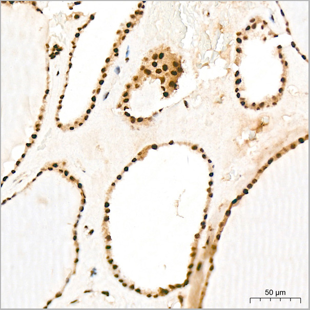
(Confocal imaging of A-431 cells using ZNF24 Rabbit PolymAb® (AAA28573, dilution 1:200) followed by a further incubation with Cy3 Goat Anti-Rabbit IgG (H+L) (AS007, dilution 1:500) (Red). The cells were counterstained with ?-Tubulin Mouse mAb (AC012, dilution 1:400) followed by incubation with ABflo® 488-conjugated Goat Anti-Mouse IgG (H+L) Ab (AS076, dilution 1:500) (Green). DAPI was used for nuclear staining (Blue). Objective: 100x.)
ICC (Immunocytochemistry)
(Confocal imaging of A-431 cells using ZNF24 Rabbit PolymAb® (AAA28573, dilution 1:200) followed by a further incubation with Cy3 Goat Anti-Rabbit IgG (H+L) (AS007, dilution 1:500) (Red). The cells were counterstained with ?-Tubulin Mouse mAb (AC012, dilution 1:400) followed by incubation with ABflo® 488-conjugated Goat Anti-Mouse IgG (H+L) Ab (AS076, dilution 1:500) (Green). DAPI was used for nuclear staining (Blue). Objective: 100x.)
ZNF24, Monoclonal Antibody (Cat# AAA28573)
Full Name
ZNF24 Rabbit PolymAb
Reactivity
Human
Applications
Western Blot, Immunohistochemistry, Immunofluorescence, Immunocytochemistry
Purity
Affinity purification
Pricing
Interleukin-35 (IL-35), Recombinant Protein (Cat# AAA13478)
Full Name
Interleukin-35 (IL-35)
Purity
>95%
Pricing
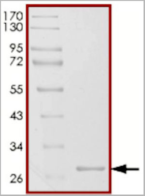 SDS-PAGE
(The purity of GST was determinedbto be >90% by densitometry.Calculated approx. MW 26kDa.Observed approx. MW 28kDa)
SDS-PAGE
(The purity of GST was determinedbto be >90% by densitometry.Calculated approx. MW 26kDa.Observed approx. MW 28kDa)
 Bioactivity
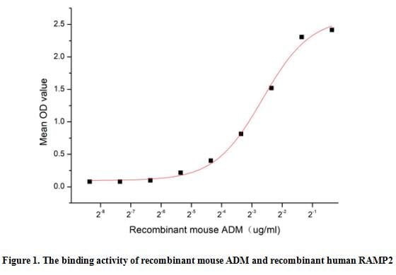
(Adrenomedullin (ADM) is a peptide hormone first discovered in 1993 in pheochromocytoma. Although AM is highly expressed in the adrenal glands, heart, lungs, and kidneys, vascular endothelium and smooth muscle are thought to be the main source of plasma AM. It is a 52 aa peptide with several functions, including vasodilation, regulation of hormone secretion, promotion of angiogenesis, and antimicrobial activity. The antimicrobial activity is antibacterial, as the peptide has been shown to kill E. coli and S. aureus at low concentration. Receptor activity-modifying protein 2 can transports the calcitonin gene-related peptide type 1 receptor (CALCRL) to the plasma membrane. Acts as a receptor for adrenomedullin (AM) together with CALCRL. Thus a functional binding ELISA assay was conducted to detect the interaction of recombinant mouse ADM and recombinant human RAMP2. Briefly, ADM was diluted serially in PBS with 0.01% BSA (pH 7.4). Duplicate samples of 100 ul were then transferred to RAMP2-coated microtiter wells and incubated for 1h at 37 degree C. Wells were washed with PBST and incubated for 1h with anti-ADM pAb, then aspirated and washed 3 times. After incubation with HRP labelled secondary antibody for 1h at 37 degree C, wells were aspirated and washed 5 times. With the addition of substrate solution, wells were incubated 15-25 minutes at 37 degree C. Finally, add 50 uL stop solution to the wells and read at 450/630nm immediately. The binding activity of recombinant mouse ADM and recombinant human RAMP2 was shown in Figure 1, the EC50 for this effect is 0.16 ug/mL.)
Bioactivity
(Adrenomedullin (ADM) is a peptide hormone first discovered in 1993 in pheochromocytoma. Although AM is highly expressed in the adrenal glands, heart, lungs, and kidneys, vascular endothelium and smooth muscle are thought to be the main source of plasma AM. It is a 52 aa peptide with several functions, including vasodilation, regulation of hormone secretion, promotion of angiogenesis, and antimicrobial activity. The antimicrobial activity is antibacterial, as the peptide has been shown to kill E. coli and S. aureus at low concentration. Receptor activity-modifying protein 2 can transports the calcitonin gene-related peptide type 1 receptor (CALCRL) to the plasma membrane. Acts as a receptor for adrenomedullin (AM) together with CALCRL. Thus a functional binding ELISA assay was conducted to detect the interaction of recombinant mouse ADM and recombinant human RAMP2. Briefly, ADM was diluted serially in PBS with 0.01% BSA (pH 7.4). Duplicate samples of 100 ul were then transferred to RAMP2-coated microtiter wells and incubated for 1h at 37 degree C. Wells were washed with PBST and incubated for 1h with anti-ADM pAb, then aspirated and washed 3 times. After incubation with HRP labelled secondary antibody for 1h at 37 degree C, wells were aspirated and washed 5 times. With the addition of substrate solution, wells were incubated 15-25 minutes at 37 degree C. Finally, add 50 uL stop solution to the wells and read at 450/630nm immediately. The binding activity of recombinant mouse ADM and recombinant human RAMP2 was shown in Figure 1, the EC50 for this effect is 0.16 ug/mL.)
Adrenomedullin (ADM), Active Protein (Cat# AAA161717)
Full Name
Active Adrenomedullin (ADM)
Gene Names
Adm; AM
Applications
Activity Assay, Cell Culture
Purity
Greater than 90% by SDS-PAGE
Pricing

 Bioactivity
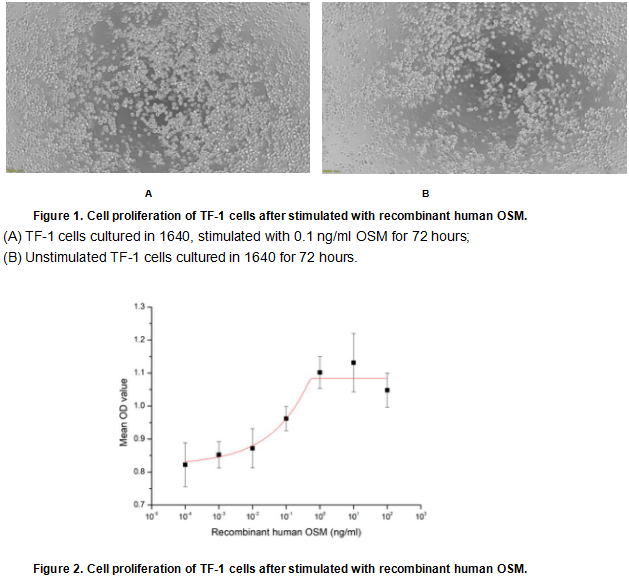
(Oncostatin M (OSM) is an approximately 30 KDa secreted cytokine belonging to the Interleukin-6 family. Like other members of the IL-6 family such as IL-11, CNTF, and Cardiotrophin-1, OSM plays crucial roles in inflammation, neuroprotection, hematopoiesis, metabolism and development. To test the effect of OSM on cell proliferation, TF-1 cells were seeded into triplicate wells of 96-well plates at a density of 20,000 cells/well with various concentrations of recombinant human OSM. After incubated for 72 hours, cells were observed by inverted microscope and cell proliferation was measured by Cell Counting Kit-8(CCK-8). Briefly, 10 ul of CCK-8 solution was added to each well of the plate, then the absorbance at 450 nm was measured using a microplate reader after incubating the plate for 1-4 hours at 37 degree C. Proliferation of TF-1 cells after incubation with OSM for 72 hours observed by inverted microscope was shown in Figure 1. Cell viability was assessed by CCK-8 (Cell Counting Kit-8) assay after incubation with recombinant human OSM for 72 hours. The result was shown in Figure 2. It was obvious that recombinant human OSM significantly increased cell viability of TF-1 cells, the EC50 was 0.08 ng/ml.)
Bioactivity
(Oncostatin M (OSM) is an approximately 30 KDa secreted cytokine belonging to the Interleukin-6 family. Like other members of the IL-6 family such as IL-11, CNTF, and Cardiotrophin-1, OSM plays crucial roles in inflammation, neuroprotection, hematopoiesis, metabolism and development. To test the effect of OSM on cell proliferation, TF-1 cells were seeded into triplicate wells of 96-well plates at a density of 20,000 cells/well with various concentrations of recombinant human OSM. After incubated for 72 hours, cells were observed by inverted microscope and cell proliferation was measured by Cell Counting Kit-8(CCK-8). Briefly, 10 ul of CCK-8 solution was added to each well of the plate, then the absorbance at 450 nm was measured using a microplate reader after incubating the plate for 1-4 hours at 37 degree C. Proliferation of TF-1 cells after incubation with OSM for 72 hours observed by inverted microscope was shown in Figure 1. Cell viability was assessed by CCK-8 (Cell Counting Kit-8) assay after incubation with recombinant human OSM for 72 hours. The result was shown in Figure 2. It was obvious that recombinant human OSM significantly increased cell viability of TF-1 cells, the EC50 was 0.08 ng/ml.)
Oncostatin M (OSM), Active Protein (Cat# AAA161690)
Full Name
Active Oncostatin M (OSM)
Applications
Activity Assay, Cell Culture
Purity
Greater than 95% by SDS-PAGE
Pricing
 IHC (Immunohiostchemistry)
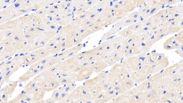
(DAB staining on IHCP;Sample: Human Cardiac Muscle Tissue; Primary Ab: 20ug/ml Mouse AntiHuman ACVR2A AntibodySecond Ab: 2ug/mL HRPLinked Caprine AntiMouse IgG Polyclonal Antibody(Catalog: SAA544Mu19))
IHC (Immunohiostchemistry)
(DAB staining on IHCP;Sample: Human Cardiac Muscle Tissue; Primary Ab: 20ug/ml Mouse AntiHuman ACVR2A AntibodySecond Ab: 2ug/mL HRPLinked Caprine AntiMouse IgG Polyclonal Antibody(Catalog: SAA544Mu19))
Activin A Receptor Type II A (ACVR2A), Monoclonal Antibody (Cat# AAA151793)
Full Name
Monoclonal Antibody to Activin A Receptor Type II A (ACVR2A)
Gene Names
ACVR2A; ACVR2; ACTRII
Reactivity
Human, Pig
Applications
Immunoprecipitation, Immunocytochemistry, Immunohistochemistry, Western Blot
Purity
Protein A + Protein G affinity chromatography
Pricing
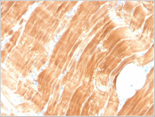
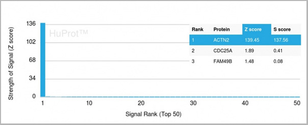 Application Data
(Analysis of Protein Array containing more than 19,000 full-length human proteins using Sarcomeric Actinin Alpha 2 Mouse Monoclonal Antibody (ACTN2/3294). Z- and S- Score: The Z-score represents the strength of a signal that a monoclonal antibody (MAb) (in combination with a fluorescently-tagged anti-IgG secondary antibody) produces when binding to a particular protein on the HuProtTM array. Z-scores are described in units of standard deviations (SD’s) above the mean value of all signals generated on that array. If targets on HuProtTM are arranged in descending order of the Z-score, the S-score is the difference (also in units of SD’s) between the Z-score. S-score therefore represents the relative target specificity of a MAb to its intended target. A MAb is considered to specific to its intended target, if the MAb has an S-score of at least 2.5. For example, if a MAb binds to protein X with a Z-score of 43 and to protein Y with a Z-score of 14, then the S-score for the binding of that MAb to protein X is equal to 29.)
Application Data
(Analysis of Protein Array containing more than 19,000 full-length human proteins using Sarcomeric Actinin Alpha 2 Mouse Monoclonal Antibody (ACTN2/3294). Z- and S- Score: The Z-score represents the strength of a signal that a monoclonal antibody (MAb) (in combination with a fluorescently-tagged anti-IgG secondary antibody) produces when binding to a particular protein on the HuProtTM array. Z-scores are described in units of standard deviations (SD’s) above the mean value of all signals generated on that array. If targets on HuProtTM are arranged in descending order of the Z-score, the S-score is the difference (also in units of SD’s) between the Z-score. S-score therefore represents the relative target specificity of a MAb to its intended target. A MAb is considered to specific to its intended target, if the MAb has an S-score of at least 2.5. For example, if a MAb binds to protein X with a Z-score of 43 and to protein Y with a Z-score of 14, then the S-score for the binding of that MAb to protein X is equal to 29.)
Sarcomeric Actinin Alpha 2/ACTN2, Monoclonal Antibody (Cat# AAA23943)
Full Name
Sarcomeric Actinin Alpha 2/ACTN2
Gene Names
ACTN2; CMH23; CMD1AA
Reactivity
Human
Applications
Western Blot, Immunohistochemistry
Purity
Purified Ab with BSA and Azide at 200ug/ml OR Purified Ab WITHOUT BSA and Azide at 1.0mg/ml
Pricing
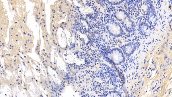
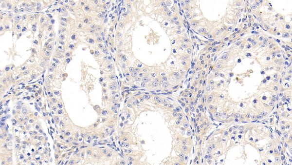
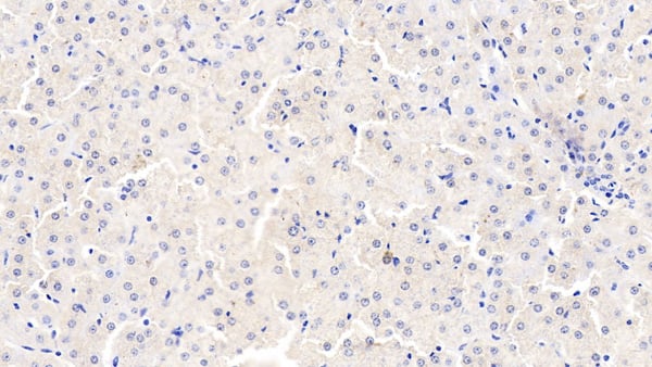 IHC (Immunohistochemisry)
(DAB staining on IHCP;Samples: Bovine Liver Tissue; Primary Ab: 10ug/ml Mouse AntiBovine TNFa AntibodySecond Ab: 2ug/mL HRPLinked Caprine AntiMouse IgG Polyclonal Antibody(Catalog: SAA544Mu19))
IHC (Immunohistochemisry)
(DAB staining on IHCP;Samples: Bovine Liver Tissue; Primary Ab: 10ug/ml Mouse AntiBovine TNFa AntibodySecond Ab: 2ug/mL HRPLinked Caprine AntiMouse IgG Polyclonal Antibody(Catalog: SAA544Mu19))
Tumor Necrosis Factor Alpha (TNFa), Monoclonal Antibody (Cat# AAA151490)
Full Name
Monoclonal Antibody to Tumor Necrosis Factor Alpha (TNFa)
Gene Names
TNF; TNFa; TNF-a; TNF-alpha
Reactivity
Mouse, Bovine
Applications
Immunoprecipitation, Immunocytochemistry, Immunohistochemistry, Western Blot
Purity
Protein A + Protein G affinity chromatography
Pricing
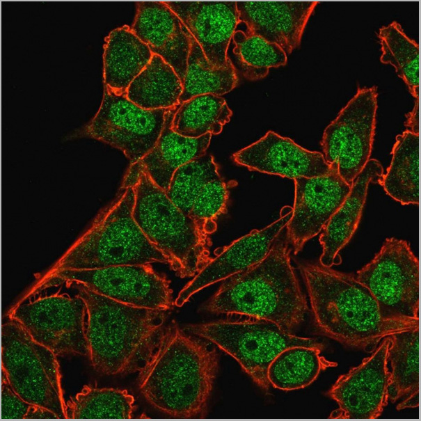
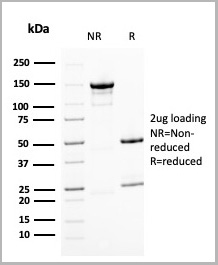
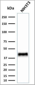
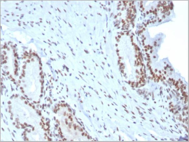
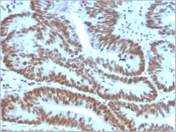
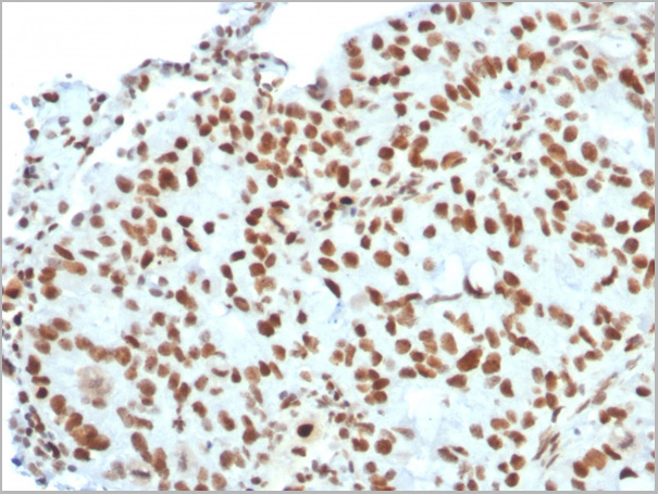
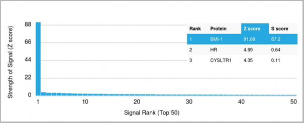 Application Data
(Analysis of Protein Array containing more than 19,000 full-length human proteins using BMI1 Mouse Monoclonal Antibody (BMI1/2823) Z- and S- Score: The Z-score represents the strength of a signal that a monoclonal antibody (MAb) (in combination with a fluorescently-tagged anti-IgG secondary antibody) produces when binding to a particular protein on the HuProtTM array. Z-scores are described in units of standard deviations (SD’s) above the mean value of all signals generated on that array. If targets on HuProtTM are arranged in descending order of the Z-score, the S-score is the difference (also in units of SD’s) between the Z-score. S-score therefore represents the relative target specificity of a MAb to its intended target. A MAb is considered to specific to its intended target, if the MAb has an S-score of at least 2.5. For example, if a MAb binds to protein X with a Z-score of 43 and to protein Y with a Z-score of 14, then the S-score for the binding of that MAb to protein X is equal to 29.)
Application Data
(Analysis of Protein Array containing more than 19,000 full-length human proteins using BMI1 Mouse Monoclonal Antibody (BMI1/2823) Z- and S- Score: The Z-score represents the strength of a signal that a monoclonal antibody (MAb) (in combination with a fluorescently-tagged anti-IgG secondary antibody) produces when binding to a particular protein on the HuProtTM array. Z-scores are described in units of standard deviations (SD’s) above the mean value of all signals generated on that array. If targets on HuProtTM are arranged in descending order of the Z-score, the S-score is the difference (also in units of SD’s) between the Z-score. S-score therefore represents the relative target specificity of a MAb to its intended target. A MAb is considered to specific to its intended target, if the MAb has an S-score of at least 2.5. For example, if a MAb binds to protein X with a Z-score of 43 and to protein Y with a Z-score of 14, then the S-score for the binding of that MAb to protein X is equal to 29.)
BMI1, Monoclonal Antibody (Cat# AAA23935)
Full Name
BMI1 (Oncogene and Stem Cell Marker)
Gene Names
BMI1; PCGF4; RNF51; FLVI2/BMI1; flvi-2/bmi-1
Reactivity
Human, Mouse
Applications
Western Blot, Immunofluorescence, Immunohistochemistry
Purity
Purified Ab with BSA and Azide at 200ug/ml OR Purified Ab WITHOUT BSA and Azide at 1.0mg/ml
Pricing
 Bioactivity
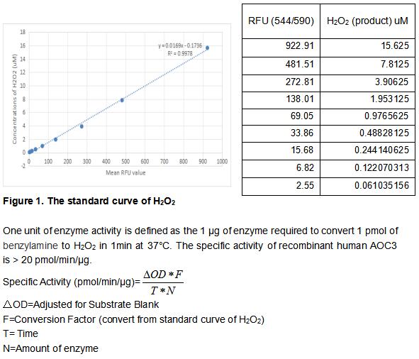
(Amine Oxidase, Copper Containing 3 (AOC3) is a copper amine oxidase with a topaquinone cofactor. AOC3 is a Type II integral membrane protein, but a soluble form of the enzyme is present in human serum, and its level increases in diabetes and some inflammatory liver diseases. AOC3 catalyzes the oxidative deamination of small primary amines such as methylamine, benzylamine, and aminoacetone in a reaction that produces an aldehyde, ammonia, and H2O2. The enzyme is sensitive to inhibition by semicarbazide. AOC3 expression is highest in the endothelium of lung, heart, and intestine, but low in tissues such as brain, spleen, kidney, and liver. The activity of recombinant human AOC3 was measured by its ability to produce hydrogen peroxide during the oxidation of benzylamine. The reaction was performed in 50 mM HEPES, pH 7.5 (assay buffer), initiated by addition 50 uL of various concentrations of AOC3 (diluted by assay buffer) to 50 ul substrate mixture of 2 mM Benzylamine, 2 units/mL HRP and 100 uM AUR. Read at excitation and emission wavelengths of 544 nm and 590 nm (top read), respectively in kinetic mode for 5 minutes.)
Bioactivity
(Amine Oxidase, Copper Containing 3 (AOC3) is a copper amine oxidase with a topaquinone cofactor. AOC3 is a Type II integral membrane protein, but a soluble form of the enzyme is present in human serum, and its level increases in diabetes and some inflammatory liver diseases. AOC3 catalyzes the oxidative deamination of small primary amines such as methylamine, benzylamine, and aminoacetone in a reaction that produces an aldehyde, ammonia, and H2O2. The enzyme is sensitive to inhibition by semicarbazide. AOC3 expression is highest in the endothelium of lung, heart, and intestine, but low in tissues such as brain, spleen, kidney, and liver. The activity of recombinant human AOC3 was measured by its ability to produce hydrogen peroxide during the oxidation of benzylamine. The reaction was performed in 50 mM HEPES, pH 7.5 (assay buffer), initiated by addition 50 uL of various concentrations of AOC3 (diluted by assay buffer) to 50 ul substrate mixture of 2 mM Benzylamine, 2 units/mL HRP and 100 uM AUR. Read at excitation and emission wavelengths of 544 nm and 590 nm (top read), respectively in kinetic mode for 5 minutes.)
Amine Oxidase, Copper Containing 3 (AOC3), Active Protein (Cat# AAA161882)
Full Name
Active Amine Oxidase, Copper Containing 3 (AOC3)
Gene Names
AOC3; HPAO; SSAO; VAP1; VAP-1
Applications
Activity Assay, Cell Culture
Purity
Greater than 90% by SDS-PAGE
Pricing
 FCM (Flow Cytometry)
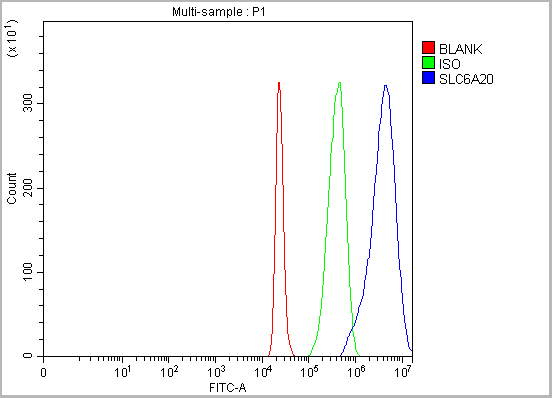
(Figure 14. Flow Cytometry analysis of 293T cells using anti-SLC6A20 antibody (AAA19934).Overlay histogram showing 293T cells stained with AAA19934 (Blue line). The cells were fixed with 4% paraformaldehyde and blocked with 10% normal goat serum. And then incubated with rabbit anti-SLC6A20 Antibody (AAA19934, 1ug/1x106 cells) for 30 min at 20 degree C. DyLight®488 conjugated goat anti-rabbit IgG (BA1127, 5-10ug/1x106 cells) was used as secondary antibody for 30 minutes at 20 degree C. Isotype control antibody (Green line) was rabbit IgG (1ug/1x106) used under the same conditions. Unlabelled sample (Red line) was also used as a control.)
FCM (Flow Cytometry)
(Figure 14. Flow Cytometry analysis of 293T cells using anti-SLC6A20 antibody (AAA19934).Overlay histogram showing 293T cells stained with AAA19934 (Blue line). The cells were fixed with 4% paraformaldehyde and blocked with 10% normal goat serum. And then incubated with rabbit anti-SLC6A20 Antibody (AAA19934, 1ug/1x106 cells) for 30 min at 20 degree C. DyLight®488 conjugated goat anti-rabbit IgG (BA1127, 5-10ug/1x106 cells) was used as secondary antibody for 30 minutes at 20 degree C. Isotype control antibody (Green line) was rabbit IgG (1ug/1x106) used under the same conditions. Unlabelled sample (Red line) was also used as a control.)
SLC6A20, Polyclonal Antibody (Cat# AAA19934)
Full Name
Anti-SLC6A20 Antibody Picoband
Gene Names
SLC6A20; XT3; SIT1; Xtrp3
Reactivity
Human, Mouse, Rat
Applications
Western Blot, Immunohistochemistry, Flow Cytometry
Purity
Immunogen affinity purified.
Pricing
 Bioactivity
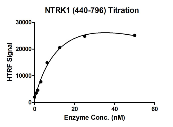
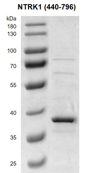
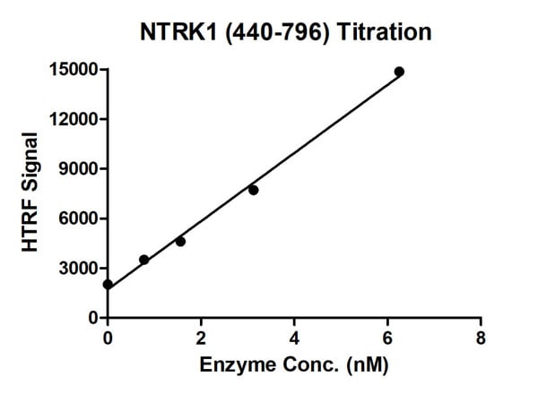
(HTRF assay for NTRK1 (440-796) activity 1 uM TK substrate was incubated with different concentrations of NTRK1 (440-796) protein in a 10 ul reaction system containing 1×Enzymatic Buffer, 5 mM MgCl2, 1 mM DTT, and 100 uM ATP for 1 hour. Then 10 ul detection reagents containing anti-TK antibody (1:2) and SA-XL665 (1:100) diluted with 1× Detection Buffer were added and incubated with the reactions for 30 min. All the operations and reactions were performed at room temperature. HTRF assay was used for detection.)
Bioactivity
(HTRF assay for NTRK1 (440-796) activity 1 uM TK substrate was incubated with different concentrations of NTRK1 (440-796) protein in a 10 ul reaction system containing 1×Enzymatic Buffer, 5 mM MgCl2, 1 mM DTT, and 100 uM ATP for 1 hour. Then 10 ul detection reagents containing anti-TK antibody (1:2) and SA-XL665 (1:100) diluted with 1× Detection Buffer were added and incubated with the reactions for 30 min. All the operations and reactions were performed at room temperature. HTRF assay was used for detection.)
NTRK1, Active Protein (Cat# AAA60324)
Full Name
Recombinant NTRK1 (440-796) protein
Purity
>=90%
Pricing
 IP (Immunoprecipitation)
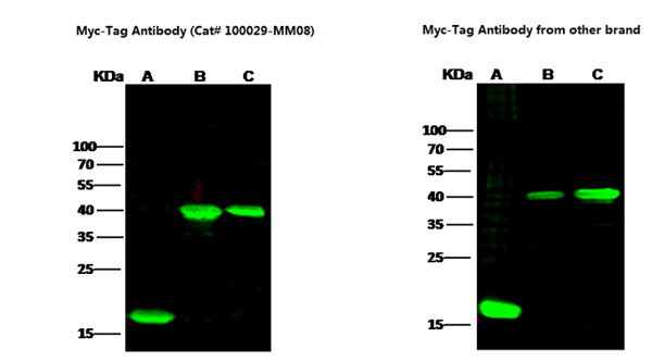
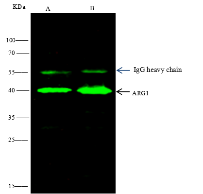
(MYC was immunoprecipitated using:Lane A:0.5 mg HA-ARG1-myc Whole Cell LysateLane B:0.5 mg myc-ARG1-HA Whole Cell Lysate4 uL anti-MYC rabbit polyclonal antibody and 60 ug of Immunomagnetic beads Protein A/G.Primary antibody:Anti-MYC rabbit polyclonal antibody,at 1:100 dilutionSecondary antibody:Goat Anti-Rabbit IgG (H+L)/HRP at 1/10000 dilutionDeveloped using the ECL technique.Performed under reducing conditions.Predicted band size: 40 kDaObserved band size :40 kDa)
IP (Immunoprecipitation)
(MYC was immunoprecipitated using:Lane A:0.5 mg HA-ARG1-myc Whole Cell LysateLane B:0.5 mg myc-ARG1-HA Whole Cell Lysate4 uL anti-MYC rabbit polyclonal antibody and 60 ug of Immunomagnetic beads Protein A/G.Primary antibody:Anti-MYC rabbit polyclonal antibody,at 1:100 dilutionSecondary antibody:Goat Anti-Rabbit IgG (H+L)/HRP at 1/10000 dilutionDeveloped using the ECL technique.Performed under reducing conditions.Predicted band size: 40 kDaObserved band size :40 kDa)
Myc, Monoclonal Antibody (Cat# AAA254119)
Full Name
Anti-Myc tag Antibody, Mouse Monoclonal
Applications
Immunoprecipitation, Western Blot
Purity
Protein A
Pricing
 WB (Western Blot)
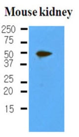
(The extracts of mouse kidney (50 ug) were resolved by SDS-PAGE, transferred to NC membrane and probed with anti-human PDPN (1:500). Proteins were visualized using a goat anti-mouse secondary antibody conjugated to HRP and an ECL detection system.)
WB (Western Blot)
(The extracts of mouse kidney (50 ug) were resolved by SDS-PAGE, transferred to NC membrane and probed with anti-human PDPN (1:500). Proteins were visualized using a goat anti-mouse secondary antibody conjugated to HRP and an ECL detection system.)
PDPN / Podoplanin, Monoclonal Antibody (Cat# AAA51836)
Full Name
Mouse Monoclonal [clone 5E2] (IgG2b,k) to Human PDPN / Podoplanin
Gene Names
PDPN; T1A; GP36; GP40; Gp38; OTS8; T1A2; TI1A; T1A-2; AGGRUS; HT1A-1; PA2.26
Reactivity
Human, Mouse
Applications
Western Blot, Immunohistochemistry
Purity
Protein G Purified
Pricing
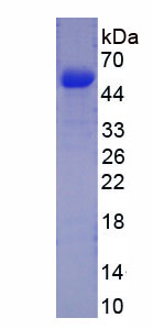 SDS-PAGE
Procollagen III N-Terminal Propeptide, Recombinant Protein (Cat# AAA150208)
Full Name
Recombinant Procollagen III N-Terminal Propeptide (PIIINP)
Gene Names
Col3a1; Tsk2; Tsk-2; Col3a-1; AW550625
Applications
Western Blot
Purity
> 90%
Pricing
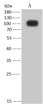
 IHC (Immunohistochemisry)
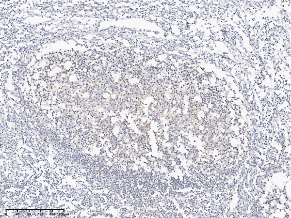
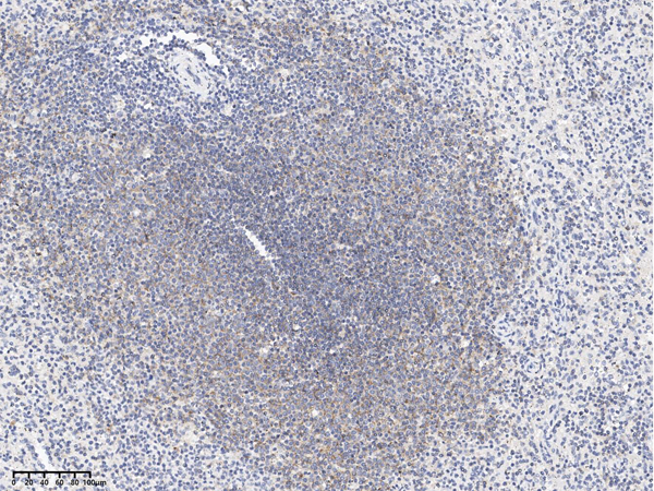
(Immunochemical staining of human CD19 in human spleen with rabbit polyclonal antibody at 1:5000 dilution, formalin-fixed paraffin embedded sections.)
IHC (Immunohistochemisry)
(Immunochemical staining of human CD19 in human spleen with rabbit polyclonal antibody at 1:5000 dilution, formalin-fixed paraffin embedded sections.)
CD19, Polyclonal Antibody (Cat# AAA258899)
Full Name
Anti-CD19 Antibody, Rabbit Polyclonal
Reactivity
Human
Applications
Immunohistochemistry, Western Blot
Purity
Protein A & Antigen Affinity
Pricing
 Application Data
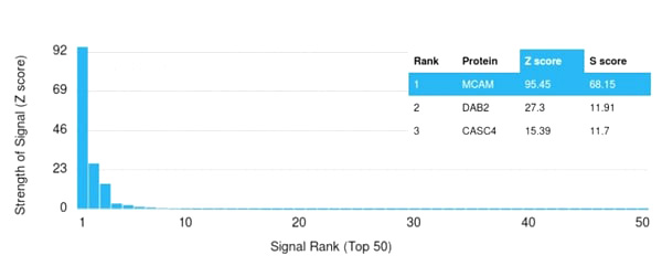
(Analysis of Protein Array containing more than 19,000 full-length human proteins using MCAM Mouse Monoclonal Antibody (MCAM/3179). Z- and S- Score: The Z-score represents the strength of a signal that a monoclonal antibody (MAb) (in combination with a fluorescently-tagged anti-IgG secondary antibody) produces when binding to a particular protein on the HuProtTM array. Z-scores are described in units of standard deviations (SD's) above the mean value of all signals generated on that array. If targets on HuProtTM are arranged in descending order of the Z-score, the S-score is the difference (also in units of SD's) between the Z-score. S-score therefore represents the relative target specificity of a MAb to its intended target. A MAb is considered to specific to its intended target, if the MAb has an S-score of at least 2.5. For example, if a MAb binds to protein X with a Z-score of 43 and to protein Y with a Z-score of 14, then the S-score for the binding of that MAb to protein X is equal to 29.)
Application Data
(Analysis of Protein Array containing more than 19,000 full-length human proteins using MCAM Mouse Monoclonal Antibody (MCAM/3179). Z- and S- Score: The Z-score represents the strength of a signal that a monoclonal antibody (MAb) (in combination with a fluorescently-tagged anti-IgG secondary antibody) produces when binding to a particular protein on the HuProtTM array. Z-scores are described in units of standard deviations (SD's) above the mean value of all signals generated on that array. If targets on HuProtTM are arranged in descending order of the Z-score, the S-score is the difference (also in units of SD's) between the Z-score. S-score therefore represents the relative target specificity of a MAb to its intended target. A MAb is considered to specific to its intended target, if the MAb has an S-score of at least 2.5. For example, if a MAb binds to protein X with a Z-score of 43 and to protein Y with a Z-score of 14, then the S-score for the binding of that MAb to protein X is equal to 29.)
MCAM (Melanoma Cell Adhesion Molecule)/MUC18/CD146, Monoclonal Antibody (Cat# AAA215079)
Full Name
MCAM (Melanoma Cell Adhesion Molecule)/MUC18/CD146
Gene Names
MCAM; CD146; MUC18; HEMCAM; METCAM; MelCAM
Reactivity
Human
Applications
ELISA
Purity
Purified Ab with BSA and Azide at 200ug/ml OR Purified Ab WITHOUT BSA and Azide at 1.0mg/ml
Pricing
 DB (Dot Blot)
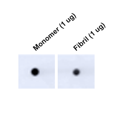
(Dot Blot analysis using Mouse Anti-Tau Monoclonal Antibody, Clone 3D4 (SMC-608). Tissue: Recombinant Protein. Species: Human. Primary Antibody: Mouse Anti-Tau Monoclonal Antibody (SMC-608) at 1:1000 for 2 hours at RT with shaking. Secondary Antibody: Goat anti-mouse IgG:HRP at 1:5000 for 1 hour at RT with shaking.)
DB (Dot Blot)
(Dot Blot analysis using Mouse Anti-Tau Monoclonal Antibody, Clone 3D4 (SMC-608). Tissue: Recombinant Protein. Species: Human. Primary Antibody: Mouse Anti-Tau Monoclonal Antibody (SMC-608) at 1:1000 for 2 hours at RT with shaking. Secondary Antibody: Goat anti-mouse IgG:HRP at 1:5000 for 1 hour at RT with shaking.)
Tau, Monoclonal Antibody (Cat# AAA253995)
Full Name
Tau Antibody, Clone 3D4: HRP
Gene Names
MAPT; TAU; MSTD; PPND; DDPAC; MAPTL; MTBT1; MTBT2; FTDP-17; PPP1R103
Reactivity
Human, Mouse, Rat
Applications
Immunofluorescence, Immunocytochemistry, Dot Blot, Western Blot
Purity
Protein G Purified
Pricing

 SDS_PAGE
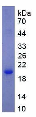
(Figure 3. SDS-PAGESample: Active recombinant CAM, Human)
SDS_PAGE
(Figure 3. SDS-PAGESample: Active recombinant CAM, Human)
Calmodulin (CAM), Active Protein (Cat# AAA149238)
Full Name
Active Calmodulin (CAM)
Gene Names
CALM1; caM; CAM2; CAM3; CAMB; CAMC; CAMI; PHKD; CPVT4; DD132; LQT14; CALML2; CAMIII
Reactivity
Homo sapiens (Human)
Applications
Activity Assay, Cell Culture
Purity
>92%
Pricing
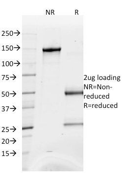
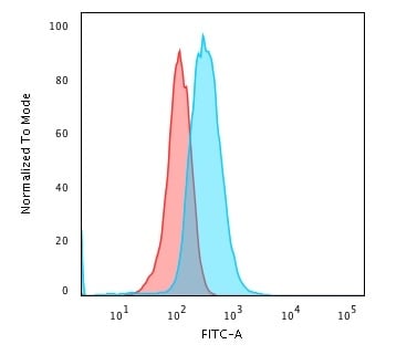
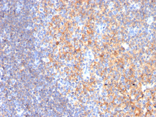
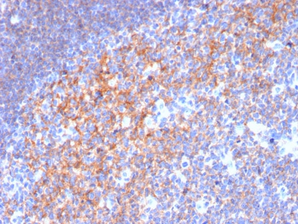
 Application Data
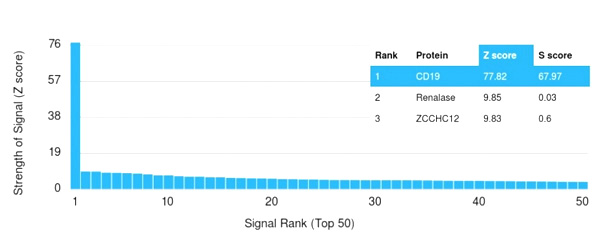
(Analysis of Protein Array containing more than 19,000 full-length human proteins using CD19 Monospecific Mouse Monoclonal Antibody (CD19/3116). Z- and S- Score: The Z-score represents the strength of a signal that a monoclonal antibody (MAb) (in combination with a fluorescently-tagged anti-IgG secondary antibody) produces when binding to a particular protein on the HuProtTM array. Z-scores are described in units of standard deviations (SD's) above the mean value of all signals generated on that array. If targets on HuProtTM are arranged in descending order of the Z-score, the S-score is the difference (also in units of SD's) between the Z-score. S-score therefore represents the relative target specificity of a MAb to its intended target. A MAb is considered to specific to its intended target, if the MAb has an S-score of at least 2.5. For example, if a MAb binds to protein X with a Z-score of 43 and to protein Y with a Z-score of 14, then the S-score for the binding of that MAb to protein X is equal to 29.)
Application Data
(Analysis of Protein Array containing more than 19,000 full-length human proteins using CD19 Monospecific Mouse Monoclonal Antibody (CD19/3116). Z- and S- Score: The Z-score represents the strength of a signal that a monoclonal antibody (MAb) (in combination with a fluorescently-tagged anti-IgG secondary antibody) produces when binding to a particular protein on the HuProtTM array. Z-scores are described in units of standard deviations (SD's) above the mean value of all signals generated on that array. If targets on HuProtTM are arranged in descending order of the Z-score, the S-score is the difference (also in units of SD's) between the Z-score. S-score therefore represents the relative target specificity of a MAb to its intended target. A MAb is considered to specific to its intended target, if the MAb has an S-score of at least 2.5. For example, if a MAb binds to protein X with a Z-score of 43 and to protein Y with a Z-score of 14, then the S-score for the binding of that MAb to protein X is equal to 29.)
CD19, Monoclonal Antibody (Cat# AAA215292)
Full Name
CD19 (B-Lymphocyte Marker)
Gene Names
CD19; B4; CVID3
Reactivity
Human
Applications
Immunohistochemistry, Flow Cytometry
Purity
Purified Ab WITHOUT BSA and Azide
Pricing
 WB (Western Blot)
(Western Blot; Sample: Lane1: Human Plasma; Lane2: Rat Plasma Primary Ab: 2ug/ml Mouse AntiHuman DBP Antibody Second Ab: 0.2ug/mL HRPLinked Caprine AntiMouse IgG Polyclonal Antibody (Catalog: SAA544Mu19))
WB (Western Blot)
(Western Blot; Sample: Lane1: Human Plasma; Lane2: Rat Plasma Primary Ab: 2ug/ml Mouse AntiHuman DBP Antibody Second Ab: 0.2ug/mL HRPLinked Caprine AntiMouse IgG Polyclonal Antibody (Catalog: SAA544Mu19))
Vitamin D Binding Protein (DBP), Monoclonal Antibody (Cat# AAA151751)
Full Name
Monoclonal Antibody to Vitamin D Binding Protein (DBP)
Gene Names
GC; DBP; GRD3; VDBG; VDBP; DBP/GC
Reactivity
Human, Rat, Pig
Applications
Immunoprecipitation, Immunocytochemistry, Immunohistochemistry, Western Blot
Purity
Protein A + Protein G affinity chromatography
Pricing
 Application Data
(Analysis of Protein Array containing more than 19, 000 full-length human proteins using p53 Mouse Monoclonal Antibody (TP53/1739). Z- and S- Score: The Z-score represents the strength of a signal that a monoclonal antibody (MAb) (in combination with a fluorescently-tagged anti-IgG secondary antibody) produces when binding to a particular protein on the HuProtTM array. Z-scores are described in units of standard deviations (SD's) above the mean value of all signals generated on that array. If targets on HuProtTM are arranged in descending order of the Z-score, the S-score is the difference (also in units of SD's) between the Z-score. S-score therefore represents the relative target specificity of a MAb to its intended target. A MAb is considered to specific to its intended target, if the MAb has an S-score of at least 2.5. For example, if a MAb binds to protein X with a Z-score of 43 and to protein Y with a Z-score of 14, then the S-score for the binding of that MAb to protein X is equal to 29.)
Application Data
(Analysis of Protein Array containing more than 19, 000 full-length human proteins using p53 Mouse Monoclonal Antibody (TP53/1739). Z- and S- Score: The Z-score represents the strength of a signal that a monoclonal antibody (MAb) (in combination with a fluorescently-tagged anti-IgG secondary antibody) produces when binding to a particular protein on the HuProtTM array. Z-scores are described in units of standard deviations (SD's) above the mean value of all signals generated on that array. If targets on HuProtTM are arranged in descending order of the Z-score, the S-score is the difference (also in units of SD's) between the Z-score. S-score therefore represents the relative target specificity of a MAb to its intended target. A MAb is considered to specific to its intended target, if the MAb has an S-score of at least 2.5. For example, if a MAb binds to protein X with a Z-score of 43 and to protein Y with a Z-score of 14, then the S-score for the binding of that MAb to protein X is equal to 29.)
p53 Tumor Suppressor Protein, Monoclonal Antibody (Cat# AAA214543)
Full Name
p53 Tumor Suppressor Protein
Gene Names
TP53; P53; BCC7; LFS1; TRP53
Reactivity
Human. Others not known
Applications
Immunohistochemistry, Western Blot, Immunofluorescence, Flow Cytometry
Pricing
 Bioactivity
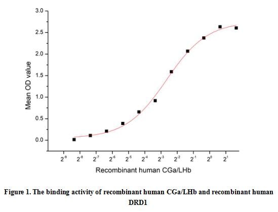
(Luteinizing Hormone (LH) is a 42 kDa heterodimer belonging to the glycoprotein hormone family. It is composed of noncovalently linked glycosylated alpha and beta chains. The alpha subunit (CG alpha) is also a component of Follicle-Stimulating Hormone (FSH), Thyroid-Stimulating Hormone, and Chorionic Gonadotropin. The unique beta subunit confers the protein's specific biological action and is responsible for the interaction with its receptor. LH is produced and secreted by the anterior pituitary gland. Its secretion is controlled by Gonadotropin-Releasing Hormone from the hypothalamus; however, LH secretion can also be stimulated by estradiol. A functional binding ELISA assay was conducted to detect the interaction of recombinant human CGa/LHb and recombinant human Dopamine Receptor D1 (DRD1). Briefly, biotin-linked CGa/LHb were diluted serially in PBS, with 0.01% BSA (pH 7.4). Duplicate samples of 100 ul were then transferred to DRD1-coated microtiter wells and incubated for 1h at 37 degree C. Wells were washed with PBST 3 times and incubation with Streptavidin-HRP for 30min, then wells were aspirated and washed 5 times. With the addition of substrate solution, wells were incubated 15-25 minutes at 37 degree C. Finally, add 50 ul stop solution to the wells and read at 450 nm immediately. The binding activity of CGa/LHb and DRD1 was shown in Figure 1, the EC50 for this effect is 0.644 ug/mL.)
Bioactivity
(Luteinizing Hormone (LH) is a 42 kDa heterodimer belonging to the glycoprotein hormone family. It is composed of noncovalently linked glycosylated alpha and beta chains. The alpha subunit (CG alpha) is also a component of Follicle-Stimulating Hormone (FSH), Thyroid-Stimulating Hormone, and Chorionic Gonadotropin. The unique beta subunit confers the protein's specific biological action and is responsible for the interaction with its receptor. LH is produced and secreted by the anterior pituitary gland. Its secretion is controlled by Gonadotropin-Releasing Hormone from the hypothalamus; however, LH secretion can also be stimulated by estradiol. A functional binding ELISA assay was conducted to detect the interaction of recombinant human CGa/LHb and recombinant human Dopamine Receptor D1 (DRD1). Briefly, biotin-linked CGa/LHb were diluted serially in PBS, with 0.01% BSA (pH 7.4). Duplicate samples of 100 ul were then transferred to DRD1-coated microtiter wells and incubated for 1h at 37 degree C. Wells were washed with PBST 3 times and incubation with Streptavidin-HRP for 30min, then wells were aspirated and washed 5 times. With the addition of substrate solution, wells were incubated 15-25 minutes at 37 degree C. Finally, add 50 ul stop solution to the wells and read at 450 nm immediately. The binding activity of CGa/LHb and DRD1 was shown in Figure 1, the EC50 for this effect is 0.644 ug/mL.)
Luteinizing Hormone (LH), Active Protein (Cat# AAA161734)
Full Name
Active Luteinizing Hormone (LH)
Gene Names
CGA; HCG; LHA; FSHA; GPHa; TSHA; GPHA1; CG-ALPHA
Applications
Activity Assay, Cell Culture
Purity
Greater than 80% by SDS-PAGE
Pricing
 WB (Western Blot)
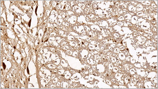
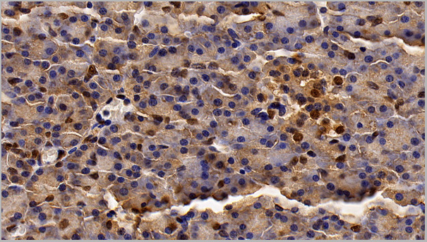
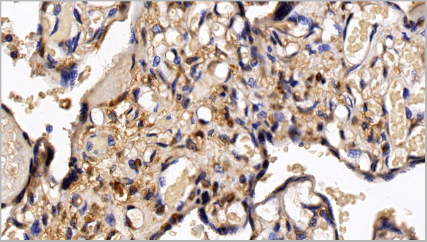
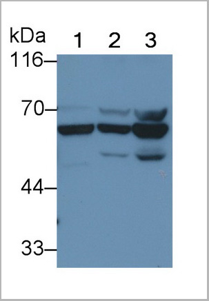
(Western Blot; Sample: Lane1: Human Lung lysate; Lane2: A549 cell lysate; Lane3: HepG2 cell lysate Primary Ab: 0.2ug/ml Mouse AntiHuman HDAC1 Antibody Second Ab: 0.2ug/mL HRPLinked Rabbit AntiMouse IgG Polyclonal Antibody (Catalog: SAA544Mu19))
WB (Western Blot)
(Western Blot; Sample: Lane1: Human Lung lysate; Lane2: A549 cell lysate; Lane3: HepG2 cell lysate Primary Ab: 0.2ug/ml Mouse AntiHuman HDAC1 Antibody Second Ab: 0.2ug/mL HRPLinked Rabbit AntiMouse IgG Polyclonal Antibody (Catalog: SAA544Mu19))
Histone Deacetylase 1 (HDAC1), Monoclonal Antibody (Cat# AAA21205)
Full Name
Monoclonal Antibody to Histone Deacetylase 1 (HDAC1)
Gene Names
HDAC1; HD1; RPD3; GON-10; RPD3L1
Reactivity
Human, Pig
Applications
Western Blot, Immunohistochemistry, Immunocytochemistry, Immunoprecipitation
Purity
Protein A + Protein G affinity chromatography
Pricing
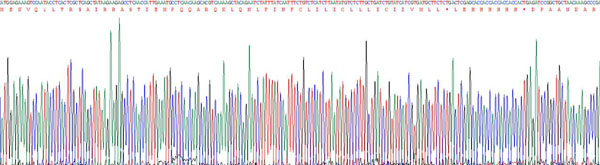
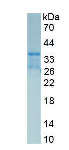
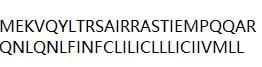 Amino Acid Info
Phospholamban (PLN), Recombinant Protein (Cat# AAA153489)
Full Name
Recombinant Phospholamban (PLN)
Gene Names
Pln; Plb
Applications
Western Blot
Purity
Greater than 80% by SDS-PAGE
Pricing
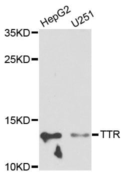
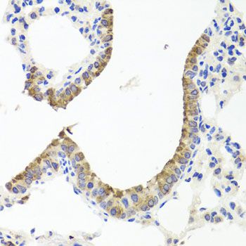 IHC (Immunohiostchemistry)
(Immunohistochemistry of paraffin-embedded mouse lung using TTR antibody at dilution of 1:100 (40x lens).)
IHC (Immunohiostchemistry)
(Immunohistochemistry of paraffin-embedded mouse lung using TTR antibody at dilution of 1:100 (40x lens).)
TTR, Polyclonal Antibody (Cat# AAA37306)
Full Name
TTR Polyclonal Antibody
Gene Names
TTR; CTS; CTS1; PALB; TBPA; HEL111; HsT2651
Reactivity
Human, Mouse, Rat
Applications
Immunohistochemistry, Western Blot
Purity
Affinity Purification
Pricing
 Bioactivity
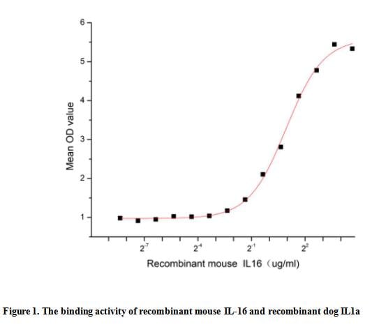
(Interleukin 16 (IL-16), formerly known as lymphocyte chemoattractant factor or LCF, is a pro-inflammatory cytokine that is chemotactic for CD4 T lymphocytes, monocytes, and eosinophils. In addition to inducing chemotaxis, IL-16 can upregulate IL-2 receptor and HLA-DR4 expression, inhibit T cell receptor (TcR)/CD3-dependent activation, and promote repression of HIV-1 transcription.Besides,Interleukin 1 Alpha (IL1a) has been identified as an interactor of IL-16, thus a functional binding ELISA assay was conducted to detect the interaction of recombinant mouse IL-16 and recombinant dog IL1a. Briefly, IL-16 was diluted serially in PBS with 0.01% BSA (pH 7.4). Duplicate samples of 100 ul were then transferred to IL1a-coated microtiter wells and incubated for 1h at 37 degree C. Wells were washed with PBST and incubated for 1h with anti-IL-16 pAb, then aspirated and washed 3 times. After incubation with HRP labelled secondary antibody for 1h at 37 degree C, wells were aspirated and washed 5 times. With the addition of substrate solution, wells were incubated 15-25 minutes at 37 degree C. Finally, add 50 uL stop solution to the wells and read at 450/630 nm immediately. The binding activity of recombinant mouse IL-16 and recombinant dog IL1a was shown in Figure 1, the EC50 for this effect is 1.9 ug/mL.)
Bioactivity
(Interleukin 16 (IL-16), formerly known as lymphocyte chemoattractant factor or LCF, is a pro-inflammatory cytokine that is chemotactic for CD4 T lymphocytes, monocytes, and eosinophils. In addition to inducing chemotaxis, IL-16 can upregulate IL-2 receptor and HLA-DR4 expression, inhibit T cell receptor (TcR)/CD3-dependent activation, and promote repression of HIV-1 transcription.Besides,Interleukin 1 Alpha (IL1a) has been identified as an interactor of IL-16, thus a functional binding ELISA assay was conducted to detect the interaction of recombinant mouse IL-16 and recombinant dog IL1a. Briefly, IL-16 was diluted serially in PBS with 0.01% BSA (pH 7.4). Duplicate samples of 100 ul were then transferred to IL1a-coated microtiter wells and incubated for 1h at 37 degree C. Wells were washed with PBST and incubated for 1h with anti-IL-16 pAb, then aspirated and washed 3 times. After incubation with HRP labelled secondary antibody for 1h at 37 degree C, wells were aspirated and washed 5 times. With the addition of substrate solution, wells were incubated 15-25 minutes at 37 degree C. Finally, add 50 uL stop solution to the wells and read at 450/630 nm immediately. The binding activity of recombinant mouse IL-16 and recombinant dog IL1a was shown in Figure 1, the EC50 for this effect is 1.9 ug/mL.)
Interleukin 16 (IL16), Active Protein (Cat# AAA161675)
Full Name
Active Interleukin 16 (IL16)
Gene Names
Il16; mKIAA4048
Applications
Activity Assay, Cell Culture
Purity
Greater than 90% by SDS-PAGE
Pricing
 Bioactivity
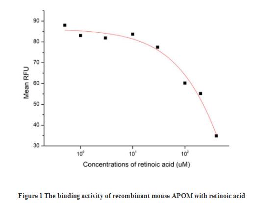
(Apolipoprotein M (APOM) is an approximately 25 kDa variably glycosylated protein that adopts a beta-barrel structure characteristic of lipocalin family proteins. It functions as a component of lipoprotein particles which play essential roles in fatty acid and cholesterol transport and metabolism. Alternative splicing generates a short isoform that lacks the N-terminal 72 amino acids. APOM is produced primarily by hepatocytes but also by renal tubule epithelial cells. The signal peptide is not cleaved and is required for APOM association with lipoprotein particles as well as Megalin mediated reabsorption by the kidney. The activity of recombinant mouse APOM was measured by its ability to bind all-trans retinoic acid. The binding of retinoic acid results in the quenching of Trp fluorescence in APOM. APOM was diluted to 50 ug/ml in 50 mM Tris, 10 mM CaCl2, 150 mM NaCl, pH 7.5 (assay buffer) and the retinoic acid was diluted to 400, 200, 100, 30, 10, 3, 1 and 0.5 uM in 95% ethanol. Mixing 112.5 uL of 50 ug/mL rmAPOM and 12.5 uL of retinoic acid serial dilutions in microtubes and a blank containing 112.5 uL of 50 ug/mL rmAPOM and 12.5 uL of 95% ethanol, then incubate at room temperature for 30 minutes. Loading 100 ul of the reaction mixtures and blank and read at excitation and emission wavelengths of 280 nm and 340 nm (top read), respectively, in endpoint mode. The result was shown in figure 1, the 50% binding concentration (BC50) is >300 uM.)
Bioactivity
(Apolipoprotein M (APOM) is an approximately 25 kDa variably glycosylated protein that adopts a beta-barrel structure characteristic of lipocalin family proteins. It functions as a component of lipoprotein particles which play essential roles in fatty acid and cholesterol transport and metabolism. Alternative splicing generates a short isoform that lacks the N-terminal 72 amino acids. APOM is produced primarily by hepatocytes but also by renal tubule epithelial cells. The signal peptide is not cleaved and is required for APOM association with lipoprotein particles as well as Megalin mediated reabsorption by the kidney. The activity of recombinant mouse APOM was measured by its ability to bind all-trans retinoic acid. The binding of retinoic acid results in the quenching of Trp fluorescence in APOM. APOM was diluted to 50 ug/ml in 50 mM Tris, 10 mM CaCl2, 150 mM NaCl, pH 7.5 (assay buffer) and the retinoic acid was diluted to 400, 200, 100, 30, 10, 3, 1 and 0.5 uM in 95% ethanol. Mixing 112.5 uL of 50 ug/mL rmAPOM and 12.5 uL of retinoic acid serial dilutions in microtubes and a blank containing 112.5 uL of 50 ug/mL rmAPOM and 12.5 uL of 95% ethanol, then incubate at room temperature for 30 minutes. Loading 100 ul of the reaction mixtures and blank and read at excitation and emission wavelengths of 280 nm and 340 nm (top read), respectively, in endpoint mode. The result was shown in figure 1, the 50% binding concentration (BC50) is >300 uM.)
Apolipoprotein M (APOM), Active Protein (Cat# AAA161884)
Full Name
Active Apolipoprotein M (APOM)
Gene Names
Apom; G3a; NG20; 1190010O19Rik
Applications
Activity Assay, Cell Culture
Purity
Greater than 95% by SDS-PAGE
Pricing
 Application Data
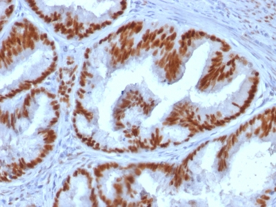
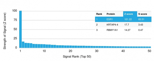
(Analysis of Protein Array containing more than 19, 000 full-length human proteins using Estrogen Receptor alpha Mouse Monoclonal Antibody (ESR1/1935) Z- and S- Score: The Z-score represents the strength of a signal that a monoclonal antibody (MAb) (in combination with a fluorescently-tagged anti-IgG secondary antibody) produces when binding to a particular protein on the HuProtTM array. Z-scores are described in units of standard deviations (SD's) above the mean value of all signals generated on that array. If targets on HuProtTM are arranged in descending order of the Z-score, the S-score is the difference (also in units of SD's) between the Z-score. S-score therefore represents the relative target specificity of a MAb to its intended target. A MAb is considered to specific to its intended target, if the MAb has an S-score of at least 2.5. For example, if a MAb binds to protein X with a Z-score of 43 and to protein Y with a Z-score of 14, then the S-score for the binding of that MAb to protein X is equal to 29.)
Application Data
(Analysis of Protein Array containing more than 19, 000 full-length human proteins using Estrogen Receptor alpha Mouse Monoclonal Antibody (ESR1/1935) Z- and S- Score: The Z-score represents the strength of a signal that a monoclonal antibody (MAb) (in combination with a fluorescently-tagged anti-IgG secondary antibody) produces when binding to a particular protein on the HuProtTM array. Z-scores are described in units of standard deviations (SD's) above the mean value of all signals generated on that array. If targets on HuProtTM are arranged in descending order of the Z-score, the S-score is the difference (also in units of SD's) between the Z-score. S-score therefore represents the relative target specificity of a MAb to its intended target. A MAb is considered to specific to its intended target, if the MAb has an S-score of at least 2.5. For example, if a MAb binds to protein X with a Z-score of 43 and to protein Y with a Z-score of 14, then the S-score for the binding of that MAb to protein X is equal to 29.)
Estrogen Receptor, alpha, Monoclonal Antibody (Cat# AAA214645)
Full Name
Estrogen Receptor, alpha (Marker of Estrogen Dependence)
Gene Names
ESR1; ER; ESR; Era; ESRA; ESTRR; NR3A1
Reactivity
Human. Others not known.
Applications
Immunohistochemistry, Western Blot
Pricing
 Application Data
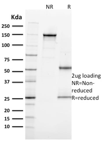
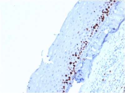
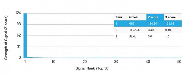
(Analysis of Protein Array containing more than 19, 000 full-length human proteins using Ki67 Mouse Monoclonal Antibody (MKI67/2462).Z- and S- Score: The Z-score represents the strength of a signal that a monoclonal antibody (MAb) (in combination with a fluorescently-tagged anti-IgG secondary antibody) produces when binding to a particular protein on the HuProtTM array. Z-scores are described in units of standard deviations (SD's) above the mean value of all signals generated on that array. If targets on HuProtTM are arranged in descending order of the Z-score, the S-score is the difference (also in units of SD's) between the Z-score. S-score therefore represents the relative target specificity of a MAb to its intended target. A MAb is considered to specific to its intended target, if the MAb has an S-score of at least 2.5. For example, if a MAb binds to protein X with a Z-score of 43 and to protein Y with a Z-score of 14, then the S-score for the binding of that MAb to protein X is equal to 29.)
Application Data
(Analysis of Protein Array containing more than 19, 000 full-length human proteins using Ki67 Mouse Monoclonal Antibody (MKI67/2462).Z- and S- Score: The Z-score represents the strength of a signal that a monoclonal antibody (MAb) (in combination with a fluorescently-tagged anti-IgG secondary antibody) produces when binding to a particular protein on the HuProtTM array. Z-scores are described in units of standard deviations (SD's) above the mean value of all signals generated on that array. If targets on HuProtTM are arranged in descending order of the Z-score, the S-score is the difference (also in units of SD's) between the Z-score. S-score therefore represents the relative target specificity of a MAb to its intended target. A MAb is considered to specific to its intended target, if the MAb has an S-score of at least 2.5. For example, if a MAb binds to protein X with a Z-score of 43 and to protein Y with a Z-score of 14, then the S-score for the binding of that MAb to protein X is equal to 29.)
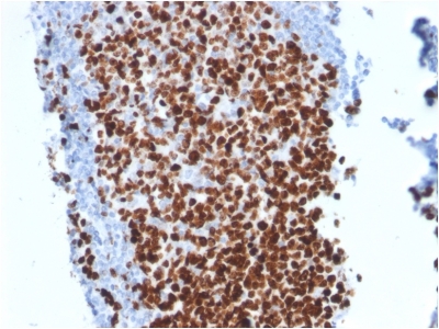
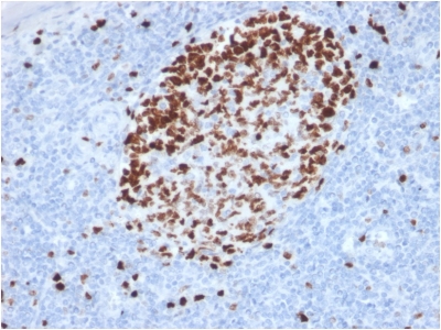
Ki-67, Monoclonal Antibody (Cat# AAA214665)
Full Name
Ki-67 (Proliferating Cell Marker)
Gene Names
MKI67; KIA; MIB-; MIB-1; PPP1R105
Reactivity
Human. Others not known.
Applications
Immunohistochemistry
Pricing
 Bioactivity
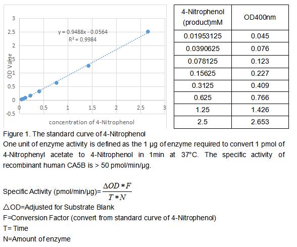
(Carbonic Anhydrase (CA) catalyzes the reversible reaction of CO2 H2O = HCO3- H, which is fundamental to many processes such as respiration, renal tubular acidification and bone resorption. Carbonic Anhydrase VB encoded by the CA5B gene is a mitochondrial protein. In comparison with another mitochondrial CA (CA5A), CA5B has different tissue distribution and chromosomal location. The activity of recombinant human CA5B was measured by its ability to hydrolyze 4-Nitrophenyl acetate (4-NPA) to 4-Nitrophenol. The reaction was performed in 12.5 mM Tris, 75 mM NaCl, pH 7.5 (assay buffer), initiated by addition 50 uL of various concentrations of CA5B (diluted by assay buffer) to 50 uL of 2 mM substrate 4-NPA (100 mM stock in Acetone, diluted by assay buffer). Incubated at 37 degree C for 5min, then read at a wavelength of 400 nm.)
Bioactivity
(Carbonic Anhydrase (CA) catalyzes the reversible reaction of CO2 H2O = HCO3- H, which is fundamental to many processes such as respiration, renal tubular acidification and bone resorption. Carbonic Anhydrase VB encoded by the CA5B gene is a mitochondrial protein. In comparison with another mitochondrial CA (CA5A), CA5B has different tissue distribution and chromosomal location. The activity of recombinant human CA5B was measured by its ability to hydrolyze 4-Nitrophenyl acetate (4-NPA) to 4-Nitrophenol. The reaction was performed in 12.5 mM Tris, 75 mM NaCl, pH 7.5 (assay buffer), initiated by addition 50 uL of various concentrations of CA5B (diluted by assay buffer) to 50 uL of 2 mM substrate 4-NPA (100 mM stock in Acetone, diluted by assay buffer). Incubated at 37 degree C for 5min, then read at a wavelength of 400 nm.)
Carbonic Anhydrase VB, Mitochondrial (CA5B), Active Protein (Cat# AAA161903)
Full Name
Active Carbonic Anhydrase VB, Mitochondrial (CA5B)
Gene Names
CA5B; CA-VB
Applications
Activity Assay, Cell Culture
Purity
Greater than 90% by SDS-PAGE
Pricing