
Filters
Clonality
Type
Reactivity
Gene Name
Isotype
Host
Application
Clone
10000 results for "Recombinant Proteins" - showing 2150-2200
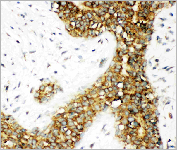
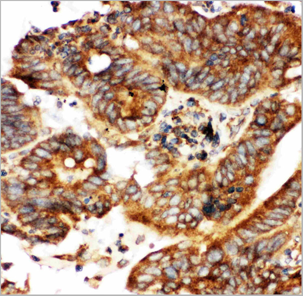
 FCM (Flow Cytometry)
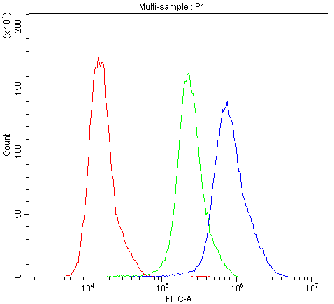
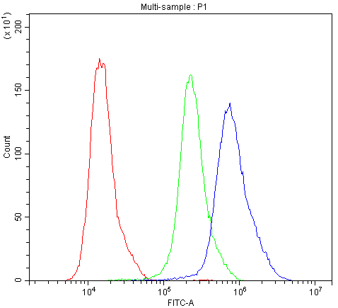
(Figure 7. Flow Cytometry analysis of Hela cells using anti-Calbindin antibody (AAA11603).Overlay histogram showing Hela cells stained with AAA11603 (Blue line).The cells were blocked with 10% normal goat serum. And then incubated with rabbit anti-Calbindin Antibody (AAA11603,1ug/1x10^6 cells) for 30 min at 20 degree C. DyLight®488 conjugated goat anti-rabbit IgG (5-10ug/1x10^6 cells) was used as secondary antibody for 30 minutes at 20 degree C. Isotype control antibody (Green line) was rabbit IgG (1ug/1x106) used under the same conditions. Unlabelled sample (Red line) was also used as a control.)
FCM (Flow Cytometry)
(Figure 7. Flow Cytometry analysis of Hela cells using anti-Calbindin antibody (AAA11603).Overlay histogram showing Hela cells stained with AAA11603 (Blue line).The cells were blocked with 10% normal goat serum. And then incubated with rabbit anti-Calbindin Antibody (AAA11603,1ug/1x10^6 cells) for 30 min at 20 degree C. DyLight®488 conjugated goat anti-rabbit IgG (5-10ug/1x10^6 cells) was used as secondary antibody for 30 minutes at 20 degree C. Isotype control antibody (Green line) was rabbit IgG (1ug/1x106) used under the same conditions. Unlabelled sample (Red line) was also used as a control.)
Calbindin, Polyclonal Antibody (Cat# AAA11603)
Full Name
Anti-Calbindin Antibody
Gene Names
CALB1; CALB
Reactivity
Human, Mouse, Rat
Applications
Western Blot, Immunohistochemistry
Purity
Immunogen affinity purified.
Pricing
 Bioactivity
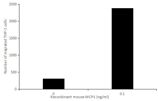
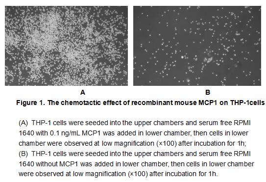
(Monocyte Chemotactic Protein 1 (MCP1), also known as C-C motif chemokine 2(CCL2), is a member of the beta (C-C) subfamily of chemokines that is a chemoattractant for monocytes and basophils. Mouse CCL2 is secreted as a 14 kDa glycoprotein monomer but nonc)
Bioactivity
(Monocyte Chemotactic Protein 1 (MCP1), also known as C-C motif chemokine 2(CCL2), is a member of the beta (C-C) subfamily of chemokines that is a chemoattractant for monocytes and basophils. Mouse CCL2 is secreted as a 14 kDa glycoprotein monomer but nonc)
Monocyte Chemotactic Protein 1 (MCP1), Active Protein (Cat# AAA152975)
Full Name
Active Monocyte Chemotactic Protein 1 (MCP1)
Gene Names
Ccl2; JE; HC11; MCAF; MCP1; MCP-1; Scya2; Sigje; SMC-CF; AI323594
Applications
Activity Assay, Cell Culture
Purity
Greater than 90% by SDS-PAGE
Pricing
 Bioactivity
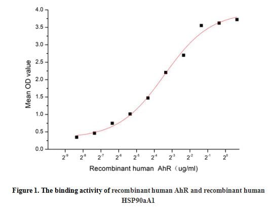
(Aryl Hydrocarbon Receptor (AhR), also known as bHLHE76, is a 110 kDa member of the bHLH/PAS transcription factor family. It is widely expressed (breast, lung, liver), and serves many functions. It is a ligand-activated transcription factor best known for its role in sensing xenobiotics and regulating the expression of genes involved in xenobiotic metabolism, including drug transporters and drug metabolizing enzymes. Heat Shock Protein 90kDa Alpha A1 (HSP90aA1) has been identified as an interactor of AhR, thus a functional binding ELISA assay was conducted to detect the interaction of recombinant human AhR and recombinant human HSP90aA1. Briefly, AhR were diluted serially in PBS, with 0.01% BSA (pH 7.4). Duplicate samples of 100 ul were then transferred to HSP90aA1-coated microtiter wells and incubated for 1h at 37 degree C. Wells were washed with PBST and incubated for 1h with anti-AhR pAb, then aspirated and washed 3 times. After incubation with HRP labelled secondary antibody, wells were aspirated and washed 5 times. With the addition of substrate solution, wells were incubated 15-25 minutes at 37 degree C. Finally, add 50 uL stop solution to the wells and read at 450 nm immediately. The binding activity of AhR and HSP90aA1 was shown in Figure 1, and this effect was in a dose dependent manner, the EC50 for this effect is 0.09 ug/mL.)
Bioactivity
(Aryl Hydrocarbon Receptor (AhR), also known as bHLHE76, is a 110 kDa member of the bHLH/PAS transcription factor family. It is widely expressed (breast, lung, liver), and serves many functions. It is a ligand-activated transcription factor best known for its role in sensing xenobiotics and regulating the expression of genes involved in xenobiotic metabolism, including drug transporters and drug metabolizing enzymes. Heat Shock Protein 90kDa Alpha A1 (HSP90aA1) has been identified as an interactor of AhR, thus a functional binding ELISA assay was conducted to detect the interaction of recombinant human AhR and recombinant human HSP90aA1. Briefly, AhR were diluted serially in PBS, with 0.01% BSA (pH 7.4). Duplicate samples of 100 ul were then transferred to HSP90aA1-coated microtiter wells and incubated for 1h at 37 degree C. Wells were washed with PBST and incubated for 1h with anti-AhR pAb, then aspirated and washed 3 times. After incubation with HRP labelled secondary antibody, wells were aspirated and washed 5 times. With the addition of substrate solution, wells were incubated 15-25 minutes at 37 degree C. Finally, add 50 uL stop solution to the wells and read at 450 nm immediately. The binding activity of AhR and HSP90aA1 was shown in Figure 1, and this effect was in a dose dependent manner, the EC50 for this effect is 0.09 ug/mL.)
Aryl Hydrocarbon Receptor (AhR), Active Protein (Cat# AAA161844)
Full Name
Active Aryl Hydrocarbon Receptor (AhR)
Gene Names
AHR; bHLHe76
Applications
Activity Assay, Cell Culture
Purity
Greater than 90% by SDS-PAGE
Pricing
 Bioactivity
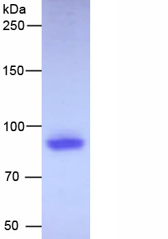
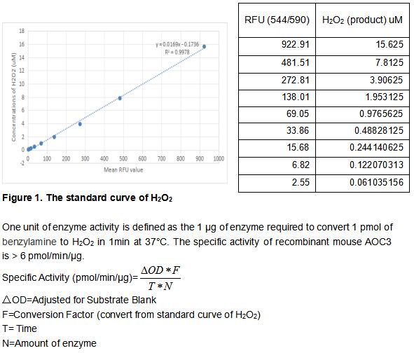
(Amine Oxidase, Copper Containing 3 (AOC3) is a copper amine oxidase with a topaquinone cofactor. AOC3 is a Type II integral membrane protein, but a soluble form of the enzyme is present in human serum, and its level increases in diabetes and some inflammatory liver diseases. AOC3 catalyzes the oxidative deamination of small primary amines such as methylamine, benzylamine, and aminoacetone in a reaction that produces an aldehyde, ammonia, and H2O2. The enzyme is sensitive to inhibition by semicarbazide. AOC3 expression is highest in the endothelium of lung, heart, and intestine, but low in tissues such as brain, spleen, kidney, and liver. The activity of recombinant mouse AOC3 was measured by its ability to produce hydrogen peroxide during the oxidation of benzylamine. The reaction was performed in 50 mM HEPES, pH 7.5 (assay buffer), initiated by addition 50 uL of various concentrations of AOC3 (diluted by assay buffer) to 50 ul substrate mixture of 2 mM Benzylamine, 2 units/mL HRP and 100 uM AUR. Read at excitation and emission wavelengths of 544 nm and 590 nm (top read), respectively in kinetic mode for 5 minutes.)
Bioactivity
(Amine Oxidase, Copper Containing 3 (AOC3) is a copper amine oxidase with a topaquinone cofactor. AOC3 is a Type II integral membrane protein, but a soluble form of the enzyme is present in human serum, and its level increases in diabetes and some inflammatory liver diseases. AOC3 catalyzes the oxidative deamination of small primary amines such as methylamine, benzylamine, and aminoacetone in a reaction that produces an aldehyde, ammonia, and H2O2. The enzyme is sensitive to inhibition by semicarbazide. AOC3 expression is highest in the endothelium of lung, heart, and intestine, but low in tissues such as brain, spleen, kidney, and liver. The activity of recombinant mouse AOC3 was measured by its ability to produce hydrogen peroxide during the oxidation of benzylamine. The reaction was performed in 50 mM HEPES, pH 7.5 (assay buffer), initiated by addition 50 uL of various concentrations of AOC3 (diluted by assay buffer) to 50 ul substrate mixture of 2 mM Benzylamine, 2 units/mL HRP and 100 uM AUR. Read at excitation and emission wavelengths of 544 nm and 590 nm (top read), respectively in kinetic mode for 5 minutes.)
Amine Oxidase, Copper Containing 3 (AOC3), Active Protein (Cat# AAA161883)
Full Name
Active Amine Oxidase, Copper Containing 3 (AOC3)
Gene Names
Aoc3; SSAO; VAP1
Applications
Activity Assay, Cell Culture
Purity
Greater than 90% by SDS-PAGE
Pricing
 WB (Western Blot)
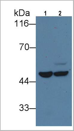
(Western Blot; Sample: Lane1: Human Liver lysate; Lane2: Porcine Liver lysate Primary Ab: 5ug/ml Mouse AntiHuman IDH1 Antibody Second Ab: 0.2ug/mL HRPLinked Caprine AntiMouse IgG Polyclonal Antibody (Catalog: SAA544Mu19))
WB (Western Blot)
(Western Blot; Sample: Lane1: Human Liver lysate; Lane2: Porcine Liver lysate Primary Ab: 5ug/ml Mouse AntiHuman IDH1 Antibody Second Ab: 0.2ug/mL HRPLinked Caprine AntiMouse IgG Polyclonal Antibody (Catalog: SAA544Mu19))
Isocitrate Dehydrogenase 1, Soluble (IDH1), Monoclonal Antibody (Cat# AAA21211)
Full Name
Monoclonal Antibody to Isocitrate Dehydrogenase 1, Soluble (IDH1)
Gene Names
IDH1; IDH; IDP; IDCD; IDPC; PICD; HEL-216; HEL-S-26
Reactivity
Human, Pig
Applications
Western Blot, Immunohistochemistry, Immunocytochemistry, Immunoprecipitation
Purity
Protein A + Protein G affinity chromatography
Pricing

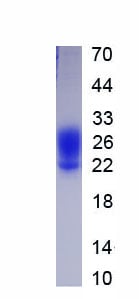
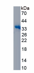
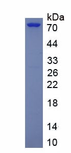
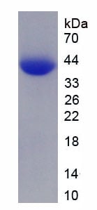 SDS-PAGE
(Figure. SDS-PAGE)
SDS-PAGE
(Figure. SDS-PAGE)
Histone H4 (H4), Recombinant Protein (Cat# AAA150665)
Full Name
Recombinant Histone H4 (H4)
Applications
Western Blot
Purity
>90%
Pricing
 IF (Imunofluorescence)
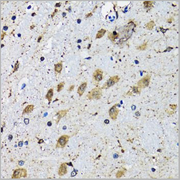
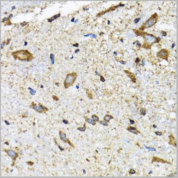
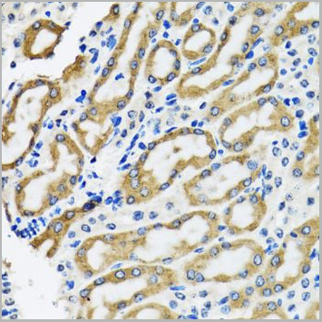
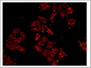
(Immunofluorescence analysis of MCF7 cells using SLC23A2 antibody (AAA28167))
IF (Imunofluorescence)
(Immunofluorescence analysis of MCF7 cells using SLC23A2 antibody (AAA28167))
SLC23A2, Polyclonal Antibody (Cat# AAA28167)
Full Name
SLC23A2 Polyclonal Antibody
Gene Names
SLC23A2; NBTL1; SVCT2; YSPL2; SLC23A1
Reactivity
Human, Mouse, Rat
Applications
Western Blot, Immunohistochemistry, Immunofluorescence
Purity
Affinity Purification
Pricing
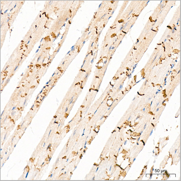 IHC (Immunohistchemistry)
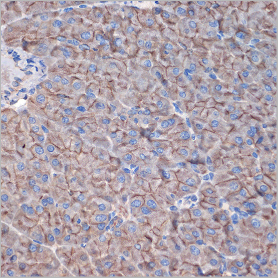
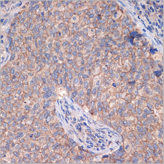
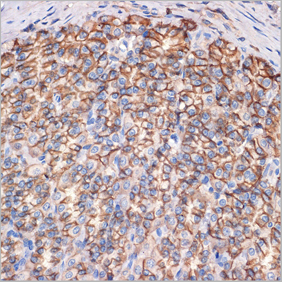
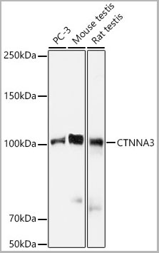
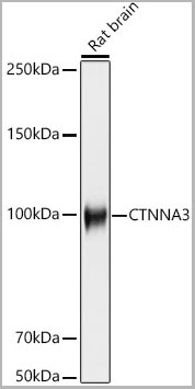
(Immunohistochemistry analysis of paraffin-embedded Mouse heart tissue using CTNNA3 Rabbit mAb (AAA28498) at a dilution of 1:200 (40x lens). High pressure antigen retrieval was performed with 0.01 M citrate buffer (pH 6.0) prior to IHC staining.)
IHC (Immunohistchemistry)
(Immunohistochemistry analysis of paraffin-embedded Mouse heart tissue using CTNNA3 Rabbit mAb (AAA28498) at a dilution of 1:200 (40x lens). High pressure antigen retrieval was performed with 0.01 M citrate buffer (pH 6.0) prior to IHC staining.)
CTNNA3, Monoclonal Antibody (Cat# AAA28498)
Full Name
CTNNA3 Rabbit mAb
Reactivity
Human
Applications
Western Blot, Immunohistochemistry
Purity
Affinity purification
Pricing
 Bioactivity
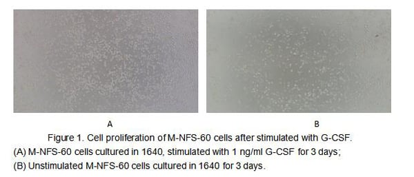
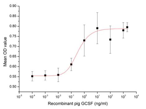
(Figure 2. Cell proliferation of M-NFS-60 cells after stimulated with G-CSF.)
Bioactivity
(Figure 2. Cell proliferation of M-NFS-60 cells after stimulated with G-CSF.)
Colony Stimulating Factor 3, Granulocyte (GCSF), Active Protein (Cat# AAA161665)
Full Name
Active Colony Stimulating Factor 3, Granulocyte (GCSF)
Gene Names
CSF3; G-CSF
Applications
Activity Assay, Cell Culture
Purity
Greater than 80% by SDS-PAGE
Pricing
 Bioactivity
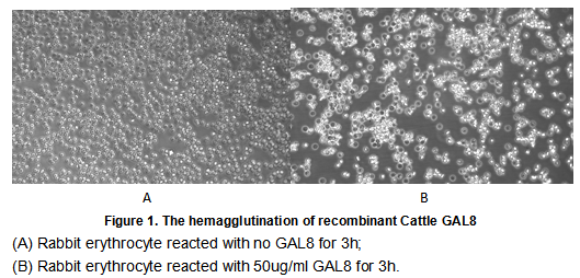
(Galectin 8 (GAL8), also known as prostate carcinoma tumor antigen 1 (PCTA1) in human, is a tandem repeat-type galectin. is a member of the lectin family, of which 14 mammalian galectins have been identified. It is also a member of the beta-galactoside-binding protein family that plays an important role in cell-cell adhesion, cell-matrix interactions, macrophage activation, angiogenesis, metastasis, apoptosis. In this case, we chose rabbit erythrocyte (RaE) to assay its ability of agglutination. A general procedure for hemagglutination assay (or haemagglutination assay; HA) is as follows, two-fold dilute the recombinant Bovine GAL8 with 0.9% sodium chloride injection, add 50uL a serial dilution of GAL8 to each well of a U or V-bottom shaped 96-well microtiter plate. The final well serves as a negative control with no GAL8, replace with 50uL 0.9% sodium chloride injection. Then add 50uL 1% rabbit erythrocyte to each well and mixed gently. The plate is incubated for 3 hours at room temperature. The results are shown in Figure 1. It was obvious that the minimal effective concentration of GAL8 is 0.781 ug/mL.)
Bioactivity
(Galectin 8 (GAL8), also known as prostate carcinoma tumor antigen 1 (PCTA1) in human, is a tandem repeat-type galectin. is a member of the lectin family, of which 14 mammalian galectins have been identified. It is also a member of the beta-galactoside-binding protein family that plays an important role in cell-cell adhesion, cell-matrix interactions, macrophage activation, angiogenesis, metastasis, apoptosis. In this case, we chose rabbit erythrocyte (RaE) to assay its ability of agglutination. A general procedure for hemagglutination assay (or haemagglutination assay; HA) is as follows, two-fold dilute the recombinant Bovine GAL8 with 0.9% sodium chloride injection, add 50uL a serial dilution of GAL8 to each well of a U or V-bottom shaped 96-well microtiter plate. The final well serves as a negative control with no GAL8, replace with 50uL 0.9% sodium chloride injection. Then add 50uL 1% rabbit erythrocyte to each well and mixed gently. The plate is incubated for 3 hours at room temperature. The results are shown in Figure 1. It was obvious that the minimal effective concentration of GAL8 is 0.781 ug/mL.)
Galectin 8 (GAL8), Active Protein (Cat# AAA152177)
Full Name
Active Galectin 8 (GAL8)
Gene Names
LGALS8; galectin-8
Applications
Activity Assay, Cell Culture
Purity
Greater than 90% by SDS-PAGE
Pricing
 WB (Western Blot)
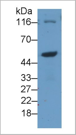
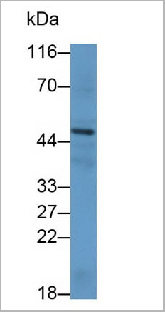
(Western Blot; Sample: Human Lung lysatePrimary Ab: 0.2ug/ml Mouse AntiHuman SHH AntibodySecond Ab: 0.2ug/mL HRPLinked Caprine AntiMouse IgG Polyclonal Antibody(Catalog: SAA544Mu19))
WB (Western Blot)
(Western Blot; Sample: Human Lung lysatePrimary Ab: 0.2ug/ml Mouse AntiHuman SHH AntibodySecond Ab: 0.2ug/mL HRPLinked Caprine AntiMouse IgG Polyclonal Antibody(Catalog: SAA544Mu19))
Hedgehog Homolog, Sonic (SHH), Monoclonal Antibody (Cat# AAA21198)
Full Name
Monoclonal Antibody to Hedgehog Homolog, Sonic (SHH)
Gene Names
SHH; TPT; HHG1; HLP3; HPE3; SMMCI; TPTPS; MCOPCB5
Reactivity
Human, Rat
Applications
Western Blot, Immunohistochemistry, Immunocytochemistry, Immunoprecipitation
Purity
Protein A + Protein G affinity chromatography
Pricing
 Bioactivity
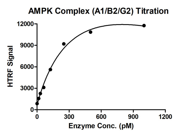
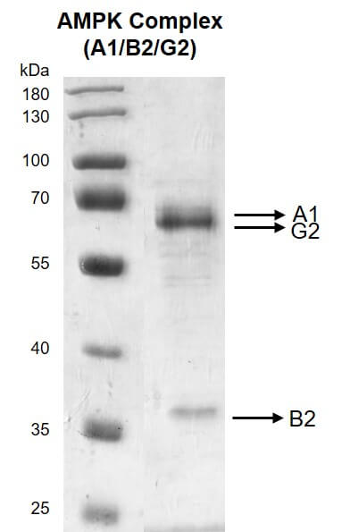
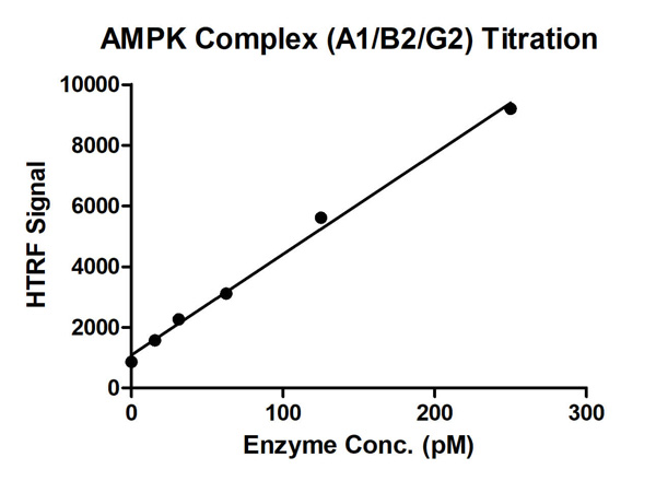
(HTRF assay for AMPK Complex (A1+B2+G2) activity 1 uM STK S1 substrate was incubated with different concentrations of AMPK Complex (A1+B2+G2) protein in a 10 ul reaction system containing 1×Enzymatic Buffer, 5 mM MgCl2, 1 mM DTT, 50 uM AMP and 100 uM ATP for 1 hr. The 10 ul detection reagents containing anti-STK antibody (1:100) and SA-XL665 (1:100) diluted with 1× Detection Buffer were added and incubated with the reactions for 30 min. All the operations and reactions were performed at room temperature. HTRF assay was used for detection.)
Bioactivity
(HTRF assay for AMPK Complex (A1+B2+G2) activity 1 uM STK S1 substrate was incubated with different concentrations of AMPK Complex (A1+B2+G2) protein in a 10 ul reaction system containing 1×Enzymatic Buffer, 5 mM MgCl2, 1 mM DTT, 50 uM AMP and 100 uM ATP for 1 hr. The 10 ul detection reagents containing anti-STK antibody (1:100) and SA-XL665 (1:100) diluted with 1× Detection Buffer were added and incubated with the reactions for 30 min. All the operations and reactions were performed at room temperature. HTRF assay was used for detection.)
AMPK Complex (A1+B2+G2), Active Protein (Cat# AAA60318)
Full Name
Recombinant AMPK Complex (A1+B2+G2) protein
Purity
>=90%
Pricing
 Bioactivity
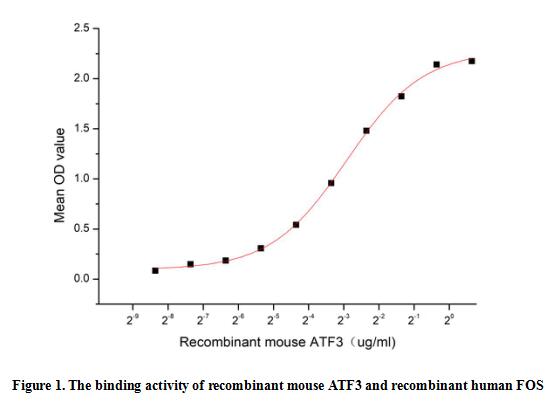
(Activating transcription factor 3 (ATF3) is a stress-induced transcription factor that plays vital roles in modulating metabolism, immunity and oncogenesis. ATF3 acts as a hub of the cellular adaptive-response network. Multiple extracellular signals, such as endoplasmic reticulum (ER) stress, cytokines, chemokines, and LPS, are connected to ATF3 induction. Proto-oncogene c-Fos can bind to ATF3 to regulate signal transduction, cell proliferation and differentiation. Thus a functional ELISA assay was conducted to detect the interaction of recombinant mouse ATF3 and recombinant human FOS. Briefly, ATF3 was diluted serially in PBS with 0.01% BSA (pH 7.4). Duplicate samples of 100 ul were then transferred to FOS-coated microtiter wells and incubated for 1h at 37 degree C. Wells were washed with PBST and incubated for 1h with anti-ATF3 pAb, then aspirated and washed 3 times. After incubation with HRP labelled secondary antibody for 1h at 37 degree C, wells were aspirated and washed 5 times. With the addition of substrate solution, wells were incubated 15-25 minutes at 37 degree C. Finally, add 50 uL stop solution to the wells and read at 450/630nm immediately. The binding activity of recombinant mouse ATF3 and recombinant human FOS was shown in Figure 1, the EC50 for this effect is 0.13 ug/mL.)
Bioactivity
(Activating transcription factor 3 (ATF3) is a stress-induced transcription factor that plays vital roles in modulating metabolism, immunity and oncogenesis. ATF3 acts as a hub of the cellular adaptive-response network. Multiple extracellular signals, such as endoplasmic reticulum (ER) stress, cytokines, chemokines, and LPS, are connected to ATF3 induction. Proto-oncogene c-Fos can bind to ATF3 to regulate signal transduction, cell proliferation and differentiation. Thus a functional ELISA assay was conducted to detect the interaction of recombinant mouse ATF3 and recombinant human FOS. Briefly, ATF3 was diluted serially in PBS with 0.01% BSA (pH 7.4). Duplicate samples of 100 ul were then transferred to FOS-coated microtiter wells and incubated for 1h at 37 degree C. Wells were washed with PBST and incubated for 1h with anti-ATF3 pAb, then aspirated and washed 3 times. After incubation with HRP labelled secondary antibody for 1h at 37 degree C, wells were aspirated and washed 5 times. With the addition of substrate solution, wells were incubated 15-25 minutes at 37 degree C. Finally, add 50 uL stop solution to the wells and read at 450/630nm immediately. The binding activity of recombinant mouse ATF3 and recombinant human FOS was shown in Figure 1, the EC50 for this effect is 0.13 ug/mL.)
Activating Transcription Factor 3 (ATF3), Active Protein (Cat# AAA161885)
Full Name
Active Activating Transcription Factor 3 (ATF3)
Gene Names
Atf3; LRG-21
Applications
Activity Assay, Cell Culture
Purity
Greater than 90% by SDS-PAGE
Pricing
 Bioactivity
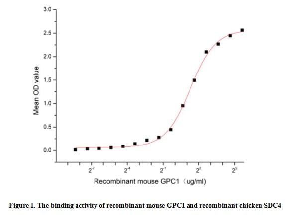
(Glypican 1 (GPC1) is one of the six glycosylphosphatidylinositol-anchored, cell surface heparan sulfate proteoglycans that acts as a growth factor signaling. Cells known to express GPC-1 include neurons, smooth and skeletal muscle cells, keratinocytes, osteoblasts, Schwann cells, immature dendritic cells, and tumor, plus tumor-associated vascular endothelial cells. The function of GPC-1 is complex and varied, it can modulate various signaling pathways including Heparin Binding Epidermal Growth Factor Like Growth Factor (HBEGF), fibroblast growth factors (FGF), vascular endothelial growth factor-A (VEGF-A), transforming growth factor-beta (TGF-beta), Wnt, Hedgehog (Hh), and bone morphogenic protein (BMP) through specific interactions with pathway ligands and receptors. Besides, Syndecan 4 (SDC4) has been also identified as an interactor of GPC1, thus a functional binding ELISA assay was conducted to detect the interaction of recombinant mouse GPC1 and recombinant chicken SDC4. Briefly, GPC1 was diluted serially in PBS with 0.01% BSA (pH 7.4). Duplicate samples of 100 ul were then transferred to SDC4-coated microtiter wells and incubated for 1h at 37 degree C. Wells were washed with PBST and incubated for 1h with anti-GPC1 pAb, then aspirated and washed 3 times. After incubation with HRP labelled secondary antibody for 1h at 37 degree C, wells were aspirated and washed 5 times. With the addition of substrate solution, wells were incubated 15-25 minutes at 37 degree C. Finally, add 50 uL stop solution to the wells and read at 450/630 nm immediately. The binding activity of recombinant mouse GPC1 and recombinant chicken SDC4 was shown in Figure 1, the EC50 for this effect is 2.5 ug/mL.)
Bioactivity
(Glypican 1 (GPC1) is one of the six glycosylphosphatidylinositol-anchored, cell surface heparan sulfate proteoglycans that acts as a growth factor signaling. Cells known to express GPC-1 include neurons, smooth and skeletal muscle cells, keratinocytes, osteoblasts, Schwann cells, immature dendritic cells, and tumor, plus tumor-associated vascular endothelial cells. The function of GPC-1 is complex and varied, it can modulate various signaling pathways including Heparin Binding Epidermal Growth Factor Like Growth Factor (HBEGF), fibroblast growth factors (FGF), vascular endothelial growth factor-A (VEGF-A), transforming growth factor-beta (TGF-beta), Wnt, Hedgehog (Hh), and bone morphogenic protein (BMP) through specific interactions with pathway ligands and receptors. Besides, Syndecan 4 (SDC4) has been also identified as an interactor of GPC1, thus a functional binding ELISA assay was conducted to detect the interaction of recombinant mouse GPC1 and recombinant chicken SDC4. Briefly, GPC1 was diluted serially in PBS with 0.01% BSA (pH 7.4). Duplicate samples of 100 ul were then transferred to SDC4-coated microtiter wells and incubated for 1h at 37 degree C. Wells were washed with PBST and incubated for 1h with anti-GPC1 pAb, then aspirated and washed 3 times. After incubation with HRP labelled secondary antibody for 1h at 37 degree C, wells were aspirated and washed 5 times. With the addition of substrate solution, wells were incubated 15-25 minutes at 37 degree C. Finally, add 50 uL stop solution to the wells and read at 450/630 nm immediately. The binding activity of recombinant mouse GPC1 and recombinant chicken SDC4 was shown in Figure 1, the EC50 for this effect is 2.5 ug/mL.)
Glypican 1 (GPC1), Active Protein (Cat# AAA161826)
Full Name
Active Glypican 1 (GPC1)
Gene Names
Gpc1; AI462976
Applications
Activity Assay, Cell Culture
Purity
Greater than 90% by SDS-PAGE
Pricing
 IP (Immunoprecipitation)
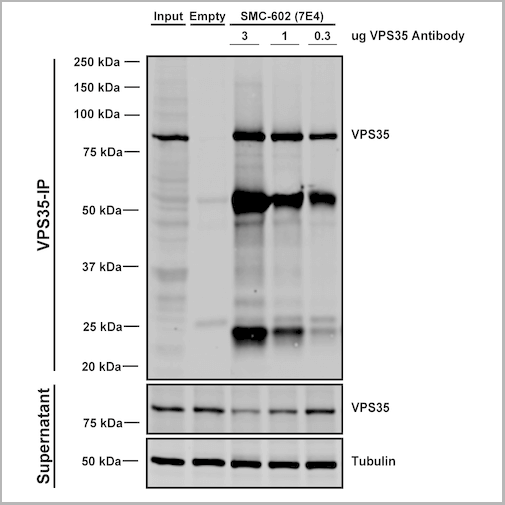
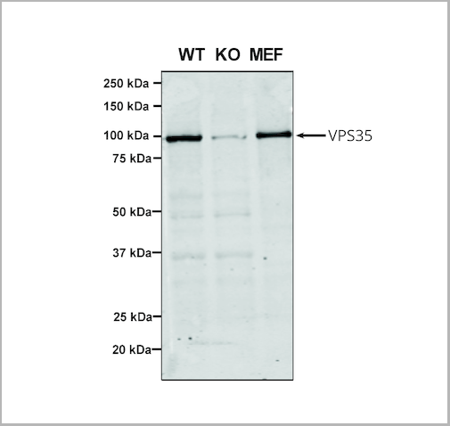
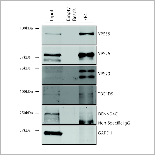
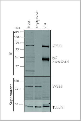
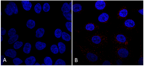
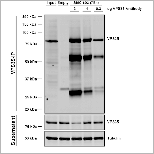
(Immunoprecipitation analysis using Mouse Anti-VPS35 Monoclonal Antibody, Clone 7E4. Tissue: embryonic fibroblast. Species: Mouse. Primary Antibody: Mouse Anti-VPS35 Monoclonal Antibody. Three amounts (3, 1 and 0.3 ug) were non-covalently coupled to 10uL of A/G sepharose beads for 1 hour at 4 degree C and next incubated with 250ug of MEF lysate for 2 hours at 4 degree C.)
IP (Immunoprecipitation)
(Immunoprecipitation analysis using Mouse Anti-VPS35 Monoclonal Antibody, Clone 7E4. Tissue: embryonic fibroblast. Species: Mouse. Primary Antibody: Mouse Anti-VPS35 Monoclonal Antibody. Three amounts (3, 1 and 0.3 ug) were non-covalently coupled to 10uL of A/G sepharose beads for 1 hour at 4 degree C and next incubated with 250ug of MEF lysate for 2 hours at 4 degree C.)
VPS35, Monoclonal Antibody (Cat# AAA27666)
Full Name
VPS35 Antibody, Clone 7E4: ATTO 488
Gene Names
VPS35; MEM3; PARK17
Reactivity
Human, Mouse, Rat
Applications
Western Blot, Immunocytochemistry, Immunofluorescence, Immunoprecipitation
Purity
Protein G Purified
Pricing
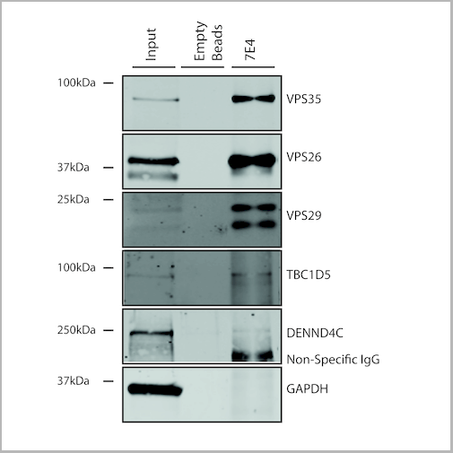
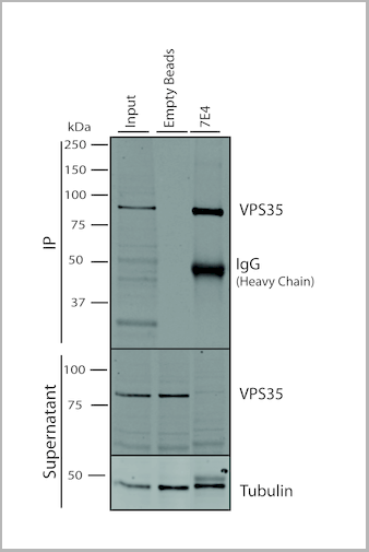
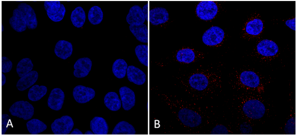
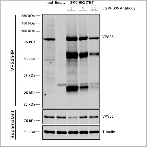 IP (Immunoprecipitation)
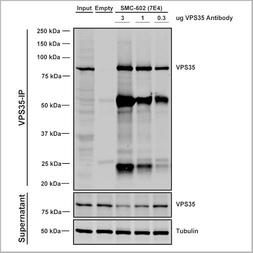
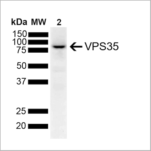
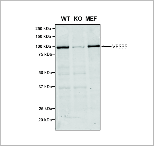
(Immunoprecipitation analysis using Mouse Anti-VPS35 Monoclonal Antibody, Clone 7E4. Tissue: embryonic fibroblast. Species: Mouse. Primary Antibody: Mouse Anti-VPS35 Monoclonal Antibody. Three amounts (3, 1 and 0.3 ug) were non-covalently coupled to 10uL of A/G sepharose beads for 1 hour at 4 degree C and next incubated with 250ug of MEF lysate for 2 hours at 4 degree C.)
IP (Immunoprecipitation)
(Immunoprecipitation analysis using Mouse Anti-VPS35 Monoclonal Antibody, Clone 7E4. Tissue: embryonic fibroblast. Species: Mouse. Primary Antibody: Mouse Anti-VPS35 Monoclonal Antibody. Three amounts (3, 1 and 0.3 ug) were non-covalently coupled to 10uL of A/G sepharose beads for 1 hour at 4 degree C and next incubated with 250ug of MEF lysate for 2 hours at 4 degree C.)
VPS35, Monoclonal Antibody (Cat# AAA27667)
Full Name
VPS35 Antibody, Clone 7E4: ATTO 594
Gene Names
VPS35; MEM3; PARK17
Reactivity
Human, Mouse, Rat
Applications
Western Blot, Immunocytochemistry, Immunofluorescence, Immunoprecipitation
Purity
Protein G Purified
Pricing
 Bioactivity
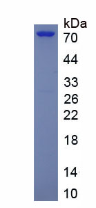
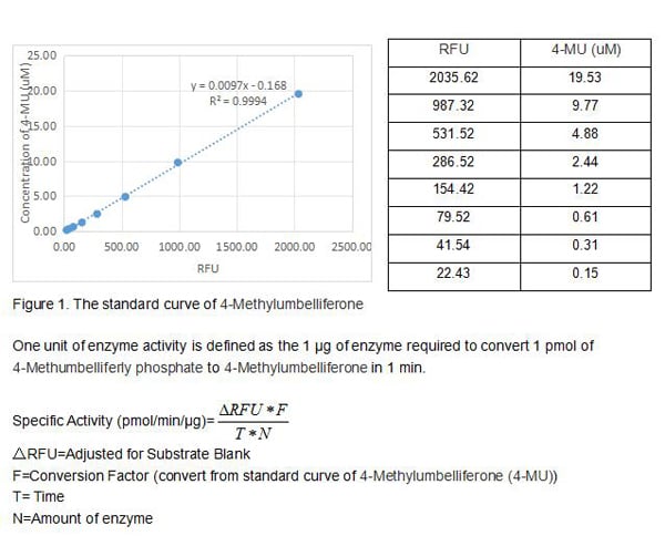
(Four distinct genes encode alkaline phosphatases (APs) in humans. The ALPL gene encodes the liver/bone/kidney isozyme, also known as the tissuenonspecific AP (TNAP). In comparison, ALPI, ALPP and ALPPL2 encode intestinal, placental and placental-like or germ cell APs, respectively. The serum levels of human APs are useful tumor markers. There are many mutations in the ALPL gene, leading to different forms of hypophosphatasia, characterized by poorly mineralized cartilage and bones. The native ALPL is a glycosylated homodimer attached to the membrane through a GPI-anchor. The activity assay of recombinant human ALPL was measured by its ability to cleave a peptide substrate, 4-Methumbelliferly phosphate. The reaction was performed in 50 mM Tris, 1 mM MgCl2, pH 9.0 (assay buffer), ainitiated by addition 50 uL of 0.625 ug/ml ALPL (diluted by assay buffer) to 50 uL of 50 uM substrate. Read at excitation and emission wavelengths of 365 nm and 445 nm (top read), respectively, in kinetic mode for 5 minutes. The specific activity of recombinant human ALPL is >11000 pmol/min/ug.)
Bioactivity
(Four distinct genes encode alkaline phosphatases (APs) in humans. The ALPL gene encodes the liver/bone/kidney isozyme, also known as the tissuenonspecific AP (TNAP). In comparison, ALPI, ALPP and ALPPL2 encode intestinal, placental and placental-like or germ cell APs, respectively. The serum levels of human APs are useful tumor markers. There are many mutations in the ALPL gene, leading to different forms of hypophosphatasia, characterized by poorly mineralized cartilage and bones. The native ALPL is a glycosylated homodimer attached to the membrane through a GPI-anchor. The activity assay of recombinant human ALPL was measured by its ability to cleave a peptide substrate, 4-Methumbelliferly phosphate. The reaction was performed in 50 mM Tris, 1 mM MgCl2, pH 9.0 (assay buffer), ainitiated by addition 50 uL of 0.625 ug/ml ALPL (diluted by assay buffer) to 50 uL of 50 uM substrate. Read at excitation and emission wavelengths of 365 nm and 445 nm (top read), respectively, in kinetic mode for 5 minutes. The specific activity of recombinant human ALPL is >11000 pmol/min/ug.)
Alkaline Phosphatase, Tissue-nonspecific (ALPL), Active Protein (Cat# AAA161829)
Full Name
Active Alkaline Phosphatase, Tissue-nonspecific (ALPL)
Gene Names
ALPL; HOPS; TNAP; APTNAP; TNSALP; AP-TNAP
Applications
Activity Assay, Cell Culture
Purity
Greater than 90% by SDS-PAGE
Pricing
 WB (Western Blot)
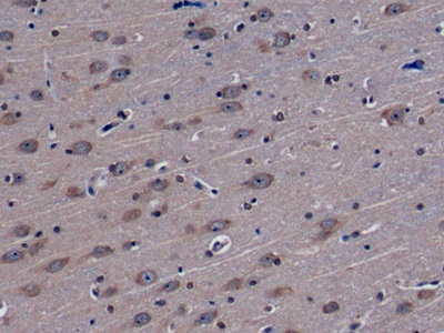
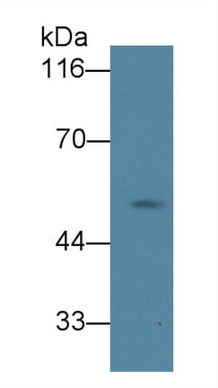
(Western Blot; Sample: Human Serum Primary Ab: 2ug/ml Mouse AntiHuman NRG1 Antibody Second Ab: 0.2ug/mL HRPLinked Caprine AntiMouse IgG Polyclonal Antibody (Catalog: SAA544Mu19))
WB (Western Blot)
(Western Blot; Sample: Human Serum Primary Ab: 2ug/ml Mouse AntiHuman NRG1 Antibody Second Ab: 0.2ug/mL HRPLinked Caprine AntiMouse IgG Polyclonal Antibody (Catalog: SAA544Mu19))
Neuregulin 1 (NRG1), Monoclonal Antibody (Cat# AAA151762)
Full Name
Monoclonal Antibody to Neuregulin 1 (NRG1)
Gene Names
NRG1; GGF; HGL; HRG; NDF; ARIA; GGF2; HRG1; HRGA; SMDF; MST131
Reactivity
Human, Pig
Applications
Immunoprecipitation, Immunocytochemistry, Immunohistochemistry, Western Blot
Purity
Protein A + Protein G affinity chromatography
Pricing
 Bioactivity
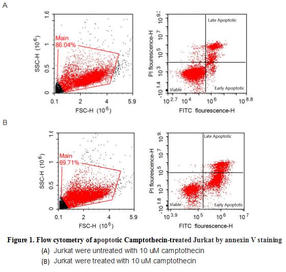
(Annexin V (ANXA5) is a multifunctional protein that is highly expressed on the apical surfaces of syncytiotrophoblasts, and plays an important role in haemostatic regulations, maintaining blood fluidity of the placenta. Lower ANXA5 levels have been observed in M2/ANXA5 haplotype carrying chorion. The association found between the maternal carriage of the M2/ANXA5 haplotype and an elevated risk of IUGR and/or PE supports the hypothesis that carrier status of this haplotype and the consequently reduced placental ANXA5 expression might be responsible, at least partially, for the onset of these gestational vascular complications. Annexin V is a calcium-dependent phospholipid binding protein that can be used to bind Phosphatidylserine (PS) during an early apoptosis event where the PS becomes exposed at the cell surface. Jurkat cells were treated with 10 uM camptothecin for 4h, 2*105 cells which were resuspended in binding buffer were stained with 5 ug recombinant human Annexin V-GFP and 10 ul Propidium iodide (PI) for 20min in dark room temperature. The flow cytometry was used to detect the early apoptotic and late apoptotic of camptothecin-treated Jurkat cells (Figure 1), the combination of Annexin V-GFP and propidium iodide allows for the distinction between early apoptotic cells (Annexin V-FITC positive and propidium iodide negative), late apoptotic and/or necrotic cells (Annexin V-FITC and propidium iodide positive), and viable cells (unstained). Thus, the recombinant human Annexin V-GFP can bind Phosphatidylserine (PS) at early apoptosis of Jurkat.)
Bioactivity
(Annexin V (ANXA5) is a multifunctional protein that is highly expressed on the apical surfaces of syncytiotrophoblasts, and plays an important role in haemostatic regulations, maintaining blood fluidity of the placenta. Lower ANXA5 levels have been observed in M2/ANXA5 haplotype carrying chorion. The association found between the maternal carriage of the M2/ANXA5 haplotype and an elevated risk of IUGR and/or PE supports the hypothesis that carrier status of this haplotype and the consequently reduced placental ANXA5 expression might be responsible, at least partially, for the onset of these gestational vascular complications. Annexin V is a calcium-dependent phospholipid binding protein that can be used to bind Phosphatidylserine (PS) during an early apoptosis event where the PS becomes exposed at the cell surface. Jurkat cells were treated with 10 uM camptothecin for 4h, 2*105 cells which were resuspended in binding buffer were stained with 5 ug recombinant human Annexin V-GFP and 10 ul Propidium iodide (PI) for 20min in dark room temperature. The flow cytometry was used to detect the early apoptotic and late apoptotic of camptothecin-treated Jurkat cells (Figure 1), the combination of Annexin V-GFP and propidium iodide allows for the distinction between early apoptotic cells (Annexin V-FITC positive and propidium iodide negative), late apoptotic and/or necrotic cells (Annexin V-FITC and propidium iodide positive), and viable cells (unstained). Thus, the recombinant human Annexin V-GFP can bind Phosphatidylserine (PS) at early apoptosis of Jurkat.)
Annexin V (ANXA5), Active Protein (Cat# AAA161724)
Full Name
Active Annexin V (ANXA5)
Gene Names
ANXA5; PP4; ANX5; ENX2; RPRGL3; HEL-S-7
Applications
Activity Assay, Cell Culture
Purity
Greater than 95% by SDS-PAGE
Pricing
 IF (Immunofluorescence)
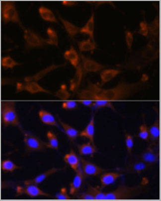
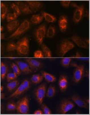
(Immunofluorescence analysis of U-2 OS cells using CAD antibody (AAA28198) at dilution of 1:100. Blue: DAPI for nuclear staining.)
IF (Immunofluorescence)
(Immunofluorescence analysis of U-2 OS cells using CAD antibody (AAA28198) at dilution of 1:100. Blue: DAPI for nuclear staining.)
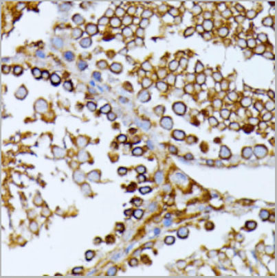
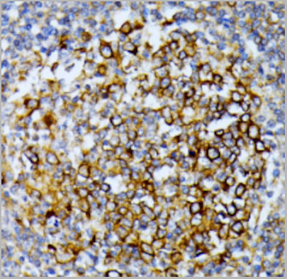
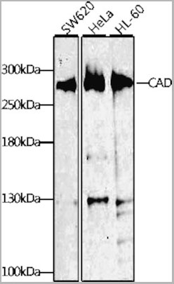
CAD, Polyclonal Antibody (Cat# AAA28198)
Full Name
CAD Polyclonal Antibody
Applications
Western Blot, Immunohistochemistry, Immunofluorescence, Immunocytochemistry
Purity
Affinity Purification
Pricing
 WB (Western Blot)
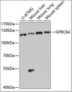
(Western blot analysis of extracts of various cell lines, using GPRC6A antibody (AAA28200) at 1:1000 dilution.Secondary antibody: HRP Goat Anti-Rabbit IgG (H+L) at 1:10000 dilution.Lysates/proteins: 25ug per lane.Blocking buffer: 3% nonfat dry milk in TBST.Detection: ECL Basic Kit.Exposure time: 90s.)
WB (Western Blot)
(Western blot analysis of extracts of various cell lines, using GPRC6A antibody (AAA28200) at 1:1000 dilution.Secondary antibody: HRP Goat Anti-Rabbit IgG (H+L) at 1:10000 dilution.Lysates/proteins: 25ug per lane.Blocking buffer: 3% nonfat dry milk in TBST.Detection: ECL Basic Kit.Exposure time: 90s.)
GPRC6A, Polyclonal Antibody (Cat# AAA28200)
Full Name
GPRC6A Polyclonal Antibody
Gene Names
GPRC6A; GPCR; bA86F4.3
Reactivity
Human, Mouse
Applications
Western Blot
Purity
Affinity Purification
Pricing
 IP (Immunoprecipitation)
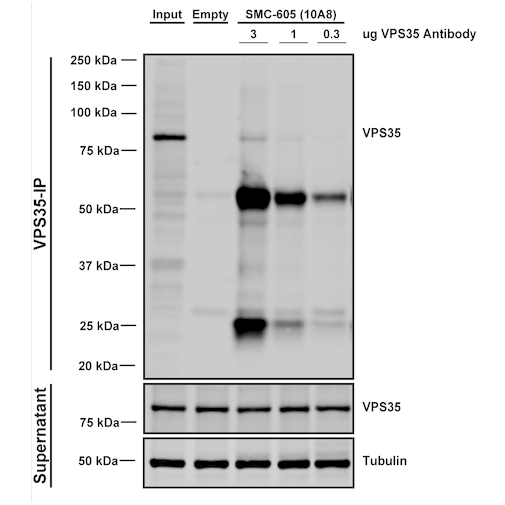
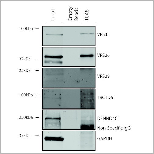
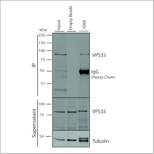
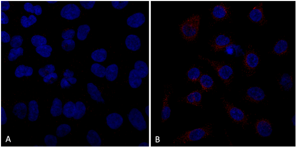
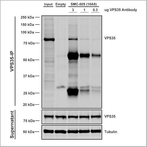
(Immunoprecipitation analysis using Mouse Anti-VPS35 Monoclonal Antibody, Clone 10A8. Tissue: embryonic fibroblast. Species: Mouse. Primary Antibody: Mouse Anti-VPS35 Monoclonal Antibody. Three amounts of (3, 1 and 0.3 ug) were non-covalently coupled to 10uL of A/G sepharose beads for 1 hour at 4 degree C and next incubated with 250ug of MEF lysate for 2 hours at 4 degree C.)
IP (Immunoprecipitation)
(Immunoprecipitation analysis using Mouse Anti-VPS35 Monoclonal Antibody, Clone 10A8. Tissue: embryonic fibroblast. Species: Mouse. Primary Antibody: Mouse Anti-VPS35 Monoclonal Antibody. Three amounts of (3, 1 and 0.3 ug) were non-covalently coupled to 10uL of A/G sepharose beads for 1 hour at 4 degree C and next incubated with 250ug of MEF lysate for 2 hours at 4 degree C.)
VPS35, Monoclonal Antibody (Cat# AAA27696)
Full Name
VPS35 Antibody, Clone 10A8: FITC
Gene Names
VPS35; MEM3; PARK17
Reactivity
Human, Mouse, Rat
Applications
Western Blot, Immunocytochemistry, Immunofluorescence, Immunoprecipitation
Purity
Protein G Purified
Pricing


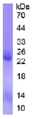 SDS-PAGE
Chromodomain Helicase DNA Binding Protein 3 (CHD3), Recombinant Protein (Cat# AAA135551)
Full Name
Recombinant Chromodomain Helicase DNA Binding Protein 3 (CHD3)
Gene Names
CHD3; ZFH; Mi-2a; Mi2-ALPHA
Reactivity
Homo sapiens (Human)
Applications
Western Blot
Purity
> 95%
Pricing
 Bioactivity
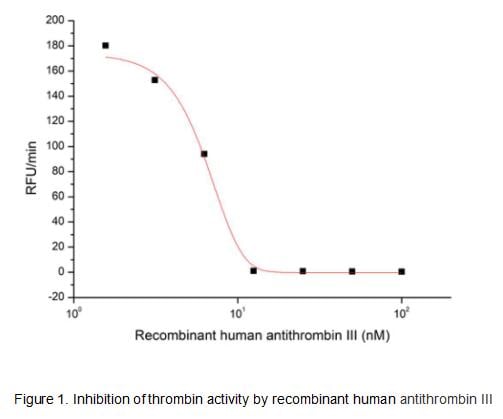
(Antithrombin III, also known as Serpin C1, is a member of the Serpin superfamily of the serine protease inhibitors. It is the principal plasma Serpin of blood clotting proteases and inhibits thrombin as well as several factors such as Xa. Similar to Serpins A5 and D1, its thrombin inhibitory activity is enhanced by heparin. Hereditary and acquired Serpin C1 deficiency is the cause of an increased thrombotic tendency in many cases. For example, acquired Serpin C1 deficiency is a common condition in sepsis, after major trauma or surgery. The activity of recombinant human antithrombin III was measured by its ability to inhibit thrombin cleavage of a fluorogenic peptide substrate Boc-VPR-AMC in the assay buffer 50 mM Tris, 10 mM CaCl2, 150 mM NaCl, 0.05% (w/v) Brij-35, pH 7.5. Thrombin was diluted to 0.5 U with heparin at 50 ug/ml in the assay buffer and 10 ul different concentrations of recombinant human antithrombin III (MW: 50 KD) was incubated with 10 ul diluted thrombin at 37 degree C for 30 minutes. Loading 50 uL of the incubated mixtures which were diluted five-fold in assay buffer into empty wells of a plate, and start the reaction by adding 50 uL of 200 uM substrate. Include a substrate blank containing 50 uL of assay buffer and 50 uL of 200 uM substrate. Then read at excitiation and emission wavelengths of 380 nm and 460 nm, respectively, in kinetic mode for 5 minutes. The result was shown in Figure 1 and it was obvious that recombinant human antithrombin III significantly decreased thrombin activity. The inhibition IC50 was )
Bioactivity
(Antithrombin III, also known as Serpin C1, is a member of the Serpin superfamily of the serine protease inhibitors. It is the principal plasma Serpin of blood clotting proteases and inhibits thrombin as well as several factors such as Xa. Similar to Serpins A5 and D1, its thrombin inhibitory activity is enhanced by heparin. Hereditary and acquired Serpin C1 deficiency is the cause of an increased thrombotic tendency in many cases. For example, acquired Serpin C1 deficiency is a common condition in sepsis, after major trauma or surgery. The activity of recombinant human antithrombin III was measured by its ability to inhibit thrombin cleavage of a fluorogenic peptide substrate Boc-VPR-AMC in the assay buffer 50 mM Tris, 10 mM CaCl2, 150 mM NaCl, 0.05% (w/v) Brij-35, pH 7.5. Thrombin was diluted to 0.5 U with heparin at 50 ug/ml in the assay buffer and 10 ul different concentrations of recombinant human antithrombin III (MW: 50 KD) was incubated with 10 ul diluted thrombin at 37 degree C for 30 minutes. Loading 50 uL of the incubated mixtures which were diluted five-fold in assay buffer into empty wells of a plate, and start the reaction by adding 50 uL of 200 uM substrate. Include a substrate blank containing 50 uL of assay buffer and 50 uL of 200 uM substrate. Then read at excitiation and emission wavelengths of 380 nm and 460 nm, respectively, in kinetic mode for 5 minutes. The result was shown in Figure 1 and it was obvious that recombinant human antithrombin III significantly decreased thrombin activity. The inhibition IC50 was )
Antithrombin (AT), Active Protein (Cat# AAA161886)
Full Name
Active Antithrombin (AT)
Gene Names
SERPINC1; AT3; AT3D; ATIII; THPH7
Applications
Activity Assay, Cell Culture
Purity
Greater than 90% by SDS-PAGE
Pricing
 Bioactivity
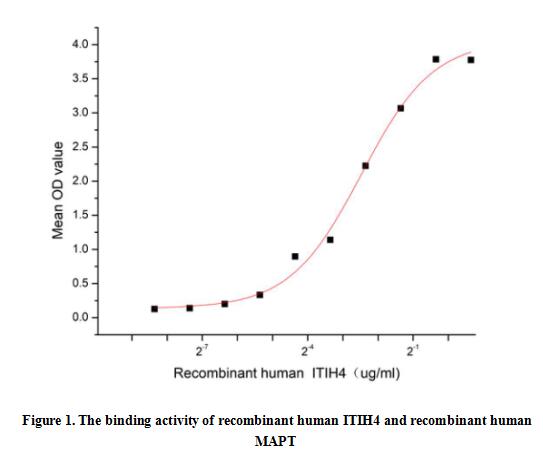
(Inter Alpha-Globulin Inhibitor H4 (ITIH4) is secreted into the blood, where it is cleaved by plasma kallikrein into two smaller forms. ITIH4 has been detected only in liver, and it seems to be upregulated during surgical trauma.It may also play a role in liver development or regeneration. Besides, Tau Protein (MAPT) has been identified as an interactor of ITIH4, thus a binding ELISA assay was conducted to detect the interaction of recombinant human ITIH4 and recombinant human MAPT. Briefly, ITIH4 were diluted serially in PBS, with 0.01% BSA (pH 7.4). Duplicate samples of 100 ul were then transferred to MAPT-coated microtiter wells and incubated for 2h at 37 degree C. Wells were washed with PBST and incubated for 1h with anti-ITIH4 pAb, then aspirated and washed 3 times. After incubation with HRP labelled secondary antibody, wells were aspirated and washed 5 times. With the addition of substrate solution, wells were incubated 15-25 minutes at 37 degree C. Finally, add 50 uL stop solution to the wells and read at 450 nm immediately. The binding activity of ITIH4 and MAPT was shown in Figure 1, the EC50 for this effect is 0.18 ug/mL.)
Bioactivity
(Inter Alpha-Globulin Inhibitor H4 (ITIH4) is secreted into the blood, where it is cleaved by plasma kallikrein into two smaller forms. ITIH4 has been detected only in liver, and it seems to be upregulated during surgical trauma.It may also play a role in liver development or regeneration. Besides, Tau Protein (MAPT) has been identified as an interactor of ITIH4, thus a binding ELISA assay was conducted to detect the interaction of recombinant human ITIH4 and recombinant human MAPT. Briefly, ITIH4 were diluted serially in PBS, with 0.01% BSA (pH 7.4). Duplicate samples of 100 ul were then transferred to MAPT-coated microtiter wells and incubated for 2h at 37 degree C. Wells were washed with PBST and incubated for 1h with anti-ITIH4 pAb, then aspirated and washed 3 times. After incubation with HRP labelled secondary antibody, wells were aspirated and washed 5 times. With the addition of substrate solution, wells were incubated 15-25 minutes at 37 degree C. Finally, add 50 uL stop solution to the wells and read at 450 nm immediately. The binding activity of ITIH4 and MAPT was shown in Figure 1, the EC50 for this effect is 0.18 ug/mL.)
Inter Alpha-Globulin Inhibitor H4 (ITIH4), Active Protein (Cat# AAA161928)
Full Name
Active Inter Alpha-Globulin Inhibitor H4 (ITIH4)
Gene Names
ITIH4; H4P; IHRP; GP120; PK120; ITIHL1; PK-120; ITI-HC4
Applications
Activity Assay, Cell Culture
Purity
Greater than 97% by SDS-PAGE
Pricing
 WB (Western Blot)
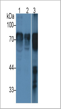
(Western Blot; Sample: Lane1: Rat Serum; Lane2: Rat Plasma; Lane3: Rat Liver lysate Primary Ab: 5ug/ml Mouse AntiRat HRG Antibody Second Ab: 0.2ug/mL HRPLinked Caprine AntiMouse IgG Polyclonal Antibody (Catalog: SAA544Mu19))
WB (Western Blot)
(Western Blot; Sample: Lane1: Rat Serum; Lane2: Rat Plasma; Lane3: Rat Liver lysate Primary Ab: 5ug/ml Mouse AntiRat HRG Antibody Second Ab: 0.2ug/mL HRPLinked Caprine AntiMouse IgG Polyclonal Antibody (Catalog: SAA544Mu19))
Histidine Rich Glycoprotein (HRG), Monoclonal Antibody (Cat# AAA21208)
Full Name
Monoclonal Antibody to Histidine Rich Glycoprotein (HRG)
Gene Names
LOC681544; Hrg; RNHRG2
Reactivity
Rat
Applications
Western Blot, Immunohistochemistry, Immunocytochemistry, Immunoprecipitation
Purity
Protein A + Protein G affinity chromatography
Pricing
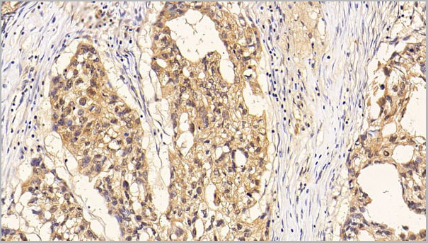
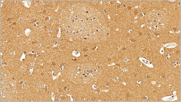
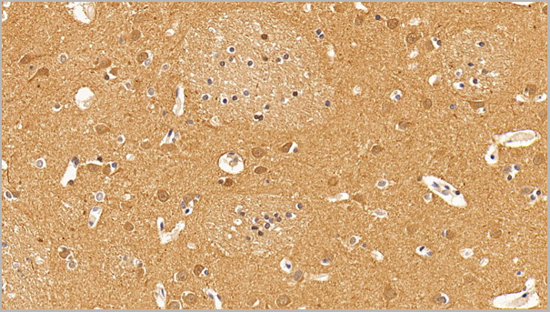
 WB (Western Blot)
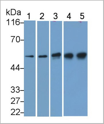
(Western Blot; Sample: Lane1: Hela cell lysate; Lane2: U87MG cell lysate; Lane3: Porcine Cerebrum lysate; Lane4: Rat Cerebrum lysate; Lane5: Bovine Cerebrum lysatePrimary Ab: 0.003ug/ml Mouse AntiHuman TUBb AntibodySecond Ab: 0.2ug/mL HRPLinked Caprine AntiMouse IgG Polyclonal Antibody(Catalog: SAA544Mu19))
WB (Western Blot)
(Western Blot; Sample: Lane1: Hela cell lysate; Lane2: U87MG cell lysate; Lane3: Porcine Cerebrum lysate; Lane4: Rat Cerebrum lysate; Lane5: Bovine Cerebrum lysatePrimary Ab: 0.003ug/ml Mouse AntiHuman TUBb AntibodySecond Ab: 0.2ug/mL HRPLinked Caprine AntiMouse IgG Polyclonal Antibody(Catalog: SAA544Mu19))
Tubulin Beta (TUBb), Monoclonal Antibody (Cat# AAA21201)
Full Name
Monoclonal Antibody to Tubulin Beta (TUBb)
Gene Names
TUBB; M40; TUBB1; TUBB5; OK/SW-cl.56
Reactivity
Human, Mouse, Rat, Pig, Bovine
Applications
Western Blot, Immunohistochemistry, Immunocytochemistry, Immunoprecipitation
Purity
Protein A + Protein G affinity chromatography
Pricing
 Application Data
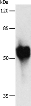
(Gel: 10%SDS-PAGELysate:2ug Recombinant HA tagged proteinPrimary antibody: 1/1000 dilutionSecondary antibody dilution: 1/8000Exposure time: 1 second)
Application Data
(Gel: 10%SDS-PAGELysate:2ug Recombinant HA tagged proteinPrimary antibody: 1/1000 dilutionSecondary antibody dilution: 1/8000Exposure time: 1 second)
c-Myc, Polyclonal Antibody (Cat# AAA301372)
Full Name
c-Myc Tag Antibody
Gene Names
MYC; MRTL; MYCC; c-Myc; bHLHe39
Applications
Western Blot
Purity
Antigen affinity purification.
Pricing
 IHC (Immunohiostchemistry)
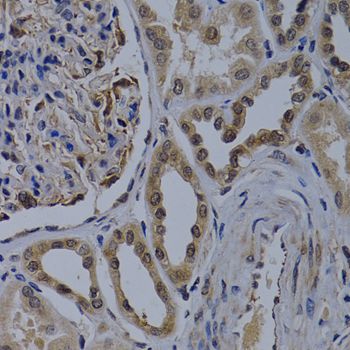
(Immunohistochemistry of paraffin-embedded human kidney cancer using WDR5 antibody at dilution of 1:200 (40x lens).)
IHC (Immunohiostchemistry)
(Immunohistochemistry of paraffin-embedded human kidney cancer using WDR5 antibody at dilution of 1:200 (40x lens).)
WDR5, Polyclonal Antibody (Cat# AAA37430)
Full Name
WDR5 Polyclonal Antibody
Gene Names
WDR5; SWD3; BIG-3; CFAP89
Reactivity
Human
Applications
Immunofluorescence, Immunohistochemistry, Western Blot
Purity
Affinity Purification
Pricing
Alpha Fetoprotein, Recombinant Protein (Cat# AAA10804)
Full Name
Recombinant Human Alpha Fetoprotein
Gene Names
AFP; AFPD; FETA; HPAFP
Purity
Greater than 95.0% as determined by SDS-PAGE.
Pricing

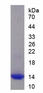 SDS-PAGE
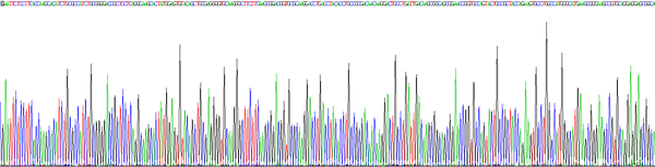
(Figure. SDS-PAGE)
SDS-PAGE
(Figure. SDS-PAGE)
Retinoid X Receptor Alpha (RXRa), Recombinant Protein (Cat# AAA150700)
Full Name
Recombinant Retinoid X Receptor Alpha (RXRa)
Applications
Western Blot
Purity
>95%
Pricing

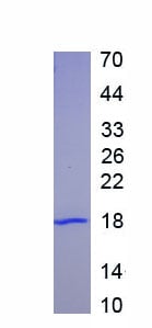 SDS-PAGE
(SDS-PAGE)
SDS-PAGE
(SDS-PAGE)
Carbohydrate Antigen 125 (CA125), Recombinant Protein (Cat# AAA152130)
Full Name
Recombinant Carbohydrate Antigen 125 (CA125)
Gene Names
MUC16; CA125
Applications
Western Blot
Purity
Greater than 95% by SDS-PAGE
Pricing
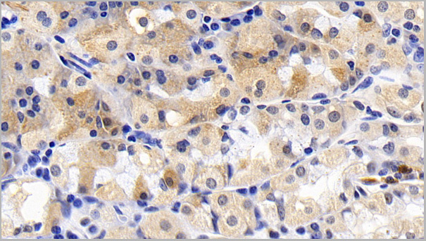
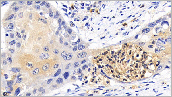
 IHC (Immunohistochemistry)
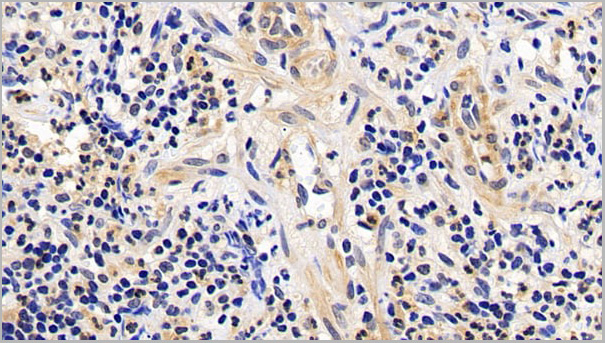
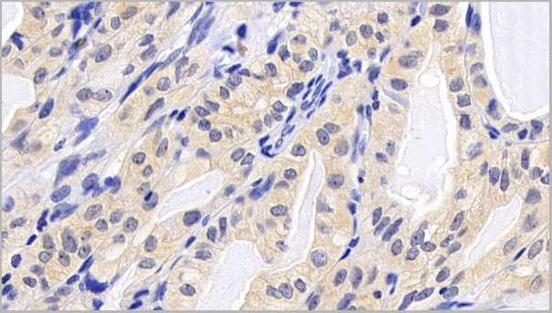
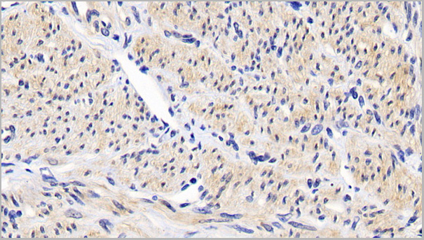
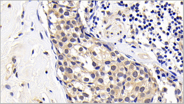
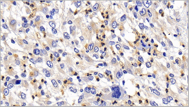
(DAB staining on IHCP;Sample: Human Liver cancer Tissue; Primary Ab: 30ug/ml Mouse AntiHuman Bcl2 AntibodySecond Ab: 2ug/mL HRPLinked Caprine AntiMouse IgG Polyclonal Antibody(Catalog: SAA544Mu19))
IHC (Immunohistochemistry)
(DAB staining on IHCP;Sample: Human Liver cancer Tissue; Primary Ab: 30ug/ml Mouse AntiHuman Bcl2 AntibodySecond Ab: 2ug/mL HRPLinked Caprine AntiMouse IgG Polyclonal Antibody(Catalog: SAA544Mu19))
BCell Leukemia/Lymphoma 2 (Bcl2), Monoclonal Antibody (Cat# AAA21188)
Full Name
Monoclonal Antibody to BCell Leukemia/Lymphoma 2 (Bcl2)
Gene Names
BCL2; Bcl-2; PPP1R50
Reactivity
Human
Applications
Western Blot, Immunohistochemistry, Immunocytochemistry, Immunoprecipitation
Purity
Protein A + Protein G affinity chromatography
Pricing
 FCM (Flow Cytometry)
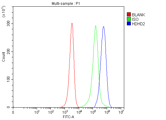
(Figure 8. Flow Cytometry analysis of JK cells using anti-HDHD2 antibody (AAA20005).Overlay histogram showing JK cells stained with AAA20005(Blue line). The cells were fixed with 4% paraformaldehyde and blocked with 10% normal goat serum. And then incubated with rabbit anti-HDHD2 Antibody (AAA20005, 1ug/1x106 cells) for 30 min at 20 degree C. DyLight488 conjugated goat anti-rabbit IgG was used as secondary antibody for 30 minutes at 20 degree C. Isotype control antibody (Green line) was rabbit IgG (1ug/1x106) used under the same conditions. Unlabelled sample (Red line) was also used as a control.)
FCM (Flow Cytometry)
(Figure 8. Flow Cytometry analysis of JK cells using anti-HDHD2 antibody (AAA20005).Overlay histogram showing JK cells stained with AAA20005(Blue line). The cells were fixed with 4% paraformaldehyde and blocked with 10% normal goat serum. And then incubated with rabbit anti-HDHD2 Antibody (AAA20005, 1ug/1x106 cells) for 30 min at 20 degree C. DyLight488 conjugated goat anti-rabbit IgG was used as secondary antibody for 30 minutes at 20 degree C. Isotype control antibody (Green line) was rabbit IgG (1ug/1x106) used under the same conditions. Unlabelled sample (Red line) was also used as a control.)
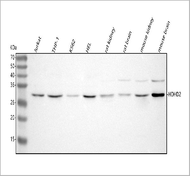
HDHD2, Polyclonal Antibody (Cat# AAA20005)
Full Name
Anti-HDHD2 Antibody Picoband
Gene Names
HDHD2; HEL-S-301; 3110052N05Rik
Reactivity
Human, Mouse, Rat
Applications
Western Blot, Immunohistochemistry, Flow Cytometry
Purity
Immunogen affinity purified.
Pricing
 Standard Curve (Sample)
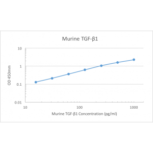
TGF-beta1, ELISA Kit (Cat# AAA79362)
Full Name
Murine TGF-betaPre-Coated ELISA Kit
Gene Names
Tgfb1; Tgfb; Tgfb-1; TGFbeta1; TGF-beta1
Reactivity
Murine
Pricing
 Bioactivity
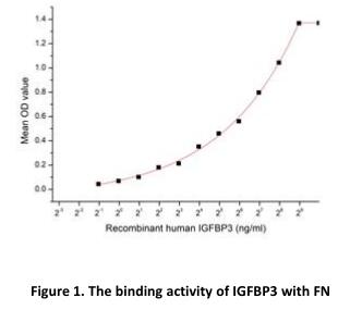
(Insulin-like growth factor-binding protein 3, also known as IGFBP3 is one of sixIGF binding proteins (IGFBP1 to IGFBP6) that have highly conserved structuresand bind the insulin-like growth factors IGF-1 and IGF-2 with high affinity. Withintissues, IGFBP3 can bind IGF1 and IGF2 released by many cell types, and blocktheir access to the IGF-1 receptor (IGF1R), which is activated by both IGFs.IGFBP3 also interacts with cell-surface proteins, affecting cell signaling fromoutside the cell or after internalization, and also enters the cell nucleus where itbinds to nuclear hormone receptors and other ligands. Besides, Fibronectin (FN)has been identified as an interactor of IGFBP3, thus a binding ELISA assay wasconducted to detect the interaction of recombinant human IGFBP3 andrecombinant human FN.Briefly, IGFBP3 were diluted serially in PBS, with 0.01% BSA (pH 7.4). Duplicatesamples of 100ul were then transferred to FN-coated microtiter wells andincubated for 1h at 37 degree C. Wells were washed with PBST and incubated for 1hwith anti-IGFBP3 pAb, then aspirated and washed 3 times. After incubation withHRP labelled secondary antibody, wells were aspirated and washed 5 times.With the addition of substrate solution, wells were incubated 15-25 minutes at37 degree C. Finally, add 50uL stop solution to the wells and read at 450nm immediately.The binding activity of IGFBP3 and FN was shown in Figure 1, the EC50 was 78ng/ml.)
Bioactivity
(Insulin-like growth factor-binding protein 3, also known as IGFBP3 is one of sixIGF binding proteins (IGFBP1 to IGFBP6) that have highly conserved structuresand bind the insulin-like growth factors IGF-1 and IGF-2 with high affinity. Withintissues, IGFBP3 can bind IGF1 and IGF2 released by many cell types, and blocktheir access to the IGF-1 receptor (IGF1R), which is activated by both IGFs.IGFBP3 also interacts with cell-surface proteins, affecting cell signaling fromoutside the cell or after internalization, and also enters the cell nucleus where itbinds to nuclear hormone receptors and other ligands. Besides, Fibronectin (FN)has been identified as an interactor of IGFBP3, thus a binding ELISA assay wasconducted to detect the interaction of recombinant human IGFBP3 andrecombinant human FN.Briefly, IGFBP3 were diluted serially in PBS, with 0.01% BSA (pH 7.4). Duplicatesamples of 100ul were then transferred to FN-coated microtiter wells andincubated for 1h at 37 degree C. Wells were washed with PBST and incubated for 1hwith anti-IGFBP3 pAb, then aspirated and washed 3 times. After incubation withHRP labelled secondary antibody, wells were aspirated and washed 5 times.With the addition of substrate solution, wells were incubated 15-25 minutes at37 degree C. Finally, add 50uL stop solution to the wells and read at 450nm immediately.The binding activity of IGFBP3 and FN was shown in Figure 1, the EC50 was 78ng/ml.)
Insulin Like Growth Factor Binding Protein 3 (IGFBP3), Active Protein (Cat# AAA161673)
Full Name
Active Insulin Like Growth Factor Binding Protein 3 (IGFBP3)
Gene Names
IGFBP3; IBP3; BP-53
Applications
Activity Assay, Cell Culture
Purity
Greater than 95% by SDS-PAGE
Pricing
 IP (Immunoprecipitation)
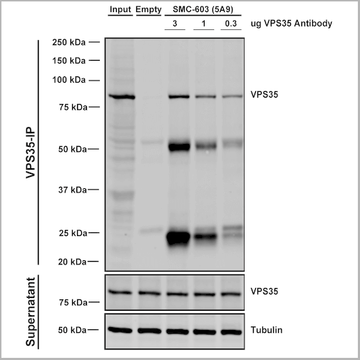
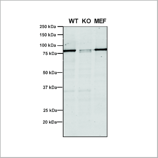
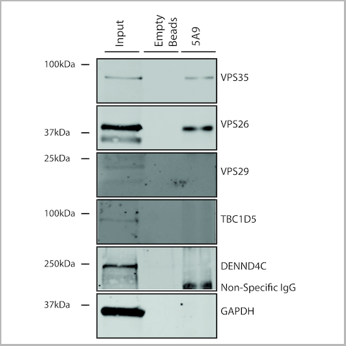
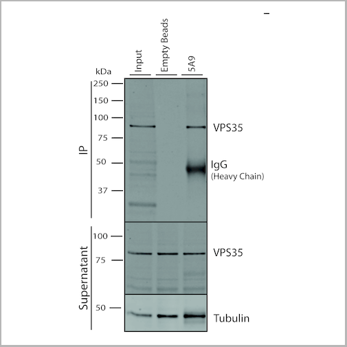
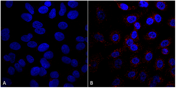
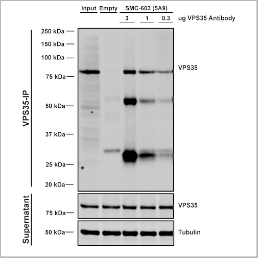
(Immunoprecipitation analysis using Mouse Anti-VPS35 Monoclonal Antibody, Clone 5A9. Tissue: embryonic fibroblast. Species: Mouse. Primary Antibody: Mouse Anti-VPS35 Monoclonal Antibody. Three amounts of (3, 1 and 0.3 ug) were non-covalently coupled to 10uL of A/G sepharose beads for 1 hour at 4 degree C and next incubated with 250ug of MEF lysate for 2 hours at 4 degree C.)
IP (Immunoprecipitation)
(Immunoprecipitation analysis using Mouse Anti-VPS35 Monoclonal Antibody, Clone 5A9. Tissue: embryonic fibroblast. Species: Mouse. Primary Antibody: Mouse Anti-VPS35 Monoclonal Antibody. Three amounts of (3, 1 and 0.3 ug) were non-covalently coupled to 10uL of A/G sepharose beads for 1 hour at 4 degree C and next incubated with 250ug of MEF lysate for 2 hours at 4 degree C.)
VPS35, Monoclonal Antibody (Cat# AAA27676)
Full Name
VPS35 Antibody, Clone 5A9: ATTO 594
Gene Names
VPS35; MEM3; PARK17
Reactivity
Human, Mouse, Rat
Applications
Western Blot, Immunocytochemistry, Immunofluorescence, Immunoprecipitation
Purity
Protein G Purified
Pricing
 Bioactivity
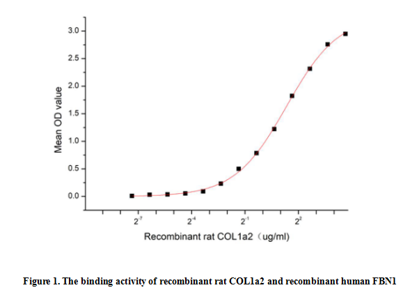
(Collagen Type I Alpha 2 (COL1a2) is a major component of collagen type I found in skin, bone, muscle, tendons, ligaments and other connective tissues.The COL1a2 protein is essential for the assembly, secretion, and stability of collagen fibers, which are vital for tissue development and repair.Mutations in COL1A2 are associated with a variety of genetic disorders, such as osteogenesis imperfecta (dysplasia of the bones) and cutis laxa. These diseases are often accompanied by weak bones and loss of skin elasticity. Besides, Fibrillin 1 (FBN1) has been identified as an interactor of COL1a2, thus a functional binding ELISA assay was conducted to detect the interaction of recombinant rat COL1a2 and recombinant human FBN1. Briefly, COL1a2 was diluted serially in PBS with 0.01% BSA (pH 7.4). Duplicate samples of 100 ul were then transferred to FBN1-coated microtiter wells and incubated for 1h at 37 degree C. Wells were washed with PBST and incubated for 1h with anti-COL1a2 pAb, then aspirated and washed 3 times. After incubation with HRP labelled secondary antibody for 1h at 37 degree C, wells were aspirated and washed 5 times. With the addition of substrate solution, wells were incubated 15-25 minutes at 37 degree C. Finally, add 50 uL stop solution to the wells and read at 450/630 nm immediately. The binding activity of recombinant rat COL1a2 and recombinant human FBN1 was shown in Figure 1, the EC50 for this effect is 2.53 ug/mL.)
Bioactivity
(Collagen Type I Alpha 2 (COL1a2) is a major component of collagen type I found in skin, bone, muscle, tendons, ligaments and other connective tissues.The COL1a2 protein is essential for the assembly, secretion, and stability of collagen fibers, which are vital for tissue development and repair.Mutations in COL1A2 are associated with a variety of genetic disorders, such as osteogenesis imperfecta (dysplasia of the bones) and cutis laxa. These diseases are often accompanied by weak bones and loss of skin elasticity. Besides, Fibrillin 1 (FBN1) has been identified as an interactor of COL1a2, thus a functional binding ELISA assay was conducted to detect the interaction of recombinant rat COL1a2 and recombinant human FBN1. Briefly, COL1a2 was diluted serially in PBS with 0.01% BSA (pH 7.4). Duplicate samples of 100 ul were then transferred to FBN1-coated microtiter wells and incubated for 1h at 37 degree C. Wells were washed with PBST and incubated for 1h with anti-COL1a2 pAb, then aspirated and washed 3 times. After incubation with HRP labelled secondary antibody for 1h at 37 degree C, wells were aspirated and washed 5 times. With the addition of substrate solution, wells were incubated 15-25 minutes at 37 degree C. Finally, add 50 uL stop solution to the wells and read at 450/630 nm immediately. The binding activity of recombinant rat COL1a2 and recombinant human FBN1 was shown in Figure 1, the EC50 for this effect is 2.53 ug/mL.)
Collagen Type I Alpha 2 (COL1a2), Active Protein (Cat# AAA161716)
Full Name
Active Collagen Type I Alpha 2 (COL1a2)
Applications
Activity Assay, Cell Culture
Purity
Greater than 90% by SDS-PAGE
Pricing
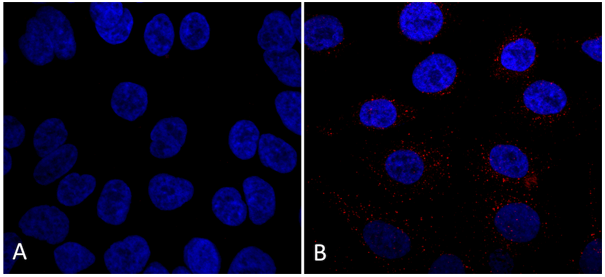
 IP (Immunoprecipitation)
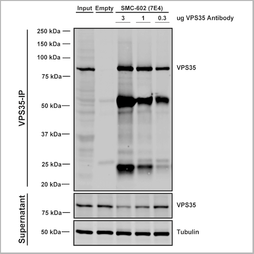
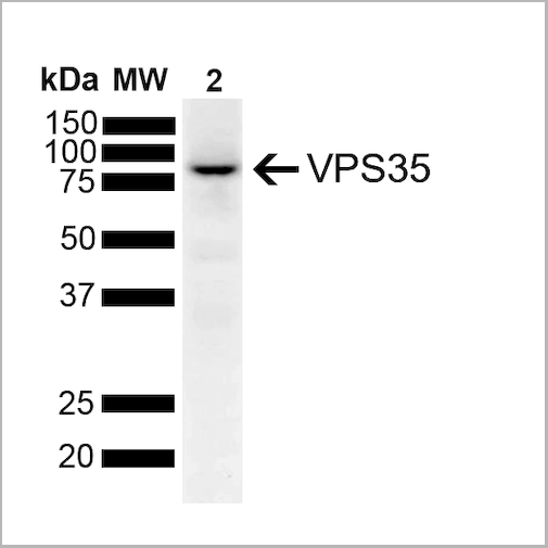
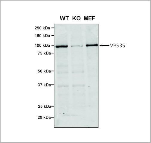
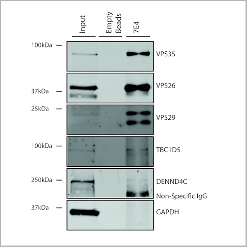
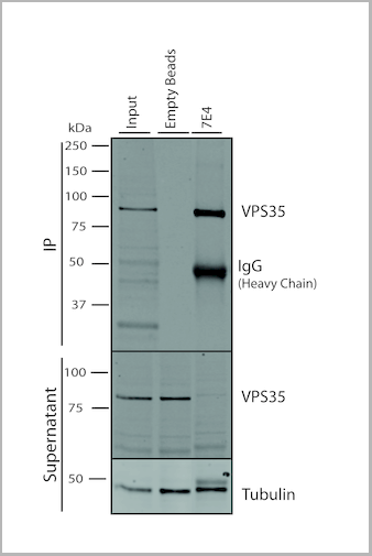
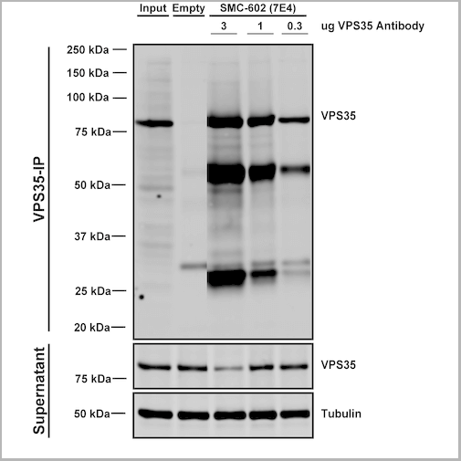
(Immunoprecipitation analysis using Mouse Anti-VPS35 Monoclonal Antibody, Clone 7E4. Tissue: embryonic fibroblast. Species: Mouse. Primary Antibody: Mouse Anti-VPS35 Monoclonal Antibody. Three amounts (3, 1 and 0.3 ug) were non-covalently coupled to 10uL of A/G sepharose beads for 1 hour at 4 degree C and next incubated with 250ug of MEF lysate for 2 hours at 4 degree C.)
IP (Immunoprecipitation)
(Immunoprecipitation analysis using Mouse Anti-VPS35 Monoclonal Antibody, Clone 7E4. Tissue: embryonic fibroblast. Species: Mouse. Primary Antibody: Mouse Anti-VPS35 Monoclonal Antibody. Three amounts (3, 1 and 0.3 ug) were non-covalently coupled to 10uL of A/G sepharose beads for 1 hour at 4 degree C and next incubated with 250ug of MEF lysate for 2 hours at 4 degree C.)
VPS35, Monoclonal Antibody (Cat# AAA27668)
Full Name
VPS35 Antibody, Clone 7E4: Biotin
Gene Names
VPS35; MEM3; PARK17
Reactivity
Human, Mouse, Rat
Applications
Western Blot, Immunocytochemistry, Immunofluorescence, Immunoprecipitation
Purity
Protein G Purified
Pricing
 Bioactivity
(Neutrophil elastase (NE), also known as polymorphonuclear leukocyte elastase, is a major protease in the primary granules of neutrophils, is involved in microbicidal activity. It is located primarily in the azurophil granules of polymorphonuclear leukocytes. NE is an important factor promoting inflammation, has bactericidal effects, and shortens the inflammatory process. NE also regulates tumor growth by promoting metastasis and tumor microenvironment remodeling. However, NE plays a role in killing tumors under certain conditions and promotes other diseases such as pulmonary ventilation dysfunction. Additionally, it plays a complex role in various physiological processes and mediates several diseases. Myeloperoxidase (MPO) has been identified as an interactor of NE, thus a functional binding ELISA assay was conducted to detect the interaction of recombinant human NE and recombinant rat MPO. Briefly, biotin-linked NE were diluted serially in PBS, with 0.01% BSA (pH 7.4). Duplicate samples of 100 ul were then transferred to MPO-coated microtiter wells and incubated for 1h at 37 degree C. Wells were washed with PBST 3 times and incubation with Streptavidin-HRP for 30min, then wells were aspirated and washed 5 times. With the addition of substrate solution, wells were incubated 15-25 minutes at 37 degree C. Finally, add 50 ul stop solution to the wells and read at 450 nm immediately. The binding activity of recombinant human NE and recombinant rat MPO was shown in Figure 1, the EC50 for this effect is 0.66 ug/mL.)
Bioactivity
(Neutrophil elastase (NE), also known as polymorphonuclear leukocyte elastase, is a major protease in the primary granules of neutrophils, is involved in microbicidal activity. It is located primarily in the azurophil granules of polymorphonuclear leukocytes. NE is an important factor promoting inflammation, has bactericidal effects, and shortens the inflammatory process. NE also regulates tumor growth by promoting metastasis and tumor microenvironment remodeling. However, NE plays a role in killing tumors under certain conditions and promotes other diseases such as pulmonary ventilation dysfunction. Additionally, it plays a complex role in various physiological processes and mediates several diseases. Myeloperoxidase (MPO) has been identified as an interactor of NE, thus a functional binding ELISA assay was conducted to detect the interaction of recombinant human NE and recombinant rat MPO. Briefly, biotin-linked NE were diluted serially in PBS, with 0.01% BSA (pH 7.4). Duplicate samples of 100 ul were then transferred to MPO-coated microtiter wells and incubated for 1h at 37 degree C. Wells were washed with PBST 3 times and incubation with Streptavidin-HRP for 30min, then wells were aspirated and washed 5 times. With the addition of substrate solution, wells were incubated 15-25 minutes at 37 degree C. Finally, add 50 ul stop solution to the wells and read at 450 nm immediately. The binding activity of recombinant human NE and recombinant rat MPO was shown in Figure 1, the EC50 for this effect is 0.66 ug/mL.)
Neutrophil Elastase (NE), Active Protein (Cat# AAA161712)
Full Name
Active Neutrophil Elastase (NE)
Gene Names
ELANE; GE; NE; HLE; HNE; ELA2; SCN1; PMN-E
Applications
Activity Assay, Cell Culture
Purity
Greater than 95% by SDS-PAGE
Pricing

 WB (Western Blot)
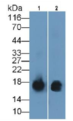
(Western Blot; Sample: Lane1: Human Leukocyte lysate; Lane2: Human Lymphocyte lysate Primary Ab: 2ug/ml Mouse Anti-Human S100A9 Antibody Second Ab: 0.2µg/mL HRP-Linked Caprine Anti-Mouse IgG Polyclonal Antibody)
WB (Western Blot)
(Western Blot; Sample: Lane1: Human Leukocyte lysate; Lane2: Human Lymphocyte lysate Primary Ab: 2ug/ml Mouse Anti-Human S100A9 Antibody Second Ab: 0.2µg/mL HRP-Linked Caprine Anti-Mouse IgG Polyclonal Antibody)
S100 Calcium Binding Protein A9 (S100A9), Monoclonal Antibody (Cat# AAA151748)
Full Name
Monoclonal Antibody to S100 Calcium Binding Protein A9 (S100A9)
Reactivity
Human, Pig
Applications
Immunoprecipitation, Immunocytochemistry, Immunohistochemistry, Western Blot
Purity
Protein A + Protein G affinity chromatography
Pricing
 Bioactivity
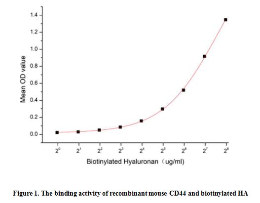
(Homing Associated Cell Adhesion Molecule (HCAM), also known as CD44, is a ubiquitous multistructural and multifunctional cells surface adhesion molecule involved in cell-cell and cell-matrix interactions. CD44 is broadly expressed, including in the membranes of B cells, granulocytes, monocytes, and erythrocytes as well as on many thymocytes and mature T cells, besides it is highly expressed in many cancers and regulates metastasis via recruitment of CD44 to the cell surface. This protein is a receptor for hyaluronic acid (HA) and can also interact with other ligands, such as osteopontin, collagens, and matrix metalloproteinases (MMPs). Thus a functional binding ELISA assay was conducted to detect the interaction of recombinant mouse HCAM and biotinylated hyaluronan (HA). Briefly, biotin-linked HA was diluted serially in PBS, with 0.01% BSA (pH 7.4). Duplicate samples of 100 ul were then transferred to HCAM-coated microtiter wells and incubated for 2h at 37 degree C. Wells were washed with PBST 3 times and incubation with Streptavidin-HRP for 1 hour, then wells were aspirated and washed 5 times. With the addition of substrate solution, wells were incubated 15-25 minutes at 37 degree C. Finally, add 50 ul stop solution to the wells and read at 450/630 nm immediately. The binding activity of recombinant mouse HCAM and biotinylated HA was shown in Figure 1, and this effect was in a dose dependent manner.)
Bioactivity
(Homing Associated Cell Adhesion Molecule (HCAM), also known as CD44, is a ubiquitous multistructural and multifunctional cells surface adhesion molecule involved in cell-cell and cell-matrix interactions. CD44 is broadly expressed, including in the membranes of B cells, granulocytes, monocytes, and erythrocytes as well as on many thymocytes and mature T cells, besides it is highly expressed in many cancers and regulates metastasis via recruitment of CD44 to the cell surface. This protein is a receptor for hyaluronic acid (HA) and can also interact with other ligands, such as osteopontin, collagens, and matrix metalloproteinases (MMPs). Thus a functional binding ELISA assay was conducted to detect the interaction of recombinant mouse HCAM and biotinylated hyaluronan (HA). Briefly, biotin-linked HA was diluted serially in PBS, with 0.01% BSA (pH 7.4). Duplicate samples of 100 ul were then transferred to HCAM-coated microtiter wells and incubated for 2h at 37 degree C. Wells were washed with PBST 3 times and incubation with Streptavidin-HRP for 1 hour, then wells were aspirated and washed 5 times. With the addition of substrate solution, wells were incubated 15-25 minutes at 37 degree C. Finally, add 50 ul stop solution to the wells and read at 450/630 nm immediately. The binding activity of recombinant mouse HCAM and biotinylated HA was shown in Figure 1, and this effect was in a dose dependent manner.)
Homing Associated Cell Adhesion Molecule (HCAM), Active Protein (Cat# AAA161765)
Full Name
Active Homing Associated Cell Adhesion Molecule (HCAM)
Gene Names
Cd44; Ly-24; Pgp-1; HERMES; AU023126; AW121933; AW146109
Applications
Activity Assay, Cell Culture
Purity
Greater than 80% by SDS-PAGE
Pricing

 WB (Western Blot)
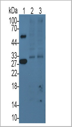
(Western Blot; Sample: Lane1: Rat Plasma; Lane2: Hela cell lysate; Lane3: HepG2 cell lysatePrimary Ab: 3ug/ml Mouse AntiRat TFPI AntibodySecond Ab: 0.2ug/mL HRPLinked Caprine AntiMouse IgG Polyclonal Antibody(Catalog: SAA544Mu19))
WB (Western Blot)
(Western Blot; Sample: Lane1: Rat Plasma; Lane2: Hela cell lysate; Lane3: HepG2 cell lysatePrimary Ab: 3ug/ml Mouse AntiRat TFPI AntibodySecond Ab: 0.2ug/mL HRPLinked Caprine AntiMouse IgG Polyclonal Antibody(Catalog: SAA544Mu19))
Tissue Factor Pathway Inhibitor (TFPI), Monoclonal Antibody (Cat# AAA21180)
Full Name
Monoclonal Antibody to Tissue Factor Pathway Inhibitor (TFPI)
Reactivity
Rat
Applications
Western Blot, Immunohistochemistry, Immunocytochemistry, Immunoprecipitation
Purity
Protein A + Protein G affinity chromatography
Pricing
 Standard Curve (Sample)
GRO/MGSA, CLIA Kit (Cat# AAA318531)
Full Name
GRO/MGSA (Human) LumiAb ELISA Kit
Gene Names
CXCL1; FSP; GRO1; GROa; MGSA; NAP-3; SCYB1; MGSA-a
Reactivity
Human
Pricing
Mycoplasma hominis, p120, Monoclonal Antibody (Cat# AAA13388)
Full Name
MAb to Mycoplasma hominis
Applications
Immunofluorescence
Purity
>90% pure. Protein A chromatography
Pricing
 SDS-PAGE
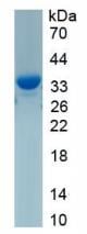
(SDS-PAGE Analysis Serum Amyloid A Rabbit Recombinant Monoclonal Antibody (SAA/2868R). Confirmation of Purity and Integrity of Antibody)
SDS-PAGE
(SDS-PAGE Analysis Serum Amyloid A Rabbit Recombinant Monoclonal Antibody (SAA/2868R). Confirmation of Purity and Integrity of Antibody)
Serum Amyloid A, Monoclonal Antibody (Cat# AAA214814)
Full Name
Serum Amyloid A
Gene Names
SAA1; SAA; PIG4; SAA2; TP53I4
Reactivity
Human. Others not known.
Applications
Immunohistochemistry
Purity
Purified Ab with BSA and Azide at 200ug/ml OR Purified Ab WITHOUT BSA and Azide at 1.0mg/ml
Pricing
 Structure
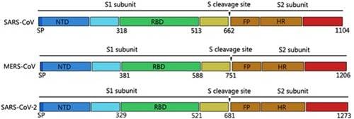
(Structural diagrams of spike glycoproteins of SARS-CoV, MERS-CoV, and SARS-CoV-2. All spike proteins of coronaviruses contain S1 subunit and S2 subunit, which were divided by the S cleavage sites. FP, fusion peptide; HR, heptad repeat 1 and heptad repeat 2; RBD, receptor-binding domain, contains core binding motif in the external subdomain; SP, signal peptide. Adapted from Composition and divergence of coronavirus spike proteins and host ACE2 receptors predict potential intermediate hosts of SARS-CoV-2. Liu Z, et al. J Med Virol. 2020 Feb 26. doi: 10.1002/jmv.25726.)
Structure
(Structural diagrams of spike glycoproteins of SARS-CoV, MERS-CoV, and SARS-CoV-2. All spike proteins of coronaviruses contain S1 subunit and S2 subunit, which were divided by the S cleavage sites. FP, fusion peptide; HR, heptad repeat 1 and heptad repeat 2; RBD, receptor-binding domain, contains core binding motif in the external subdomain; SP, signal peptide. Adapted from Composition and divergence of coronavirus spike proteins and host ACE2 receptors predict potential intermediate hosts of SARS-CoV-2. Liu Z, et al. J Med Virol. 2020 Feb 26. doi: 10.1002/jmv.25726.)
COVID 19 Spike S1 Coronavirus, Recombinant Protein (Cat# AAA78531)
Full Name
Recombinant 2019-nCoV S1 Protein (Active)
Applications
Functional Assay
Purity
>85% as determined by reducing SDS-PAGE.
Pricing
 Bioactivity
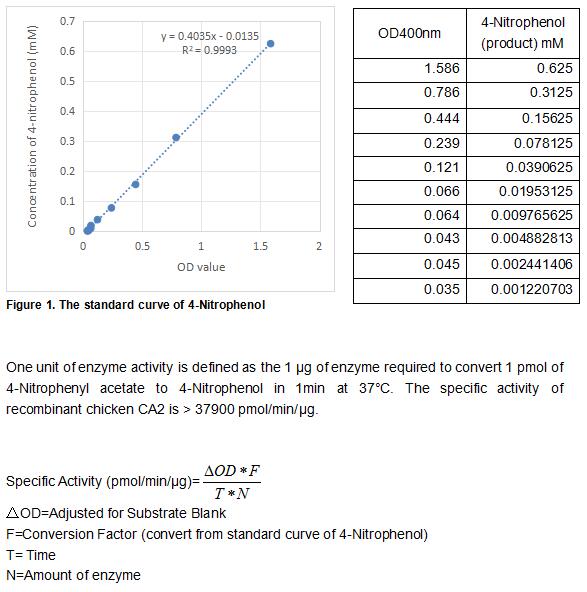
(Carbonic Anhydrase (CA) catalyzes the reversible reaction of CO2 H2O = HCO3- H, which is fundamental to many processes such as respiration, renal tubular acidification and bone resorption. CA2 is a cytosolic enzyme with the highest activity among all known CAs. Mutations in the CA2 gene result in the CA II deficiency syndrome, an autosomal recessive disorder that produces osteopetrosis, renal tubular acidosis and cerebral calcification. The activity of recombinant chicken CA2 was measured by its ability to hydrolyze 4-Nitrophenyl acetate (4-NPA) to 4-Nitrophenol. The reaction was performed in 12.5 mM Tris, 75 mM NaCl, pH 7.5 (assay buffer), initiated by addition 50 uL of various concentrations of CA2 (diluted by assay buffer) to 50 uL of 2 mM substrate 4-NPA (100 mM stock in Acetone, diluted by assay buffer). Incubated at 37 degree C for 5min, then read at a wavelength of 400 nm.)
Bioactivity
(Carbonic Anhydrase (CA) catalyzes the reversible reaction of CO2 H2O = HCO3- H, which is fundamental to many processes such as respiration, renal tubular acidification and bone resorption. CA2 is a cytosolic enzyme with the highest activity among all known CAs. Mutations in the CA2 gene result in the CA II deficiency syndrome, an autosomal recessive disorder that produces osteopetrosis, renal tubular acidosis and cerebral calcification. The activity of recombinant chicken CA2 was measured by its ability to hydrolyze 4-Nitrophenyl acetate (4-NPA) to 4-Nitrophenol. The reaction was performed in 12.5 mM Tris, 75 mM NaCl, pH 7.5 (assay buffer), initiated by addition 50 uL of various concentrations of CA2 (diluted by assay buffer) to 50 uL of 2 mM substrate 4-NPA (100 mM stock in Acetone, diluted by assay buffer). Incubated at 37 degree C for 5min, then read at a wavelength of 400 nm.)
Carbonic Anhydrase II (CA2), Active Protein (Cat# AAA161775)
Full Name
Active Carbonic Anhydrase II (CA2)
Applications
Activity Assay, Cell Culture
Purity
Greater than 90% by SDS-PAGE
Pricing
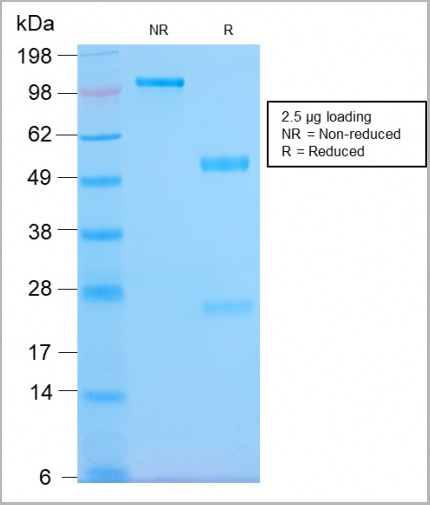
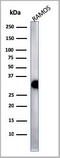
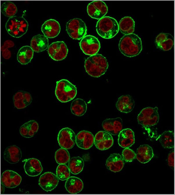
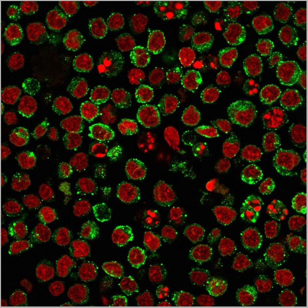
 Application Data
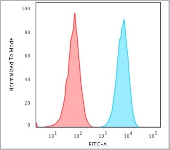
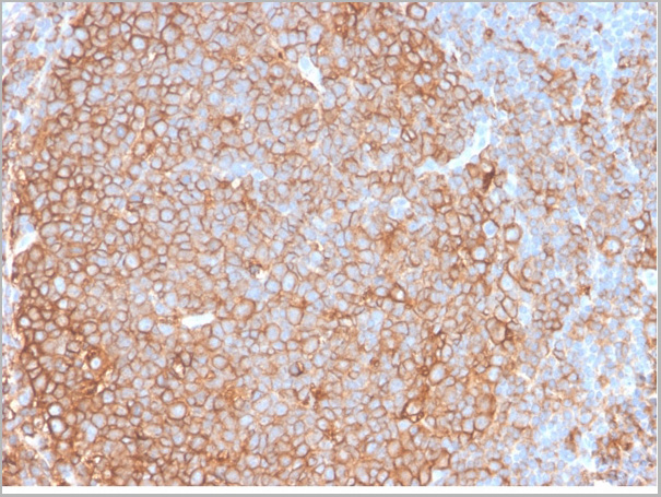
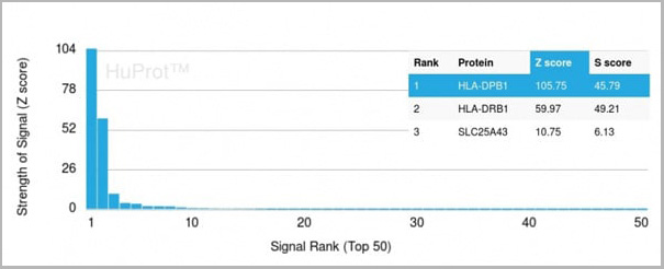
(Analysis of Protein Array containing more than 19,000 full-length human proteins using HLA-Pan Rabbit Recombinant Monoclonal Antibody (HLA-Pan/2967R). Z- and S- Score: The Z-score represents the strength of a signal that a monoclonal antibody (MAb) (in combination with a fluorescently-tagged anti-IgG secondary antibody) produces when binding to a particular protein on the HuProtTM array. Z-scores are described in units of standard deviations (SD’s) above the mean value of all signals generated on that array. If targets on HuProtTM are arranged in descending order of the Z-score, the S-score is the difference (also in units of SD’s) between the Z-score. S-score therefore represents the relative target specificity of a MAb to its intended target. A MAb is considered to specific to its intended target, if the MAb has an S-score of at least 2.5. For example, if a MAb binds to protein X with a Z-score of 43 and to protein Y with a Z-score of 14, then the S-score for the binding of that MAb to protein X is equal to 29.)
Application Data
(Analysis of Protein Array containing more than 19,000 full-length human proteins using HLA-Pan Rabbit Recombinant Monoclonal Antibody (HLA-Pan/2967R). Z- and S- Score: The Z-score represents the strength of a signal that a monoclonal antibody (MAb) (in combination with a fluorescently-tagged anti-IgG secondary antibody) produces when binding to a particular protein on the HuProtTM array. Z-scores are described in units of standard deviations (SD’s) above the mean value of all signals generated on that array. If targets on HuProtTM are arranged in descending order of the Z-score, the S-score is the difference (also in units of SD’s) between the Z-score. S-score therefore represents the relative target specificity of a MAb to its intended target. A MAb is considered to specific to its intended target, if the MAb has an S-score of at least 2.5. For example, if a MAb binds to protein X with a Z-score of 43 and to protein Y with a Z-score of 14, then the S-score for the binding of that MAb to protein X is equal to 29.)
HLA-Pan, Antibody (Cat# AAA23950)
Full Name
HLA-Pan (MHC II)
Gene Names
HLA-DPB1; DPB1; HLA-DP; HLA-DPB; HLA-DP1B
Reactivity
Human
Applications
Flow Cytometry, Immunofluorescence, Western Blot, Immunohistochemistry
Purity
Purified Ab with BSA and Azide at 200ug/ml OR Purified Ab WITHOUT BSA and Azide at 1.0mg/ml
Pricing
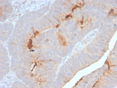
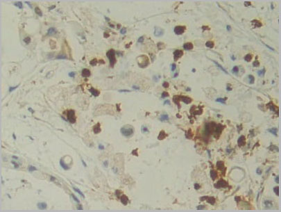 IHC (Immunohistchemistry)
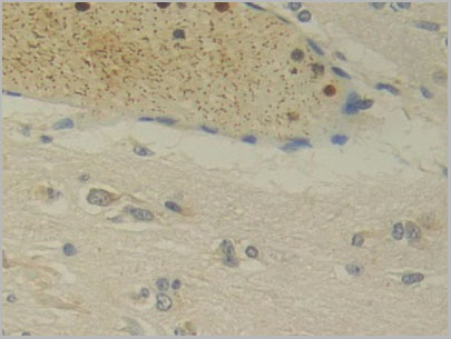
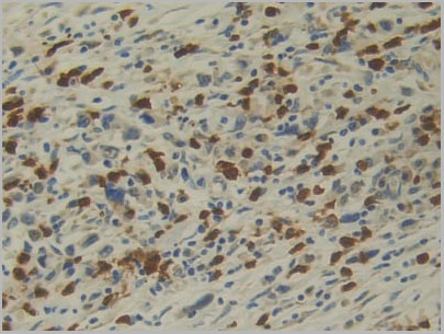
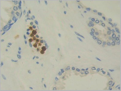
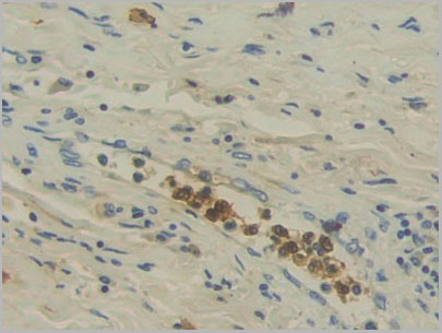
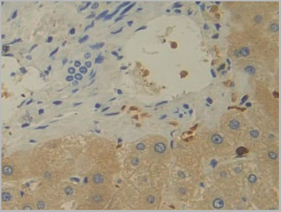
(DAB staining on IHCP;Sample: Human Bile duct cancer Tissue; Primary Ab: 30ug/ml Mouse AntiHuman IL12A AntibodySecond Ab: 2ug/mL HRPLinked Caprine AntiMouse IgG Polyclonal Antibody(Catalog: SAA544Mu19))
IHC (Immunohistchemistry)
(DAB staining on IHCP;Sample: Human Bile duct cancer Tissue; Primary Ab: 30ug/ml Mouse AntiHuman IL12A AntibodySecond Ab: 2ug/mL HRPLinked Caprine AntiMouse IgG Polyclonal Antibody(Catalog: SAA544Mu19))
Interleukin 12A (IL12A), Monoclonal Antibody (Cat# AAA21177)
Full Name
Monoclonal Antibody to Interleukin 12A (IL12A)
Gene Names
IL12A; P35; CLMF; NFSK; NKSF1; IL-12A
Reactivity
Human
Applications
Western Blot, Immunohistochemistry, Immunocytochemistry, Immunoprecipitation
Purity
Protein A + Protein G affinity chromatography
Pricing