
Filters
Clonality
Type
Reactivity
Gene Name
Isotype
Host
Application
Clone
10000 results for "Recombinant Proteins" - showing 2350-2400
Hepatitis C Virus (HCV) E2, Monoclonal Antibody (Cat# AAA13364)
Full Name
MAb to HCV E2
Applications
Immunofluorescence, Western Blot
Purity
>90% pure. Protein A chromatography
Pricing
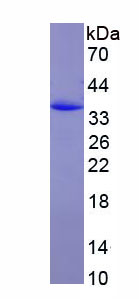
 Amino Acid Info
Chondroitin Sulfate Proteoglycan 4 (CSPG4), Recombinant Protein (Cat# AAA153429)
Full Name
Recombinant Chondroitin Sulfate Proteoglycan 4 (CSPG4)
Gene Names
Cspg4; AN2; NG2; 4732461B14Rik
Applications
Western Blot
Purity
Greater than 97% by SDS-PAGE
Pricing
 Application Data
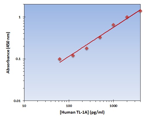
(The OmniKine Human TL-1A ELISA Kit allows for the detection and quantification of endogenous levels of natural and/or recombinant Human TL-1A proteins within the range of 62-4000 pg/ml.)
Application Data
(The OmniKine Human TL-1A ELISA Kit allows for the detection and quantification of endogenous levels of natural and/or recombinant Human TL-1A proteins within the range of 62-4000 pg/ml.)
TL-1A, ELISA Kit (Cat# AAA315656)
Full Name
TL-1A (Human) OmniKine ELISA Kit
Gene Names
TNFSF15; TL1; TL1A; VEGI; TNLG1B; VEGI192A
Reactivity
Human
Pricing
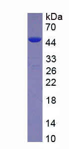
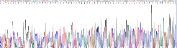
 Amino Acid Info
(Amino Acid info)
Amino Acid Info
(Amino Acid info)
Adiponectin (ADPN), Recombinant Protein (Cat# AAA153366)
Full Name
Recombinant Adiponectin (ADPN)
Gene Names
Adipoq; Ad; APN; Acdc; apM1; 30kDa; GBP28; adipo; Acrp30
Applications
Western Blot
Purity
Greater than 95% by SDS-PAGE
Pricing
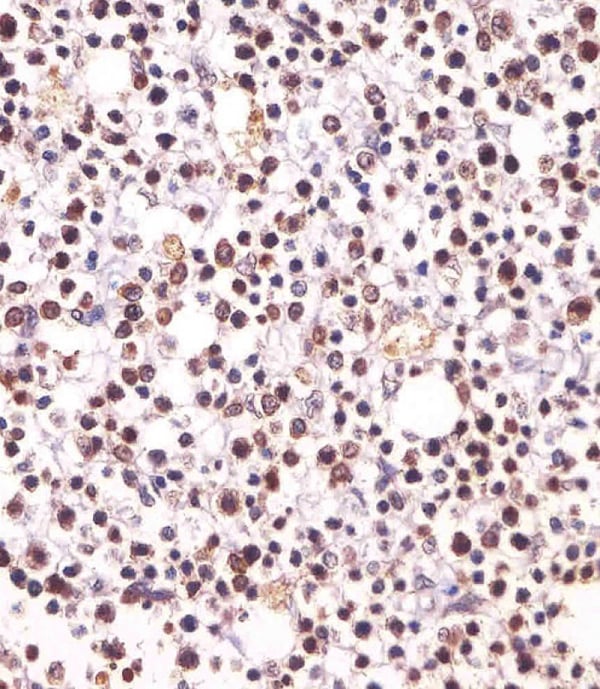
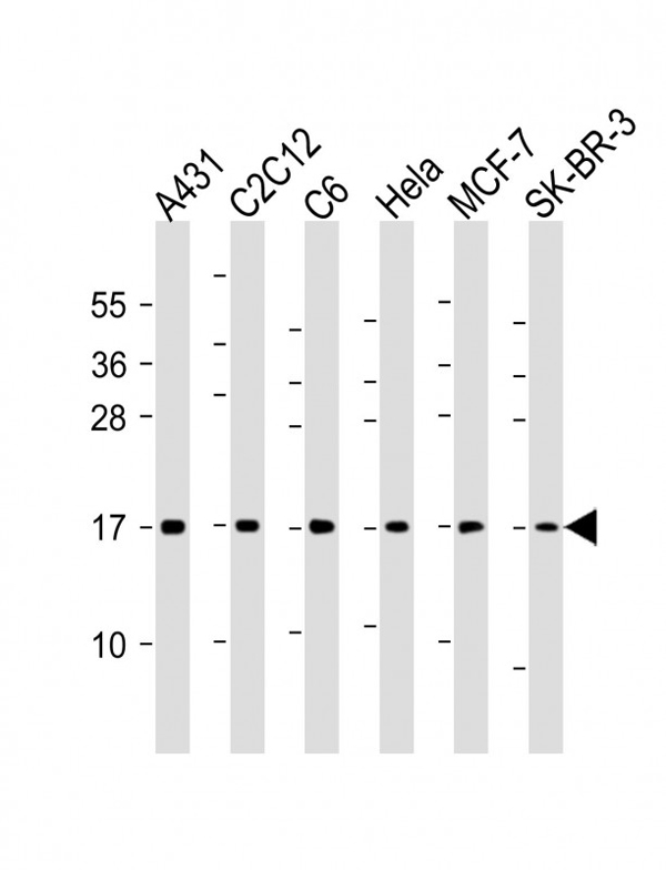
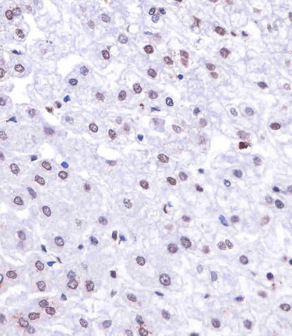 IHC (Immunohistochemisry)
(AM8433 staining Histone H3 in human liver tissue sections by Immunohistochemistry (IHC-P - paraformaldehyde-fixed, paraffin-embedded sections). Tissue was fixed with formaldehyde and blocked with 3% BSA for 0. 5 hour at room temperature; antigen retrieval was by heat mediation with a citrate buffer (pH6). Samples were incubated with primary antibody (1/25) for 1 hours at 37°C. A undiluted biotinylated goat polyvalent antibody was used as the secondary antibody.)
IHC (Immunohistochemisry)
(AM8433 staining Histone H3 in human liver tissue sections by Immunohistochemistry (IHC-P - paraformaldehyde-fixed, paraffin-embedded sections). Tissue was fixed with formaldehyde and blocked with 3% BSA for 0. 5 hour at room temperature; antigen retrieval was by heat mediation with a citrate buffer (pH6). Samples were incubated with primary antibody (1/25) for 1 hours at 37°C. A undiluted biotinylated goat polyvalent antibody was used as the secondary antibody.)
Histone H3, Monoclonal Antibody (Cat# AAA290961)
Full Name
Histone H3 Antibody
Gene Names
HIST1H3A; H3/A; H3FA
Reactivity
Human, Mouse, Rat
Predicted Reactivity: Bovine
Predicted Reactivity: Bovine
Applications
Immunohistochemistry, Western Blot
Pricing

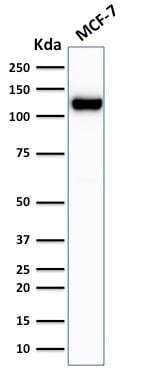 SDS-PAGE
(SDS-PAGE Analysis of Purified E-Cadherin Mouse Recombinant Monoclonal Antibody (rCDH1/1525).)
SDS-PAGE
(SDS-PAGE Analysis of Purified E-Cadherin Mouse Recombinant Monoclonal Antibody (rCDH1/1525).)
E-Cadherin (CDH1)/CD324, Monoclonal Antibody (Cat# AAA214624)
Full Name
E-Cadherin (CDH1)/CD324 (Intercellular Junction Marker)
Gene Names
CDH1; UVO; CDHE; ECAD; LCAM; Arc-1; BCDS1; CD324
Reactivity
Human.
Does not react with Mouse and Rat. Others not known.
Does not react with Mouse and Rat. Others not known.
Applications
Immunohistochemistry, Western Blot, Immunofluorescence, Flow Cytometry
Pricing
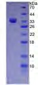
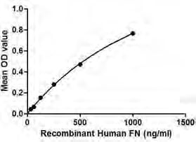
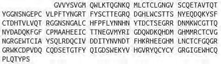
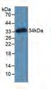 WB (Western Blot)
(Sample: Recombinant Fibronectin, Human; Antibody: Rabbit Anti-Human Fibronectin Ab)
WB (Western Blot)
(Sample: Recombinant Fibronectin, Human; Antibody: Rabbit Anti-Human Fibronectin Ab)
Fibronectin (FN), Active Protein (Cat# AAA148156)
Full Name
Active Fibronectin (FN)
Gene Names
FN1; FN; CIG; FNZ; MSF; ED-B; FINC; GFND; LETS; GFND2; SMDCF
Applications
Activity Assay, Cell Culture
Purity
>98%
Pricing
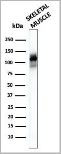
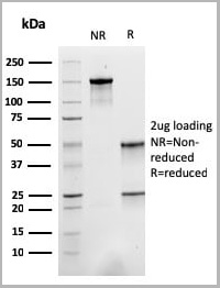
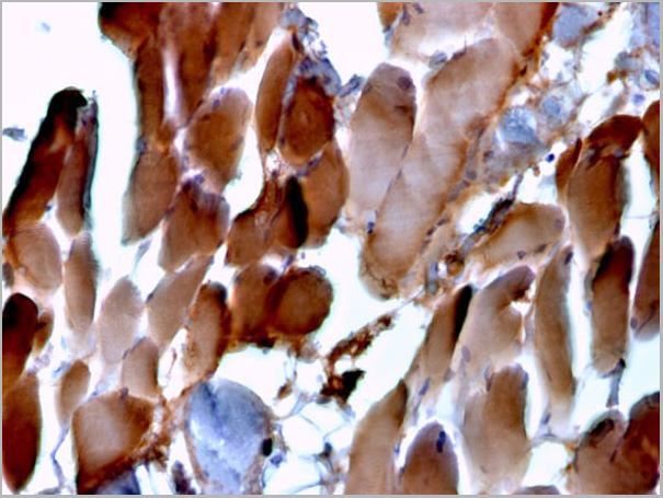
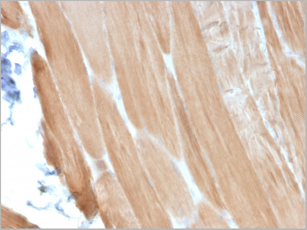
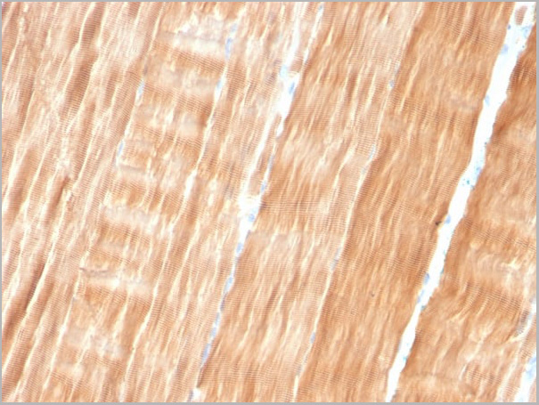
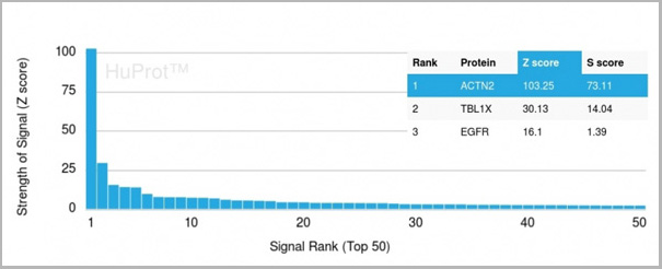 Application Data
(Analysis of Protein Array containing more than 19,000 full-length human proteins using Sarcomeric Actinin Alpha 2 Mouse Monoclonal Antibody (ACTN2/3293). Z- and S- Score: The Z-score represents the strength of a signal that a monoclonal antibody (MAb) (in combination with a fluorescently-tagged anti-IgG secondary antibody) produces when binding to a particular protein on the HuProtTM array. Z-scores are described in units of standard deviations (SD€™s) above the mean value of all signals generated on that array. If targets on HuProtTM are arranged in descending order of the Z-score, the S-score is the difference (also in units of SD€™s) between the Z-score. S-score therefore represents the relative target specificity of a MAb to its intended target. A MAb is considered to specific to its intended target, if the MAb has an S-score of at least 2.5. For example, if a MAb binds to protein X with a Z-score of 43 and to protein Y with a Z-score of 14, then the S-score for the binding of that MAb to protein X is equal to 29.)
Application Data
(Analysis of Protein Array containing more than 19,000 full-length human proteins using Sarcomeric Actinin Alpha 2 Mouse Monoclonal Antibody (ACTN2/3293). Z- and S- Score: The Z-score represents the strength of a signal that a monoclonal antibody (MAb) (in combination with a fluorescently-tagged anti-IgG secondary antibody) produces when binding to a particular protein on the HuProtTM array. Z-scores are described in units of standard deviations (SD€™s) above the mean value of all signals generated on that array. If targets on HuProtTM are arranged in descending order of the Z-score, the S-score is the difference (also in units of SD€™s) between the Z-score. S-score therefore represents the relative target specificity of a MAb to its intended target. A MAb is considered to specific to its intended target, if the MAb has an S-score of at least 2.5. For example, if a MAb binds to protein X with a Z-score of 43 and to protein Y with a Z-score of 14, then the S-score for the binding of that MAb to protein X is equal to 29.)
Sarcomeric Actinin Alpha 2/ACTN2, Monoclonal Antibody (Cat# AAA23942)
Full Name
Sarcomeric Actinin Alpha 2/ACTN2
Gene Names
ACTN2; CMH23; CMD1AA
Reactivity
Human
Applications
Western Blot, Immunohistochemistry
Purity
Purified Ab with BSA and Azide at 200ug/ml OR Purified Ab WITHOUT BSA and Azide at 1.0mg/ml
Pricing
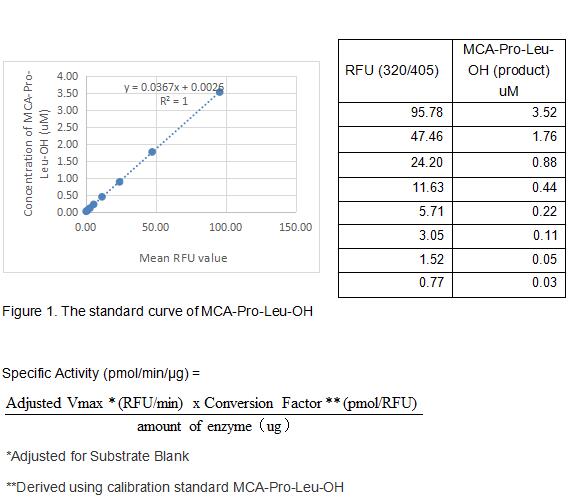 Bioactivity
(Cathepsin S (CTSS) is a lysosomal cysteine protease of the papain family. It plays a major role in the processing of the MHC class IIassociated invariant chain. It has been implicated in the pathogenesis of several diseases such as Alzheimer's disease and degenerative disorders associated with the cells of the mononuclear phagocytic system. Mouse Cathepsin S is synthesized as a preproenzyme of 340 amino acid residues consisting a signal peptide (residues 117), a pro region (residues 18122), and the mature enzyme (residues 123340). Cathepsin S is less abundant in tissues than Cathepsins B, L and H. The activity of recombinant mouse CTSS is measured by its ability to cleave a fluorogenic peptide substrate Mca-Arg-Pro-Lys-Pro-Val-Glu-Nval-Trp-Arg-Lys(Dnp)-NH2 in the assay buffer 50 mM NaOAc, 5 mM DTT, 250 mM NaCl, pH 4.5. The rmCTSS is diluted to 50 ug/ml in assay buffer, then incubated at room temperature for 2h. The incubated rmCTSS is diluted to 5 ug/mL in assay buffer. Loading into a black well plate 50 uL of 5 ug/mL rmCTSS and start the reaction by adding 50 uL of 20 uM substrate, with a substrate blank containing 50 uL assay buffer, 50 uL substrate, and no rmCTSS. Then read at excitiation and emission wavelengths of 320 nm and 405 nm, respectively, in kinetic mode for 5 minutes. The specific activity of recombinant mouse CTSS is > 10 pmol/min/ug.)
Bioactivity
(Cathepsin S (CTSS) is a lysosomal cysteine protease of the papain family. It plays a major role in the processing of the MHC class IIassociated invariant chain. It has been implicated in the pathogenesis of several diseases such as Alzheimer's disease and degenerative disorders associated with the cells of the mononuclear phagocytic system. Mouse Cathepsin S is synthesized as a preproenzyme of 340 amino acid residues consisting a signal peptide (residues 117), a pro region (residues 18122), and the mature enzyme (residues 123340). Cathepsin S is less abundant in tissues than Cathepsins B, L and H. The activity of recombinant mouse CTSS is measured by its ability to cleave a fluorogenic peptide substrate Mca-Arg-Pro-Lys-Pro-Val-Glu-Nval-Trp-Arg-Lys(Dnp)-NH2 in the assay buffer 50 mM NaOAc, 5 mM DTT, 250 mM NaCl, pH 4.5. The rmCTSS is diluted to 50 ug/ml in assay buffer, then incubated at room temperature for 2h. The incubated rmCTSS is diluted to 5 ug/mL in assay buffer. Loading into a black well plate 50 uL of 5 ug/mL rmCTSS and start the reaction by adding 50 uL of 20 uM substrate, with a substrate blank containing 50 uL assay buffer, 50 uL substrate, and no rmCTSS. Then read at excitiation and emission wavelengths of 320 nm and 405 nm, respectively, in kinetic mode for 5 minutes. The specific activity of recombinant mouse CTSS is > 10 pmol/min/ug.)
Cathepsin S (CTSS), Active Protein (Cat# AAA161867)
Full Name
Active Cathepsin S (CTSS)
Gene Names
Ctss; Cats
Applications
Activity Assay, Cell Culture
Purity
Greater than 90% by SDS-PAGE
Pricing
 Bioactivity
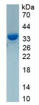
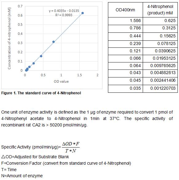
(Carbonic Anhydrase (CA) catalyzes the reversible reaction of CO2 H2O = HCO3- H, which is fundamental to many processes such as respiration, renal tubular acidification and bone resorption. CA2 is a cytosolic enzyme with the highest activity among all known CAs. Mutations in the CA2 gene result in the CA II deficiency syndrome, an autosomal recessive disorder that produces osteopetrosis, renal tubular acidosis and cerebral calcification. The activity of recombinant rat CA2 was measured by its ability to hydrolyze 4-Nitrophenyl acetate (4-NPA) to 4-Nitrophenol. The reaction was performed in 12.5 mM Tris, 75 mM NaCl, pH 7.5 (assay buffer), initiated by addition 50 uL of various concentrations of CA2 (diluted by assay buffer) to 50 uL of 2 mM substrate 4-NPA (100 mM stock in Acetone, diluted by assay buffer). Incubated at 37 degree C for 5min, then read at a wavelength of 400 nm.)
Bioactivity
(Carbonic Anhydrase (CA) catalyzes the reversible reaction of CO2 H2O = HCO3- H, which is fundamental to many processes such as respiration, renal tubular acidification and bone resorption. CA2 is a cytosolic enzyme with the highest activity among all known CAs. Mutations in the CA2 gene result in the CA II deficiency syndrome, an autosomal recessive disorder that produces osteopetrosis, renal tubular acidosis and cerebral calcification. The activity of recombinant rat CA2 was measured by its ability to hydrolyze 4-Nitrophenyl acetate (4-NPA) to 4-Nitrophenol. The reaction was performed in 12.5 mM Tris, 75 mM NaCl, pH 7.5 (assay buffer), initiated by addition 50 uL of various concentrations of CA2 (diluted by assay buffer) to 50 uL of 2 mM substrate 4-NPA (100 mM stock in Acetone, diluted by assay buffer). Incubated at 37 degree C for 5min, then read at a wavelength of 400 nm.)
Carbonic Anhydrase II (CA2), Active Protein (Cat# AAA161777)
Full Name
Active Carbonic Anhydrase II (CA2)
Gene Names
Car2; Ca2
Applications
Activity Assay, Cell Culture
Purity
Greater than 95% by SDS-PAGE
Pricing
 Bioactivity
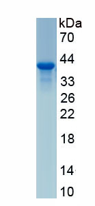
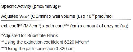
(A hallmark of most cancer cells is an altered metabolism involving a shift to aerobic glycolysis with lactate production coupled with a higher uptake of glucose as the main source of energy. Lactate dehydrogenase (LDH) is key to this shift by catalyzing the formation of lactate by reducing pyruvate with NADH, which also generates NAD() that is essential for the continuity of glycolysis. Inhibiting LDH activity has an anti-proliferative effect on cancer cells. It may reverse the resistance of tumor cells to conventional chemo- and radiotherapy. Recent research has renewed interest in LDH as an anticancer drug target. LDH enzymes have three homologous subunits LDHA, LDHB and LDHC. The activity of recombinant rat LDHC was measured by its ability to reduce pyruvate to lactate. The reaction was performed in 25 mM Tris, 100 mM NaCl, pH 7.5 (assay buffer), initiated by addition 50 uL of various concentrations of rrLDHC (diluted by assay buffer) to 50 uL of substrate mixture 1.6 mM beta-NADH and 4 mM sodium pyruvate. The final well serves as a negative control with no rrLDHC, replaced with 50 ul assay buffer. Read at a wavelength of 340 nm in kinetic mode for 5 minutes. The specific activity of recombinant rat LDHC is > 880 pmol/min/ug.)
Bioactivity
(A hallmark of most cancer cells is an altered metabolism involving a shift to aerobic glycolysis with lactate production coupled with a higher uptake of glucose as the main source of energy. Lactate dehydrogenase (LDH) is key to this shift by catalyzing the formation of lactate by reducing pyruvate with NADH, which also generates NAD() that is essential for the continuity of glycolysis. Inhibiting LDH activity has an anti-proliferative effect on cancer cells. It may reverse the resistance of tumor cells to conventional chemo- and radiotherapy. Recent research has renewed interest in LDH as an anticancer drug target. LDH enzymes have three homologous subunits LDHA, LDHB and LDHC. The activity of recombinant rat LDHC was measured by its ability to reduce pyruvate to lactate. The reaction was performed in 25 mM Tris, 100 mM NaCl, pH 7.5 (assay buffer), initiated by addition 50 uL of various concentrations of rrLDHC (diluted by assay buffer) to 50 uL of substrate mixture 1.6 mM beta-NADH and 4 mM sodium pyruvate. The final well serves as a negative control with no rrLDHC, replaced with 50 ul assay buffer. Read at a wavelength of 340 nm in kinetic mode for 5 minutes. The specific activity of recombinant rat LDHC is > 880 pmol/min/ug.)
Lactate Dehydrogenase C (LDHC), Active Protein (Cat# AAA161921)
Full Name
Active Lactate Dehydrogenase C (LDHC)
Gene Names
Ldhc; Ldh3
Applications
Activity Assay, Cell Culture
Purity
Greater than 95% by SDS-PAGE
Pricing

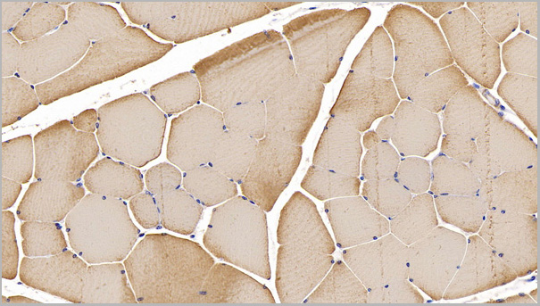
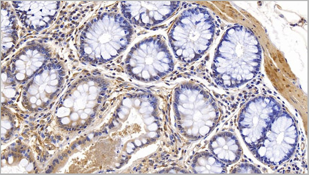
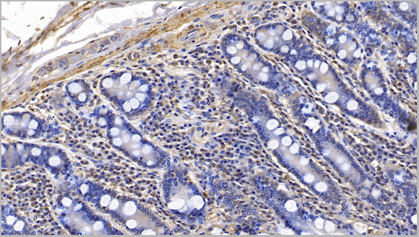
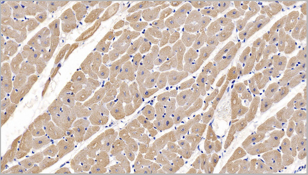
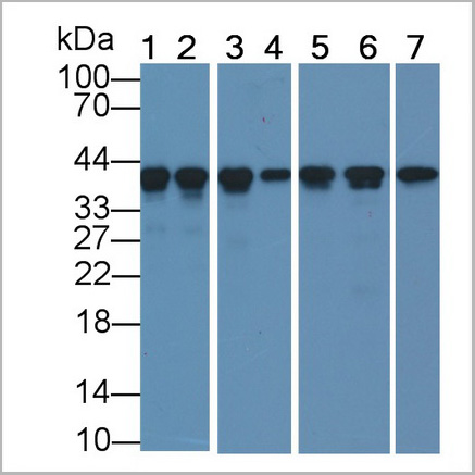 WB (Western Blot)
(Western Blot; Sample: Lane1: Mouse Skeletal muscle lysate; Lane2: Porcine Heart lysate; Lane3: Porcine Skeletal muscle lysate; Lane4: Canine Heart lysate; Lane5: Gallus Skeletal muscle lysate; Lane6: Bovine Heart lysate; Lane7: Rabbit Skeletal muscle lysatePrimary Ab: 0.5ug/ml Mouse AntiHuman ACTC1 AntibodySecond Ab: 0.2ug/mL HRPLinked Caprine AntiMouse IgG Polyclonal Antibody(Catalog: SAA544Mu19))
WB (Western Blot)
(Western Blot; Sample: Lane1: Mouse Skeletal muscle lysate; Lane2: Porcine Heart lysate; Lane3: Porcine Skeletal muscle lysate; Lane4: Canine Heart lysate; Lane5: Gallus Skeletal muscle lysate; Lane6: Bovine Heart lysate; Lane7: Rabbit Skeletal muscle lysatePrimary Ab: 0.5ug/ml Mouse AntiHuman ACTC1 AntibodySecond Ab: 0.2ug/mL HRPLinked Caprine AntiMouse IgG Polyclonal Antibody(Catalog: SAA544Mu19))
Actin Alpha 1, Cardiac Muscle (ACTC1), Monoclonal Antibody (Cat# AAA21192)
Full Name
Monoclonal Antibody to Actin Alpha 1, Cardiac Muscle (ACTC1)
Gene Names
ACTC1; ACTC; ASD5; CMD1R; CMH11; LVNC4
Reactivity
Human, Mouse, Rat, Rabbit, Dog, Pig, Bovine, Chicken
Applications
Western Blot, Immunohistochemistry, Immunocytochemistry, Immunoprecipitation
Purity
Protein A + Protein G affinity chromatography
Pricing
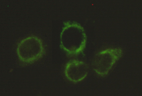
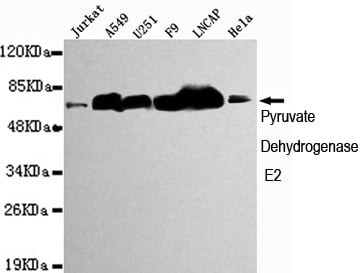 ICC (Immunocytochemistry)
(Immunocytochemistry stain of Hela using Pyruvate Dehydrogenase E2 mouse mAb (1:300).)
ICC (Immunocytochemistry)
(Immunocytochemistry stain of Hela using Pyruvate Dehydrogenase E2 mouse mAb (1:300).)
Pyruvate Dehydrogenase E2, Monoclonal Antibody (Cat# AAA320350)
Full Name
Pyruvate Dehydrogenase E2 Mouse mAb
Gene Names
DLAT; DLTA; PDCE2; PDC-E2
Reactivity
Human, Mouse
Applications
Immunoprecipitation, Immunocytochemistry, Western Blot
Purity
The antibody was affinity-purified from mouse ascites by affinity-chromatography using epitope-specific immunogen.
Pricing
 ELISA
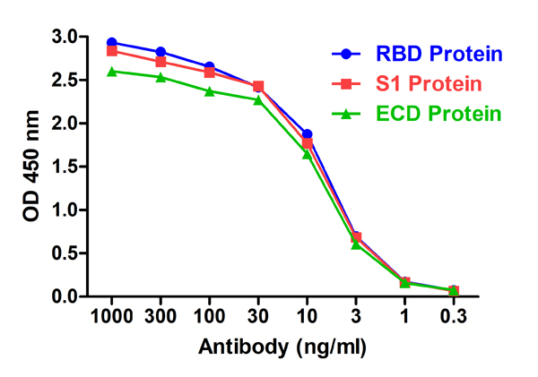
(Figure 2 ELISA Validation with SARS-CoV-2 (COVID-19) Spike RBD, S1 and ECD Proteins Antibodies: SARS-CoV-2 (COVID-19) Spike S1 RBD Antibody, AAA40979. A direct ELISA was performed using SARS-CoV-2 Spike recombinant proteins (RBD, 10-303; S1, 97-087; ECD, 11-070) as coating antigen at 1ug/mL and the anti-SARS-CoV-2 (COVID-19) Spike S1 RBD antibody (AAA40979) as the capture antibody, following by anti-cMyc-tag antibody (PM-7669) at 1ug/mL. Secondary: Goat anti-mouse IgG HRP conjugate at 1:5000 dilution. Detection range is from 0.3 ng/mL to 1000 ng/mL. SARS-CoV-2 (COVID-19) Spike S1 RBD Antibody, AAA40979 can detect spike RBD, S1 and ECD proteins at 2 ng/mL.)
ELISA
(Figure 2 ELISA Validation with SARS-CoV-2 (COVID-19) Spike RBD, S1 and ECD Proteins Antibodies: SARS-CoV-2 (COVID-19) Spike S1 RBD Antibody, AAA40979. A direct ELISA was performed using SARS-CoV-2 Spike recombinant proteins (RBD, 10-303; S1, 97-087; ECD, 11-070) as coating antigen at 1ug/mL and the anti-SARS-CoV-2 (COVID-19) Spike S1 RBD antibody (AAA40979) as the capture antibody, following by anti-cMyc-tag antibody (PM-7669) at 1ug/mL. Secondary: Goat anti-mouse IgG HRP conjugate at 1:5000 dilution. Detection range is from 0.3 ng/mL to 1000 ng/mL. SARS-CoV-2 (COVID-19) Spike S1 RBD Antibody, AAA40979 can detect spike RBD, S1 and ECD proteins at 2 ng/mL.)
SARS-CoV-2 (COVID-19) S1 RBD, Monoclonal Recombinant Antibody (Cat# AAA40979)
Full Name
SARS-CoV-2 (COVID-19) S1 RBD Single Domain Antibody [T5P4-A12]
Reactivity
Virus
Applications
ELISA
Purity
SARS-CoV-2 (COVID-19) S1 RBD Antibody [T5P4-A12] is affinity chromatography purified via Nickel column. Antibody is supplied as a His-tagged purified protein. It also contains a myc-tag for detection.
Pricing
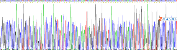
 Amino Acid Info
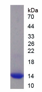
Insulin Receptor (INSR), Recombinant Protein (Cat# AAA153402)
Full Name
Recombinant Insulin Receptor (INSR)
Gene Names
INSR; HHF5; CD220
Applications
Western Blot
Purity
Greater than 90% by SDS-PAGE
Pricing
 Amino Acid Info
Anti-Mullerian Hormone (AMH), Recombinant Protein (Cat# AAA153312)
Full Name
Recombinant Anti-Mullerian Hormone (AMH)
Applications
Western Blot
Purity
Greater than 95% by SDS-PAGE
Pricing

 ELISA
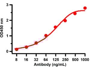
(ELISA Validation with SARS-CoV-2 (COVID-19) Nucleocapsid Recombinant Protein Antibodies: SARS-CoV-2 (COVID-19) Nucleocapsid antibody, AAA40905 (1 ug/mL). A direct ELISA was performed using SARS-CoV-2 (COVID-19) Nucleocapsid recombinant protein as coating antigen and the anti-SARSCoV-2 (COVID-19) Nucleocapsid antibody as the capture antibody. Secondary: Goat anti-rabbit IgG HRP conjugate at 1:20000 dilution. Detection range is from 8 ng/mL to 1000ng/mL.)
ELISA
(ELISA Validation with SARS-CoV-2 (COVID-19) Nucleocapsid Recombinant Protein Antibodies: SARS-CoV-2 (COVID-19) Nucleocapsid antibody, AAA40905 (1 ug/mL). A direct ELISA was performed using SARS-CoV-2 (COVID-19) Nucleocapsid recombinant protein as coating antigen and the anti-SARSCoV-2 (COVID-19) Nucleocapsid antibody as the capture antibody. Secondary: Goat anti-rabbit IgG HRP conjugate at 1:20000 dilution. Detection range is from 8 ng/mL to 1000ng/mL.)
COVID 19 Nucleocapsid (NP) Coronavirus, Polyclonal Antibody (Cat# AAA40905)
Full Name
SARS-CoV-2 (COVID-19) Nucleocapsid Antibody
Reactivity
Virus
Applications
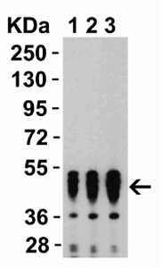
Western Blot
Purity
Affinity chromatography purified via peptide column.
Pricing
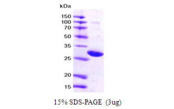 SDS-PAGE
(Figure annotation denotes ug of protein loaded and % gel used.)
SDS-PAGE
(Figure annotation denotes ug of protein loaded and % gel used.)
14.3.3 zeta, Recombinant Protein (Cat# AAA107758)
Full Name
14.3.3 zeta protein (His tag)
Gene Names
ywhaz-b; ywhaz; ywhazb; 14-3-3z; 14-3-3zeta
Applications
SDS-PAGE
Purity
> 95% pure
Pricing
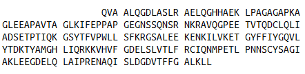
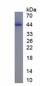 SDS-PAGE
(SDS-PAGE)
SDS-PAGE
(SDS-PAGE)
B-Cell Activating Factor (BAFF), Recombinant Protein (Cat# AAA152210)
Full Name
Recombinant B-Cell Activating Factor (BAFF)
Gene Names
TNFSF13B; DTL; BAFF; BLYS; CD257; TALL1; THANK; ZTNF4; TALL-1; TNFSF20
Applications
Western Blot
Purity
Greater than 80% by SDS-PAGE
Pricing
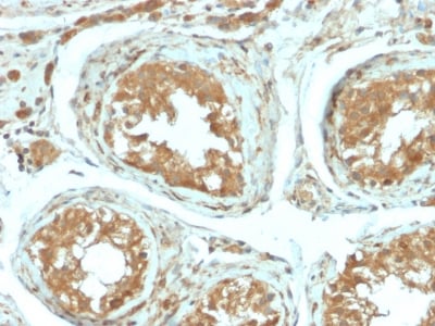
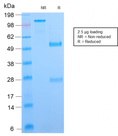 SDS-PAGE
(SDS-PAGE Analysis Purified B7-H4 Rabbit Recombinant Monoclonal Antibody (B7H4/2652R). Confirmation of Purity and Integrity of Antibody.)
SDS-PAGE
(SDS-PAGE Analysis Purified B7-H4 Rabbit Recombinant Monoclonal Antibody (B7H4/2652R). Confirmation of Purity and Integrity of Antibody.)
B7-H4, Monoclonal Antibody (Cat# AAA214839)
Full Name
B7-H4 (Immuno-Inhibitory Protein)
Gene Names
VTCN1; B7X; B7H4; B7S1; B7-H4; B7h.5; VCTN1; PRO1291
Reactivity
Human. Others not known.
Applications
Immunohistochemistry
Purity
Purified Ab with BSA and Azide at 200ug/ml OR Purified Ab WITHOUT BSA and Azide at 1.0mg/ml
Pricing
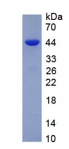
 Amino Acid Info
Eukaryotic Excitatory Amino Acid Transporter 2 (EAAT2), Protein (Cat# AAA153222)
Full Name
Eukaryotic Excitatory Amino Acid Transporter 2 (EAAT2)
Gene Names
SLC1A2; EAAT2; GLT-1
Applications
Western Blot
Purity
Greater than 90% by SDS-PAGE
Pricing
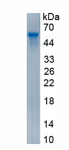
 Bioactivity
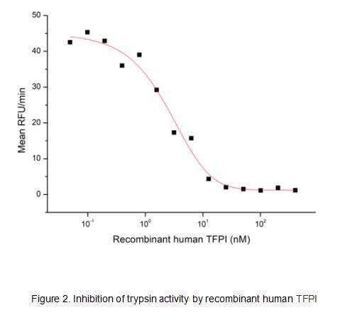
(Human TFPI, also known as lipoprotein-associated coagulation inhibitor (LACI) and extrinsic pathway inhibitor (EPI), is a physiological inhibitor of extrinsic pathway of coagulation and has biological functions of anticoagulation and anti-inflammation. It is a secreted protein with a N-terminal acidic region, three Kunitz (K) domains separated with by two linker regions, and a C-terminal basic region. The activity of recombinant human TFPI was measured by its ability to inhibit trypsin cleavage of a fluorogenic peptide substrate Mca-RPKPVE-Nval-WRK(Dnp)-NH2 in the assay buffer 50 mM Tris, 10 mM CaCl2, 150 mM NaCl, 0.05% (w/v) Brij-35, pH 7.5. Trypsin was diluted to 50 ug/ml in the assay buffer and 20 ul different concentrations of recombinant human TFPI (MW: 61.9 KD) was incubated with 20 ul diluted trypsin at 37 degree C for 15 minutes. Loading 50 uL of the incubated mixtures which were diluted five-fold in assay buffer into empty wells of a plate, and start the reaction by adding 50 uL of 20 uM substrate. Include a substrate blank containing 50 uL of assay buffer and 50 uL of 20 uM substrate. Then read at excitiation and emission wavelengths of 320 nm and 405 nm, respectively, in kinetic mode for 5 minutes. The result was shown in Figure 1 and it was obvious that recombinant human TFPI significantly decreased trypsin activity. The inhibition IC50 was )
Bioactivity
(Human TFPI, also known as lipoprotein-associated coagulation inhibitor (LACI) and extrinsic pathway inhibitor (EPI), is a physiological inhibitor of extrinsic pathway of coagulation and has biological functions of anticoagulation and anti-inflammation. It is a secreted protein with a N-terminal acidic region, three Kunitz (K) domains separated with by two linker regions, and a C-terminal basic region. The activity of recombinant human TFPI was measured by its ability to inhibit trypsin cleavage of a fluorogenic peptide substrate Mca-RPKPVE-Nval-WRK(Dnp)-NH2 in the assay buffer 50 mM Tris, 10 mM CaCl2, 150 mM NaCl, 0.05% (w/v) Brij-35, pH 7.5. Trypsin was diluted to 50 ug/ml in the assay buffer and 20 ul different concentrations of recombinant human TFPI (MW: 61.9 KD) was incubated with 20 ul diluted trypsin at 37 degree C for 15 minutes. Loading 50 uL of the incubated mixtures which were diluted five-fold in assay buffer into empty wells of a plate, and start the reaction by adding 50 uL of 20 uM substrate. Include a substrate blank containing 50 uL of assay buffer and 50 uL of 20 uM substrate. Then read at excitiation and emission wavelengths of 320 nm and 405 nm, respectively, in kinetic mode for 5 minutes. The result was shown in Figure 1 and it was obvious that recombinant human TFPI significantly decreased trypsin activity. The inhibition IC50 was )
Tissue Factor Pathway Inhibitor (TFPI), Active Protein (Cat# AAA161730)
Full Name
Active Tissue Factor Pathway Inhibitor (TFPI)
Gene Names
TFPI; EPI; TFI; LACI; TFPI1
Applications
Activity Assay, Cell Culture
Purity
Greater than 80% by SDS-PAGE
Pricing
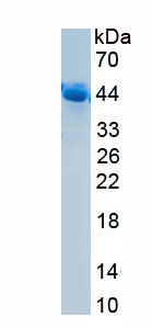
 Bioactivity
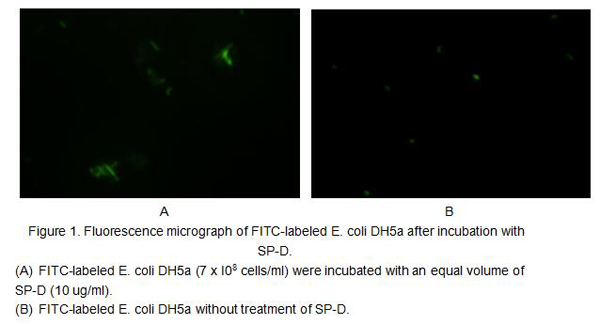
(SP-D (surfactant protein-D) is a 43 kDa member of the collectin family of innate immune modulators. It is constitutively secreted by alveolar lining cells and epithelium associated with tubular structures. Its principal components consist of a collagen-like region and a C-terminal carbohydrate recognition domain (CRD), a structure that further places it in a subset of an expanded group of proteins termed defense collagens. SP-D is known to bind both SIRP alpha and the calreticulin/CD91 complex on macrophages. When the ratio of antigen/pathogen to available CRDs is low, antigen can be bound without occupying all available CRDs. The free CRDs will bind to SIRP alpha, generating a signal that downmodulates the inflammatory response. When virtually all CRDs are occupied by ligand, however, free CRDs are not available for SIRP alpha binding. The activity of the recombinant human SPD was measured by its ability to bind fluorescein-conjugated E. coli bioparticles. The rhSPD was diluted to 10 ug/ml in assay buffer of 20 mM Tris, 137 mM NaCl, 1 mM CaC12, pH7.4 and fluorescein-conjugated E. coli was diluted to 7*108 cells/ml. Equal volume of 10 ug/ml rhSPD and fluorescein-conjugated E. coli were mixed and incubated at room temperature for 1h. 10 ul mixture was took onto the slide and observed under the fluorescence microscopy. The result was shown in figure 1, the rhSPD could bind fluorescein-conjugated E. coli bioparticles.)
Bioactivity
(SP-D (surfactant protein-D) is a 43 kDa member of the collectin family of innate immune modulators. It is constitutively secreted by alveolar lining cells and epithelium associated with tubular structures. Its principal components consist of a collagen-like region and a C-terminal carbohydrate recognition domain (CRD), a structure that further places it in a subset of an expanded group of proteins termed defense collagens. SP-D is known to bind both SIRP alpha and the calreticulin/CD91 complex on macrophages. When the ratio of antigen/pathogen to available CRDs is low, antigen can be bound without occupying all available CRDs. The free CRDs will bind to SIRP alpha, generating a signal that downmodulates the inflammatory response. When virtually all CRDs are occupied by ligand, however, free CRDs are not available for SIRP alpha binding. The activity of the recombinant human SPD was measured by its ability to bind fluorescein-conjugated E. coli bioparticles. The rhSPD was diluted to 10 ug/ml in assay buffer of 20 mM Tris, 137 mM NaCl, 1 mM CaC12, pH7.4 and fluorescein-conjugated E. coli was diluted to 7*108 cells/ml. Equal volume of 10 ug/ml rhSPD and fluorescein-conjugated E. coli were mixed and incubated at room temperature for 1h. 10 ul mixture was took onto the slide and observed under the fluorescence microscopy. The result was shown in figure 1, the rhSPD could bind fluorescein-conjugated E. coli bioparticles.)
Surfactant Associated Protein D (SPD), Active Protein (Cat# AAA161827)
Full Name
Active Surfactant Associated Protein D (SPD)
Gene Names
SFTPD; SP-D; PSP-D; SFTP4; COLEC7
Applications
Activity Assay, Cell Culture
Purity
Greater than 90% by SDS-PAGE
Pricing

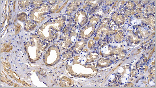
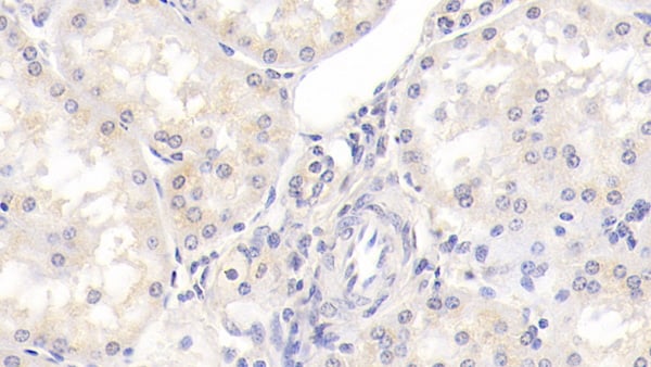 IHC (Immunohiostchemistry)
(DAB staining on IHCP;Sample: Porcine Kidney Tissue; Primary Ab: 30ug/ml Mouse AntiHuman ITGb1 AntibodySecond Ab: 2ug/mL HRPLinked Caprine AntiMouse IgG Polyclonal Antibody(Catalog: SAA544Mu19))
IHC (Immunohiostchemistry)
(DAB staining on IHCP;Sample: Porcine Kidney Tissue; Primary Ab: 30ug/ml Mouse AntiHuman ITGb1 AntibodySecond Ab: 2ug/mL HRPLinked Caprine AntiMouse IgG Polyclonal Antibody(Catalog: SAA544Mu19))
Integrin Beta 1 (ITGb1), Monoclonal Antibody (Cat# AAA151671)
Full Name
Monoclonal Antibody to Integrin Beta 1 (ITGb1)
Gene Names
ITGB1; CD29; FNRB; MDF2; VLAB; GPIIA; MSK12; VLA-BETA
Reactivity
Human, Pig
Applications
Immunoprecipitation, Immunocytochemistry, Immunohistochemistry, Western Blot
Purity
Protein A + Protein G affinity chromatography
Pricing
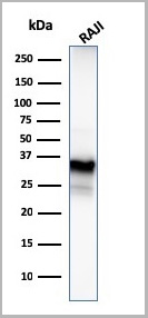
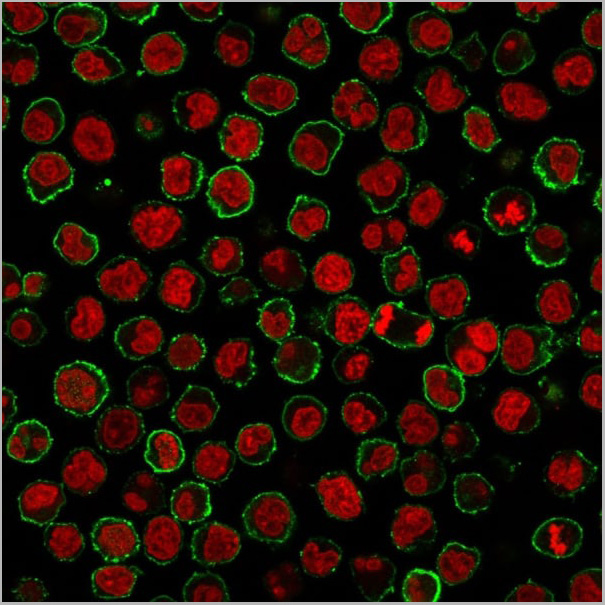
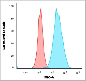
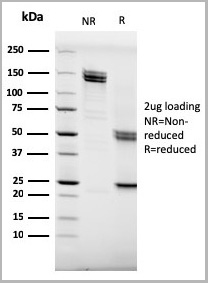
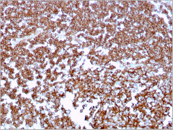
 Application Data
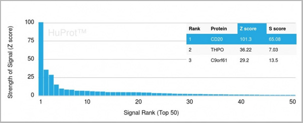
(Analysis of Protein Array containing more than 19,000 full-length human proteins using CD20 Mouse Recombinant Monoclonal Antibody (MS4A1/3409). Z- and S- Score: The Z-score represents the strength of a signal that a monoclonal antibody (MAb) (in combination with a fluorescently-tagged anti-IgG secondary antibody) produces when binding to a particular protein on the HuProtTM array. Z-scores are described in units of standard deviations (SD’s) above the mean value of all signals generated on that array. If targets on HuProtTM are arranged in descending order of the Z-score, the S-score is the difference (also in units of SD’s) between the Z-score. S-score therefore represents the relative target specificity of a MAb to its intended target. A MAb is considered to specific to its intended target, if the MAb has an S-score of at least 2.5. For example, if a MAb binds to protein X with a Z-score of 43 and to protein Y with a Z-score of 14, then the S-score for the binding of that MAb to protein X is equal to 29.)
Application Data
(Analysis of Protein Array containing more than 19,000 full-length human proteins using CD20 Mouse Recombinant Monoclonal Antibody (MS4A1/3409). Z- and S- Score: The Z-score represents the strength of a signal that a monoclonal antibody (MAb) (in combination with a fluorescently-tagged anti-IgG secondary antibody) produces when binding to a particular protein on the HuProtTM array. Z-scores are described in units of standard deviations (SD’s) above the mean value of all signals generated on that array. If targets on HuProtTM are arranged in descending order of the Z-score, the S-score is the difference (also in units of SD’s) between the Z-score. S-score therefore represents the relative target specificity of a MAb to its intended target. A MAb is considered to specific to its intended target, if the MAb has an S-score of at least 2.5. For example, if a MAb binds to protein X with a Z-score of 43 and to protein Y with a Z-score of 14, then the S-score for the binding of that MAb to protein X is equal to 29.)
CD20/MS4A1, Monoclonal Antibody (Cat# AAA23945)
Full Name
CD20/MS4A1 (B-Cell Marker)
Gene Names
MS4A1; B1; S7; Bp35; CD20; CVID5; MS4A2; LEU-16
Reactivity
Human
Applications
Flow Cytometry, Immunofluorescence, Western Blot, Immunohistochemistry
Purity
Purified Ab with BSA and Azide at 200ug/ml OR Purified Ab WITHOUT BSA and Azide at 1.0mg/ml
Pricing
 Bioactivity
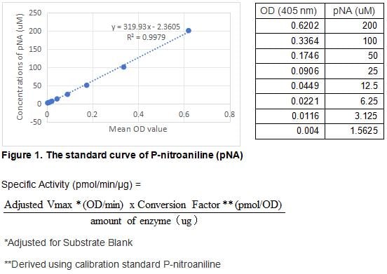
(Kallikrein 3, commonly known as prostate specific antigen (PSA), is a serine protease of the human tissue Kallikrein gene family. PSA is synthesized in the ductal and acinar epithelium of the prostate gland and secreted into the seminal plasma in high concentrations (0.5 - 2 g/L). A small portion of PSA "leaks" into the systemic circulation, the levels of which increase significantly (30-fold) from prostate cancer tissue than normal prostate tissue. PSA has become a well established tumor marker that aids the identification, staging, and follow up of prostate cancer. The activity of recombinant human KLK3 is measured by its ability to cleave a colorimetric peptide substrate Suc-Arg-Pro-Tyr-pNa in the assay buffer 50 mM Tris, 1 M NaCl, pH 8.0. The rhKLK3 is diluted to 200 ug/ml in activation buffer 50 mM Tris, 10 mM CaCl2, 150 mM NaCl, 0.05% Brij-35, pH 7.5, then activated with a final concentration of 1 ug/ml Thermolysin at 37 degree C for 5min. Adding a final concentration of 10 mM 1,10 Phenanthroline to stop the activation. The activated rhKLK3 is diluted to 50 ug/mL in assay buffer. Loading into a clear well plate 50 uL of 50 ug/mL rhKLK3 and start the reaction by adding 50 uL of 2 mM substrate, with a substrate blank containing 50 uL assay buffer, 50 uL substrate, and no rhKLK3. Then read at 405 nm in kinetic mode for 5 minutes. The specific activity of recombinant human KLK3 is > 400 pmol/min/ug.)
Bioactivity
(Kallikrein 3, commonly known as prostate specific antigen (PSA), is a serine protease of the human tissue Kallikrein gene family. PSA is synthesized in the ductal and acinar epithelium of the prostate gland and secreted into the seminal plasma in high concentrations (0.5 - 2 g/L). A small portion of PSA "leaks" into the systemic circulation, the levels of which increase significantly (30-fold) from prostate cancer tissue than normal prostate tissue. PSA has become a well established tumor marker that aids the identification, staging, and follow up of prostate cancer. The activity of recombinant human KLK3 is measured by its ability to cleave a colorimetric peptide substrate Suc-Arg-Pro-Tyr-pNa in the assay buffer 50 mM Tris, 1 M NaCl, pH 8.0. The rhKLK3 is diluted to 200 ug/ml in activation buffer 50 mM Tris, 10 mM CaCl2, 150 mM NaCl, 0.05% Brij-35, pH 7.5, then activated with a final concentration of 1 ug/ml Thermolysin at 37 degree C for 5min. Adding a final concentration of 10 mM 1,10 Phenanthroline to stop the activation. The activated rhKLK3 is diluted to 50 ug/mL in assay buffer. Loading into a clear well plate 50 uL of 50 ug/mL rhKLK3 and start the reaction by adding 50 uL of 2 mM substrate, with a substrate blank containing 50 uL assay buffer, 50 uL substrate, and no rhKLK3. Then read at 405 nm in kinetic mode for 5 minutes. The specific activity of recombinant human KLK3 is > 400 pmol/min/ug.)
Prostate Specific Antigen (PSA), Active Protein (Cat# AAA161711)
Full Name
Active Prostate Specific Antigen (PSA)
Gene Names
KLK3; APS; PSA; hK3; KLK2A1
Applications
Activity Assay, Cell Culture
Purity
Greater than 90% by SDS-PAGE
Pricing
 Bioactivity
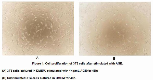
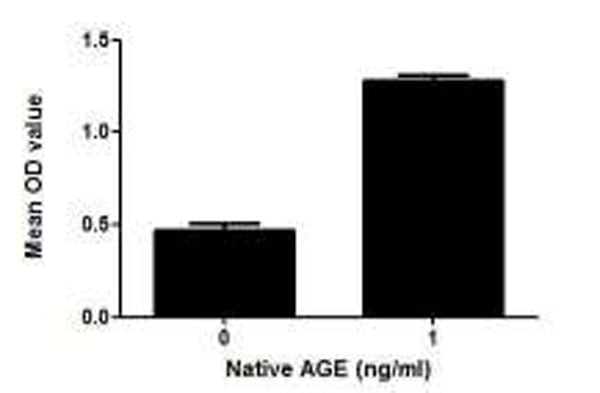
(Figure 2. Cell proliferation of 3T3 cells after stimulated with AGE.)
Bioactivity
(Figure 2. Cell proliferation of 3T3 cells after stimulated with AGE.)
Advanced Glycation End Product (AGE), Active Protein (Cat# AAA148250)
Full Name
Active Advanced Glycation End Product (AGE)
Applications
Activity Assay, Cell Culture
Purity
Purity: >90% as determined by SDS-PAGE.
Purification Methods: Salt co-precipitation and ionic-Exchange chromatography.
Purification Methods: Salt co-precipitation and ionic-Exchange chromatography.
Pricing
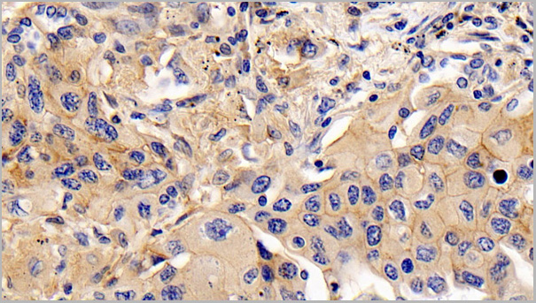
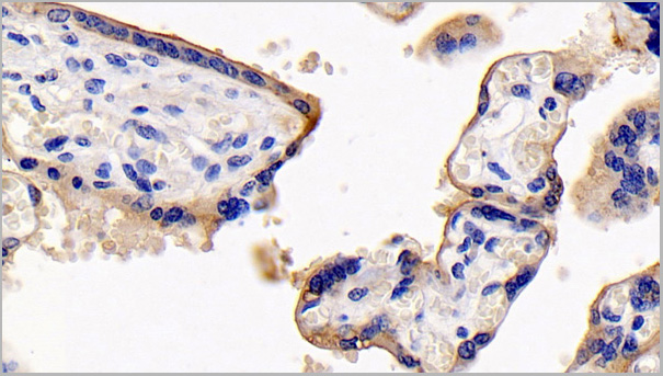
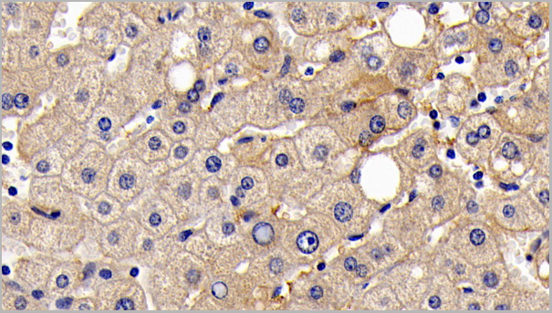 IHC (Immunohistchemistry)
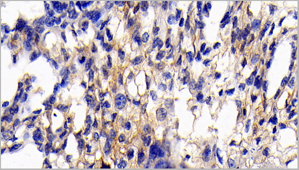
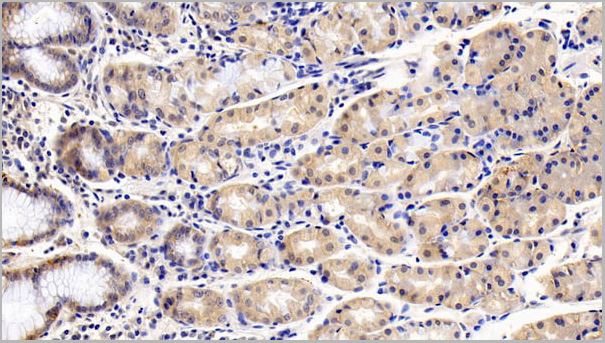
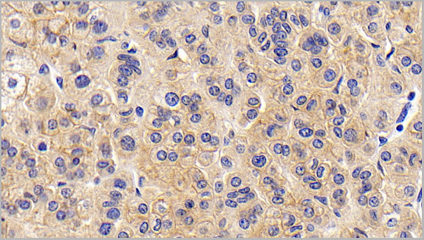
(DAB staining on IHCP;Sample: Human Liver Tissue; Primary Ab: 30ug/ml Mouse AntiHuman CD147 AntibodySecond Ab: 2ug/mL HRPLinked Caprine AntiMouse IgG Polyclonal Antibody(Catalog: SAA544Mu19))
IHC (Immunohistchemistry)
(DAB staining on IHCP;Sample: Human Liver Tissue; Primary Ab: 30ug/ml Mouse AntiHuman CD147 AntibodySecond Ab: 2ug/mL HRPLinked Caprine AntiMouse IgG Polyclonal Antibody(Catalog: SAA544Mu19))
Cluster Of Differentiation 147 (CD147), Monoclonal Antibody (Cat# AAA21197)
Full Name
Monoclonal Antibody to Cluster Of Differentiation 147 (CD147)
Gene Names
BSG; M6; OK; 5F7; TCSF; CD147; EMMPRIN
Reactivity
Human
Applications
Western Blot, Immunohistochemistry, Immunocytochemistry, Immunoprecipitation
Purity
Protein A + Protein G affinity chromatography
Pricing

 FCM/FACS (Flow Cytometry)
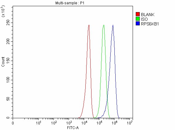
(Figure 2. Flow Cytometry analysis of U251 cells using anti-P70 S6K/RPS6KB1 antibody (AAA127060).Overlay histogram showing U251 cells stained with AAA127060 (Blue line). To facilitate intracellular staining, cells were fixed with 4% paraformaldehyde and permeabilized with permeabilization buffer. The cells were blocked with 10% normal goat serum. And then incubated with rabbit anti-P70 S6K/RPS6KB1 Antibody (AAA127060, 1ug/1x106 cells) for 30 min at 20 degree C. DyLight488 conjugated goat anti-rabbit IgG was used as secondary antibody for 30 minutes at 20 degree C. Isotype control antibody (Green line) was rabbit IgG (1ug/1x106) used under the same conditions. Unlabelled sample without incubation with primary antibody and secondary antibody (Red line) was used as a blank control.)
FCM/FACS (Flow Cytometry)
(Figure 2. Flow Cytometry analysis of U251 cells using anti-P70 S6K/RPS6KB1 antibody (AAA127060).Overlay histogram showing U251 cells stained with AAA127060 (Blue line). To facilitate intracellular staining, cells were fixed with 4% paraformaldehyde and permeabilized with permeabilization buffer. The cells were blocked with 10% normal goat serum. And then incubated with rabbit anti-P70 S6K/RPS6KB1 Antibody (AAA127060, 1ug/1x106 cells) for 30 min at 20 degree C. DyLight488 conjugated goat anti-rabbit IgG was used as secondary antibody for 30 minutes at 20 degree C. Isotype control antibody (Green line) was rabbit IgG (1ug/1x106) used under the same conditions. Unlabelled sample without incubation with primary antibody and secondary antibody (Red line) was used as a blank control.)
p70 S6K/RPS6KB1, Polyclonal Antibody (Cat# AAA127060)
Full Name
Anti-p70 S6K/RPS6KB1 Antibody Picoband
Gene Names
RPS6KB1; S6K; PS6K; S6K1; STK14A; p70-S6K; p70 S6KA; p70-alpha; S6K-beta-1; p70(S6K)-alpha
Reactivity
Human
Applications
Flow Cytometry, Western Blot
Purity
Immunogen affinity purified.
Pricing
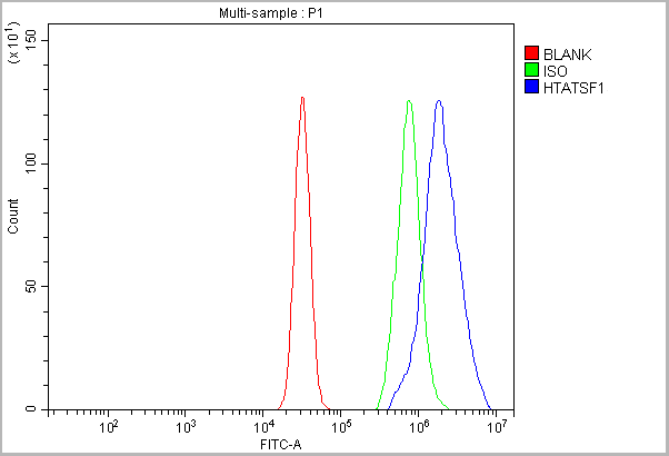
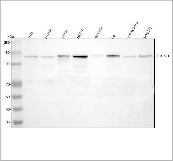
 IF (Immunofluorescence)
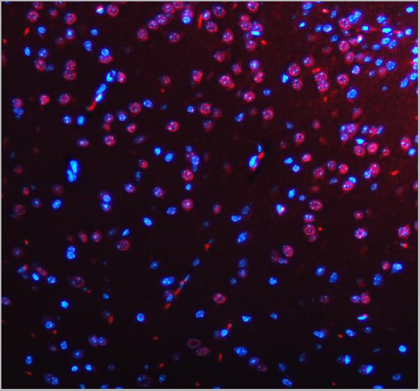
(Figure 15. IF analysis of HTATSF1 using anti-HTATSF1 antibody (AAA19895).HTATSF1 was detected in a paraffin-embedded section of mouse brain tissue. Heat mediated antigen retrieval was performed in EDTA buffer (pH 8.0, epitope retrieval solution). The tissue section was blocked with 10% goat serum. The tissue section was then incubated with 5ug/mL rabbit anti-HTATSF1 Antibody (AAA19895) overnight at 4 degree C. DyLight®594 Conjugated Goat Anti-Rabbit IgG (BA1142) was used as secondary antibody at 1:500 dilution and incubated for 30 minutes at 37 degree C. The section was counterstained with DAPI. Visualize using a fluorescence microscope and filter sets appropriate for the label used.)
IF (Immunofluorescence)
(Figure 15. IF analysis of HTATSF1 using anti-HTATSF1 antibody (AAA19895).HTATSF1 was detected in a paraffin-embedded section of mouse brain tissue. Heat mediated antigen retrieval was performed in EDTA buffer (pH 8.0, epitope retrieval solution). The tissue section was blocked with 10% goat serum. The tissue section was then incubated with 5ug/mL rabbit anti-HTATSF1 Antibody (AAA19895) overnight at 4 degree C. DyLight®594 Conjugated Goat Anti-Rabbit IgG (BA1142) was used as secondary antibody at 1:500 dilution and incubated for 30 minutes at 37 degree C. The section was counterstained with DAPI. Visualize using a fluorescence microscope and filter sets appropriate for the label used.)
HTATSF1, Polyclonal Antibody (Cat# AAA19895)
Full Name
Anti-HTATSF1 Antibody Picoband
Gene Names
HTATSF1; TATSF1; TAT-SF1; dJ196E23.2
Reactivity
Human, Mouse, Rat
Applications
Flow Cytometry, Immunofluorescence, Immunohistochemistry, Western Blot
Purity
Immunogen affinity purified.
Pricing

 SDS-PAGE
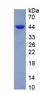
(Figure. SDS-PAGE)
SDS-PAGE
(Figure. SDS-PAGE)
Actin Alpha 2, Smooth Muscle (ACTa2), Recombinant Protein (Cat# AAA150687)
Full Name
Recombinant Actin Alpha 2, Smooth Muscle (ACTa2)
Gene Names
ACTA2; AAT6; ACTSA; MYMY5
Applications
Western Blot
Purity
>95%
Pricing


 Bioactivity
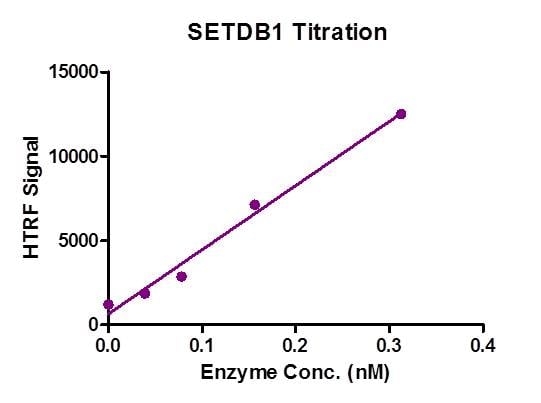
(HTRF assay for Recombinant SETDB1 protein activity. 1 uM histone peptide H3 (1-21) was incubated with SETDB1 in reaction buffer including 50 mM Tris-HCl pH 8.6, 0.02% Triton X-100, 2 mM MgCl2, 1 mM TCEP and 50 uM SAM for 3 hours at room temperature. H3K9me2 antibody was used to detect reaction product.)
Bioactivity
(HTRF assay for Recombinant SETDB1 protein activity. 1 uM histone peptide H3 (1-21) was incubated with SETDB1 in reaction buffer including 50 mM Tris-HCl pH 8.6, 0.02% Triton X-100, 2 mM MgCl2, 1 mM TCEP and 50 uM SAM for 3 hours at room temperature. H3K9me2 antibody was used to detect reaction product.)
 Amino Acid Info
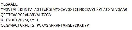
(Amino Acid info)
Amino Acid Info
(Amino Acid info)
Claudin 5 (CLDN5), Recombinant Protein (Cat# AAA153630)
Full Name
Recombinant Claudin 5 (CLDN5)
Gene Names
Cldn5; MBEC1; Tmvcf; AI854493
Applications
Western Blot
Purity
Greater than 90% by SDS-PAGE
Pricing
Dengue Virus NS1, Monoclonal Antibody (Cat# AAA13399)
Full Name
MAb to Dengue Virus NS1
Applications
Lateral Flow, Immunofluorescence
Purity
> 90% pure. Protein A Chromatography
Pricing
 SDS-PAGE
(The purity was determined to be >85% by densitometry. Approx. MW 84kDa)
SDS-PAGE
(The purity was determined to be >85% by densitometry. Approx. MW 84kDa)
MDM2, Recombinant Protein (Cat# AAA72902)
Full Name
MDM2 recombinant protein
Gene Names
MDM2; HDMX; hdm2; ACTFS
Applications
Western Blot
Purity
>85%
Pricing
 Application Data
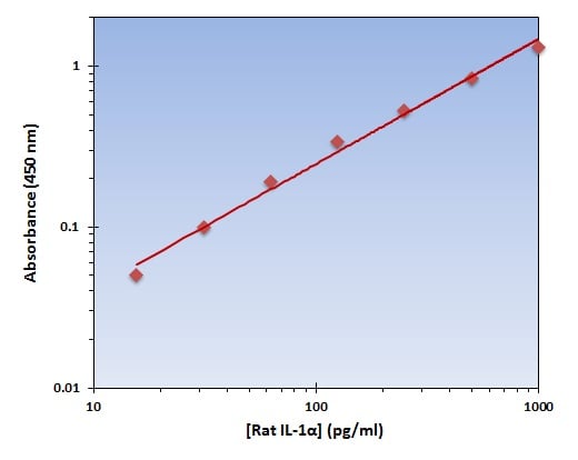
(The OmniKine Rat IL-1alpha ELISA Kit allows for the detection and quantification of endogenous levels of natural and/or recombinant Rat IL-1alpha proteins within the range of 16-1000 pg/ml.)
Application Data
(The OmniKine Rat IL-1alpha ELISA Kit allows for the detection and quantification of endogenous levels of natural and/or recombinant Rat IL-1alpha proteins within the range of 16-1000 pg/ml.)
IL-1alpha, ELISA Kit (Cat# AAA315695)
Full Name
IL-1alpha (Rat) OmniKine ELISA Kit
Gene Names
Il1a; IL-1F1; IL-1 alpha
Reactivity
Rat
Pricing
 SDS-PAGE
(The purity was determined to be >90% by densitometry . Approx. MW 94 kDa)
SDS-PAGE
(The purity was determined to be >90% by densitometry . Approx. MW 94 kDa)
HSP90 alpha, Recombinant Protein (Cat# AAA72931)
Full Name
HSP90 alpha recombinant protein
Gene Names
HSP90AA1; EL52; HSPN; LAP2; HSP86; HSPC1; HSPCA; Hsp89; Hsp90; HSP89A; HSP90A; HSP90N; HSPCAL1; HSPCAL4
Applications
Western Blot
Purity
>90%
Pricing
 SDS-PAGE
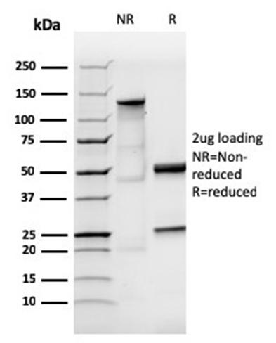
(SDS-PAGE Analysis Purified IgA Recombinant Rabbit Monoclonal Antibody (IGHA/3877R). Confirmation of Purity and Integrity of Antibody.)
SDS-PAGE
(SDS-PAGE Analysis Purified IgA Recombinant Rabbit Monoclonal Antibody (IGHA/3877R). Confirmation of Purity and Integrity of Antibody.)
IgA (Immunoglobulin Alpha Heavy Chain), Monoclonal Antibody (Cat# AAA215730)
Full Name
IgA (Immunoglobulin Alpha Heavy Chain) (B-Cell Marker) Recombinant Rabbit Monoclonal Antibody [Clone IGHA/3877R]
Gene Names
IGHA1; IgA1
Reactivity
Human
Applications
Immunohistochemistry
Pricing
 Bioactivity
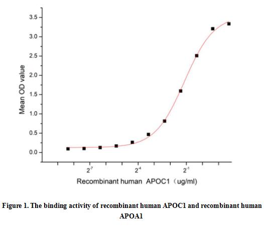
(Apolipoprotein C1 (APOC1), also known as Apo-CI; ApoC-I; apo-CIB; apoC-IB, is the smallest size apolipoprotein of all apolipoprotein C family (Mr = 6.6 kDa) and located at position 19q13.32. APOC1 is primarily expressed in the liver and activated when monocytes differentiate into macrophages. It plays important roles in the innate immune response as effector of glucocorticoid-mediated responses and regulator of the inflammatory process. It has anti-inflammatory activity and also can promote the differentiation of T-cells into Th1 cells and negatively regulates differentiation into Th2 cells. Besides, Apolipoprotein A1 (APOA1) has been identified as an interactor of APOC1, thus a functional binding ELISA assay was conducted to detect the interaction of recombinant human APOC1 and recombinant human APOA1. Briefly, APOC1 was diluted serially in PBS with 0.01% BSA (pH 7.4). Duplicate samples of 100 ul were then transferred to APOA1-coated microtiter wells and incubated for 1h at 37 degree C. Wells were washed with PBST and incubated for 1h with anti-APOC1 pAb, then aspirated and washed 3 times. After incubation with HRP labelled secondary antibody for 1h at 37 degree C, wells were aspirated and washed 5 times. With the addition of substrate solution, wells were incubated 15-25 minutes at 37 degree C. Finally, add 50 uL stop solution to the wells and read at 450/630 nm immediately. The binding activity of recombinant human APOC1 and recombinant human APOA1 was shown in Figure 1, the EC50 for this effect is 0.46 ug/mL.)
Bioactivity
(Apolipoprotein C1 (APOC1), also known as Apo-CI; ApoC-I; apo-CIB; apoC-IB, is the smallest size apolipoprotein of all apolipoprotein C family (Mr = 6.6 kDa) and located at position 19q13.32. APOC1 is primarily expressed in the liver and activated when monocytes differentiate into macrophages. It plays important roles in the innate immune response as effector of glucocorticoid-mediated responses and regulator of the inflammatory process. It has anti-inflammatory activity and also can promote the differentiation of T-cells into Th1 cells and negatively regulates differentiation into Th2 cells. Besides, Apolipoprotein A1 (APOA1) has been identified as an interactor of APOC1, thus a functional binding ELISA assay was conducted to detect the interaction of recombinant human APOC1 and recombinant human APOA1. Briefly, APOC1 was diluted serially in PBS with 0.01% BSA (pH 7.4). Duplicate samples of 100 ul were then transferred to APOA1-coated microtiter wells and incubated for 1h at 37 degree C. Wells were washed with PBST and incubated for 1h with anti-APOC1 pAb, then aspirated and washed 3 times. After incubation with HRP labelled secondary antibody for 1h at 37 degree C, wells were aspirated and washed 5 times. With the addition of substrate solution, wells were incubated 15-25 minutes at 37 degree C. Finally, add 50 uL stop solution to the wells and read at 450/630 nm immediately. The binding activity of recombinant human APOC1 and recombinant human APOA1 was shown in Figure 1, the EC50 for this effect is 0.46 ug/mL.)
Apolipoprotein C1 (APOC1), Active Protein (Cat# AAA161720)
Full Name
Active Apolipoprotein C1 (APOC1)
Applications
Activity Assay, Cell Culture
Purity
Greater than 90% by SDS-PAGE
Pricing
 Bioactivity
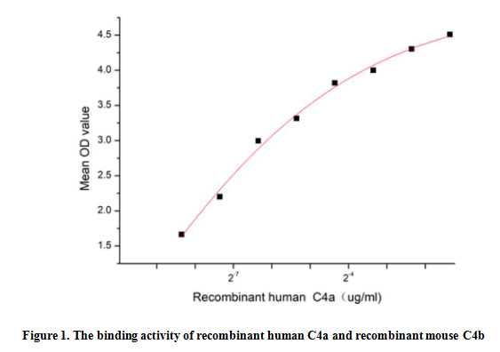
(Complement Component 4a (C4a) is a component of the complement system, which is a cleavage product of the complement C4 protein. C4a has been implicated in various inflammatory and immune responses. It acts as a chemoattractant, recruiting immune cells such as neutrophils and macrophages to the site of inflammation. Additionally, C4a can stimulate the release of pro-inflammatory cytokines and chemokines, further amplifying the immune response. C4a and C4b are the two subunits of C4, thus a functional binding ELISA assay was conducted to detect the interaction of recombinant human C4a and recombinant mouse C4b. Briefly, C4a was diluted serially in PBS with 0.01% BSA (pH 7.4). Duplicate samples of 100 ul were then transferred to C4b-coated microtiter wells and incubated for 1h at 37 degree C. Wells were washed with PBST and incubated for 1h with anti-C4a pAb, then aspirated and washed 3 times. After incubation with HRP labelled secondary antibody for 1h at 37 degree C, wells were aspirated and washed 5 times. With the addition of substrate solution, wells were incubated 15-25 minutes at 37 degree C. Finally, add 50 uL stop solution to the wells and read at 450/630 nm immediately. The binding activity of recombinant human C4a and recombinant mouse C4b was shown in Figure 1,and this effect was in a dose dependent manner.)
Bioactivity
(Complement Component 4a (C4a) is a component of the complement system, which is a cleavage product of the complement C4 protein. C4a has been implicated in various inflammatory and immune responses. It acts as a chemoattractant, recruiting immune cells such as neutrophils and macrophages to the site of inflammation. Additionally, C4a can stimulate the release of pro-inflammatory cytokines and chemokines, further amplifying the immune response. C4a and C4b are the two subunits of C4, thus a functional binding ELISA assay was conducted to detect the interaction of recombinant human C4a and recombinant mouse C4b. Briefly, C4a was diluted serially in PBS with 0.01% BSA (pH 7.4). Duplicate samples of 100 ul were then transferred to C4b-coated microtiter wells and incubated for 1h at 37 degree C. Wells were washed with PBST and incubated for 1h with anti-C4a pAb, then aspirated and washed 3 times. After incubation with HRP labelled secondary antibody for 1h at 37 degree C, wells were aspirated and washed 5 times. With the addition of substrate solution, wells were incubated 15-25 minutes at 37 degree C. Finally, add 50 uL stop solution to the wells and read at 450/630 nm immediately. The binding activity of recombinant human C4a and recombinant mouse C4b was shown in Figure 1,and this effect was in a dose dependent manner.)
Complement Component 4a (C4a), Active Protein (Cat# AAA161728)
Full Name
Active Complement Component 4a (C4a)
Gene Names
C4B; CH; C4F; CO4; C4B1; C4B2; C4B3; C4B5; C4BD; C4B12; C4B_2; CPAMD3
Applications
Activity Assay, Cell Culture
Purity
Greater than 90% by SDS-PAGE
Pricing
 Bioactivity
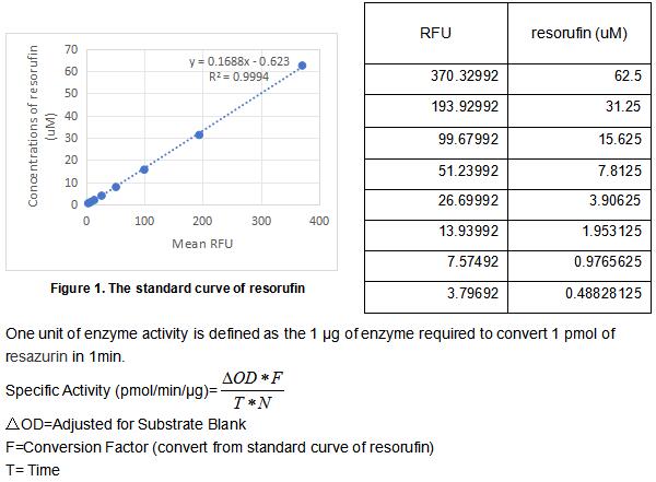
(NAD(P)H:quinone acceptor oxidoreductase 1 (NQO1), also known as DT-diaphorase, is a widely-distributed FAD-dependent flavoprotein that promotes 2-electron reductions of quinones, quinoneimines, nitroaromatics, and azo dyes. As a result it prevents the one electron reduction of quinones that results in the production of radical species. NQO1 is a highly-inducible enzyme that is regulated by the Keap1/Nrf2/ARE pathway. The increase and decrease of NQO1 levels are associated with decreased and increased susceptibilities to oxidative stress, respectively. Thus, NQO1 is a marker cytoprotective enzyme in oxidative stress. Independently of its catalytic function, NQO1 plays a role in regulating the proteosomal degradation of p53, p73a, and p33. NQO1 physically interacts with p53 and p73 in an NADH-dependent manner and protects them from 20S proteasomal degradation in a ubiquitin independent pathway. The activity assay of recombinant rat NQO1 was measured by its ability to oxidize the substrate resazurin to resorufin. The rrNQO1 was diluted to 0.05 ug/ml in the assay buffer 50 mM HEPES, 0.2 M NaCl, 5 uM FAD, 0.05% Tween 20, pH 7.5. 50 ul 0.05 ug/ml rhNQO1 was added into the microplate and start the reaction by adding 50 ul substrate mixture of 400 uM beta-NADH and 20 uM resazurin which was diluted in assay buffer. Read at excitation and emission wavelengths of 540 nm and 585 nm (top read), respectively, in kinetic mode for 5 minutes. The specific activity of recombinant rat NQO1 is >45000 pmol/min/ug.)
Bioactivity
(NAD(P)H:quinone acceptor oxidoreductase 1 (NQO1), also known as DT-diaphorase, is a widely-distributed FAD-dependent flavoprotein that promotes 2-electron reductions of quinones, quinoneimines, nitroaromatics, and azo dyes. As a result it prevents the one electron reduction of quinones that results in the production of radical species. NQO1 is a highly-inducible enzyme that is regulated by the Keap1/Nrf2/ARE pathway. The increase and decrease of NQO1 levels are associated with decreased and increased susceptibilities to oxidative stress, respectively. Thus, NQO1 is a marker cytoprotective enzyme in oxidative stress. Independently of its catalytic function, NQO1 plays a role in regulating the proteosomal degradation of p53, p73a, and p33. NQO1 physically interacts with p53 and p73 in an NADH-dependent manner and protects them from 20S proteasomal degradation in a ubiquitin independent pathway. The activity assay of recombinant rat NQO1 was measured by its ability to oxidize the substrate resazurin to resorufin. The rrNQO1 was diluted to 0.05 ug/ml in the assay buffer 50 mM HEPES, 0.2 M NaCl, 5 uM FAD, 0.05% Tween 20, pH 7.5. 50 ul 0.05 ug/ml rhNQO1 was added into the microplate and start the reaction by adding 50 ul substrate mixture of 400 uM beta-NADH and 20 uM resazurin which was diluted in assay buffer. Read at excitation and emission wavelengths of 540 nm and 585 nm (top read), respectively, in kinetic mode for 5 minutes. The specific activity of recombinant rat NQO1 is >45000 pmol/min/ug.)
NADH Dehydrogenase, Quinone 1 (NQO1), Active Protein (Cat# AAA161939)
Full Name
Active NADH Dehydrogenase, Quinone 1 (NQO1)
Gene Names
Nqo1; Dia4
Applications
Activity Assay, Cell Culture
Purity
Greater than 90% by SDS-PAGE
Pricing
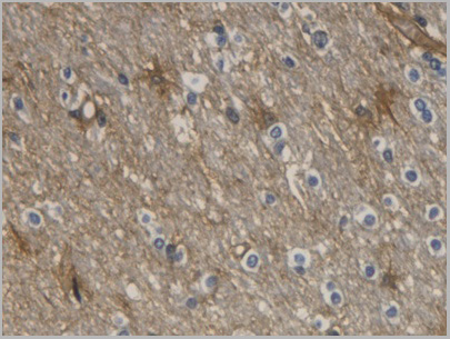
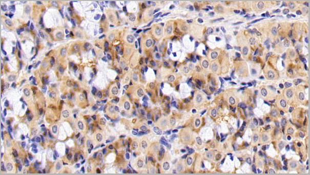 IHC (Immunohistchemistry)
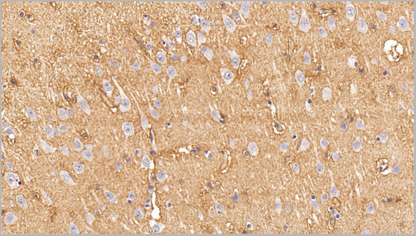
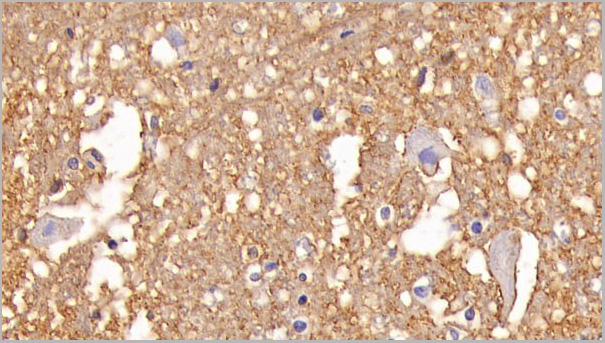
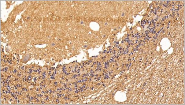
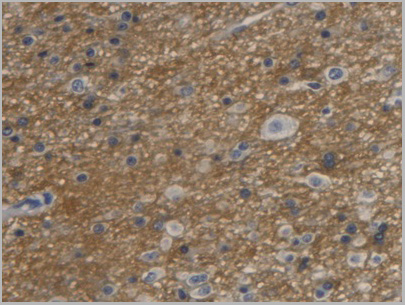
(DAB staining on IHCP;Sample: Porcine Stomach Tissue; Primary Ab: 30ug/ml Mouse AntiHuman AQP4 AntibodySecond Ab: 2ug/mL HRPLinked Caprine AntiMouse IgG Polyclonal Antibody(Catalog: SAA544Mu19))
IHC (Immunohistchemistry)
(DAB staining on IHCP;Sample: Porcine Stomach Tissue; Primary Ab: 30ug/ml Mouse AntiHuman AQP4 AntibodySecond Ab: 2ug/mL HRPLinked Caprine AntiMouse IgG Polyclonal Antibody(Catalog: SAA544Mu19))
Aquaporin 4 (AQP4), Monoclonal Antibody (Cat# AAA21184)
Full Name
Monoclonal Antibody to Aquaporin 4 (AQP4)
Gene Names
AQP4; MIWC; HMIWC2
Reactivity
Human, Pig
Applications
Western Blot, Immunohistochemistry, Immunocytochemistry, Immunoprecipitation
Purity
Protein A + Protein G affinity chromatography
Pricing
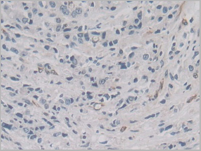
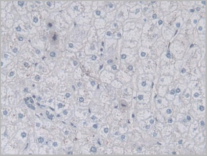
 IHC (Immunohistchemistry)
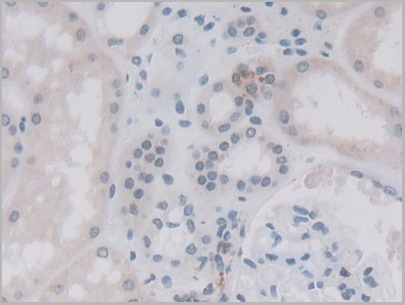
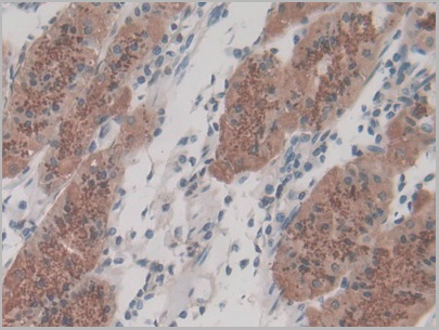
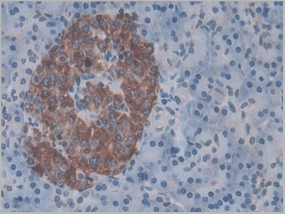
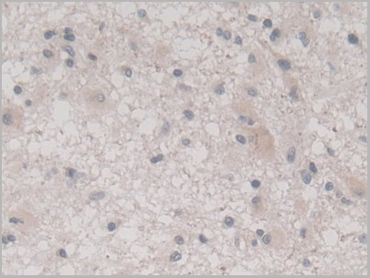
(DAB staining on IHCP;Sample: Human Glioma Tissue; Primary Ab: 30ug/ml Mouse AntiHuman VEGF121 AntibodySecond Ab: 2ug/mL HRPLinked Caprine AntiMouse IgG Polyclonal Antibody(Catalog: SAA544Mu19))
IHC (Immunohistchemistry)
(DAB staining on IHCP;Sample: Human Glioma Tissue; Primary Ab: 30ug/ml Mouse AntiHuman VEGF121 AntibodySecond Ab: 2ug/mL HRPLinked Caprine AntiMouse IgG Polyclonal Antibody(Catalog: SAA544Mu19))
Vascular Endothelial Growth Factor 121 (VEGF121), Monoclonal Antibody (Cat# AAA21199)
Full Name
Monoclonal Antibody to Vascular Endothelial Growth Factor 121 (VEGF121)
Gene Names
VEGFA; VPF; VEGF; MVCD1
Reactivity
Human
Applications
Western Blot, Immunohistochemistry, Immunocytochemistry, Immunoprecipitation
Purity
Protein A + Protein G affinity chromatography
Pricing
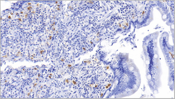
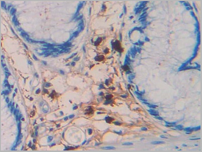
 WB (Western Blot)
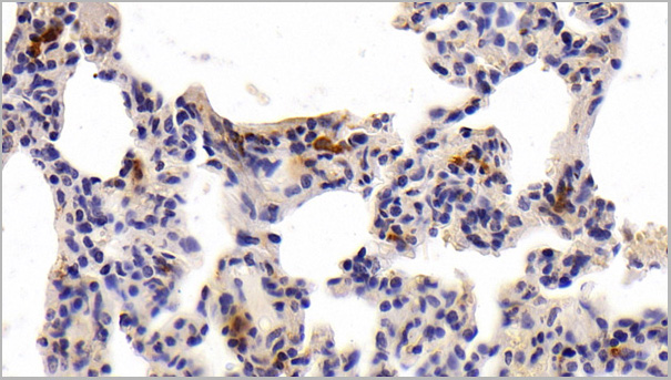
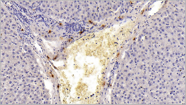
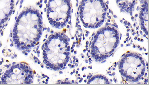
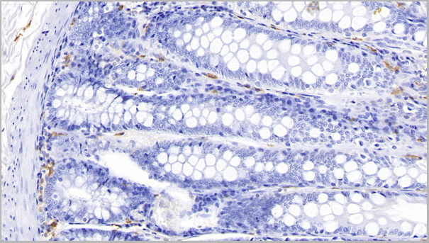
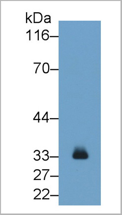
(Western Blot; Sample: Porcine Skin lysate Primary Ab: 0.2ug/ml Mouse AntiHuman TPS Antibody Second Ab: 0.2ug/mL HRPLinked Caprine AntiMouse IgG Polyclonal Antibody (Catalog: SAA544Mu19))
WB (Western Blot)
(Western Blot; Sample: Porcine Skin lysate Primary Ab: 0.2ug/ml Mouse AntiHuman TPS Antibody Second Ab: 0.2ug/mL HRPLinked Caprine AntiMouse IgG Polyclonal Antibody (Catalog: SAA544Mu19))
Tryptase (TPS), Monoclonal Antibody (Cat# AAA21190)
Full Name
Monoclonal Antibody to Tryptase (TPS)
Gene Names
TPSAB1; TPS1; TPS2; TPSB1
Reactivity
Human, Dog, Pig
Applications
Western Blot, Immunohistochemistry, Immunocytochemistry, Immunoprecipitation
Purity
Protein A + Protein G affinity chromatography
Pricing
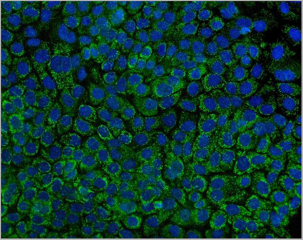
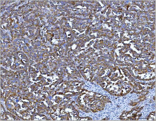
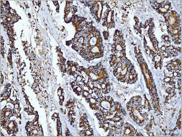
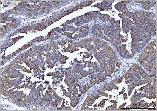
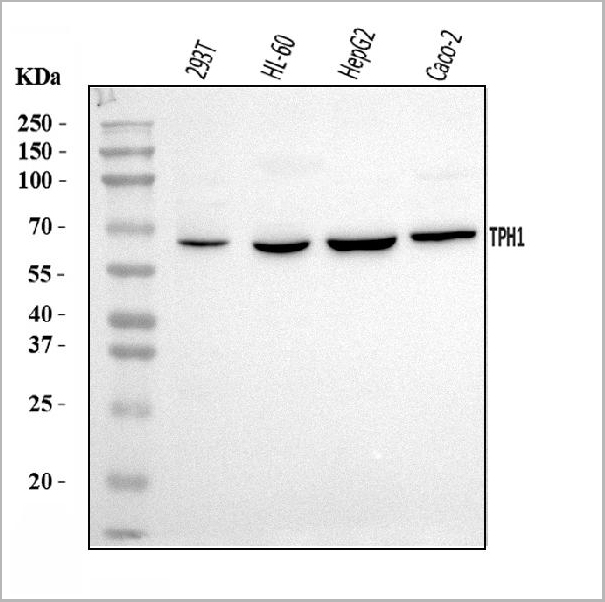
 FCM (Flow Cytometry)
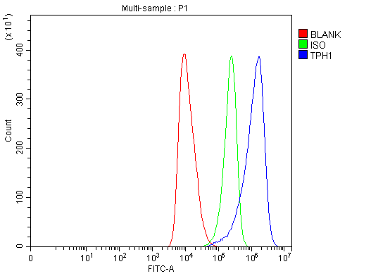
(Figure 6. Flow Cytometry analysis of HEL cells using anti-Tryptophan Hydroxylase/TPH1 antibody (AAA19426).Overlay histogram showing HEL cells stained with AAA19426 (Blue line). The cells were blocked with 10% normal goat serum. And then incubated with rabbit anti-Tryptophan Hydroxylase/TPH1 Antibody (AAA19426, 1 ug/1x10^6 cells) for 30 min at 20 degree C. DyLight488 conjugated goat anti-rabbit IgG was used as secondary antibody for 30 minutes at 20 degree C. Isotype control antibody (Green line) was rabbit IgG (1 ug/1x10^6) used under the same conditions. Unlabelled sample (Red line) was also used as a control.)
FCM (Flow Cytometry)
(Figure 6. Flow Cytometry analysis of HEL cells using anti-Tryptophan Hydroxylase/TPH1 antibody (AAA19426).Overlay histogram showing HEL cells stained with AAA19426 (Blue line). The cells were blocked with 10% normal goat serum. And then incubated with rabbit anti-Tryptophan Hydroxylase/TPH1 Antibody (AAA19426, 1 ug/1x10^6 cells) for 30 min at 20 degree C. DyLight488 conjugated goat anti-rabbit IgG was used as secondary antibody for 30 minutes at 20 degree C. Isotype control antibody (Green line) was rabbit IgG (1 ug/1x10^6) used under the same conditions. Unlabelled sample (Red line) was also used as a control.)
Tryptophan Hydroxylase/TPH1, Polyclonal Antibody (Cat# AAA19426)
Full Name
Anti-Tryptophan Hydroxylase/TPH1 Antibody Picoband
Gene Names
TPH1; TPRH; TRPH
Reactivity
Human
Applications
Flow Cytometry, Immunofluorescence, Immunohistochemistry, Immunocytochemistry, Western Blot
Purity
Immunogen affinity purified.
Pricing
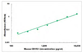 Standard Curve (Sample)
CD152, ELISA Kit (Cat# AAA105974)
Full Name
Mouse CD152 ELISA Kit
Gene Names
Ctla4; Cd152; Ly-56; Ctla-4
Reactivity
Mouse
Applications
Sandwich ELISA
Pricing
 SDS-PAGE
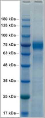
(Reduced SDS-PAGE: purified HA (aa 18-518)(A/Puerto Rico/8/1934)(H1N1) protein)
SDS-PAGE
(Reduced SDS-PAGE: purified HA (aa 18-518)(A/Puerto Rico/8/1934)(H1N1) protein)
HA, Recombinant Protein (Cat# AAA13698)
Full Name
HA (A/Puerto Rico/8/1934)(H1N1)(aa 18-518) Trimer
Purity
>95% pure by 4-20% SDS-PAGE gel
Pricing
 IF (Immunofluorescence)
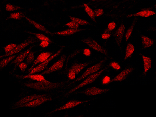
(Immunofluorescence staining of S100P in Hela cells. Cells were fixed with 4% PFA, permeabilzed with 0.1% Triton X-100 in PBS,blocked with 10% serum, and incubated with rabbit anti-Y S100P monoclonal antibody (dilution ratio 1:60) at 4 degree C overnight. Then cells were stained with the Alexa Fluor594-conjugated Goat Anti-rabbit IgG secondary antibody (red). Positive staining was localized to Nucleus and Cytoplasm.)
IF (Immunofluorescence)
(Immunofluorescence staining of S100P in Hela cells. Cells were fixed with 4% PFA, permeabilzed with 0.1% Triton X-100 in PBS,blocked with 10% serum, and incubated with rabbit anti-Y S100P monoclonal antibody (dilution ratio 1:60) at 4 degree C overnight. Then cells were stained with the Alexa Fluor594-conjugated Goat Anti-rabbit IgG secondary antibody (red). Positive staining was localized to Nucleus and Cytoplasm.)
S100P, Monoclonal Antibody (Cat# AAA255599)
Full Name
Recombinant Anti-S100P Antibody, Rabbit Monoclonal
Gene Names
S100P; MIG9
Reactivity
Human
Applications
Immunofluorescence, Immunocytochemistry, Immunohistochemistry
Purity
Protein A
Pricing
 Application Data
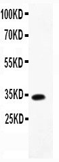
(Anti-TARC Picoband antibody, AAA45702-1.jpgAll lanes: Anti-TARC(AAA45702) at 0.5ug/mlWB: Recombinant Mouse TARC Protein 0.5ngPredicted bind size: 34KDObserved bind size: 34KD)
Application Data
(Anti-TARC Picoband antibody, AAA45702-1.jpgAll lanes: Anti-TARC(AAA45702) at 0.5ug/mlWB: Recombinant Mouse TARC Protein 0.5ngPredicted bind size: 34KDObserved bind size: 34KD)
TARC, Polyclonal Antibody (Cat# AAA45702)
Full Name
Anti-TARC Antibody
Gene Names
Ccl17; Tarc; Abcd-2; Scya17; Scya17l
Reactivity
Mouse
Applications
Western Blot
Purity
Immunogen affinity purified.
Pricing
 IF (Immunofluorescence)
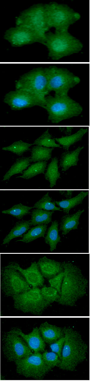
(ICC/IF analysis of IL-6 in A549 cells line, stained with DAPI (Blue) for nucleus staining and monoclonal anti-human IL-6 antibody (1:100) with goat anti-mouse IgG-Alexa fluor 488 conjugate (Green) ICC/IF analysis of IL-6 in HeLa cells line, stained with DAPI (Blue) for nucleus staining and monoclonal anti-human IL-6 antibody (1:100) with goat anti-mouse IgG-Alexa fluor 488 conjugate (Green) ICC/IF analysis of IL-6 in Hep3B cells line, stained with DAPI (Blue) for nucleus staining and monoclonal anti-human IL-6 antibody (1:100) with goat anti-mouse IgG-Alexa fluor 488 conjugate (Green))
IF (Immunofluorescence)
(ICC/IF analysis of IL-6 in A549 cells line, stained with DAPI (Blue) for nucleus staining and monoclonal anti-human IL-6 antibody (1:100) with goat anti-mouse IgG-Alexa fluor 488 conjugate (Green) ICC/IF analysis of IL-6 in HeLa cells line, stained with DAPI (Blue) for nucleus staining and monoclonal anti-human IL-6 antibody (1:100) with goat anti-mouse IgG-Alexa fluor 488 conjugate (Green) ICC/IF analysis of IL-6 in Hep3B cells line, stained with DAPI (Blue) for nucleus staining and monoclonal anti-human IL-6 antibody (1:100) with goat anti-mouse IgG-Alexa fluor 488 conjugate (Green))
IL-6, Monoclonal Antibody (Cat# AAA48239)
Full Name
IL-6 antibody
Gene Names
IL6; CDF; HGF; HSF; BSF2; IL-6; BSF-2; IFNB2; IFN-beta-2
Reactivity
Human
Applications
Immunofluorescence, Immunocytochemistry, Flow Cytometry, Western Blot
Purity
By protein-G affinity chromatography
Pricing