
Filters
Clonality
Type
Reactivity
Gene Name
Isotype
Host
Application
Clone
10000 results for "Recombinant Proteins" - showing 3050-3100
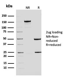
 Application Data
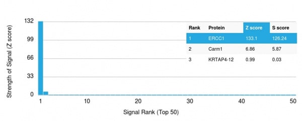
(Analysis of Protein Array containing more than 19,000 full-length human proteins using ERCC1 Mouse Monoclonal Antibody (ERCC1/2683). Z- and S- Score: The Z-score represents the strength of a signal that a monoclonal antibody (Monoclonal Antibody) (in combination with a fluorescently-tagged anti-IgG secondary antibody) produces when binding to a particular protein on the HuProtTM array. Z-scores are described in units of standard deviations (SD's) above the mean value of all signals generated on that array. If targets on HuProtTM are arranged in descending order of the Z-score, the S-score is the difference (also in units of SD's) between the Z-score. S-score therefore represents the relative target specificity of a Monoclonal Antibody to its intended target. A Monoclonal Antibody is considered to specific to its intended target, if the Monoclonal Antibody has an S-score of at least 2.5. For example, if a Monoclonal Antibody binds to protein X with a Z-score of 43 and to protein Y with a Z-score of 14, then the S-score for the binding of that Monoclonal Antibody to protein X is equal to 29.)
Application Data
(Analysis of Protein Array containing more than 19,000 full-length human proteins using ERCC1 Mouse Monoclonal Antibody (ERCC1/2683). Z- and S- Score: The Z-score represents the strength of a signal that a monoclonal antibody (Monoclonal Antibody) (in combination with a fluorescently-tagged anti-IgG secondary antibody) produces when binding to a particular protein on the HuProtTM array. Z-scores are described in units of standard deviations (SD's) above the mean value of all signals generated on that array. If targets on HuProtTM are arranged in descending order of the Z-score, the S-score is the difference (also in units of SD's) between the Z-score. S-score therefore represents the relative target specificity of a Monoclonal Antibody to its intended target. A Monoclonal Antibody is considered to specific to its intended target, if the Monoclonal Antibody has an S-score of at least 2.5. For example, if a Monoclonal Antibody binds to protein X with a Z-score of 43 and to protein Y with a Z-score of 14, then the S-score for the binding of that Monoclonal Antibody to protein X is equal to 29.)
ERCC1/RAD10, Monoclonal Antibody (Cat# AAA214950)
Full Name
ERCC1/RAD10 (Tumor Progression Marker)
Gene Names
ERCC1; UV20; COFS4; RAD10
Reactivity
Human
Applications
Western Blot
Purity
Purified Ab with BSA and Azide at 200ug/ml OR Purified Ab WITHOUT BSA and Azide at 1.0mg/ml
Pricing
 WB (Western Blot)
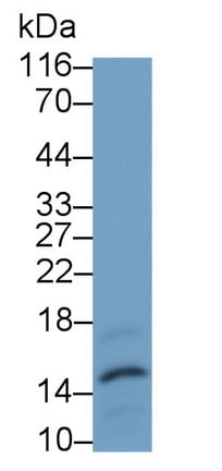
(WB Image)
WB (Western Blot)
(WB Image)
Procalcitonin (PCT), Monoclonal Antibody (Cat# AAA151602)
Full Name
Monoclonal Antibody to Procalcitonin (PCT)
Gene Names
CALCA; CT; KC; CGRP; CALC1; CGRP1; CGRP-I
Reactivity
Human
Applications
Immunoprecipitation, Immunocytochemistry, Immunohistochemistry, Western Blot
Purity
Protein A + Protein G affinity chromatography
Pricing

 Application Data
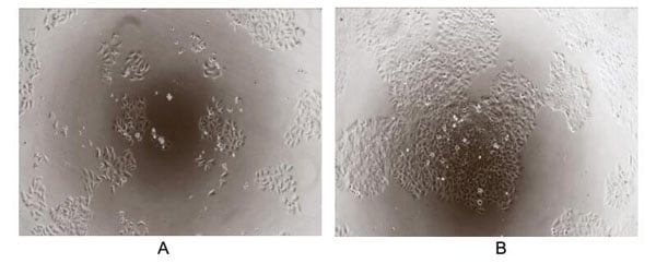
(Transforming growth factor beta (TGF-) is a multifunctional cytokine belonging to the transforming growth factor superfamily. The TGF- superfamily includes endogenous growth inhibiting proteins; an increase in expression of TGF- often correlates with the malignancy of many cancers and a defect in the cellular growth inhibition response to TGF-. Its immunosuppressive functions then come to dominate, contributing to oncogenesis. To test the effect of TGF- on inhibit HGF-dependent proliferation, HepG2 cells were seeded into triplicate wells of 96-well plates at a density of 2,000 cells/well and allowed to attach, replaced with serum-free overnight, then the medium was replaced with 2% serum standard DMEM including 1ng/mL HGF prior to the addition of various concentrations of recombinant human TGF-. After incubated for 96h, cells were observed by inverted microscope and cell proliferation was measured by Cell Counting Kit-8 (CCK-8). Briefly, 10uL of CCK-8 solution was added to each well of the plate, then the absorbance at 450nm was measured using a microplate reader after incubating the plate for 1-4 hours at 37. The inhibitory effect of TGF- on HGF-dependent proliferation of HepG2 cells observed by inverted microscope was shown in Figure 2. Cell viability was assessed by CCK-8 assay after incubation with recombinant TGF- for 96h. The result was shown in Figure 3. It was obvious that TGF- significantly decreased cell viability of HepG2 cells. (A) HepG2 cells cultured in DMEM, stimulated with 1ug/mL TGF- for 96h; (B) Unstimulated HepG2 cells cultured in DMEM for 96h.Figure.The inhibitory effect of TGF- on cell proliferation of HepG2 cells.)
Application Data
(Transforming growth factor beta (TGF-) is a multifunctional cytokine belonging to the transforming growth factor superfamily. The TGF- superfamily includes endogenous growth inhibiting proteins; an increase in expression of TGF- often correlates with the malignancy of many cancers and a defect in the cellular growth inhibition response to TGF-. Its immunosuppressive functions then come to dominate, contributing to oncogenesis. To test the effect of TGF- on inhibit HGF-dependent proliferation, HepG2 cells were seeded into triplicate wells of 96-well plates at a density of 2,000 cells/well and allowed to attach, replaced with serum-free overnight, then the medium was replaced with 2% serum standard DMEM including 1ng/mL HGF prior to the addition of various concentrations of recombinant human TGF-. After incubated for 96h, cells were observed by inverted microscope and cell proliferation was measured by Cell Counting Kit-8 (CCK-8). Briefly, 10uL of CCK-8 solution was added to each well of the plate, then the absorbance at 450nm was measured using a microplate reader after incubating the plate for 1-4 hours at 37. The inhibitory effect of TGF- on HGF-dependent proliferation of HepG2 cells observed by inverted microscope was shown in Figure 2. Cell viability was assessed by CCK-8 assay after incubation with recombinant TGF- for 96h. The result was shown in Figure 3. It was obvious that TGF- significantly decreased cell viability of HepG2 cells. (A) HepG2 cells cultured in DMEM, stimulated with 1ug/mL TGF- for 96h; (B) Unstimulated HepG2 cells cultured in DMEM for 96h.Figure.The inhibitory effect of TGF- on cell proliferation of HepG2 cells.)
Transforming Growth Factor Beta 2, Active Protein (Cat# AAA150084)
Full Name
Active Transforming Growth Factor Beta 2 (TGFb2)
Gene Names
TGFB2; LDS4; G-TSF; TGF-beta2
Applications
Activity Assay, Cell Culture
Purity
>90%
Pricing
 IF (Immunofluorescence)
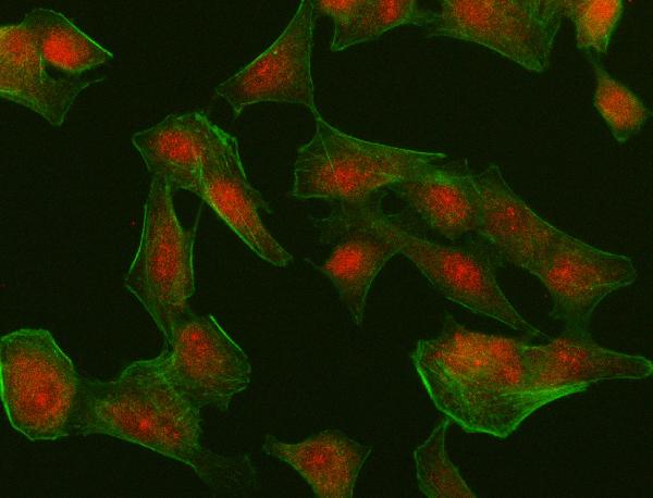
(Figure 2. IF analysis of IRF4 using anti-IRF4 antibody (AAA126988).IRF4 was detected in an immunocytochemical section of A549 cells. Enzyme antigen retrieval was performed using IHC enzyme antigen retrieval reagent for 15 mins. The cells were blocked with 10% goat serum. And then incubated with 5ug/mL rabbit anti-IRF4 Antibody (AAA126988) overnight at 4 degree C. Cy3 Conjugated Goat Anti-Rabbit IgG (BA1032) was used as secondary antibody at 1:100 dilution and incubated for 30 minutes at 37 degree C. The tissue section was developed using Phalloidin-iFluor 488 Conjugated. Visualize using a fluorescence microscope and filter sets appropriate for the label used.)
IF (Immunofluorescence)
(Figure 2. IF analysis of IRF4 using anti-IRF4 antibody (AAA126988).IRF4 was detected in an immunocytochemical section of A549 cells. Enzyme antigen retrieval was performed using IHC enzyme antigen retrieval reagent for 15 mins. The cells were blocked with 10% goat serum. And then incubated with 5ug/mL rabbit anti-IRF4 Antibody (AAA126988) overnight at 4 degree C. Cy3 Conjugated Goat Anti-Rabbit IgG (BA1032) was used as secondary antibody at 1:100 dilution and incubated for 30 minutes at 37 degree C. The tissue section was developed using Phalloidin-iFluor 488 Conjugated. Visualize using a fluorescence microscope and filter sets appropriate for the label used.)
MUM1/IRF4, Polyclonal Antibody (Cat# AAA126988)
Full Name
Anti-MUM1/IRF4 Antibody Picoband
Gene Names
IRF4; MUM1; LSIRF; NF-EM5
Reactivity
Human
Applications
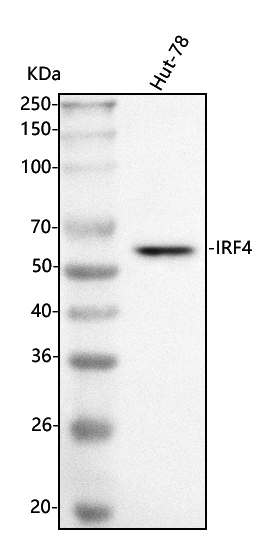
Immunofluorescence, Immunocytochemistry, Western Blot
Purity
Immunogen affinity purified.
Pricing
 Bioactivity
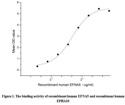
(Ephrin-A5, also known as EFNA5, is a member of the Ephrin family. EFNA5 is localized in cell membranes and was highly expressed in skin, brain, spleen, kidney, heart and fat. EFNA5 can bind to its receptors to play important physiological functions, such as participating in neurogenesis, angiogenesis, promoting cell proliferation and differentiation, balancing glucose homeostasis, maintaining lens development, regulating female mammalian reproduction, and mobilizing fat thermogenesis. EPHA10 is a receptor for members of the ephrin-A family. It binds to EFNA3, EFNA4 and EFNA5. Thus a functional binding ELISA assay was conducted to detect the interaction of recombinant human EFNA5 and recombinant human EPHA10. Briefly, EFNA5 was diluted serially in PBS with 0.01% BSA (pH7.4). Duplicate samples of 100 ul were then transferred to EPHA10-coated microtiter wells and incubated for 1h at 37 degree C. Wells were washed with PBST and incubated for 1h with anti-EFNA5 pAb, then aspirated and washed 3 times. After incubation with HRP labelled secondary antibody for 1h at 37 degree C, wells were aspirated and washed 5 times. With the addition of substrate solution, wells were incubated 15-25 minutes at 37 degree C. Finally, add 50 uL stop solution to the wells and read at 450/630 nm immediately. The binding activity of recombinant human EFNA5 and recombinant human EPHA10 was shown in Figure 1, the EC50 for this effect is 0.03 ug/mL.)
Bioactivity
(Ephrin-A5, also known as EFNA5, is a member of the Ephrin family. EFNA5 is localized in cell membranes and was highly expressed in skin, brain, spleen, kidney, heart and fat. EFNA5 can bind to its receptors to play important physiological functions, such as participating in neurogenesis, angiogenesis, promoting cell proliferation and differentiation, balancing glucose homeostasis, maintaining lens development, regulating female mammalian reproduction, and mobilizing fat thermogenesis. EPHA10 is a receptor for members of the ephrin-A family. It binds to EFNA3, EFNA4 and EFNA5. Thus a functional binding ELISA assay was conducted to detect the interaction of recombinant human EFNA5 and recombinant human EPHA10. Briefly, EFNA5 was diluted serially in PBS with 0.01% BSA (pH7.4). Duplicate samples of 100 ul were then transferred to EPHA10-coated microtiter wells and incubated for 1h at 37 degree C. Wells were washed with PBST and incubated for 1h with anti-EFNA5 pAb, then aspirated and washed 3 times. After incubation with HRP labelled secondary antibody for 1h at 37 degree C, wells were aspirated and washed 5 times. With the addition of substrate solution, wells were incubated 15-25 minutes at 37 degree C. Finally, add 50 uL stop solution to the wells and read at 450/630 nm immediately. The binding activity of recombinant human EFNA5 and recombinant human EPHA10 was shown in Figure 1, the EC50 for this effect is 0.03 ug/mL.)
Ephrin A5 (EFNA5), Active Protein (Cat# AAA161919)
Full Name
Active Ephrin A5 (EFNA5)
Gene Names
EFNA5; AF1; EFL5; RAGS; EPLG7; GLC1M; LERK7
Applications
Activity Assay, Cell Culture
Purity
Greater than 90% by SDS-PAGE
Pricing

 Bioactivity
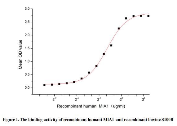
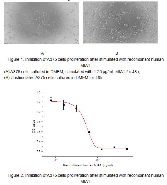
(To test the effect of MIA1 on cell apoptosis, A375 cells were seeded into triplicate wells of 96-well plates at a density of 4,000 cells/well and allowed to attach overnight, then the medium was replaced with various concentrations of recombinant human MIA1 diluted with 5% serum standard DMEM. After incubated for 48h, cells were observed by inverted microscope and cell viability was measured by Cell Counting Kit-8 (CCK-8). Briefly, 10 ul of CCK-8 solution was added to each well of the plate, then the absorbance at 450 nm was measured using a microplate reader after incubating the plate for 1-4 hours at 37 degree C. Apoptosis of A375 cells after incubation with MIA1 for 48h observed by inverted microscope was shown in Figure 1. Cell viability was assessed by CCK-8 assay after incubation with recombinant human MIA1 for 48h. The result was shown in Figure 2. It was obvious that MIA1 significantly decreased cell viability of A375 cells. The ED50 of recombinant human MIA1 is 0.53 ug/ml.)
Bioactivity
(To test the effect of MIA1 on cell apoptosis, A375 cells were seeded into triplicate wells of 96-well plates at a density of 4,000 cells/well and allowed to attach overnight, then the medium was replaced with various concentrations of recombinant human MIA1 diluted with 5% serum standard DMEM. After incubated for 48h, cells were observed by inverted microscope and cell viability was measured by Cell Counting Kit-8 (CCK-8). Briefly, 10 ul of CCK-8 solution was added to each well of the plate, then the absorbance at 450 nm was measured using a microplate reader after incubating the plate for 1-4 hours at 37 degree C. Apoptosis of A375 cells after incubation with MIA1 for 48h observed by inverted microscope was shown in Figure 1. Cell viability was assessed by CCK-8 assay after incubation with recombinant human MIA1 for 48h. The result was shown in Figure 2. It was obvious that MIA1 significantly decreased cell viability of A375 cells. The ED50 of recombinant human MIA1 is 0.53 ug/ml.)
Melanoma Inhibitory Activity Protein 1 (MIA1), Active Protein (Cat# AAA161926)
Full Name
Active Melanoma Inhibitory Activity Protein 1 (MIA1)
Gene Names
MIA; CD-RAP
Applications
Activity Assay, Cell Culture
Purity
Greater than 80% by SDS-PAGE
Pricing
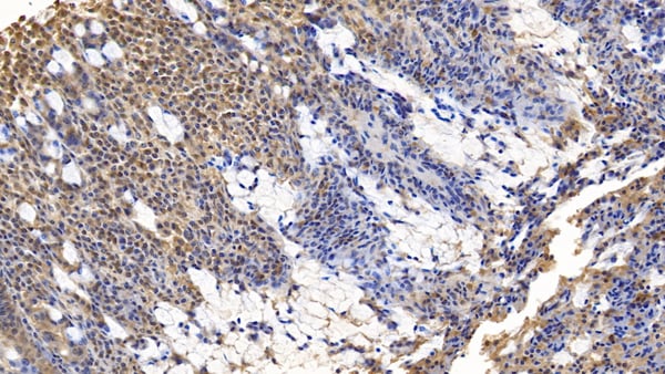 IHC (Immunohiostchemistry)
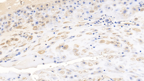
(DAB staining on IHCP;Sample: Human Small intestine Tissue; Primary Ab: 10ug/ml Mouse AntiHuman WNT3A AntibodySecond Ab: 2ug/mL HRPLinked Caprine AntiMouse IgG Polyclonal Antibody(Catalog: SAA544Mu19))
IHC (Immunohiostchemistry)
(DAB staining on IHCP;Sample: Human Small intestine Tissue; Primary Ab: 10ug/ml Mouse AntiHuman WNT3A AntibodySecond Ab: 2ug/mL HRPLinked Caprine AntiMouse IgG Polyclonal Antibody(Catalog: SAA544Mu19))
Wingless Type MMTV Integration Site Family, Member 3A (WNT3A), Monoclonal Antibody (Cat# AAA151910)
Full Name
Monoclonal Antibody to Wingless Type MMTV Integration Site Family, Member 3A (WNT3A)
Reactivity
Human, Pig
Applications
Immunoprecipitation, Immunocytochemistry, Immunohistochemistry, Western Blot
Purity
Protein A + Protein G affinity chromatography
Pricing
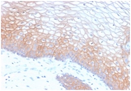
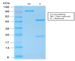 SDS-PAGE
(SDS-PAGE Analysis of Purified E-Cadherin Rabbit Recombinant Monoclonal Antibody (CDH1/2208R).)
SDS-PAGE
(SDS-PAGE Analysis of Purified E-Cadherin Rabbit Recombinant Monoclonal Antibody (CDH1/2208R).)
E-Cadherin (CDH1)/CD324, Monoclonal Antibody (Cat# AAA214625)
Full Name
E-Cadherin (CDH1)/CD324 (Intercellular Junction Marker)
Gene Names
CDH1; UVO; CDHE; ECAD; LCAM; Arc-1; BCDS1; CD324
Reactivity
Human.
Does not react with Mouse and Rat. Others not known.
Does not react with Mouse and Rat. Others not known.
Applications
Immunohistochemistry, Western Blot, Immunofluorescence, Flow Cytometry
Pricing
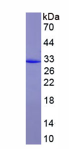
 Bioactivity
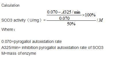
(Extracellular superoxide dismutase [Cu-Zn] is an enzyme that in humans is encoded by the SOD3 gene. This gene encodes a member of the superoxide dismutase (SOD) protein family. SODs are antioxidant enzymes that catalyze the dismutation of two superoxide radicals into hydrogen peroxide and oxygen. Acroding to the report, in a weakly alkaline buffer solution (pH=8.2) with N-tris(hydroxymethyl)amino methane-HCL, pyrogallol can occur autoxidation in the air, then SOD can inhibit this reaction. Thus, we use this way to measued the activity of recombinant human SOD3. The reaction was performed in adding 8 ul 5 mmol/L pyrogallol to 200 ul 50mmol/L Tris-HCl, rapidly mixing at 25 degree C, then read at 325 nm (using 50mmol/L Tris-HCl as blank control) in kinetic mode for 3 minutes using a microplate reader controlling the pyrogallol autoxidation rate at 0.70 OD/min. Different concentrations of recombinant human SOD3 were added into 200 ul 50 mmol/L Tris-HCl, incubated for 20 min at 25 degree C, then adding 8 ul 5 mmol/L pyrogallol to each well, rapidly mixing and read at 325 nm in kinetic mode for 3 minutes. Under these conditions, the enzyme amount of 50% inhibition of pyrogallol autooxidation per minute is defined as a unit. The specific activity of recombinant human SOD3 is 846.6 U/mg.)
Bioactivity
(Extracellular superoxide dismutase [Cu-Zn] is an enzyme that in humans is encoded by the SOD3 gene. This gene encodes a member of the superoxide dismutase (SOD) protein family. SODs are antioxidant enzymes that catalyze the dismutation of two superoxide radicals into hydrogen peroxide and oxygen. Acroding to the report, in a weakly alkaline buffer solution (pH=8.2) with N-tris(hydroxymethyl)amino methane-HCL, pyrogallol can occur autoxidation in the air, then SOD can inhibit this reaction. Thus, we use this way to measued the activity of recombinant human SOD3. The reaction was performed in adding 8 ul 5 mmol/L pyrogallol to 200 ul 50mmol/L Tris-HCl, rapidly mixing at 25 degree C, then read at 325 nm (using 50mmol/L Tris-HCl as blank control) in kinetic mode for 3 minutes using a microplate reader controlling the pyrogallol autoxidation rate at 0.70 OD/min. Different concentrations of recombinant human SOD3 were added into 200 ul 50 mmol/L Tris-HCl, incubated for 20 min at 25 degree C, then adding 8 ul 5 mmol/L pyrogallol to each well, rapidly mixing and read at 325 nm in kinetic mode for 3 minutes. Under these conditions, the enzyme amount of 50% inhibition of pyrogallol autooxidation per minute is defined as a unit. The specific activity of recombinant human SOD3 is 846.6 U/mg.)
Superoxide Dismutase 3, Extracellular (SOD3), Active Protein (Cat# AAA161691)
Full Name
Active Superoxide Dismutase 3, Extracellular (SOD3)
Gene Names
SOD3; EC-SOD
Applications
Activity Assay, Cell Culture
Purity
Greater than 90% by SDS-PAGE
Pricing
 Bioactivity
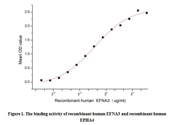
(Ephrin A3 (EFNA3), also known as EFL2, EPLG3, LERK3, Ehk1-L, is a member of the Eph/ephrin tyrosine kinase family. EFNA3 is localized in cell membranes and was highly expressed in brain, skin, esophagus, skeletal muscle, spleen, thymus, prostate, testis, ovary, small intestine, peripheral blood leukocytes. EFNA3, like most genes in the ephrin family, plays a central role in embryonic development can associate with multiple signaling pathways involved in cell growth and tumor cell metastasis. Aberrant regulation of EFNA3 is associated with the occurrence and development of various types of cancer. Ephrin Type A Receptor 4 (EPHA4) is a receptor for members of the ephrin-A family. It can bind to EFNA3. Thus a functional binding ELISA assay was conducted to detect the interaction of recombinant human EFNA3 and recombinant human EPHA4. Briefly, EFNA3 was diluted serially in PBS with 0.01% BSA (pH 7.4). Duplicate samples of 100 ul were then transferred to EPHA4-coated microtiter wells and incubated for 1h at 37 degree C. Wells were washed with PBST and incubated for 1h with anti-EFNA3 pAb, then aspirated and washed 3 times. After incubation with HRP labelled secondary antibody for 1h at 37 degree C, wells were aspirated and washed 5 times. With the addition of substrate solution, wells were incubated 15-25 minutes at 37 degree C. Finally, add 50 uL stop solution to the wells and read at 450/630 nm immediately. The binding activity of recombinant human EFNA3 and recombinant human EPHA4 was shown in Figure 1, the EC50 for this effect is 0.4 ug/mL.)
Bioactivity
(Ephrin A3 (EFNA3), also known as EFL2, EPLG3, LERK3, Ehk1-L, is a member of the Eph/ephrin tyrosine kinase family. EFNA3 is localized in cell membranes and was highly expressed in brain, skin, esophagus, skeletal muscle, spleen, thymus, prostate, testis, ovary, small intestine, peripheral blood leukocytes. EFNA3, like most genes in the ephrin family, plays a central role in embryonic development can associate with multiple signaling pathways involved in cell growth and tumor cell metastasis. Aberrant regulation of EFNA3 is associated with the occurrence and development of various types of cancer. Ephrin Type A Receptor 4 (EPHA4) is a receptor for members of the ephrin-A family. It can bind to EFNA3. Thus a functional binding ELISA assay was conducted to detect the interaction of recombinant human EFNA3 and recombinant human EPHA4. Briefly, EFNA3 was diluted serially in PBS with 0.01% BSA (pH 7.4). Duplicate samples of 100 ul were then transferred to EPHA4-coated microtiter wells and incubated for 1h at 37 degree C. Wells were washed with PBST and incubated for 1h with anti-EFNA3 pAb, then aspirated and washed 3 times. After incubation with HRP labelled secondary antibody for 1h at 37 degree C, wells were aspirated and washed 5 times. With the addition of substrate solution, wells were incubated 15-25 minutes at 37 degree C. Finally, add 50 uL stop solution to the wells and read at 450/630 nm immediately. The binding activity of recombinant human EFNA3 and recombinant human EPHA4 was shown in Figure 1, the EC50 for this effect is 0.4 ug/mL.)
Ephrin A3 (EFNA3), Active Protein (Cat# AAA161916)
Full Name
Active Ephrin A3 (EFNA3)
Gene Names
EFNA3; EFL2; EPLG3; LERK3; Ehk1-L
Applications
Activity Assay, Cell Culture
Purity
Greater than 90% by SDS-PAGE
Pricing
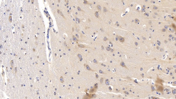
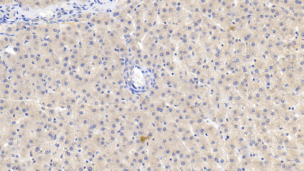 IHC (Immunohistochemisry)
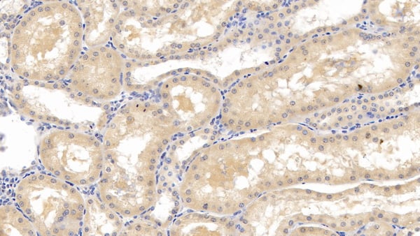
(DAB staining on IHCP;Sample: Porcine Liver Tissue; Primary Ab: 10ug/ml Mouse AntiHuman LCAT AntibodySecond Ab: 2ug/mL HRPLinked Caprine AntiMouse IgG Polyclonal Antibody(Catalog: SAA544Mu19))
IHC (Immunohistochemisry)
(DAB staining on IHCP;Sample: Porcine Liver Tissue; Primary Ab: 10ug/ml Mouse AntiHuman LCAT AntibodySecond Ab: 2ug/mL HRPLinked Caprine AntiMouse IgG Polyclonal Antibody(Catalog: SAA544Mu19))
Lecithin Cholesterol Acyltransferase (LCAT), Monoclonal Antibody (Cat# AAA151893)
Full Name
Monoclonal Antibody to Lecithin Cholesterol Acyltransferase (LCAT)
Reactivity
Human, Pig
Applications
Immunoprecipitation, Immunocytochemistry, Immunohistochemistry, Western Blot
Purity
Protein A + Protein G affinity chromatography
Pricing
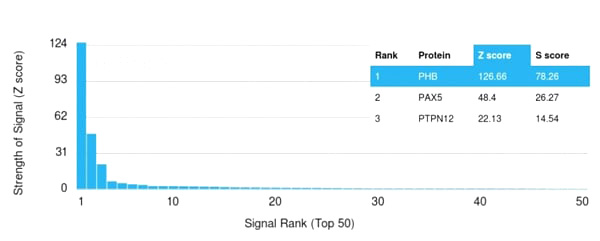
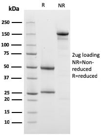
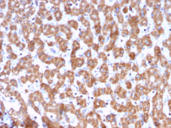
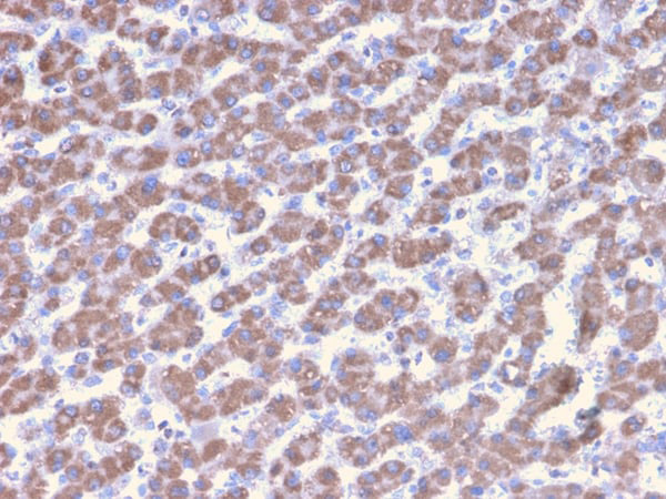
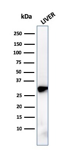 WB (Western Blot)
(Western Blot Analysis of human liver tissue lysate using Prohibitin Mouse Monoclonal Antibody (PHB/3228).)
WB (Western Blot)
(Western Blot Analysis of human liver tissue lysate using Prohibitin Mouse Monoclonal Antibody (PHB/3228).)
Prohibitin, Monoclonal Antibody (Cat# AAA215145)
Full Name
Prohibitin (Mitochondrial Marker)
Gene Names
PHB; PHB1; HEL-215; HEL-S-54e
Reactivity
Human
Applications
Immunohistochemistry, Western Blot
Purity
Purified Ab with BSA and Azide at 200ug/ml OR Purified Ab WITHOUT BSA and Azide at 1.0mg/ml
Pricing
 Application Data
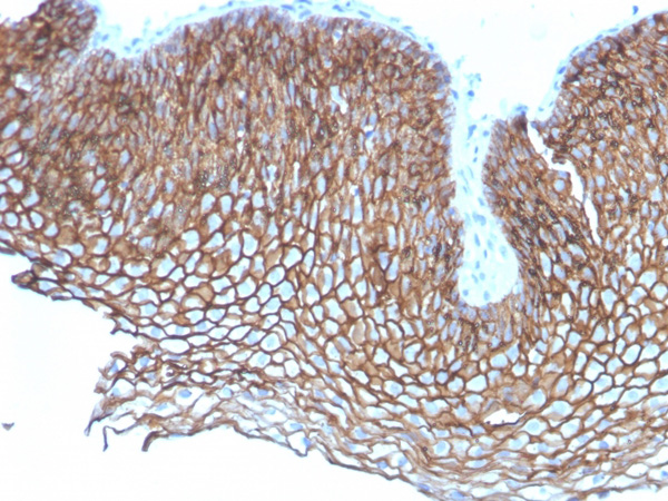
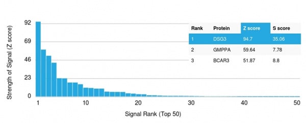
(Analysis of Protein Array containing more than 19,000 full-length human proteins using Desmoglein-3 Mouse Monoclonal Antibody (DSG3/2837) Z- and S- Score: The Z-score represents the strength of a signal that a monoclonal antibody (Monoclonal Antibody) (in combination with a fluorescently-tagged anti-IgG secondary antibody) produces when binding to a particular protein on the HuProtTM array. Z-scores are described in units of standard deviations (SD's) above the mean value of all signals generated on that array. If targets on HuProtTM are arranged in descending order of the Z-score, the S-score is the difference (also in units of SD's) between the Z-score. S-score therefore represents the relative target specificity of a Monoclonal Antibody to its intended target. A Monoclonal Antibody is considered to specific to its intended target, if the Monoclonal Antibody has an S-score of at least 2.5. For example, if a Monoclonal Antibody binds to protein X with a Z-score of 43 and to protein Y with a Z-score of 14, then the S-score for the binding of that Monoclonal Antibody to protein X is equal to 29.)
Application Data
(Analysis of Protein Array containing more than 19,000 full-length human proteins using Desmoglein-3 Mouse Monoclonal Antibody (DSG3/2837) Z- and S- Score: The Z-score represents the strength of a signal that a monoclonal antibody (Monoclonal Antibody) (in combination with a fluorescently-tagged anti-IgG secondary antibody) produces when binding to a particular protein on the HuProtTM array. Z-scores are described in units of standard deviations (SD's) above the mean value of all signals generated on that array. If targets on HuProtTM are arranged in descending order of the Z-score, the S-score is the difference (also in units of SD's) between the Z-score. S-score therefore represents the relative target specificity of a Monoclonal Antibody to its intended target. A Monoclonal Antibody is considered to specific to its intended target, if the Monoclonal Antibody has an S-score of at least 2.5. For example, if a Monoclonal Antibody binds to protein X with a Z-score of 43 and to protein Y with a Z-score of 14, then the S-score for the binding of that Monoclonal Antibody to protein X is equal to 29.)
Desmoglein-3, Monoclonal Antibody (Cat# AAA214933)
Full Name
Desmoglein-3 (Squamous Cell Marker)
Gene Names
DSG3; PVA; CDHF6
Reactivity
Human
Applications
Immunohistochemistry
Purity
Purified Ab with BSA and Azide at 200ug/ml OR Purified Ab WITHOUT BSA and Azide at 1.0mg/ml
Pricing
 Application Data
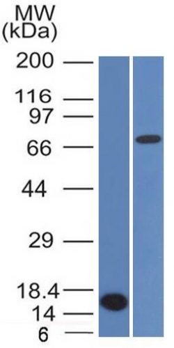
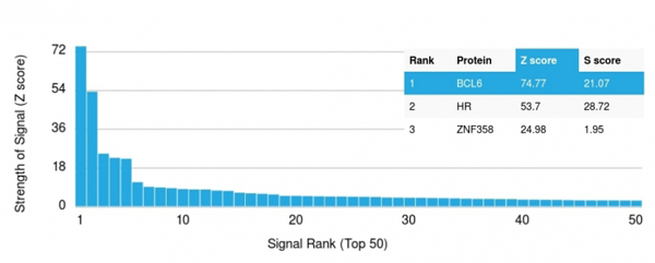
(Analysis of Protein Array containing more than 19, 000 full-length human proteins using bcl-6 Mouse Monoclonal Antibody (BCL6/1475).Z- and S- Score: The Z-score represents the strength of a signal that a monoclonal antibody (MAb) (in combination with a fluorescently-tagged anti-IgG secondary antibody) produces when binding to a particular protein on the HuProtTM array. Z-scores are described in units of standard deviations (SD's) above the mean value of all signals generated on that array. If targets on HuProtTM are arranged in descending order of the Z-score, the S-score is the difference (also in units of SD's) between the Z-score. S-score therefore represents the relative target specificity of a MAb to its intended target. A MAb is considered to specific to its intended target, if the MAb has an S-score of at least 2.5. For example, if a MAb binds to protein X with a Z-score of 43 and to protein Y with a Z-score of 14, then the S-score for the binding of that MAb to protein X is equal to 29.)
Application Data
(Analysis of Protein Array containing more than 19, 000 full-length human proteins using bcl-6 Mouse Monoclonal Antibody (BCL6/1475).Z- and S- Score: The Z-score represents the strength of a signal that a monoclonal antibody (MAb) (in combination with a fluorescently-tagged anti-IgG secondary antibody) produces when binding to a particular protein on the HuProtTM array. Z-scores are described in units of standard deviations (SD's) above the mean value of all signals generated on that array. If targets on HuProtTM are arranged in descending order of the Z-score, the S-score is the difference (also in units of SD's) between the Z-score. S-score therefore represents the relative target specificity of a MAb to its intended target. A MAb is considered to specific to its intended target, if the MAb has an S-score of at least 2.5. For example, if a MAb binds to protein X with a Z-score of 43 and to protein Y with a Z-score of 14, then the S-score for the binding of that MAb to protein X is equal to 29.)
Bcl-6, Monoclonal Antibody (Cat# AAA214506)
Full Name
Bcl-6 (Follicular Lymphoma Marker)
Gene Names
BCL6; BCL5; LAZ3; BCL6A; ZNF51; ZBTB27
Reactivity
Human. Others not known.
Applications
Western Blot
Pricing
 Bioactivity
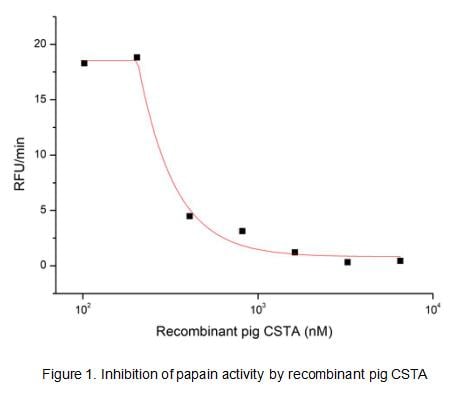
(Cystatin A (CSTA) is a member of family 1 of the cystatin superfamily. Like Cystatin B, it is an intracellular inhibitor regulating the activities of cysteine proteases of the papain family such as cathepsins B, H and L. For example, immunohistochemical analysis of Cystatin A and cathepsin L is a useful indicator for malignancy in human epidermal keratinocytes. The ratio of cathepsin B and Cystatin A can be used in the differential identification and treatment of patients with prostate carcinoma. The activity of recombinant pig Cystatin A was measured by its ability to inhibit papain cleavage of a fluorogenic peptide substrate Z-FR-AMC in the assay buffer 50 mM Tris, pH 7.0. Papain was diluted to 500 ug/ml in activation buffer 50 mM Tris, 5 mM DTT, pH 7.0 and incubated at room temperature for 15 minutes. The activated papain was diluted to 100 ug/ml in the assay buffer and 20 ul different concentrations of recombinant pig Cystatin A (MW: 40.9 KD) was incubated with 20 ul 100 ug/ml papain at 37 degree C for 10 minutes. Loading 50 uL of the incubated mixtures which were diluted five-fold in assay buffer into empty wells of a plate, and start the reaction by adding 50 uL of 200 uM substrate. Include a substrate blank containing 50 uL of assay buffer and 50 uL of 200 uM substrate. Then read at excitiation and emission wavelengths of 380 nm and 460 nm, respectively, in kinetic mode for 5 minutes. The result was shown in Figure 1 and it was obvious that recombinant pig Cystatin A significantly decreased papain activity. The inhibition IC50 was )
Bioactivity
(Cystatin A (CSTA) is a member of family 1 of the cystatin superfamily. Like Cystatin B, it is an intracellular inhibitor regulating the activities of cysteine proteases of the papain family such as cathepsins B, H and L. For example, immunohistochemical analysis of Cystatin A and cathepsin L is a useful indicator for malignancy in human epidermal keratinocytes. The ratio of cathepsin B and Cystatin A can be used in the differential identification and treatment of patients with prostate carcinoma. The activity of recombinant pig Cystatin A was measured by its ability to inhibit papain cleavage of a fluorogenic peptide substrate Z-FR-AMC in the assay buffer 50 mM Tris, pH 7.0. Papain was diluted to 500 ug/ml in activation buffer 50 mM Tris, 5 mM DTT, pH 7.0 and incubated at room temperature for 15 minutes. The activated papain was diluted to 100 ug/ml in the assay buffer and 20 ul different concentrations of recombinant pig Cystatin A (MW: 40.9 KD) was incubated with 20 ul 100 ug/ml papain at 37 degree C for 10 minutes. Loading 50 uL of the incubated mixtures which were diluted five-fold in assay buffer into empty wells of a plate, and start the reaction by adding 50 uL of 200 uM substrate. Include a substrate blank containing 50 uL of assay buffer and 50 uL of 200 uM substrate. Then read at excitiation and emission wavelengths of 380 nm and 460 nm, respectively, in kinetic mode for 5 minutes. The result was shown in Figure 1 and it was obvious that recombinant pig Cystatin A significantly decreased papain activity. The inhibition IC50 was )
Cystatin A (CSTA), Active Protein (Cat# AAA161737)
Full Name
Active Cystatin A (CSTA)
Applications
Activity Assay, Cell Culture
Purity
Greater than 90% by SDS-PAGE
Pricing
 WB (Western Blot)
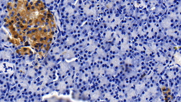
(Western Blot; Sample: Lane1: Porcine Serum; Lane2: Porcine Lung lysate; Lane3: Porcine Cerebrum lysate Primary Ab: 2ug/ml Mouse AntiPorcine CLU Antibody Second Ab: 0.2ug/mL HRPLinked Caprine AntiMouse IgG Polyclonal Antibody (Catalog: SAA544Mu19))
WB (Western Blot)
(Western Blot; Sample: Lane1: Porcine Serum; Lane2: Porcine Lung lysate; Lane3: Porcine Cerebrum lysate Primary Ab: 2ug/ml Mouse AntiPorcine CLU Antibody Second Ab: 0.2ug/mL HRPLinked Caprine AntiMouse IgG Polyclonal Antibody (Catalog: SAA544Mu19))
Clusterin (CLU), Monoclonal Antibody (Cat# AAA151690)
Full Name
Monoclonal Antibody to Clusterin (CLU)
Reactivity
Pig
Applications
Immunoprecipitation, Immunocytochemistry, Immunohistochemistry, Western Blot
Purity
Protein A + Protein G affinity chromatography
Pricing
 Application Data
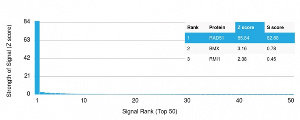
(Analysis of Protein Array containing more than 19,000 full-length human proteins using RAD51 Mouse Monoclonal Antibody (RAD51/2702). Z- and S- Score: The Z-score represents the strength of a signal that a monoclonal antibody (MAb) (in combination with a fluorescently-tagged anti-IgG secondary antibody) produces when binding to a particular protein on the HuProtTM array. Z-scores are described in units of standard deviations (SD's) above the mean value of all signals generated on that array. If targets on HuProtTM are arranged in descending order of the Z-score, the S-score is the difference (also in units of SD's) between the Z-score. S-score therefore represents the relative target specificity of a MAb to its intended target. A MAb is considered to specific to its intended target, if the MAb has an S-score of at least 2.5. For example, if a MAb binds to protein X with a Z-score of 43 and to protein Y with a Z-score of 14, then the S-score for the binding of that MAb to protein X is equal to 29.)
Application Data
(Analysis of Protein Array containing more than 19,000 full-length human proteins using RAD51 Mouse Monoclonal Antibody (RAD51/2702). Z- and S- Score: The Z-score represents the strength of a signal that a monoclonal antibody (MAb) (in combination with a fluorescently-tagged anti-IgG secondary antibody) produces when binding to a particular protein on the HuProtTM array. Z-scores are described in units of standard deviations (SD's) above the mean value of all signals generated on that array. If targets on HuProtTM are arranged in descending order of the Z-score, the S-score is the difference (also in units of SD's) between the Z-score. S-score therefore represents the relative target specificity of a MAb to its intended target. A MAb is considered to specific to its intended target, if the MAb has an S-score of at least 2.5. For example, if a MAb binds to protein X with a Z-score of 43 and to protein Y with a Z-score of 14, then the S-score for the binding of that MAb to protein X is equal to 29.)
RAD51, Monoclonal Antibody (Cat# AAA214806)
Full Name
RAD51 (Prognostic and Response to Chemotherapy Marker)
Gene Names
RAD51; RECA; BRCC5; FANCR; MRMV2; HRAD51; RAD51A; HsRad51; HsT16930
Reactivity
Human. Others not known.
Applications
Immunohistochemistry
Purity
Purified Ab with BSA and Azide at 200ug/ml OR Purified Ab WITHOUT BSA and Azide at 1.0mg/ml
Pricing
 IHC (Immunohistochemisry)
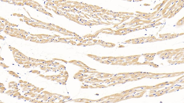
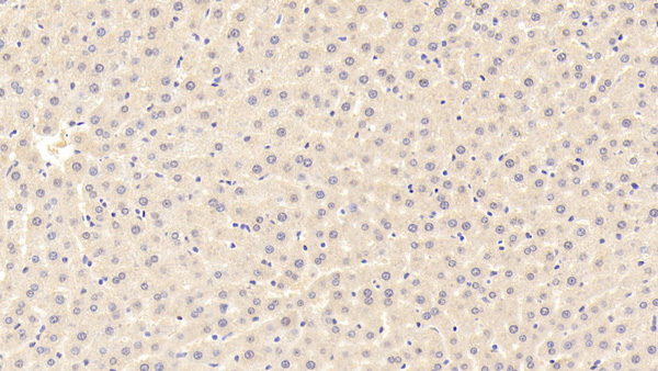
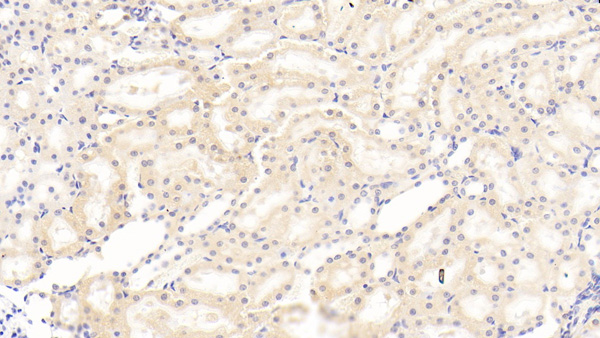
(DAB staining on IHCP;Sample: Rat Kidney Tissue; Primary Ab: 30ug/ml Mouse AntiRat ADP AntibodySecond Ab: 2ug/mL HRPLinked Caprine AntiMouse IgG Polyclonal Antibody(Catalog: SAA544Mu19))
IHC (Immunohistochemisry)
(DAB staining on IHCP;Sample: Rat Kidney Tissue; Primary Ab: 30ug/ml Mouse AntiRat ADP AntibodySecond Ab: 2ug/mL HRPLinked Caprine AntiMouse IgG Polyclonal Antibody(Catalog: SAA544Mu19))
Adiponectin (ADPN), Monoclonal Antibody (Cat# AAA151585)
Full Name
Monoclonal Antibody to Adiponectin (ADPN)
Gene Names
Adipoq; Acdc; Acrp30
Reactivity
Rat
Applications
Immunoprecipitation, Immunocytochemistry, Immunohistochemistry, Western Blot
Purity
Protein A + Protein G affinity chromatography
Pricing
 IHC (Immunohistochemistry)
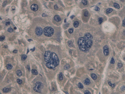
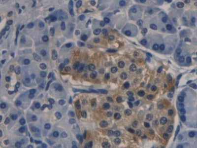
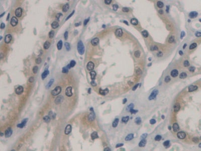
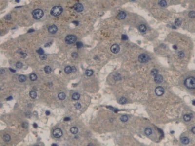
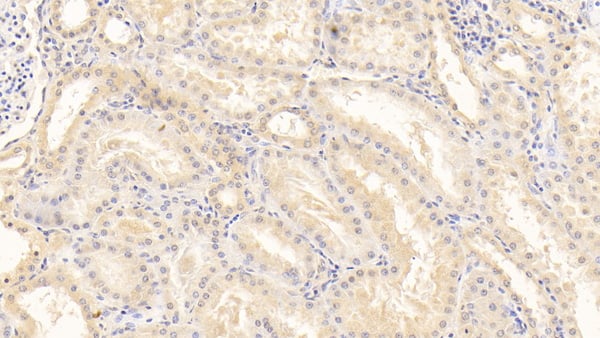
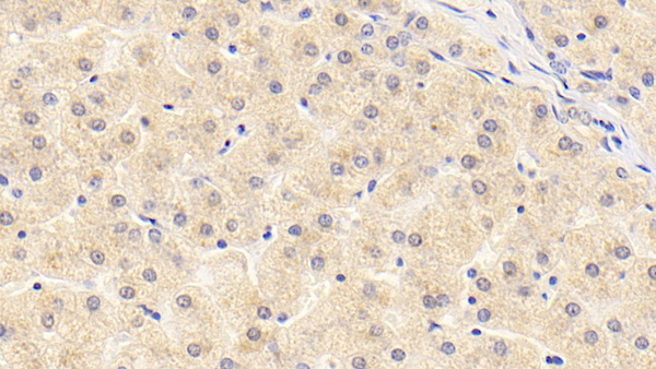
(DAB staining on IHCP;Sample: Human Liver Tissue; Primary Ab: 30ug/ml Mouse AntiHuman PCT AntibodySecond Ab: 2ug/mL HRPLinked Caprine AntiMouse IgG Polyclonal Antibody)
IHC (Immunohistochemistry)
(DAB staining on IHCP;Sample: Human Liver Tissue; Primary Ab: 30ug/ml Mouse AntiHuman PCT AntibodySecond Ab: 2ug/mL HRPLinked Caprine AntiMouse IgG Polyclonal Antibody)
Procalcitonin (PCT), Monoclonal Antibody (Cat# AAA151605)
Full Name
Monoclonal Antibody to Procalcitonin (PCT)
Gene Names
CALCA; CT; KC; CGRP; CALC1; CGRP1; CGRP-I
Reactivity
Human
Applications
Immunohistochemistry
Purity
Protein A + Protein G affinity chromatography
Pricing
 WB (Western Blot)
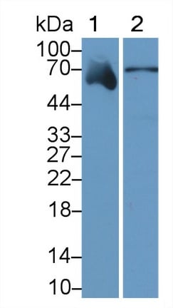
(Western Blot; Sample: Lane1: Human Serum; Lane2: Human Urine Primary Ab: 2ug/ml Mouse AntiHuman a2PI Antibody Second Ab: 0.2ug/mL HRPLinked Caprine AntiMouse IgG Polyclonal Antibody (Catalog: SAA544Mu19))
WB (Western Blot)
(Western Blot; Sample: Lane1: Human Serum; Lane2: Human Urine Primary Ab: 2ug/ml Mouse AntiHuman a2PI Antibody Second Ab: 0.2ug/mL HRPLinked Caprine AntiMouse IgG Polyclonal Antibody (Catalog: SAA544Mu19))
Alpha 2Antiplasmin (a2PI), Monoclonal Antibody (Cat# AAA151497)
Full Name
Monoclonal Antibody to Alpha 2Antiplasmin (a2PI)
Gene Names
SERPINF2; AAP; API; PLI; A2AP; ALPHA-2-PI
Reactivity
Human, Pig
Applications
Immunoprecipitation, Immunocytochemistry, Immunohistochemistry, Western Blot
Purity
Protein A + Protein G affinity chromatography
Pricing
 FCM/FACS (Flow Cytometry)
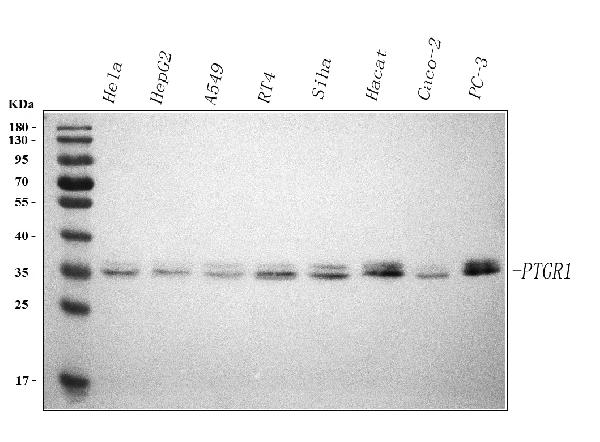
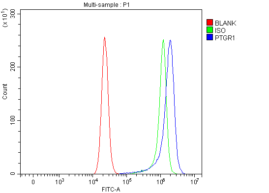
(Figure 2. Flow Cytometry analysis of RT4 cells using anti-PTGR1 antibody (AAA127533).Overlay histogram showing RT4 cells stained with AAA127533 (Blue line). To facilitate intracellular staining, cells were fixed with 4% paraformaldehyde and permeabilized with permeabilization buffer. The cells were blocked with 10% normal goat serum. And then incubated with rabbit anti-PTGR1 Antibody (AAA127533, 1ug/1x106 cells) for 30 min at 20 degree C. DyLight488 conjugated goat anti-rabbit IgG was used as secondary antibody for 30 minutes at 20 degree C. Isotype control antibody (Green line) was rabbit IgG (1ug/1x106) used under the same conditions. Unlabelled sample (Red line) was also used as a control.)
FCM/FACS (Flow Cytometry)
(Figure 2. Flow Cytometry analysis of RT4 cells using anti-PTGR1 antibody (AAA127533).Overlay histogram showing RT4 cells stained with AAA127533 (Blue line). To facilitate intracellular staining, cells were fixed with 4% paraformaldehyde and permeabilized with permeabilization buffer. The cells were blocked with 10% normal goat serum. And then incubated with rabbit anti-PTGR1 Antibody (AAA127533, 1ug/1x106 cells) for 30 min at 20 degree C. DyLight488 conjugated goat anti-rabbit IgG was used as secondary antibody for 30 minutes at 20 degree C. Isotype control antibody (Green line) was rabbit IgG (1ug/1x106) used under the same conditions. Unlabelled sample (Red line) was also used as a control.)
PTGR1, Polyclonal Antibody (Cat# AAA127533)
Full Name
Anti-PTGR1 Antibody Picoband
Gene Names
PTGR1; PGR1; ZADH3; LTB4DH
Reactivity
Human
Applications
Flow Cytometry, Western Blot
Purity
Immunogen affinity purified.
Pricing
 FCM/FACS (Flow Cytometry)
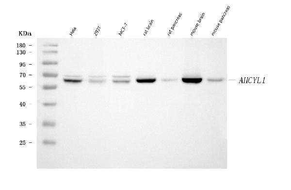
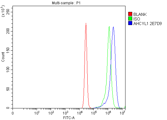
(Figure 2. Flow Cytometry analysis of K562 cells using anti-IRBIT/AHCYL1 antibody (AAA126957).Overlay histogram showing K562 cells stained with AAA126957 (Blue line). The cells were blocked with 10% normal goat serum. And then incubated with mouse anti-IRBIT/AHCYL1 Antibody (AAA126957, 1 ug/1x10^6 cells) for 30 min at 20 degree C. DyLight488 conjugated goat anti-mouse IgG was used as secondary antibody for 30 minutes at 20 degree C. Isotype control antibody (Green line) was mouse IgG (1 ug/1x10^6) used under the same conditions. Unlabelled sample (Red line) was also used as a control.)
FCM/FACS (Flow Cytometry)
(Figure 2. Flow Cytometry analysis of K562 cells using anti-IRBIT/AHCYL1 antibody (AAA126957).Overlay histogram showing K562 cells stained with AAA126957 (Blue line). The cells were blocked with 10% normal goat serum. And then incubated with mouse anti-IRBIT/AHCYL1 Antibody (AAA126957, 1 ug/1x10^6 cells) for 30 min at 20 degree C. DyLight488 conjugated goat anti-mouse IgG was used as secondary antibody for 30 minutes at 20 degree C. Isotype control antibody (Green line) was mouse IgG (1 ug/1x10^6) used under the same conditions. Unlabelled sample (Red line) was also used as a control.)
IRBIT/AHCYL1, Monoclonal Antibody (Cat# AAA126957)
Full Name
Anti-IRBIT/AHCYL1 Antibody Picoband (monoclonal, 2E7D9)
Gene Names
AHCYL1; DCAL; IRBIT; PPP1R78; PRO0233; XPVKONA
Reactivity
Human, Mouse, Rat
Applications
Flow Cytometry, Western Blot
Purity
Immunogen affinity purified.
Pricing
 IHC (Immunohistochemisry)
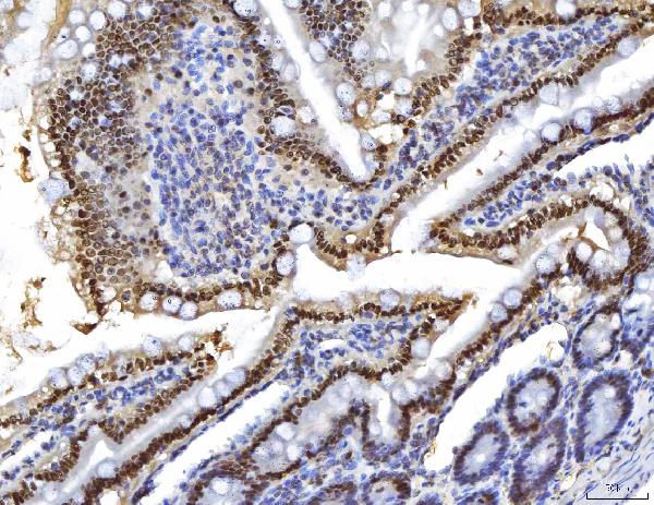
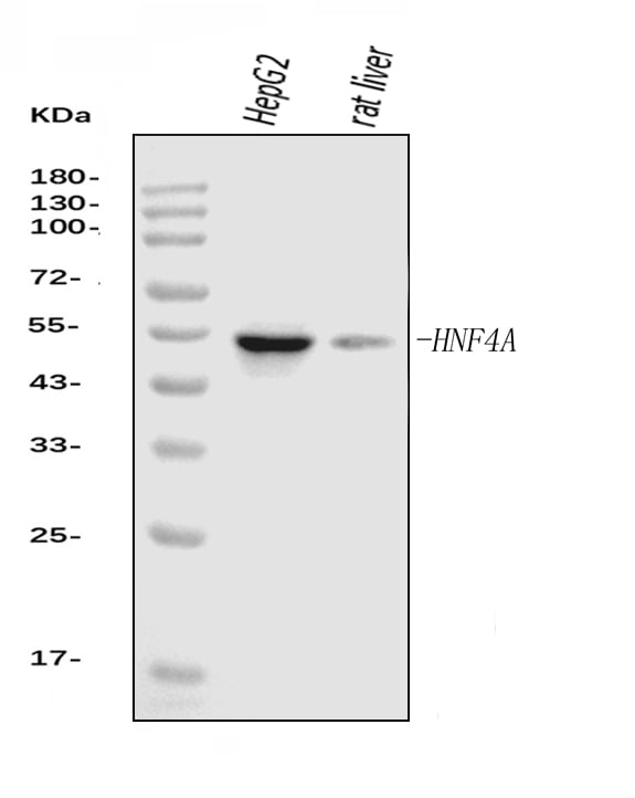
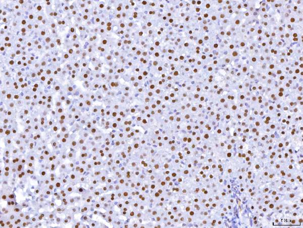
(Figure 3. IHC analysis of HNF-4-alpha using anti-HNF-4-alpha antibody (AAA126867).HNF-4-alpha was detected in a paraffin-embedded section of rat liver tissue. Heat mediated antigen retrieval was performed in EDTA buffer (pH 8.0, epitope retrieval solution). The tissue section was blocked with 10% goat serum. The tissue section was then incubated with 2 ug/ml mouse anti-HNF-4-alpha Antibody (AAA126867) overnight at 4 degree C. Peroxidase Conjugated Goat Anti-mouse IgG was used as secondary antibody and incubated for 30 minutes at 37 degree C. The tissue section was developed using HRP Conjugated Mouse IgG Super Vision Assay Kit with DAB as the chromogen.)
IHC (Immunohistochemisry)
(Figure 3. IHC analysis of HNF-4-alpha using anti-HNF-4-alpha antibody (AAA126867).HNF-4-alpha was detected in a paraffin-embedded section of rat liver tissue. Heat mediated antigen retrieval was performed in EDTA buffer (pH 8.0, epitope retrieval solution). The tissue section was blocked with 10% goat serum. The tissue section was then incubated with 2 ug/ml mouse anti-HNF-4-alpha Antibody (AAA126867) overnight at 4 degree C. Peroxidase Conjugated Goat Anti-mouse IgG was used as secondary antibody and incubated for 30 minutes at 37 degree C. The tissue section was developed using HRP Conjugated Mouse IgG Super Vision Assay Kit with DAB as the chromogen.)
HNF-4-alpha, Monoclonal Antibody (Cat# AAA126867)
Full Name
Anti-HNF-4-alpha Antibody Picoband (monoclonal, 6C8E9)
Gene Names
HNF4A; TCF; HNF4; MODY; MODY1; NR2A1; TCF14; HNF4a7; HNF4a8; HNF4a9; NR2A21; HNF4alpha
Reactivity
Human, Rat
Applications
Immunohistochemistry, Western Blot
Purity
Immunogen affinity purified.
Pricing
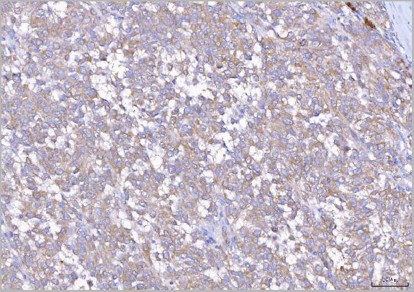
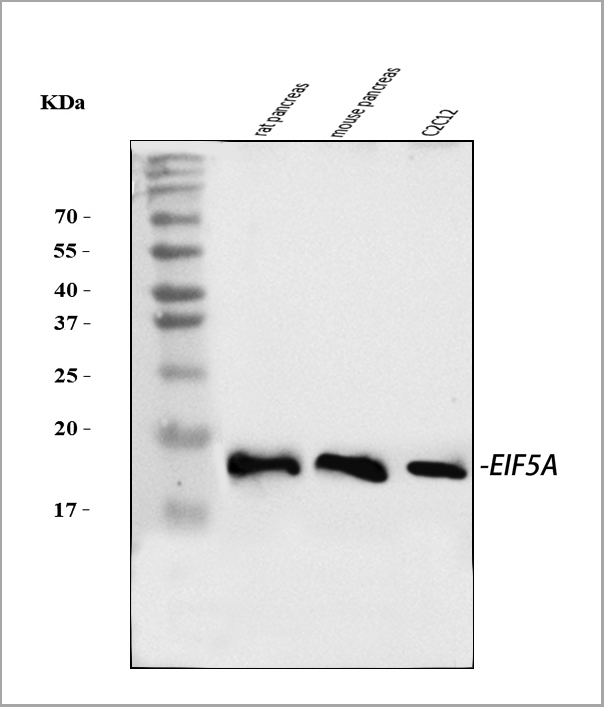
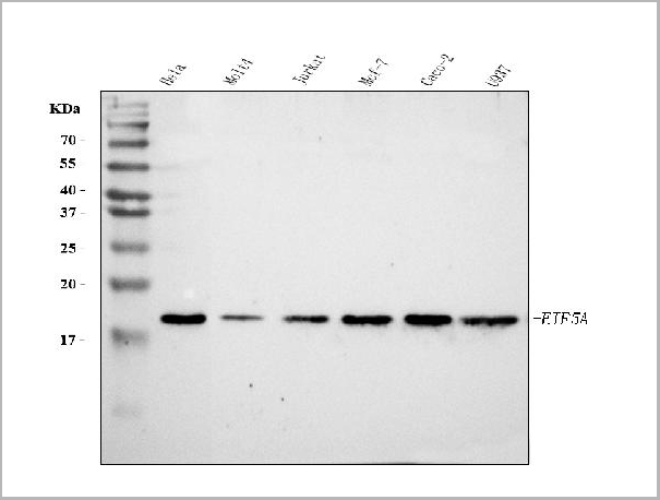
 FCM (Flow Cytometry)
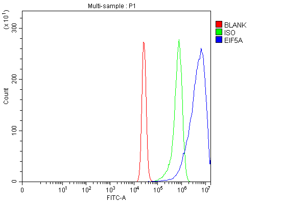
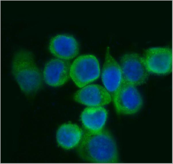
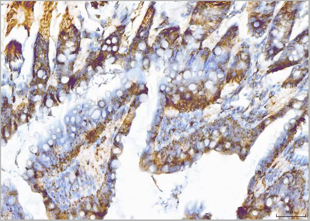
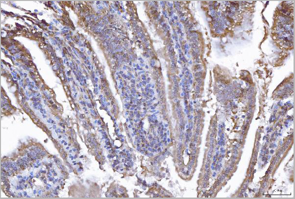
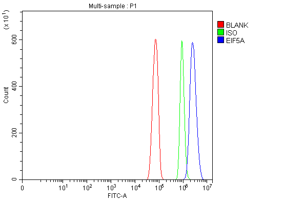
(Figure 8. Flow Cytometry analysis of RH35 cells using anti-EIF5A antibody (AAA19429).Overlay histogram showing RH35 cells stained with AAA19429 (Blue line). The cells were blocked with 10% normal goat serum. And then incubated with rabbit anti-EIF5A Antibody (AAA19429, 1 ug/1x10^6 cells) for 30 min at 20 degree C. DyLight488 conjugated goat anti-rabbit IgG was used as secondary antibody for 30 minutes at 20 degree C. Isotype control antibody (Green line) was rabbit IgG (1 ug/1x10^6) used under the same conditions. Unlabelled sample (Red line) was also used as a control.)
FCM (Flow Cytometry)
(Figure 8. Flow Cytometry analysis of RH35 cells using anti-EIF5A antibody (AAA19429).Overlay histogram showing RH35 cells stained with AAA19429 (Blue line). The cells were blocked with 10% normal goat serum. And then incubated with rabbit anti-EIF5A Antibody (AAA19429, 1 ug/1x10^6 cells) for 30 min at 20 degree C. DyLight488 conjugated goat anti-rabbit IgG was used as secondary antibody for 30 minutes at 20 degree C. Isotype control antibody (Green line) was rabbit IgG (1 ug/1x10^6) used under the same conditions. Unlabelled sample (Red line) was also used as a control.)
EIF5A, Polyclonal Antibody (Cat# AAA19429)
Full Name
Anti-EIF5A Antibody Picoband
Gene Names
EIF5A; EIF-5A; EIF5A1; eIF5AI
Reactivity
Human, Mouse, Rat
Applications
Western Blot, Immunohistochemistry, Immunocytochemistry, Immunofluorescence, Flow Cytometry
Purity
Immunogen affinity purified.
Pricing
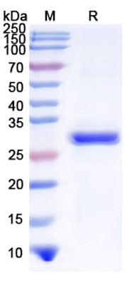 SDS-PAGE
(SDS-PAGE for Recombinant Foot-and-mouth disease virus O Capsid protein VP1 protein)
SDS-PAGE
(SDS-PAGE for Recombinant Foot-and-mouth disease virus O Capsid protein VP1 protein)
Capsid protein VP1, Recombinant Protein (Cat# AAA120776)
Full Name
Recombinant FMDV Capsid protein VP1 Protein, N-His
Applications
Western Blot
Purity
>90% as determined by SDS-PAGE.
Pricing
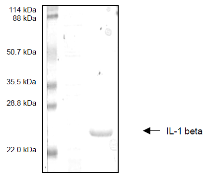 SDS-PAGE
Interleukin-1 Beta (IL-1B), Recombinant Protein (Cat# AAA58752)
Full Name
Interleukin-1 Beta (IL-1B)
Gene Names
IL1B; IL1-BETA
Purity
>95% by SDS-PAGE
Pricing

 ELISA
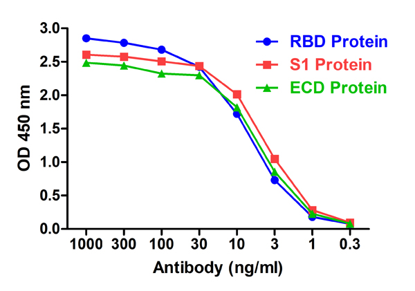
(Figure 2 ELISA Validation with SARS-CoV-2 (COVID-19) Spike RBD, S1 and ECD Proteins Antibodies: SARS-CoV-2 (COVID-19) Spike S1 RBD Antibody, AAA40983. A direct ELISA was performed using SARS-CoV-2 Spike recombinant proteins (RBD, 10-303; S1, 97-087; ECD, 11-070) as coating antigen at 1ug/mL and the anti-SARS-CoV-2 (COVID-19) Spike S1 RBD antibody (AAA40983) as the capture antibody, following by anti-cMyc-tag antibody (PM-7669) at 1ug/mL. Secondary: Goat anti-mouse IgG HRP conjugate at 1:5000 dilution. Detection range is from 0.3 ng/mL to 1000 ng/mL. SARS-CoV-2 (COVID-19) Spike S1 RBD Antibody, AAA40983 can detect spike RBD, S1 and ECD proteins at 2 ng/mL.)
ELISA
(Figure 2 ELISA Validation with SARS-CoV-2 (COVID-19) Spike RBD, S1 and ECD Proteins Antibodies: SARS-CoV-2 (COVID-19) Spike S1 RBD Antibody, AAA40983. A direct ELISA was performed using SARS-CoV-2 Spike recombinant proteins (RBD, 10-303; S1, 97-087; ECD, 11-070) as coating antigen at 1ug/mL and the anti-SARS-CoV-2 (COVID-19) Spike S1 RBD antibody (AAA40983) as the capture antibody, following by anti-cMyc-tag antibody (PM-7669) at 1ug/mL. Secondary: Goat anti-mouse IgG HRP conjugate at 1:5000 dilution. Detection range is from 0.3 ng/mL to 1000 ng/mL. SARS-CoV-2 (COVID-19) Spike S1 RBD Antibody, AAA40983 can detect spike RBD, S1 and ECD proteins at 2 ng/mL.)
SARS-CoV-2 (COVID-19) S1 RBD, Monoclonal Recombinant Antibody (Cat# AAA40983)
Full Name
SARS-CoV-2 (COVID-19) S1 RBD Single Domain Antibody [T3P1-C8]
Reactivity
Virus
Applications
ELISA
Purity
SARS-CoV-2 (COVID-19) S1 RBD Antibody [T3P1-C8] is affinity chromatography purified via Nickel column. Antibody is supplied as a His-tagged purified protein. It also contains a myc-tag for detection.
Pricing

 SDS-PAGE
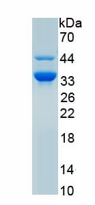
(Figure. SDS-PAGE)
SDS-PAGE
(Figure. SDS-PAGE)
Eosinophil Peroxidase (EPX), Recombinant Protein (Cat# AAA162017)
Full Name
Recombinant Eosinophil Peroxidase (EPX)
Gene Names
Epx; EPO
Applications
Western Blot
Purity
Greater than 95% by SDS-PAGE
Pricing
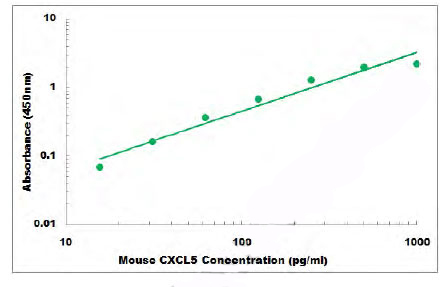 Standard Curve (Sample)
CXCL5, ELISA Kit (Cat# AAA105697)
Full Name
Mouse CXCL5 ELISA Kit
Gene Names
Cxcl5; LIX; GCP-2; Scyb5; Scyb6; ENA-78; AMCF-II
Reactivity
Mouse
Applications
Sandwich ELISA
Pricing
 SDS-PAGE
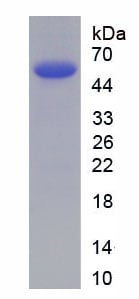
(SDS PAGE for Recombinant Human AR/Androgen receptor protein)
SDS-PAGE
(SDS PAGE for Recombinant Human AR/Androgen receptor protein)
AR/Androgen receptor, Recombinant Protein (Cat# AAA120613)
Full Name
Recombinant Human AR/Androgen receptor Protein, N-His
Gene Names
AR; KD; AIS; TFM; DHTR; SBMA; HYSP1; NR3C4; SMAX1; HUMARA
Applications
Western Blot
Purity
>90% as determined by SDS-PAGE
Pricing
 Amino Acid Info
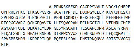
(Amino Acid info)
Amino Acid Info
(Amino Acid info)
Coagulation Factor XII (F12), Recombinant Protein (Cat# AAA153382)
Full Name
Recombinant Coagulation Factor XII (F12)
Gene Names
F12; HAF
Applications
Western Blot
Purity
Greater than 90% by SDS-PAGE
Pricing
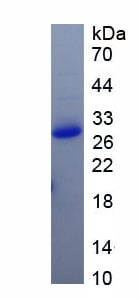
 Amino Acid Info
(Amino Acid info)
Amino Acid Info
(Amino Acid info)
Platelet Factor 4 (PF4), Recombinant Protein (Cat# AAA153300)
Full Name
Recombinant Platelet Factor 4 (PF4)
Applications
Western Blot
Purity
Greater than 95% by SDS-PAGE
Pricing


 IF (Immunofluorescence)
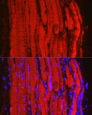
(Immunofluorescence analysis of mouse skeletal muscle using Fbx32/FBOX32 Rabbit pAb (A6825) at dilution of 1:100 (40x lens). Blue: DAPI for nuclear staining.)
IF (Immunofluorescence)
(Immunofluorescence analysis of mouse skeletal muscle using Fbx32/FBOX32 Rabbit pAb (A6825) at dilution of 1:100 (40x lens). Blue: DAPI for nuclear staining.)
FBXO32, Polyclonal Antibody (Cat# AAA37616)
Full Name
FBXO32 Polyclonal Antibody
Gene Names
FBXO32; Fbx32; MAFbx
Applications
Immunofluorescence, Western Blot
Purity
Affinity Purification
Pricing
 Bioactivity
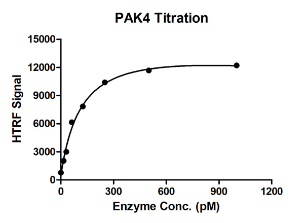
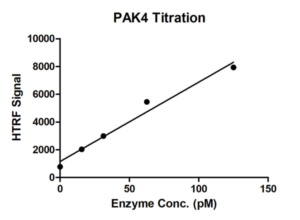
(HTRF Assay for Recombinant PAK4 activity. 1 uM STK S2 substrate was incubated with different concentrations of PAK4 protein in 10 ul reaction system containing 1×Enzymatic Buffer, 5 mM MgCl2, 1 mM DTT and 100 uM ATP for 1 hour. The 10 ul detection reagents containing anti-STK antibody (1:2) and SA-XL665 (1:100) diluted with 1× Detection Buffer were added and incubated with the reactions for 30 min. All the operations and reactions were performed at room temperature. HTRF assay was used for detection.)
Bioactivity
(HTRF Assay for Recombinant PAK4 activity. 1 uM STK S2 substrate was incubated with different concentrations of PAK4 protein in 10 ul reaction system containing 1×Enzymatic Buffer, 5 mM MgCl2, 1 mM DTT and 100 uM ATP for 1 hour. The 10 ul detection reagents containing anti-STK antibody (1:2) and SA-XL665 (1:100) diluted with 1× Detection Buffer were added and incubated with the reactions for 30 min. All the operations and reactions were performed at room temperature. HTRF assay was used for detection.)
 Bioactivity
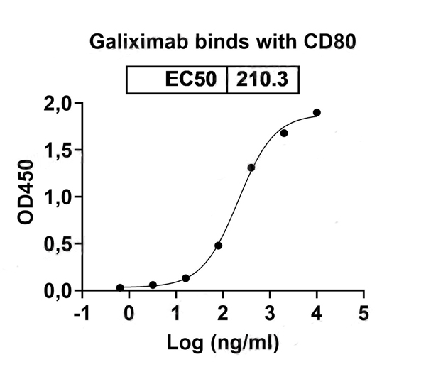
(Detects Galiximab in indirect ELISAs.)
Bioactivity
(Detects Galiximab in indirect ELISAs.)
CD80/B7-1, Recombinant Protein (Cat# AAA120474)
Full Name
Recombinant Human CD80/B7-1, C-His
Gene Names
CD80; B7; BB1; B7-1; B7.1; LAB7; CD28LG; CD28LG1
Applications
Western Blot
Purity
>90% as determined by SDS-PAGE
Pricing
 Amino Acid Info
(Amino Acid info)
Amino Acid Info
(Amino Acid info)
Insulin Like Protein 3 (INSL3), Recombinant Protein (Cat# AAA153590)
Full Name
Recombinant Insulin Like Protein 3 (INSL3)
Gene Names
Insl3; Rlf; Rlnl
Applications
Western Blot
Purity
Greater than 95% by SDS-PAGE
Pricing
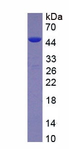
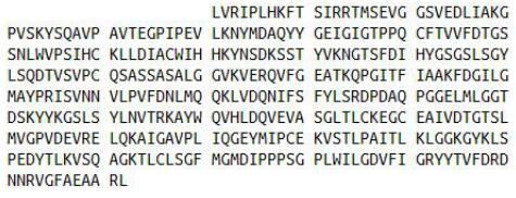 Amino Acid Info
(Amino Acid info)
Amino Acid Info
(Amino Acid info)
Cathepsin D (CTSD), Recombinant Protein (Cat# AAA153443)
Full Name
Recombinant Cathepsin D (CTSD)
Gene Names
CTSD; CPSD; CLN10
Applications
Western Blot
Purity
Greater than 95% by SDS-PAGE
Pricing
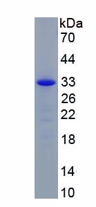
 Amino Acid Info
Caspase 3 (CASP3), Recombinant Protein (Cat# AAA153367)
Full Name
Recombinant Caspase 3 (CASP3)
Gene Names
Casp3; CC3; AC-3; Lice; Yama; mldy; CPP32; SCA-1; CASP-3; CPP-32; Caspase-3; A830040C14Rik
Applications
Western Blot
Purity
Greater than 90% by SDS-PAGE
Pricing
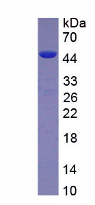
 Amino Acid Info
Creatine Kinase, Muscle (CKM), Recombinant Protein (Cat# AAA153276)
Full Name
Recombinant Creatine Kinase, Muscle (CKM)
Applications
Western Blot
Purity
Greater than 95% by SDS-PAGE
Pricing
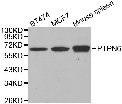
 IF (Immunofluorescence)
(Immunofluorescence analysis of U2OS cells using PTPN6 antibody. Blue: DAPI for nuclear staining.)
IF (Immunofluorescence)
(Immunofluorescence analysis of U2OS cells using PTPN6 antibody. Blue: DAPI for nuclear staining.)
PTPN6, Polyclonal Antibody (Cat# AAA37289)
Full Name
PTPN6 Polyclonal Antibody
Gene Names
PTPN6; HCP; HCPH; SHP1; SHP-1; HPTP1C; PTP-1C; SHP-1L; SH-PTP1
Reactivity
Human, Mouse
Applications
Immunofluorescence, Immunohistochemistry, Western Blot
Purity
Affinity Purification
Pricing
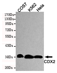
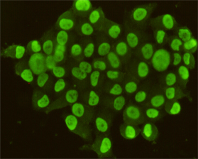
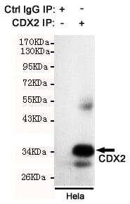 IP (Immunoprecipitation)
(Immunoprecipitation analysis of Hela cell lysate using CDX2 antibody.)
IP (Immunoprecipitation)
(Immunoprecipitation analysis of Hela cell lysate using CDX2 antibody.)
CDX2, Monoclonal Antibody (Cat# AAA300839)
Full Name
CDX2 Monoclonal Antibody
Gene Names
CDX2; CDX3; CDX-3; CDX2/AS
Reactivity
Human, Monkey
Applications
Immunocytochemistry, Immunoprecipitation, Western Blot
Purity
Affinity purified
Pricing
 SDS-PAGE
(SDS PAGE for Recombinant Felis catus (Cat) (Felis silvestris catus) Fel d 1-A/CH1/Allergen Fel dI protein)
SDS-PAGE
(SDS PAGE for Recombinant Felis catus (Cat) (Felis silvestris catus) Fel d 1-A/CH1/Allergen Fel dI protein)
Fel d 1-A/CH1/Allergen Fel dI, Recombinant Protein (Cat# AAA120614)
Full Name
Recombinant Cat Fel d 1-A/CH1/Allergen Fel dI Protein, N-GST & C-His
Gene Names
LOC677877; CH1
Applications
Western Blot
Purity
>90% as determined by SDS-PAGE
Pricing
 Bioactivity
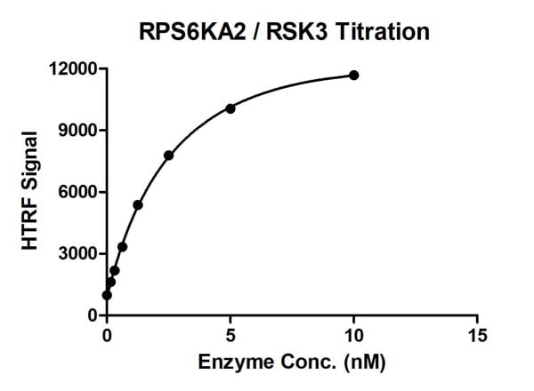
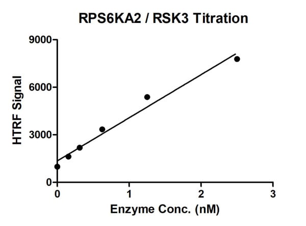
(HTRF assay for RPS6KA2/RSK3 protein activity 1 uM STK S1 substrate was incubated with different concentrations of RPS6KA2/RSK3 protein in a 10 ul reaction system containing 1×Enzymatic Buffer, 5 mM MgCl2, 1 mM DTT, and 100 uM ATP for 1 hour. The 10 ul detection reagents containing anti-STK antibody (1:2) and SA-XL665 (1:100) diluted with 1× Detection Buffer were added and incubated with the reactions for 30 min. All the operations and reactions were performed at room temperature. HTRF assay was used for detection.)
Bioactivity
(HTRF assay for RPS6KA2/RSK3 protein activity 1 uM STK S1 substrate was incubated with different concentrations of RPS6KA2/RSK3 protein in a 10 ul reaction system containing 1×Enzymatic Buffer, 5 mM MgCl2, 1 mM DTT, and 100 uM ATP for 1 hour. The 10 ul detection reagents containing anti-STK antibody (1:2) and SA-XL665 (1:100) diluted with 1× Detection Buffer were added and incubated with the reactions for 30 min. All the operations and reactions were performed at room temperature. HTRF assay was used for detection.)
RPS6KA2/RSK3, Active Protein (Cat# AAA60321)
Full Name
Recombinant RPS6KA2/RSK3 protein
Purity
>=90%
Pricing

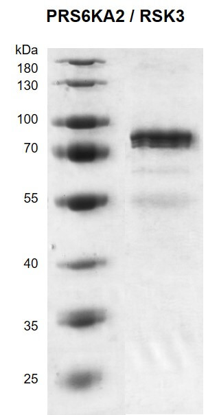
 SDS-PAGE
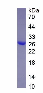
(SDS-PAGE)
SDS-PAGE
(SDS-PAGE)
Calretinin (CR), Recombinant Protein (Cat# AAA152194)
Full Name
Eukaryotic Calretinin (CR)
Applications
Western Blot
Purity
Greater than 95% by SDS-PAGE
Pricing

 SDS-PAGE
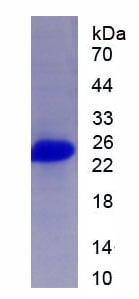
(SDS-PAGE)
SDS-PAGE
(SDS-PAGE)
Ras Related C3 Botulinum Toxin Substrate 1 (Rac1), Recombinant Protein (Cat# AAA152288)
Full Name
Recombinant Ras Related C3 Botulinum Toxin Substrate 1 (Rac1)
Applications
Western Blot
Purity
Greater than 95% by SDS-PAGE
Pricing
 Application Data
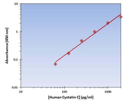
(The OmniKine Human Cystatin C ELISA Kit allows for the detection and quantification of endogenous levels of natural and/or recombinant Human Cystatin C proteins within the range of 63-2000 pg/ml.)
Application Data
(The OmniKine Human Cystatin C ELISA Kit allows for the detection and quantification of endogenous levels of natural and/or recombinant Human Cystatin C proteins within the range of 63-2000 pg/ml.)
Cystatin C, ELISA Kit (Cat# AAA315805)
Full Name
Cystatin C (Human) OmniKine ELISA Kit
Gene Names
CST3; ARMD11; HEL-S-2
Reactivity
Human
Pricing
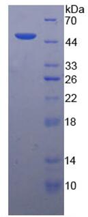
 SDS-PAGE
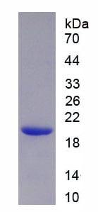
(SDS-PAGE)
SDS-PAGE
(SDS-PAGE)
Programmed Cell Death Protein 1 (PD1), Recombinant Protein (Cat# AAA152039)
Full Name
Recombinant Programmed Cell Death Protein 1 (PD1)
Gene Names
Pdcd1; PD-1; Pdc1; Ly101
Applications
Western Blot
Purity
Greater than 95% by SDS-PAGE
Pricing
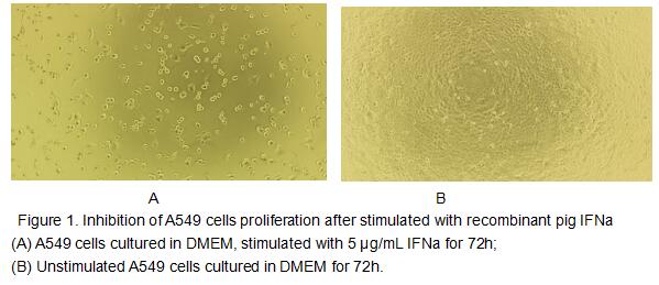
 Bioactivity
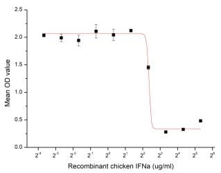
(Figure 2. Inhibition of A549 cells proliferation after stimulated with recombinant chicken IFN-alpha.)
Bioactivity
(Figure 2. Inhibition of A549 cells proliferation after stimulated with recombinant chicken IFN-alpha.)
Interferon Alpha (IFNa), Active Protein (Cat# AAA152952)
Full Name
Active Interferon Alpha (IFNa)
Gene Names
IFNA3; IFNA; IFNA1; IFNA2; IFNA6; IFN-alpha; IFN-gamma
Applications
Activity Assay, Cell Culture
Purity
Greater than 95% by SDS-PAGE
Pricing
 Standard Curve (Sample)
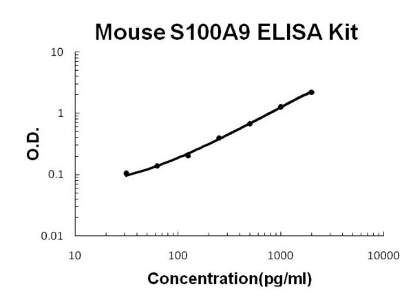
(Mouse S100A9 PicoKine ELISA Kit standard curve)
Standard Curve (Sample)
(Mouse S100A9 PicoKine ELISA Kit standard curve)
Protein S100-A9, ELISA Kit (Cat# AAA46121)
Full Name
Mouse S100A9 PicoKine ELISA Kit
Gene Names
S100a9; p14; Cagb; GAGB; L1Ag; BEE22; MRP14; 60B8Ag; AW546964
Reactivity
Mouse. There is no detectable cross-reactivity with other relevant proteins.
Pricing
 Bioactivity
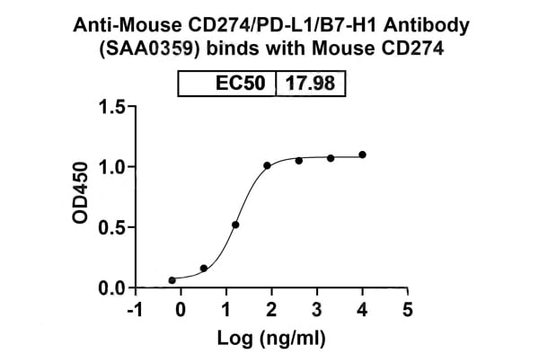
(Detects Mouse CD274/PD-L1/B7-H1 in indirect ELISAs.)
Bioactivity
(Detects Mouse CD274/PD-L1/B7-H1 in indirect ELISAs.)
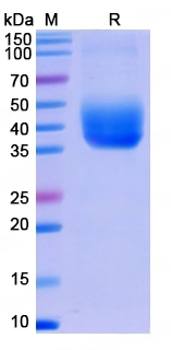
CD274/PD-L1/B7-H1, Recombinant Protein (Cat# AAA120595)
Full Name
Recombinant Mouse CD274/PD-L1/B7-H1 Protein, C-His
Gene Names
Cd274; B7h1; Pdl1; Pdcd1l1; Pdcd1lg1; A530045L16Rik
Applications
Western Blot
Purity
>90% as determined by SDS-PAGE
Pricing