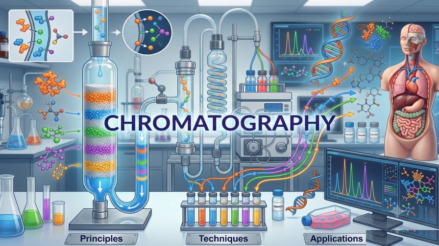
Epitope Mapping: Methods and Why It's Critical for Antibody Development
In this Article
- What Is an Antigenic Determinant?
- Why Epitope Mapping Is Critical for Antibody Development
- Core Epitope Mapping Techniques
- Linear vs. Conformational Epitopes: Choosing the Right Tool
- The Role of Ab Quality in Epitope Mapping
- Computational Epitope Prediction: A Complementary Layer
- Practical Considerations When Selecting Epitope Mapping Services
- Conclusion
All of the products listed in AAA Biotech’s catalog are strictly for research-use only (RUO).

Summary
- Epitope mapping identifies the exact site on an antigen where an antibody binds, enabling precise antibody characterization.
- Core techniques include peptide ELISA, ala-scan epitope mapping, HDX-MS, X-ray crystallography, and cryo-EM.
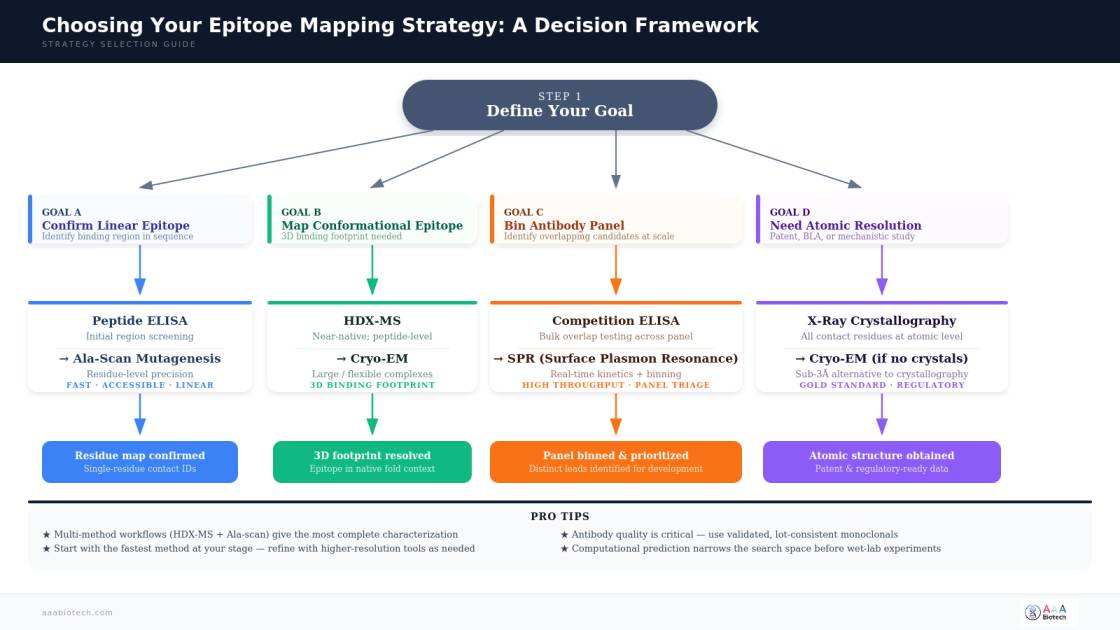
- Choosing the right method depends on epitope type (linear vs. conformational), resolution needed, and development stage.
- Epitope data guides antibody engineering, improves specificity, supports regulatory submissions, and streamlines panel selection.
- High-quality, validated antibodies are essential inputs for any reliable epitope mapping workflow.
Every time an antibody binds a pathogen, a tumor marker, or a cytokine receptor, it does so by contacting a chemically defined region on its target. That region is the epitope, and knowing precisely where an antibody lands is one of the most informative things a researcher can learn about it.
Epitope mapping is the systematic process of identifying and characterizing the binding site. It answers the questions that sit at the core of antibody development: Is this antibody hitting the right location? Does it overlap with a functionally critical domain? Will it compete with or complement other antibodies in a therapeutic combination?
This guide covers what an epitope is, why mapping it matters, the major epitope mapping techniques available today, and the practical considerations that determine which method to use and when.
What Is an Antigenic Determinant?
An antigenic determinant, more commonly called an epitope, is the specific portion of an antigen recognized and physically contacted by an antibody's complementarity-determining regions (CDRs). Most protein antigens carry multiple distinct epitopes, meaning different antibodies raised against the same antigen may bind entirely separate sites.
They fall into two broad categories:
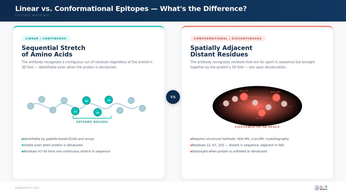
01. Linear (continuous) epitopes: A sequential stretch of amino acids that the Ab recognizes regardless of the antigen's three-dimensional conformation. These are more straightforward to identify with peptide-based methods.
02. Conformational (discontinuous) epitopes: Amino acid residues that are distant in the primary sequence but spatially adjacent in the folded protein. These require structural or biophysical methods to map accurately.
Understanding which category applies to the Ab under study is the essential first step in selecting the right approach.
Why Epitope Mapping Is Critical for Antibody Development

The epitope of an antibody is not merely a molecular address; it governs virtually every aspect of how that Ab performs. Here's why mapping it is non-negotiable:
- Confirming Target Specificity
An antibody may bind a target protein, but if it binds to a structurally conserved domain shared across other proteins in the proteome, cross-reactivity becomes a serious concern. Defining the epitope antigenic confirms whether the antibody is engaging a unique, functionally relevant site or landing somewhere that risks off-target effects.
- Guiding Antibody Engineering and Optimization
Therapeutic antibodies must do specific things, such as block a receptor, neutralize a ligand, or recruit effector cells. The epitope location determines whether an antibody can perform these functions. Mapping informs affinity maturation, humanization, and bispecific antibody design.
- Panel Binning and De-Duplication
When generating large Ab panels against a single antigen, researchers need to identify which candidates bind distinct sites versus overlapping ones. Competitive epitope mapping and epitope binning allow efficient prioritization, so development resources focus on genuinely diverse leads.
- Intellectual Property Differentiation
Demonstrating that a novel therapeutic antibody binds a distinct, previously uncharacterized site is often central to a patent filing strategy in competitive biopharmaceutical development.
- Supporting Regulatory Submissions
Regulatory agencies increasingly expect detailed mechanistic characterization of therapeutic antibodies, including epitope data. This information contributes directly to the scientific rationale required in IND and BLA submissions.
Core Epitope Mapping Techniques
Method selection depends on epitope type, required resolution, throughput, and available infrastructure. Below is a breakdown of the most widely used techniques.
1. Peptide-Based ELISA and Peptide Arrays
Overlapping synthetic peptides, typically 10–20 amino acids in length, spanning the full antigen sequence, are screened for antibody binding by ELISA. Reactive peptides define the approximate binding region.
- Strengths: High throughput, relatively low cost, accessible in most labs.
- Limitations: Resolves only linear epitopes; misses conformational binding sites entirely.
- Best for: Initial screening; confirming linear epitope location in monoclonal Ab characterization.
2. Ala-Scan Epitope Mapping (Alanine Scanning Mutagenesis)
Ala-scan epitope mapping involves systematically substituting individual residues within a candidate epitope region with Alanine, a small, chemically inert amino acid that does not introduce steric or electrostatic artifacts. When antibody binding is disrupted by a specific substitution, that residue is flagged as a key contact.
This approach delivers single-residue resolution and is most powerful when an approximate epitope region has already been identified. It is commonly used after initial peptide ELISA screening to sharpen the epitope definition to functionally critical residues.
- Strengths: Residue-level resolution; identifies key contact residues directly.
- Limitations: Labor-intensive; requires prior knowledge of the approximate epitope location.
- Best for: Therapeutic antibody characterization; lead optimization; mapping functional hotspots.
3. Hydrogen–Deuterium Exchange Mass Spectrometry (HDX-MS)
HDX-MS exploits the natural exchange of backbone amide hydrogens with deuterium in solution. When an antibody binds its antigen, it physically protects the epitope region from solvent exchange. By comparing deuterium incorporation in the bound versus unbound antigen, the binding footprint is resolved at the peptide level, under near-native solution conditions.
- Strengths: Works under physiological conditions; suitable for conformational epitopes; no labeled antigens required.
- Limitations: Peptide-level (not single-residue) resolution; requires specialized mass spectrometry infrastructure.
- Best for: Conformational mapping; early-stage therapeutic candidates.
4. X-Ray Crystallography
The gold standard for structural epitope mapping. Crystallography resolves the atomic-level three-dimensional structure of the antibody–antigen complex, unambiguously identifying all contact residues and the precise geometry of the binding interface.
- Strengths: Atomic resolution; definitive for both linear and conformational epitopes.
- Limitations: Time-consuming; technically demanding; requires crystallizable complexes; may not reflect native solution conditions.
- Best for: Late-stage therapeutic development; mechanistic studies; patent support.
5. Cryo-Electron Microscopy (Cryo-EM)
Cryo-EM has emerged as a powerful alternative for large, flexible, or membrane-associated antigen–antibody complexes that resist crystallization. Recent technological advances have brought cryo-EM resolution below 3 Å for well-behaved samples.
- Strengths: No crystallization required; handles large, flexible complexes.
- Limitations: Still requires significant infrastructure; lower throughput than peptide methods.
- Best for: Large antigen targets such as viral glycoproteins; complexes that cannot be crystallized.
6. Epitope Binning by Competition Assay
Competition ELISAs or surface plasmon resonance (SPR) experiments determine whether two antibodies can bind the target simultaneously. Those that block each other are grouped into overlapping "bins."
- Strengths: High throughput; excellent for panel prioritization.
- Limitations: Does not reveal the actual epitope location, only relative overlap between antibodies.
- Best for: Large-scale Ab panel screening; early-stage hit selection and triage.
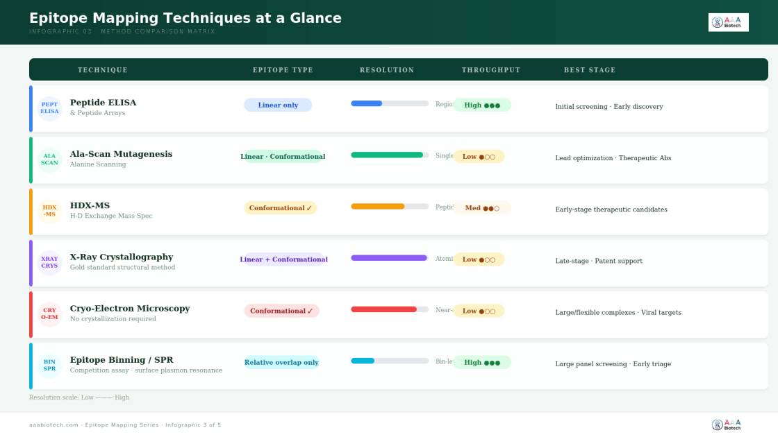
An Overview of the Major Techniques
Linear vs. Conformational Epitopes: Choosing the Right Tool
The distinction between epitope types has direct, practical consequences for method selection. A peer-reviewed study published in mAbs comparing six mapping methods across five therapeutic antibody–antigen pairs found that peptide arrays successfully identified partial epitopes in only 2 of 5 cases, while hydrogen–deuterium exchange and structural methods revealed binding regions that peptide arrays missed entirely.
This underscores a critical principle: no single method covers all types, and multi-method workflows are often necessary for comprehensive characterization of a therapeutic candidate.
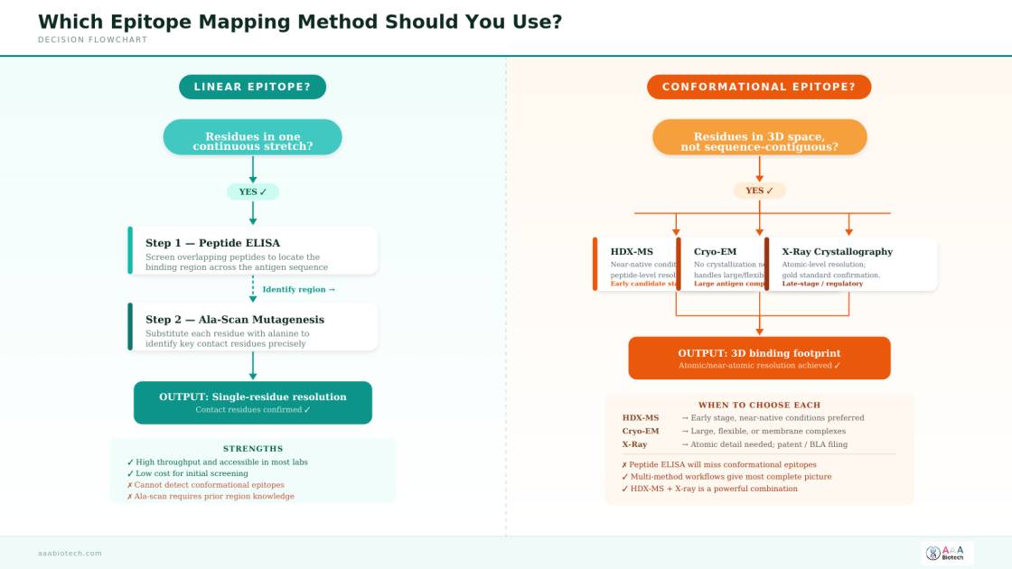
For conformational epitopes, which represent a large and functionally important proportion of antibody binding sites in drug development, only methods that preserve the native three-dimensional architecture of the antigen will deliver reliable results. HDX-MS, cryo-EM, and crystallography are the appropriate primary tools in this space.
The Role of Ab Quality in Epitope Mapping
Epitope mapping data are only as reliable as the antibody being characterized. Poorly validated or lot-inconsistent antibodies introduce significant noise into any mapping experiment, making results difficult to reproduce or interpret.
Before beginning any workflow, the antibody should be:
- Rigorously validated for specificity in the intended application. Understanding how antibody validation improves experimental reproducibility is directly relevant to mapping workflow design.
- Well-characterized for isotype, binding affinity (Kd), and kinetics.
- Lot-consistent, particularly when mapping data will be used across multiple labs or longitudinal studies.
Monoclonal antibodies are the preferred starting material for rigorous mapping experiments. Their defined, single-clone specificity eliminates the ambiguity introduced by the polyclonal mixture of binding events present in serum preparations, giving cleaner, more interpretable results.
For structural methods such as X-ray crystallography or cryo-EM, the purity (typically >95% by SDS-PAGE), conformational homogeneity, and stability of both the Ab (usually as a Fab fragment) and the antigen are critical determinants of whether high-resolution structures can be obtained.
Computational Epitope Prediction: A Complementary Layer
Experimental mapping is resource-intensive by nature. Computational tools can serve as a valuable complementary layer by narrowing the search space before committing to wet-lab experiments.
Machine learning algorithms trained on structurally resolved antibody–antigen complexes from the Protein Data Bank can predict likely epitope residues based on surface accessibility, evolutionary conservation, and physicochemical properties.
A 2024 review in Frontiers in Immunology demonstrated that integrating machine learning with experimental epitope mapping improves efficiency and enhances prediction of conformational epitopes, particularly for therapeutically relevant targets where off-target binding carries serious consequences.
The key caveat: computational predictions are hypothesis-generating tools, not hypothesis-confirming ones. They direct attention toward likely binding regions — experimental methods confirm which residues actually matter.
Practical Considerations When Selecting Epitope Mapping Services
Many research and development teams do not maintain in-house structural biology or mass spectrometry infrastructure, making it practical and common to work with specialized epitope mapping services. Key factors to evaluate include:
- Technique breadth: Does the provider offer multiple methods, including structural approaches for conformational epitopes?
- Antigen complexity: Can the team handle glycosylated, membrane-associated, or otherwise challenging target proteins?
- Turnaround time: Are timelines compatible with the stage of development?
- Data deliverables: Are results provided at residue-level resolution, with full structural data files where applicable?
- Integration with downstream workflows: Can the mapping data feed directly into antibody engineering or formulation decisions?
Here is a Quick Glance:
Regardless of provider, the starting point is always a clearly defined scientific question. Are you confirming a hypothesized binding site, distinguishing competing antibodies, investigating a cross-reactivity problem, or generating data for a regulatory filing? Researchers must also explore the range of recombinant antibodies available as well-characterized starting points for mapping workflows.
Conclusion
Epitope mapping sits at the intersection of structural biology, immunology, and pharmaceutical development. Whether the goal is confirming why an antibody works, optimizing it for a therapeutic application, or building a characterization package for a regulatory submission, knowing exactly where an Ab binds is foundational knowledge, not optional context.
The field has expanded well beyond peptide scans. Today's toolkit, spanning ala-scan epitope mapping, HDX-MS, cryo-EM, machine learning-assisted prediction, and atomic-resolution crystallography, gives research and development teams the resolution and throughput to answer questions at every stage of the pipeline.
High-quality, rigorously characterized antibodies remain the essential raw material for any of these approaches. The time invested in thorough characterization pays back in cleaner data, fewer late-stage surprises, and a stronger scientific foundation for the programs that follow.
Faq's
What is the difference between an epitope and a paratope?
An epitope is the specific region on the antigen that an antibody recognizes. A paratope is the structurally complementary region on the antibody itself, formed by the CDR loops, that physically contacts the epitope. The two are mirror images of each other at the molecular level: the paratope is shaped to fit the epitope, much like a key fits a lock.
Can the same antigen have multiple epitopes recognized by different antibodies?
Yes. Most protein antigens contain several distinct epitopes accessible to antibodies. It is entirely possible for two antibodies raised against the same protein to bind completely non-overlapping sites, which is why epitope binning and panel diversification are important considerations in antibody development programs.
How does epitope mapping differ in its goals between therapeutic and diagnostic antibody development?
In therapeutic development, the epitope location often directly determines the mechanism of action, for example, whether an antibody blocks a protein–protein interaction or merely binds without functional consequence. In diagnostics, the priority shifts to identifying an accessible, unique epitope that gives a robust signal in complex biological matrices without cross-reacting with structurally related proteins.
Is computational epitope prediction reliable enough to replace experimental validation?
Not at present. Computational tools are most useful for generating hypotheses and narrowing the experimental search space. They cannot reliably resolve conformational epitopes or identify functionally critical contact residues with the precision required for therapeutic development or regulatory filings. Experimental validation, through techniques such as alanine scanning, HDX-MS, or crystallography, remains essential.
What impact does the antibody fragment type (IgG vs. Fab) have on structural epitope mapping?
For X-ray crystallography and cryo-EM, the Fab fragment (rather than intact IgG) is typically preferred because its smaller, more rigid structure is more amenable to crystallization and produces higher-resolution EM reconstructions. The Fc region of intact IgG adds flexibility and size that can complicate structural studies. Using Fab fragments does not affect epitope characterization since the CDRs, which are the actual binding regions, are fully preserved.
Lead Clinical Research Coordinator (LCRC)
Cynthia Lee is the President of AAA Biotech and specializes in understanding highly validated and characterized monoclonal/polyclonal antibodies, recombinant proteins, and ELISA kits.
Related Posts