Filters
▼Clonality
▼Type
▼Reactivity
▼Gene Name
▼Isotype
▼Host
▼Application
▼Clone
▼Polyclonal Antibodies
At AAA Biotech also known as AAA Bio or AAABio, we provide a broad range of purified polyclonal antibodies (pAbs) that are able to all be browsed online through our website. Due to their high specificity and strong binding affinity, these antibodies are ideal for wide swathes of research and experimental applications.
Our polyclonal antibodies can easily support your work, whether you use them for Western Blotting, Immunocytochemistry (with or without Immunofluorescence used in conjunction), Immunohistochemistry, Immunoprecipitation, and ELISA tests. We highly encourage you to browse our range of pAbs and choose the one that best suits your experimental model.
Viewing 1250-1300 of 118597 product results
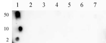
 DB (Dot Blot)
(Histone H4 monomethyl Lys31 pAb tested by dot blot analysis. Dot blot analysis was used to confirm the specificity of Histone H4 monomethyl Lys31 pAb for monomethyl Lys31 histone H4. Methylated peptides corresponding to the immunogen were spotted onto PVDF and probed with the antibody at 1:5,000. The amount of peptide (picomoles) spotted is indicated next to each row. Lane 1: Unmodified peptide corresponding to amino acids 28-36 of human histone H4 Lane 2: Monomethyl Lys31 Lane 3: Dimethyl Lys31; Lane 4: Trimethyl Lys31 Lane 5: Unmodified peptide corresponding to amino acids 41-49 of human histone H4 Lane 6: Monomethyl Lys44 Lane 7: Dimethyl-Lys44 Lane 8: Trimethyl Lys44. No detection of peptides (mono-, di-, or trimethylated) corresponding to lysine 4, 9, 18, 23 or 27 of histone H3 was observed with this antibody (data not shown).)
DB (Dot Blot)
(Histone H4 monomethyl Lys31 pAb tested by dot blot analysis. Dot blot analysis was used to confirm the specificity of Histone H4 monomethyl Lys31 pAb for monomethyl Lys31 histone H4. Methylated peptides corresponding to the immunogen were spotted onto PVDF and probed with the antibody at 1:5,000. The amount of peptide (picomoles) spotted is indicated next to each row. Lane 1: Unmodified peptide corresponding to amino acids 28-36 of human histone H4 Lane 2: Monomethyl Lys31 Lane 3: Dimethyl Lys31; Lane 4: Trimethyl Lys31 Lane 5: Unmodified peptide corresponding to amino acids 41-49 of human histone H4 Lane 6: Monomethyl Lys44 Lane 7: Dimethyl-Lys44 Lane 8: Trimethyl Lys44. No detection of peptides (mono-, di-, or trimethylated) corresponding to lysine 4, 9, 18, 23 or 27 of histone H3 was observed with this antibody (data not shown).)
Histone H4K31me1, Polyclonal Antibody (Cat# AAA59869)
 DB (Dot Blot)
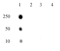
(Histone H3 acetyl Lys9 pAb tested by dot blot analysis. Dot blot analysis was used to confirm the specificity of Histone H3 acetyl Lys9 pAb for acetyl Lys9 histone H3. Acetylated peptides corresponding to the immunogen and related peptides were spotted onto PVDF and probed with the antibody at a dilution of 1:5,000. The amount of peptide (picomoles) spotted is indicated next to each row. Lane 1: acetyl-Lys9 peptide. Lane 2: unmodified Lys9 peptide. Lane 3: acetyl-Lys14 peptide. Lane 4: unmodified Lys14 peptide. Lane 5: acetyl-Lys18 peptide. Lane 6: acetyl-Lys23 peptide. Lane 7: acetyl-Lys27 peptide.)
DB (Dot Blot)
(Histone H3 acetyl Lys9 pAb tested by dot blot analysis. Dot blot analysis was used to confirm the specificity of Histone H3 acetyl Lys9 pAb for acetyl Lys9 histone H3. Acetylated peptides corresponding to the immunogen and related peptides were spotted onto PVDF and probed with the antibody at a dilution of 1:5,000. The amount of peptide (picomoles) spotted is indicated next to each row. Lane 1: acetyl-Lys9 peptide. Lane 2: unmodified Lys9 peptide. Lane 3: acetyl-Lys14 peptide. Lane 4: unmodified Lys14 peptide. Lane 5: acetyl-Lys18 peptide. Lane 6: acetyl-Lys23 peptide. Lane 7: acetyl-Lys27 peptide.)
Histone H3K9ac, Polyclonal Antibody (Cat# AAA59887)
 DB (Dot Blot)
(STAT3 phospho Tyr705 pAb tested by dot blot analysis. Dot blot analysis was used to confirm the specificity of STAT3 phospho Tyr705 pAb for STAT3 phospho Tyr705. Phosphorylated peptides corresponding to the immunogen and related peptides were spotted onto PVDF and probed with the antibody at 1:10,000. The amount of peptide (picomoles) spotted is indicated next to each row. Lane 1: Unmodified Ser727 STAT1 peptide. Lane 2: Phospho Ser727 STAT1 peptide. Lane 3: Unmodified Tyr689 STAT2 peptide. Lane 4: Phospho Tyr689 STAT2 peptide. Lane 5: Unmodified Ser727 STAT3 peptide. Lane 6: Phospho Ser727 STAT3 peptide. Lane 7: Unmodified Tyr705 STAT3 peptide. Lane 8: Phospho Tyr705 STAT3 peptide. Lane 9: Unmodified Ser726 STAT5A/Ser731 STAT5B peptide. Lane 10: Phospho Ser726 STAT5A/Ser731 STAT5B peptide. Lane 11: Unmodified Tyr694 STAT5A/Tyr699 STAT5B peptide. Lane 12: Phospho Tyr694 STAT5A/Tyr699 STAT5B peptide.)
DB (Dot Blot)
(STAT3 phospho Tyr705 pAb tested by dot blot analysis. Dot blot analysis was used to confirm the specificity of STAT3 phospho Tyr705 pAb for STAT3 phospho Tyr705. Phosphorylated peptides corresponding to the immunogen and related peptides were spotted onto PVDF and probed with the antibody at 1:10,000. The amount of peptide (picomoles) spotted is indicated next to each row. Lane 1: Unmodified Ser727 STAT1 peptide. Lane 2: Phospho Ser727 STAT1 peptide. Lane 3: Unmodified Tyr689 STAT2 peptide. Lane 4: Phospho Tyr689 STAT2 peptide. Lane 5: Unmodified Ser727 STAT3 peptide. Lane 6: Phospho Ser727 STAT3 peptide. Lane 7: Unmodified Tyr705 STAT3 peptide. Lane 8: Phospho Tyr705 STAT3 peptide. Lane 9: Unmodified Ser726 STAT5A/Ser731 STAT5B peptide. Lane 10: Phospho Ser726 STAT5A/Ser731 STAT5B peptide. Lane 11: Unmodified Tyr694 STAT5A/Tyr699 STAT5B peptide. Lane 12: Phospho Tyr694 STAT5A/Tyr699 STAT5B peptide.)
STAT3 phospho Tyr705, Polyclonal Antibody (Cat# AAA59891)
 DB (Dot Blot)
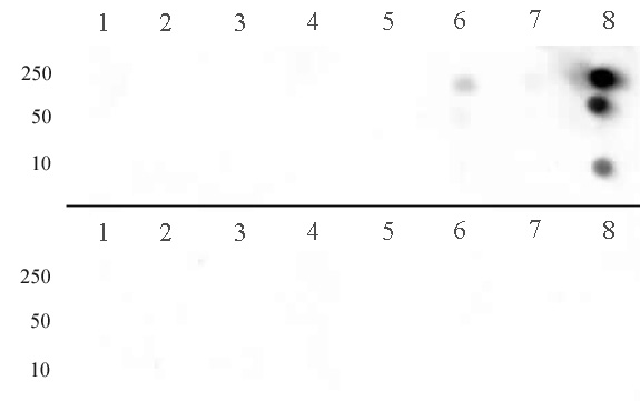
(Histone H3 dimethyl Arg8 asymmetric pAb tested by dot blot analysis. Dot blot analysis was used to confirm the specificity of Histone H3 dimethyl Arg8 asymmetric pAb for dimethyl-arginine 8 of histone H3. Peptides corresponding to the immunogen and related peptides were spotted onto PVDF and probed with Histone H3 dimethyl Arg8 asymmetric pAb at 1:30,000. The amount of peptide (picomoles) spotted is indicated next to each row. Top Panel: Lane 1: unmod Arg2. Lane 2: monomethyl-Arg2 H3. Lane 3: dimethyl-Arg2 H3 (sym). Lane 4: dimethyl-Arg2 H3 (asym). Lane 5: unmod Arg8. Lane 6: monomethyl-Arg8 H3. Lane 7: dimethyl-Arg8 H3 (sym). Lane 8: dimethyl-Arg8 H3 (asym). Bottom Panel: Lane 1: unmod Arg17 H3. Lane 2: monomethyl-Arg17. Lane 3: dimethyl-Arg17 H3 (sym). Lane 4: dimethyl-Arg17 H3 (asym). Lane 5: unmod Arg26. Lane 6: monomethyl-Arg26 H3. Lane 7: dimethyl-Arg26 H3 (sym). Lane 8: dimethyl-Arg26 H3 (asymmetric). No detection of peptides (unmodified, mono-, di-, or tri-methylated) corresponding to Lys4, Lys9, Lys18, Lys23 and Lys27 of histone H3 was observed.)
DB (Dot Blot)
(Histone H3 dimethyl Arg8 asymmetric pAb tested by dot blot analysis. Dot blot analysis was used to confirm the specificity of Histone H3 dimethyl Arg8 asymmetric pAb for dimethyl-arginine 8 of histone H3. Peptides corresponding to the immunogen and related peptides were spotted onto PVDF and probed with Histone H3 dimethyl Arg8 asymmetric pAb at 1:30,000. The amount of peptide (picomoles) spotted is indicated next to each row. Top Panel: Lane 1: unmod Arg2. Lane 2: monomethyl-Arg2 H3. Lane 3: dimethyl-Arg2 H3 (sym). Lane 4: dimethyl-Arg2 H3 (asym). Lane 5: unmod Arg8. Lane 6: monomethyl-Arg8 H3. Lane 7: dimethyl-Arg8 H3 (sym). Lane 8: dimethyl-Arg8 H3 (asym). Bottom Panel: Lane 1: unmod Arg17 H3. Lane 2: monomethyl-Arg17. Lane 3: dimethyl-Arg17 H3 (sym). Lane 4: dimethyl-Arg17 H3 (asym). Lane 5: unmod Arg26. Lane 6: monomethyl-Arg26 H3. Lane 7: dimethyl-Arg26 H3 (sym). Lane 8: dimethyl-Arg26 H3 (asymmetric). No detection of peptides (unmodified, mono-, di-, or tri-methylated) corresponding to Lys4, Lys9, Lys18, Lys23 and Lys27 of histone H3 was observed.)
Histone H3R8me2a, Polyclonal Antibody (Cat# AAA59906)
 DB (Dot Blot)
(HP1 gamma phospho Ser93 pAb tested by dot blot analysis. Dot blot analysis was used to confirm the specificity of HP1 gamma phospho Ser93 pAb for HP1 gamma phospho Ser93. Phosphorylated peptides corresponding to the immunogen and related peptides were spotted onto PVDF and probed with HP1 gamma phospho Ser93 pAb at 1:5,000. The amount of peptide (picomoles) spotted is indicated next to each row. Lane 1: unmodified HP1 gamma peptide. Lane 2: phospho Ser93 HP1 gamma peptide. Lane 3: phospho Ser92 HP1 alpha peptide. Lane 4: phospho Ser89 HP1 beta peptide.)
DB (Dot Blot)
(HP1 gamma phospho Ser93 pAb tested by dot blot analysis. Dot blot analysis was used to confirm the specificity of HP1 gamma phospho Ser93 pAb for HP1 gamma phospho Ser93. Phosphorylated peptides corresponding to the immunogen and related peptides were spotted onto PVDF and probed with HP1 gamma phospho Ser93 pAb at 1:5,000. The amount of peptide (picomoles) spotted is indicated next to each row. Lane 1: unmodified HP1 gamma peptide. Lane 2: phospho Ser93 HP1 gamma peptide. Lane 3: phospho Ser92 HP1 alpha peptide. Lane 4: phospho Ser89 HP1 beta peptide.)
HP1 gamma phospho Ser93, Polyclonal Antibody (Cat# AAA59908)
 DB (Dot Blot)
(RNA Pol II CTD phospho Thr4 pAb tested by dot blot analysis. Dot blot analysis was used to confirm the specificity of RNA Pol II CTD phospho Thr4 pAb. Peptides corresponding to the immunogen and related peptides were spotted onto PVDF and probed with the antibody at a dilution of 1 ug/ml. The amount of peptide (picomoles) spotted is indicated next to each row. Lane 1: Phospho Thr4 of RNA Pol II CTD peptide. Lane 2: Unmodified Thr4 of RNA Pol II CTD peptide. Lane 3: Phospho Ser2 of RNA Pol II CTD peptide. Lane 4: Phospho Ser5 of RNA Pol II CTD peptide.)
DB (Dot Blot)
(RNA Pol II CTD phospho Thr4 pAb tested by dot blot analysis. Dot blot analysis was used to confirm the specificity of RNA Pol II CTD phospho Thr4 pAb. Peptides corresponding to the immunogen and related peptides were spotted onto PVDF and probed with the antibody at a dilution of 1 ug/ml. The amount of peptide (picomoles) spotted is indicated next to each row. Lane 1: Phospho Thr4 of RNA Pol II CTD peptide. Lane 2: Unmodified Thr4 of RNA Pol II CTD peptide. Lane 3: Phospho Ser2 of RNA Pol II CTD peptide. Lane 4: Phospho Ser5 of RNA Pol II CTD peptide.)
RNA pol II CTD phospho Thr4, Polyclonal Antibody (Cat# AAA60008)
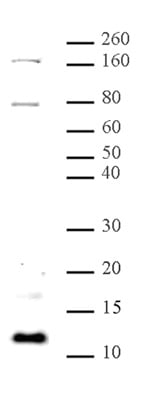
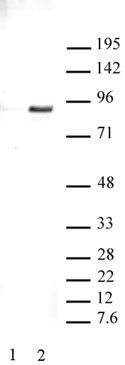
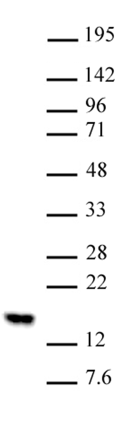
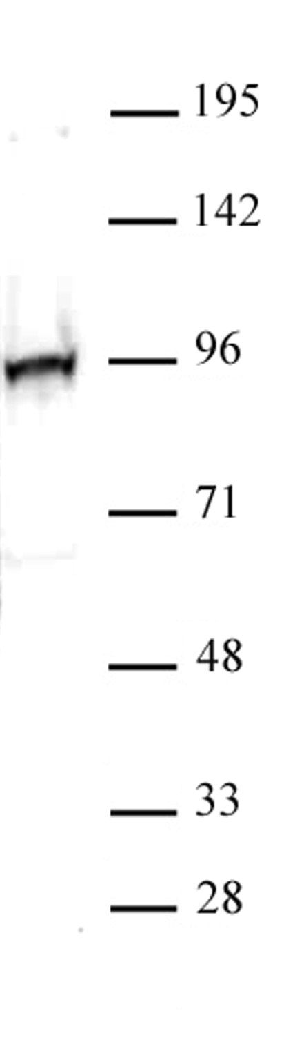
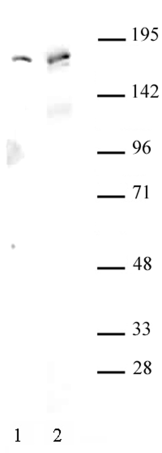
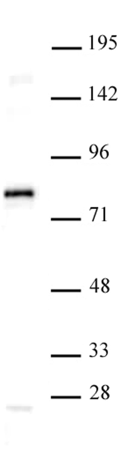
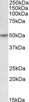
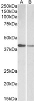
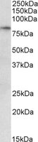
 WB (Western Blot)
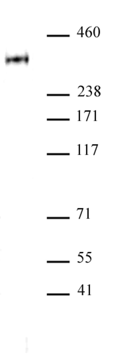
(ATAD2 antibody (pAb) tested by Western blot. ATAD2 detection by Western blot. The analysis was performed using 40 ug Jurkat nuclear cell extract and CENP-B at a 1:1,000 dilution.)
WB (Western Blot)
(ATAD2 antibody (pAb) tested by Western blot. ATAD2 detection by Western blot. The analysis was performed using 40 ug Jurkat nuclear cell extract and CENP-B at a 1:1,000 dilution.)
ATAD2, Polyclonal Antibody (Cat# AAA60014)
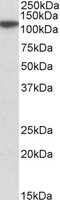
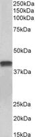
 WB (Western Blot)
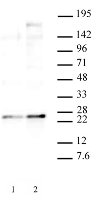
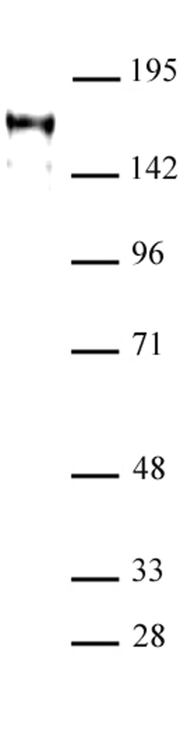
(JunD antibody (pAb) tested by Western blot. K-562 nuclear extract (20 ug) probed with JunD antibody at a dilution of 1:1,000.)
WB (Western Blot)
(JunD antibody (pAb) tested by Western blot. K-562 nuclear extract (20 ug) probed with JunD antibody at a dilution of 1:1,000.)
JunD, Polyclonal Antibody (Cat# AAA60021)
 DB (Dot Blot)
(Histone H2BK5me1 (pAb) tested by dot blot analysis. Dot blot analysis was used to confirm the specificity of Histone H2BK5me1 pAb for monomethyl-Lys5 of histone H2B. Decreasing amounts of modified and unmodified peptides were spotted onto PVDF and probed with the antibody at a dilution of 1:10,000. Lane 1: Unmodified lysine 5 peptide. Lane 2: Peptide monomethylated at lysine 5 of H2B. Lane 3: Peptide dimethylated at lysine 5 of H2B. Lane 4: Peptide trimethylated at lysine 5 of H2B.)
DB (Dot Blot)
(Histone H2BK5me1 (pAb) tested by dot blot analysis. Dot blot analysis was used to confirm the specificity of Histone H2BK5me1 pAb for monomethyl-Lys5 of histone H2B. Decreasing amounts of modified and unmodified peptides were spotted onto PVDF and probed with the antibody at a dilution of 1:10,000. Lane 1: Unmodified lysine 5 peptide. Lane 2: Peptide monomethylated at lysine 5 of H2B. Lane 3: Peptide dimethylated at lysine 5 of H2B. Lane 4: Peptide trimethylated at lysine 5 of H2B.)
Histone H2BK5me1, Polyclonal Antibody (Cat# AAA60028)

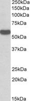
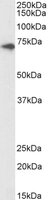
 WB (Western Blot)
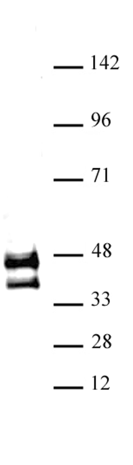
(MED15 antibody (pAb) tested by Western blot. HeLa whole-cell extract (20 ug) probed with MED15 antibody at a dilution of 1:1,000.)
WB (Western Blot)
(MED15 antibody (pAb) tested by Western blot. HeLa whole-cell extract (20 ug) probed with MED15 antibody at a dilution of 1:1,000.)
MED15, Polyclonal Antibody (Cat# AAA60052)
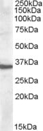
 WB (Western Blot)
(NRF2 antibody (pAb) tested by Western blot. Nuclear extract of HepG2 cells (25 ug) treated with tHBQ and probed with NRF2 antibody at a dilution of 1:500.)
WB (Western Blot)
(NRF2 antibody (pAb) tested by Western blot. Nuclear extract of HepG2 cells (25 ug) treated with tHBQ and probed with NRF2 antibody at a dilution of 1:500.)
NRF2, Polyclonal Antibody (Cat# AAA60053)
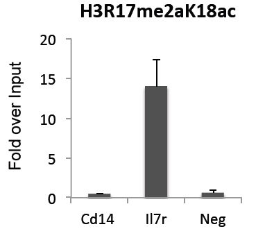
 DB (Dot Blot)
(Histone H3R17me2aK18ac antibody (pAb) tested by dot blot analysis. Dot blot analysis was used to confirm the specificity of Histone H3R17me2aK18ac antibody for asymmetric dimethyl-arginine 17 and acetyl-lysine 18 of histone H3. Peptides corresponding to the immunogen and related peptides were spotted onto PVDF and probed with H3R17me2aK18ac antibody at a 1:500 dilution. The amount of peptide (picomoles) spotted is indicated next to each row. Lane 1: Peptide containing asymmetric dimethyl-Arg17 and acetyl-Lys18 of Histone H3 . Lane 2: unmodified Histone H3 peptide. Lane 3: Asymmetric dimethyl-Arg17 of Histone H3 peptide. Lane 4: Acetyl-Lys18 of Histone H3 peptide.)
DB (Dot Blot)
(Histone H3R17me2aK18ac antibody (pAb) tested by dot blot analysis. Dot blot analysis was used to confirm the specificity of Histone H3R17me2aK18ac antibody for asymmetric dimethyl-arginine 17 and acetyl-lysine 18 of histone H3. Peptides corresponding to the immunogen and related peptides were spotted onto PVDF and probed with H3R17me2aK18ac antibody at a 1:500 dilution. The amount of peptide (picomoles) spotted is indicated next to each row. Lane 1: Peptide containing asymmetric dimethyl-Arg17 and acetyl-Lys18 of Histone H3 . Lane 2: unmodified Histone H3 peptide. Lane 3: Asymmetric dimethyl-Arg17 of Histone H3 peptide. Lane 4: Acetyl-Lys18 of Histone H3 peptide.)
Histone H3R17me2aK18ac, Polyclonal Antibody (Cat# AAA60055)
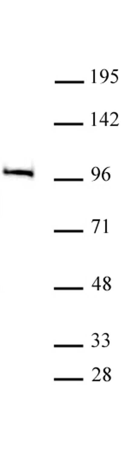
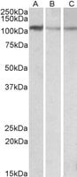
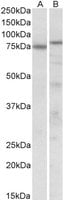
 WB (Western Blot)
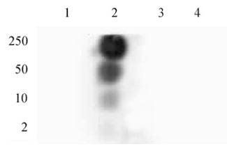
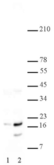
(AIB1 antibody (pAb) tested by Western blot. Detection of AIB1 by Western blot. Lane 1: HeLa whole-cell extract (30 ug). Lane 2: Whole cell extract (30 ug) of MCF-7 cells. Both probed with AIB1 antibody at a 1:500 dilution.)
WB (Western Blot)
(AIB1 antibody (pAb) tested by Western blot. Detection of AIB1 by Western blot. Lane 1: HeLa whole-cell extract (30 ug). Lane 2: Whole cell extract (30 ug) of MCF-7 cells. Both probed with AIB1 antibody at a 1:500 dilution.)
AIB1/SRC-3, Polyclonal Antibody (Cat# AAA59931)
 WB (Western Blot)
(GATA-6 antibody (pAb) tested by Western blot. Nuclear extract (20 ug) of SW48 cells probed with GATA-6 antibody at a dilution of 1:500.)
WB (Western Blot)
(GATA-6 antibody (pAb) tested by Western blot. Nuclear extract (20 ug) of SW48 cells probed with GATA-6 antibody at a dilution of 1:500.)
GATA-6, Polyclonal Antibody (Cat# AAA59971)
 WB (Western Blot)
(Western blot of TCF7L1 / TCF3 antibody. Nuclear extract of PANC-1 cells (20 ?g) probed with TCF7L1 / TCF3 antibody (1:500).)
WB (Western Blot)
(Western blot of TCF7L1 / TCF3 antibody. Nuclear extract of PANC-1 cells (20 ?g) probed with TCF7L1 / TCF3 antibody (1:500).)
TCF7L1/TCF3, Polyclonal Antibody (Cat# AAA59982)
 Application Data
(Histone H4R3me2a (asymmetric) antibody (pAb) specificity tested by peptide array analysis. Peptide array analysis was used to confirm the specificity of this antibody for its intended modification. Histone H4R3me2a antibody was applied at a dilution of 1:20,000 to MODified Histone Peptide Array . The arrays were scanned with ArrayAnalysis Software 16 and the results plotted. Specificity data is shown for the most reactive peptides and those related to the immunogen.)
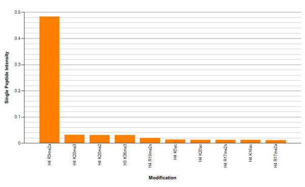
Application Data
(Histone H4R3me2a (asymmetric) antibody (pAb) specificity tested by peptide array analysis. Peptide array analysis was used to confirm the specificity of this antibody for its intended modification. Histone H4R3me2a antibody was applied at a dilution of 1:20,000 to MODified Histone Peptide Array . The arrays were scanned with ArrayAnalysis Software 16 and the results plotted. Specificity data is shown for the most reactive peptides and those related to the immunogen.)
Histone H4R3me2a, Polyclonal Antibody (Cat# AAA59792)
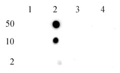
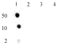
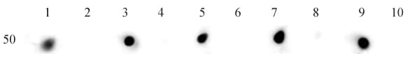 DB (Dot Blot)
(Histone H3ac (pan-acetyl) antibody (pAb) tested by dot blot analysis. Dot blot analysis was used to confirm the specificity of Histone H3ac antibody. Acetylated peptides corresponding to the immunogen and related peptides were spotted onto PVDF and probed with the antibody at a dilution of 1:1,000. The amount of peptide (picomoles) spotted is indicated (50 picomoles). Lane 1: H3K4ac peptide. Lane 2: unmodified H3K4 peptide. Lane 3: H3K9ac peptide. Lane 4: unmodified H3K9 peptide. Lane 5: H3K18ac peptide. Lane 6: unmodified H3K18 peptide. Lane 7: H3K23ac peptide. Lane 8: unmodified H3K23 peptide. Lane 9: H3K27ac peptide. Lane 10: unmodified H3K27 peptide.)
DB (Dot Blot)
(Histone H3ac (pan-acetyl) antibody (pAb) tested by dot blot analysis. Dot blot analysis was used to confirm the specificity of Histone H3ac antibody. Acetylated peptides corresponding to the immunogen and related peptides were spotted onto PVDF and probed with the antibody at a dilution of 1:1,000. The amount of peptide (picomoles) spotted is indicated (50 picomoles). Lane 1: H3K4ac peptide. Lane 2: unmodified H3K4 peptide. Lane 3: H3K9ac peptide. Lane 4: unmodified H3K9 peptide. Lane 5: H3K18ac peptide. Lane 6: unmodified H3K18 peptide. Lane 7: H3K23ac peptide. Lane 8: unmodified H3K23 peptide. Lane 9: H3K27ac peptide. Lane 10: unmodified H3K27 peptide.)
Histone H3ac, Polyclonal Antibody (Cat# AAA59800)
 DB (Dot Blot)
(Histone H2A/H4 phospho Ser1 pAb tested by dot blot analysis. Dot blot analysis was used to confirm the specificity of Histone H2A/H4 phospho Ser1 pAb for phospho Ser1 of Histone H2A/H4. Modified and unmodified peptides were spotted onto PVDF and probed with the antibody at a 1:10,000 dilution. The amount of peptide spotted (in picomoles) is indicated next to each row. Lane 1: Peptide phosphorylated at Ser1. Lane 2: Unmodified Ser1 peptide.)
DB (Dot Blot)
(Histone H2A/H4 phospho Ser1 pAb tested by dot blot analysis. Dot blot analysis was used to confirm the specificity of Histone H2A/H4 phospho Ser1 pAb for phospho Ser1 of Histone H2A/H4. Modified and unmodified peptides were spotted onto PVDF and probed with the antibody at a 1:10,000 dilution. The amount of peptide spotted (in picomoles) is indicated next to each row. Lane 1: Peptide phosphorylated at Ser1. Lane 2: Unmodified Ser1 peptide.)
Histone H2A/H4S1ph, Polyclonal Antibody (Cat# AAA59830)
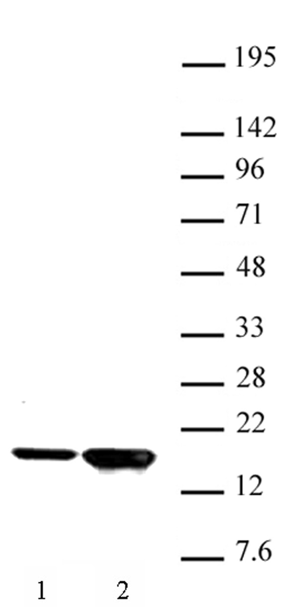
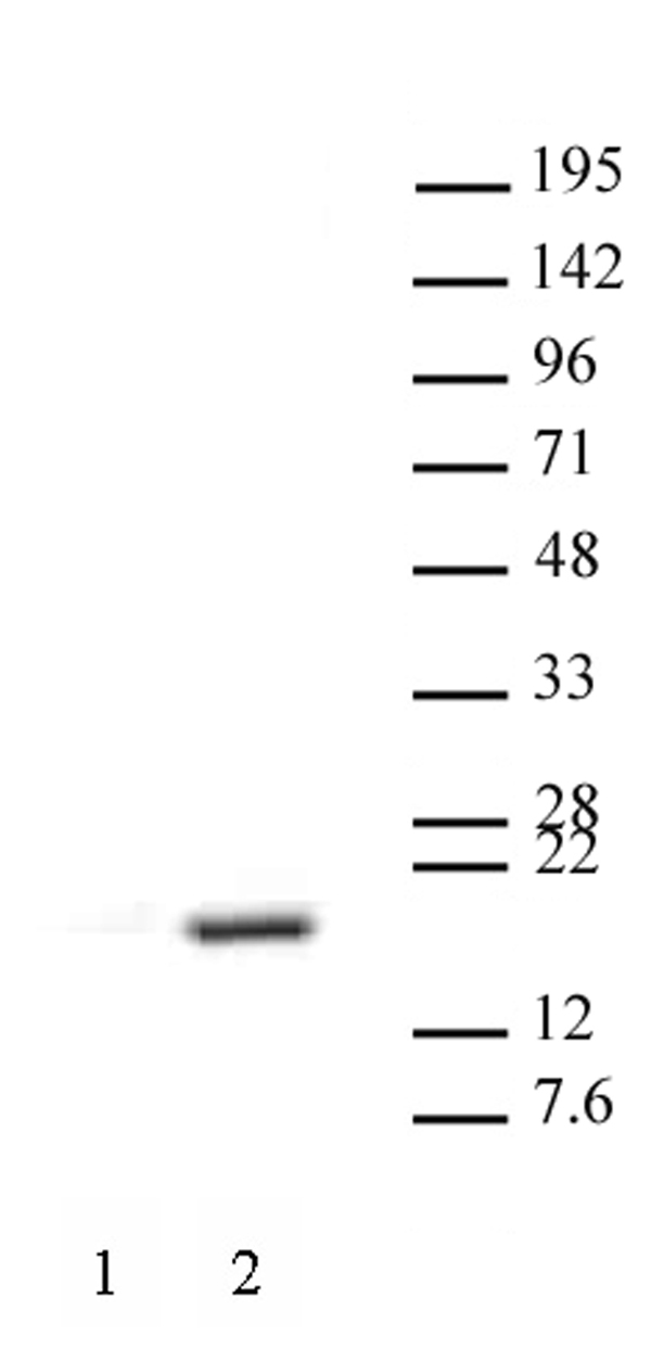
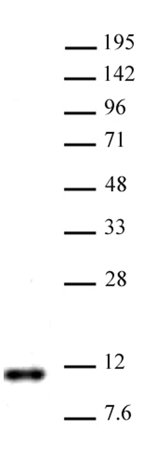
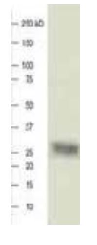
 WB (Western Blot)
(Western blot detection of purified human immunodeficiency virus integrase (HIV-1 Integrase). Primary antibody (1:10,000 dilution) and AP-conjugated anti-rabbit IgG secondary antibody. (Invitrogen; 1:10,000 dilution).)
WB (Western Blot)
(Western blot detection of purified human immunodeficiency virus integrase (HIV-1 Integrase). Primary antibody (1:10,000 dilution) and AP-conjugated anti-rabbit IgG secondary antibody. (Invitrogen; 1:10,000 dilution).)
HIV-1 Integrase, Polyclonal Antibody (Cat# AAA60706)
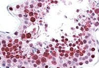
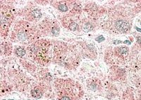
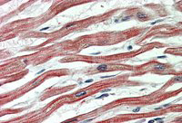
 IHC (Immunohistochemistry)
(Immunohistochemical staining of human heart tissue using Acid Ceramidase 1 antibody at 10 ug/ml.)
IHC (Immunohistochemistry)
(Immunohistochemical staining of human heart tissue using Acid Ceramidase 1 antibody at 10 ug/ml.)
Acid Ceramidase 1, Polyclonal Antibody (Cat# AAA60495)
EBNA 3C, Polyclonal Antibody (Cat# AAA60501)
b3 Calcium Channel, Polyclonal Antibody (Cat# AAA60503)
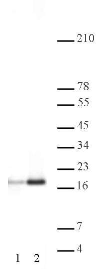
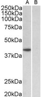
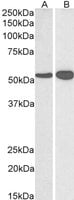
 WB (Western Blot)
((0.5ug/ml) staining of Mouse fetal Brain lysate (35ug protein in RIPA buffer). Primary incubation was 1 hour. Detected by chemiluminescence.)
WB (Western Blot)
((0.5ug/ml) staining of Mouse fetal Brain lysate (35ug protein in RIPA buffer). Primary incubation was 1 hour. Detected by chemiluminescence.)
Orexin Receptor 2, Polyclonal Antibody (Cat# AAA61592)
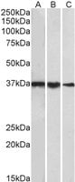
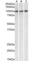
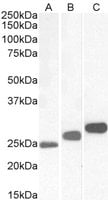
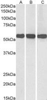
 WB (Western Blot)
((0.1ug/ml) staining of NIH3T3 (A), Mouse Brain (B) and Rat Brain (C) lysates (35ug protein in RIPA buffer). Primary incubation was 1 hour. Detected by chemiluminescence.)
WB (Western Blot)
((0.1ug/ml) staining of NIH3T3 (A), Mouse Brain (B) and Rat Brain (C) lysates (35ug protein in RIPA buffer). Primary incubation was 1 hour. Detected by chemiluminescence.)
MTHFD1L, Polyclonal Antibody (Cat# AAA61597)
 IHC (Immunohiostchemistry)
(1 hour. Detected by chemiluminescence.)
IHC (Immunohiostchemistry)
(1 hour. Detected by chemiluminescence.)
HSPC117, Polyclonal Antibody (Cat# AAA61602)
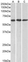
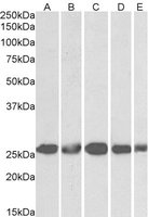
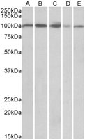 WB (Western Blot)
((0.3ug/ml) staining of Human Spleen (A), Human Thymus (B), Mouise Spleen (C), Mouse Thymus (D) and Rat Thymus (E) lysates (35ug protein in RIPA buffer). Primary incubation was 1 hour. Detected by chemiluminescence.)
WB (Western Blot)
((0.3ug/ml) staining of Human Spleen (A), Human Thymus (B), Mouise Spleen (C), Mouse Thymus (D) and Rat Thymus (E) lysates (35ug protein in RIPA buffer). Primary incubation was 1 hour. Detected by chemiluminescence.)
SIDT1, Polyclonal Antibody (Cat# AAA61624)
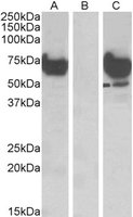
 WB (Western Blot)
((1ug/ml) staining of cell line NIH3T3 (A), Mouse Testis (B) and Rat Testis (C) lysates (35ug protein in RIPA buffer). Primary incubation was 1 hour. Detected by chemiluminescence)
WB (Western Blot)
((1ug/ml) staining of cell line NIH3T3 (A), Mouse Testis (B) and Rat Testis (C) lysates (35ug protein in RIPA buffer). Primary incubation was 1 hour. Detected by chemiluminescence)
TBP /Transcription factor IID, Polyclonal Antibody (Cat# AAA61625)
 WB (Western Blot)
((2ug/ml) staining of NIH3T3 nuclear lysate (35ug protein in RIPA buffer). Primary incubation was 1 hour. Detected by chemiluminescence.)
WB (Western Blot)
((2ug/ml) staining of NIH3T3 nuclear lysate (35ug protein in RIPA buffer). Primary incubation was 1 hour. Detected by chemiluminescence.)
RFX5, Polyclonal Antibody (Cat# AAA61643)
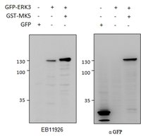
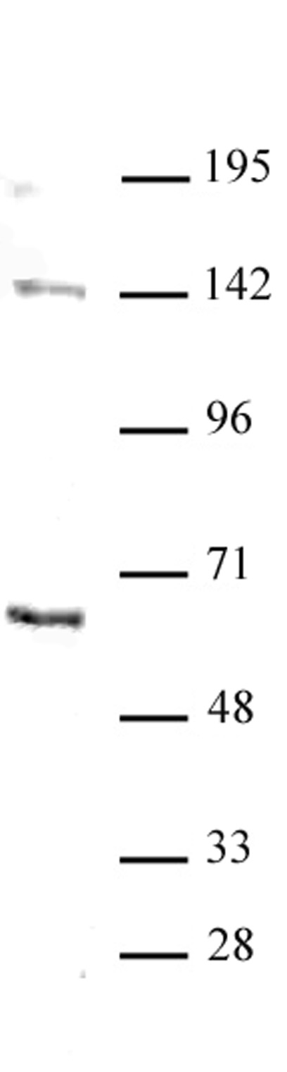
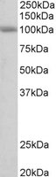
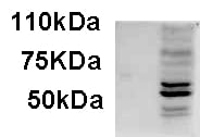
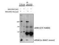 WB (Western Blot)
((1.5ug) immunoprecipitation from lysates of MK5/ERK3 double knockout MEFs, with (third lane) and without (fourth lane) rescued MK5/ERK3 expression through retroviral transduction. The corresponding lysates (first and second lane resp. ) were analyzed in parallel in this Western blot labelled with rabbit anti-Erk3 (and co-precipitation was measured using mouse anti-MK5/PRAK in the lower panel).)
WB (Western Blot)
((1.5ug) immunoprecipitation from lysates of MK5/ERK3 double knockout MEFs, with (third lane) and without (fourth lane) rescued MK5/ERK3 expression through retroviral transduction. The corresponding lysates (first and second lane resp. ) were analyzed in parallel in this Western blot labelled with rabbit anti-Erk3 (and co-precipitation was measured using mouse anti-MK5/PRAK in the lower panel).)
ERK3/MAPK6, Polyclonal Antibody (Cat# AAA61660)
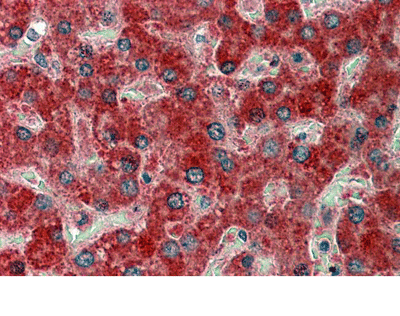 IHC (Immunohiostchemistry)
(AAA61662 (5ug/ml) staining of paraffin embedded Human Liver. Steamed antigen retrieval with citrate buffer pH 6, AP-staining.)
IHC (Immunohiostchemistry)
(AAA61662 (5ug/ml) staining of paraffin embedded Human Liver. Steamed antigen retrieval with citrate buffer pH 6, AP-staining.)
MMP14, Polyclonal Antibody (Cat# AAA61662)
Expected from Sequence similarity: Human, Mouse, Rat, Dog, Pig, Cow
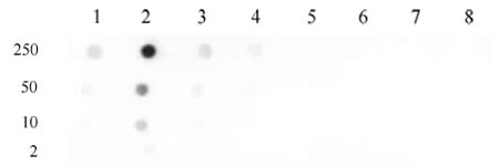
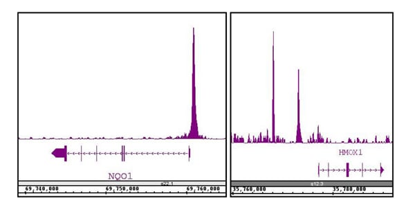
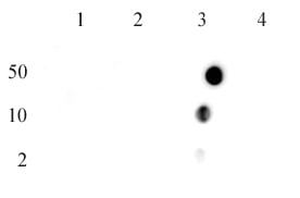
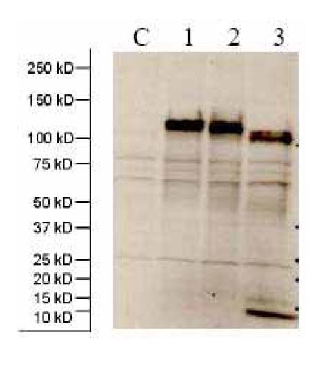 Application Data
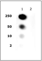
(C: 293 cell extract control; 1: 293 cell expressing gp140 (Clade A); 2: 293 cell expressing gp140 (Clade B); 3: 293 cell expressing gp140 (Clade C))
Application Data
(C: 293 cell extract control; 1: 293 cell expressing gp140 (Clade A); 2: 293 cell expressing gp140 (Clade B); 3: 293 cell expressing gp140 (Clade C))
gp120/160 (HIV-1/Pan), Polyclonal Antibody (Cat# AAA62103)
 Application Data
M1 (A/California/06/2009)(H1N1), Polyclonal Antibody (Cat# AAA62119)
 WB (Western Blot)
((0.1ug/ml) staining of Mouse Spleen lysate (35ug protein in RIPA buffer). Primary incubation was 1 hour. Detected by chemiluminescence.)
WB (Western Blot)
((0.1ug/ml) staining of Mouse Spleen lysate (35ug protein in RIPA buffer). Primary incubation was 1 hour. Detected by chemiluminescence.)
VPS16, Polyclonal Antibody (Cat# AAA61755)
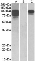
 WB (Western Blot)
((0.3ug/ml) staining of Mouse (A), Rat (B) and Pig (C) Testis lysates (35ug protein in RIPA buffer). Primary incubation was 1 hour. Detected by chemiluminescence.)
WB (Western Blot)
((0.3ug/ml) staining of Mouse (A), Rat (B) and Pig (C) Testis lysates (35ug protein in RIPA buffer). Primary incubation was 1 hour. Detected by chemiluminescence.)
CSNK2B, Polyclonal Antibody (Cat# AAA61766)
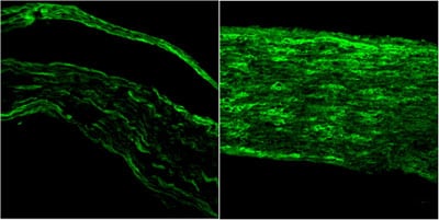
 IHC (Immunohiostchemistry)
(AAA61776 (3ug/ml) staining of paraffin embedded Human Cortex. Steamed antigen retrieval with citrate buffer pH 6, AP-staining.)
IHC (Immunohiostchemistry)
(AAA61776 (3ug/ml) staining of paraffin embedded Human Cortex. Steamed antigen retrieval with citrate buffer pH 6, AP-staining.)
CHRNA7, Polyclonal Antibody (Cat# AAA61776)
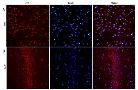
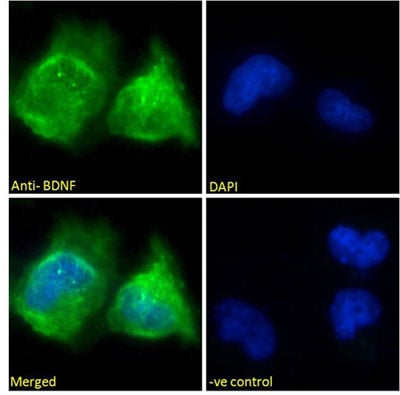
 IHC (Immunohistochemistry)
((3.75ug/ml) staining of paraffin embedded Human Cortex. Steamed antigen retrieval with citrate buffer pH 6, AP-staining.)
IHC (Immunohistochemistry)
((3.75ug/ml) staining of paraffin embedded Human Cortex. Steamed antigen retrieval with citrate buffer pH 6, AP-staining.)
BDNF, Polyclonal Antibody (Cat# AAA61777)
 WB (Western Blot)
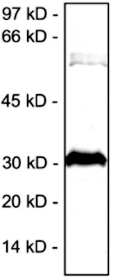
((0.1ug/ml) staining of Pig Heart lysate (35ug protein in RIPA buffer). Primary incubation was 1 hour. Detected by chemiluminescence.)
WB (Western Blot)
((0.1ug/ml) staining of Pig Heart lysate (35ug protein in RIPA buffer). Primary incubation was 1 hour. Detected by chemiluminescence.)
cardiac troponin T, Polyclonal Antibody (Cat# AAA61784)
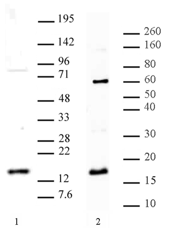
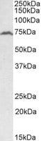
 WB (Western Blot)
(Biotinylated (1ug/ml) staining of Human Kidney lysate (35ug protein in RIPA buffer), exactly mirroring its parental non-biotinylated product. Primary incubation was 1 hour. Detected by chemiluminescence, using streptavidin-HRP and using NAP blocker as a substitute for skimmed milk.)
WB (Western Blot)
(Biotinylated (1ug/ml) staining of Human Kidney lysate (35ug protein in RIPA buffer), exactly mirroring its parental non-biotinylated product. Primary incubation was 1 hour. Detected by chemiluminescence, using streptavidin-HRP and using NAP blocker as a substitute for skimmed milk.)
ABCD3, Polyclonal Antibody (Cat# AAA61797)
Tested: Human
Expected from sequence similarity: Human, Mouse, Rat
 IHC (Immunohiostchemistry)
((3.8ug/ml) staining of paraffin embedded Human Liver. Steamed antigen retrieval with citrate buffer pH 6, AP-staining.)
IHC (Immunohiostchemistry)
((3.8ug/ml) staining of paraffin embedded Human Liver. Steamed antigen retrieval with citrate buffer pH 6, AP-staining.)
Apolipoprotein F, Polyclonal Antibody (Cat# AAA61446)
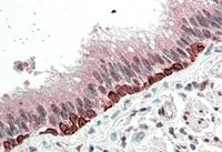 IHC (Immunohistochemistry)
((5ug/ml) staining of paraffin embedded Human Bronchus. Steamed antigen retrieval with citrate buffer pH 6, AP-staining.)
IHC (Immunohistochemistry)
((5ug/ml) staining of paraffin embedded Human Bronchus. Steamed antigen retrieval with citrate buffer pH 6, AP-staining.)
FOXI3/Forkhead box I3, Polyclonal Antibody (Cat# AAA61456)
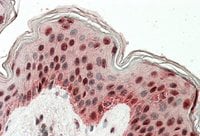 IHC (Immunohiostchemistry)
((3.8ug/ml) staining of paraffin embedded Human Skin. Steamed antigen retrieval with citrate buffer pH 6, AP-staining.)
IHC (Immunohiostchemistry)
((3.8ug/ml) staining of paraffin embedded Human Skin. Steamed antigen retrieval with citrate buffer pH 6, AP-staining.)
TIAL1 & TIA, Polyclonal Antibody (Cat# AAA61472)
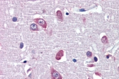
 IHC (Immunohistochemisry)
((3.8ug/ml) staining of paraffin embedded Human Cerebral Cortex. Steamed antigen retrieval with citrate buffer pH 6, AP-staining.)
IHC (Immunohistochemisry)
((3.8ug/ml) staining of paraffin embedded Human Cerebral Cortex. Steamed antigen retrieval with citrate buffer pH 6, AP-staining.)
Glutamate Dehydrogenase, Polyclonal Antibody (Cat# AAA61480)
 IHC (Immunohiostchemistry)
((3.8ug/ml) staining of paraffin embedded Human Spleen. Steamed antigen retrieval with citrate buffer pH 6, AP-staining.)
IHC (Immunohiostchemistry)
((3.8ug/ml) staining of paraffin embedded Human Spleen. Steamed antigen retrieval with citrate buffer pH 6, AP-staining.)
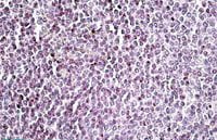
AIM2, Polyclonal Antibody (Cat# AAA61487)
Npc1, Polyclonal Antibody (Cat# AAA61500)
 IHC (Immunohiostchemistry)
((3.8ug/ml) staining of paraffin embedded Human Liver. Steamed antigen retrieval with citrate buffer pH 6, AP-staining.)
IHC (Immunohiostchemistry)
((3.8ug/ml) staining of paraffin embedded Human Liver. Steamed antigen retrieval with citrate buffer pH 6, AP-staining.)
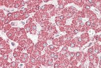
ETFB, Polyclonal Antibody (Cat# AAA61509)
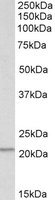
 WB (Western Blot)
((0.1ug/ml) staining of Human Heart lysate (35ug protein in RIPA buffer). Primary incubation was 1 hour. Detected by chemiluminescence.)
WB (Western Blot)
((0.1ug/ml) staining of Human Heart lysate (35ug protein in RIPA buffer). Primary incubation was 1 hour. Detected by chemiluminescence.)
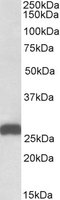
NDUFS2, Polyclonal Antibody (Cat# AAA61511)
 WB (Western Blot)
((0.3ug/ml) staining of Rat Thymus lysate (35ug protein in RIPA buffer). Primary incubation was 1 hour. Detected by chemiluminescence.)
WB (Western Blot)
((0.3ug/ml) staining of Rat Thymus lysate (35ug protein in RIPA buffer). Primary incubation was 1 hour. Detected by chemiluminescence.)
PSMB9, Polyclonal Antibody (Cat# AAA61524)
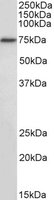
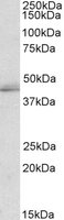
 WB (Western Blot)
(HEK293 lysate (10ug protein in RIPA buffer) overexpressing Human PCSK9 with C-terminal MYC tag probed (1ug/ml) in Lane A and probed with anti-MYC Tag (1/1000) in lane C. Mock-transfected HEK293 probed (1mg/ml) in Lane B. Primary incubations were for 1 hour. Detected by chemiluminescence.)
WB (Western Blot)
(HEK293 lysate (10ug protein in RIPA buffer) overexpressing Human PCSK9 with C-terminal MYC tag probed (1ug/ml) in Lane A and probed with anti-MYC Tag (1/1000) in lane C. Mock-transfected HEK293 probed (1mg/ml) in Lane B. Primary incubations were for 1 hour. Detected by chemiluminescence.)
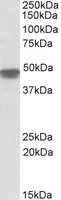
PCSK9, Polyclonal Antibody (Cat# AAA61535)
Expected from sequence similarity: Human, Mouse, Rat
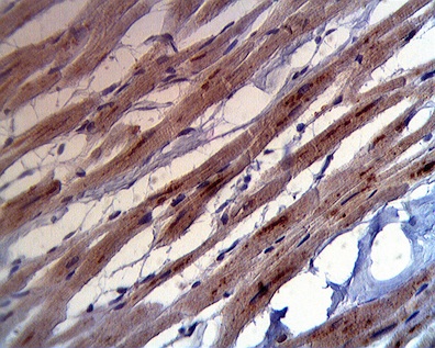
 IHC (Immunohiostchemistry)
((5ug/ml) staining of paraffin embedded Human Heart. Steamed antigen retrieval with citrate buffer pH 6, AP-staining.)
IHC (Immunohiostchemistry)
((5ug/ml) staining of paraffin embedded Human Heart. Steamed antigen retrieval with citrate buffer pH 6, AP-staining.)
EPM2AIP1, Polyclonal Antibody (Cat# AAA61557)
 WB (Western Blot)
((0.3ug/ml) staining of Mouse (A) and Rat (B) Heart lysate (35ug protein in RIPA buffer). Primary incubation was 1 hour. Detected by chemiluminescence.)
WB (Western Blot)
((0.3ug/ml) staining of Mouse (A) and Rat (B) Heart lysate (35ug protein in RIPA buffer). Primary incubation was 1 hour. Detected by chemiluminescence.)
TAp63alpha/deltaNp63alpha, Polyclonal Antibody (Cat# AAA61559)
What are Polyclonal Antibodies?
Polyclonal antibodies are antibodies that come from multiple B cell clones of a host animal. The typical hosts used for the majority of polyclonal antibody production are rabbits, goats, sheep, and donkeys. These polyclonal antibodies, once having identified their target, will bind to different epitopes located at different regions or sequences on the same protein/antigen. This ability to bind multiple epitopes is what makes polyclonal antibodies highly sensitive, as explained in our detailed guide on polyclonal antibodies and why they matter.
As a result, they are ideal at locating and binding to the target, even if the target is in very low concentrations (due to many different antibodies being able to bind to the same target molecule, which allows for significant amplification of a downstream signal).
Polyclonal antibodies are typically produced by injecting an antigen into a host animal, which causes the animal’s immune system to attack the foreign antigen by mass generating antibodies against it. After a period of time, serum is collected from the animal and purified using physicochemical fractionation, class-specific affinity purification, and/or antigen-affinity purification.
Key Uses of Polyclonal Antibodies
- Western Blotting: This method is used to find specific proteins in biological samples after separating them by size.
- Immunohistochemistry: IHC helps visualize the location of proteins in tissue sections using various staining techniques.
- ELISA: (Enzyme-Linked Immunosorbent Assay) is typically used to identify specific protein quantities in a sample. ELISAs can be either “Quantitative” or “Qualitative”.
- Flow Cytometry: technique that identifies and measures the specific protein on the surface or inside the cells in a fluid suspension.
- Immunoprecipitation: IP isolates and studies a specific protein from a complex mixture using antibodies.
Why Buy Polyclonal Antibodies from AAA Biotech?
1. Ideal for Various Applications
Our antibodies are generally going to be validated for use in multiple types of assays, including ELISA, Western Blotting, Immunohistochemistry, Immunoprecipitation, amongst others. They are ideal for a wide range of research applications.
2. Rigorous Quality Control
All of the antibodies in our catalog undergo strict quality testing to ensure specificity, sensitivity, and consistent performance. We are confident in the ability of our antibodies to provide you with accurate results.
3. Wide Assortment of Antibodies
Antibodies in our catalog can be found for both common and exotic species, and these antibodies are also available in both conjugated and recombinant forms to suit many diverse experimental needs.
4. Highly Purified
Our antibodies are available in purified forms with over 85% purity, as confirmed by SDS-PAGE. They are also available with tags such as His, Flag, GST, or MBP. We cater to customers worldwide.
FAQ
1. How are polyclonal antibodies produced?
Traditionally, polyclonal antibodies are produced by injecting an antigen into a host animal (such as a rabbit or goat), which then triggers an immune response from the host animal. The animal’s B cells produce antibodies that will recognize different parts of the injected antigen. These antibodies are then collected from the animal’s blood and purified for use.
2. How do polyclonal antibodies differ from monoclonal antibodies?
Polyclonal antibodies are a mix of antibodies that bind to different locations (epitopes) of the same antigen, while monoclonal antibodies are identical and bind to just one specific epitope. This makes polyclonal antibodies more versatile and better at detecting proteins that may be present in low quantities or in altered/modified forms.
3. How should I store polyclonal antibodies?
Polyclonal antibodies should be stored at 4°C for short-term use (up to a few weeks) and at -20°C or -80°C for long-term storage. Avoid repeated freeze-thaw cycles by dividing them into small aliquots. Always check the datasheet for specific storage instructions.