Filters
▼Clonality
▼Type
▼Reactivity
▼Gene Name
▼Isotype
▼Host
▼Application
▼Clone
▼Polyclonal Antibodies
At AAA Biotech also known as AAA Bio or AAABio, we provide a broad range of purified polyclonal antibodies (pAbs) that are able to all be browsed online through our website. Due to their high specificity and strong binding affinity, these antibodies are ideal for wide swathes of research and experimental applications.
Our polyclonal antibodies can easily support your work, whether you use them for Western Blotting, Immunocytochemistry (with or without Immunofluorescence used in conjunction), Immunohistochemistry, Immunoprecipitation, and ELISA tests. We highly encourage you to browse our range of pAbs and choose the one that best suits your experimental model.
Viewing 1050-1100 of 118597 product results
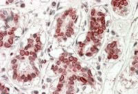
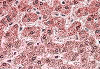
TAS1R3/T1R3, Polyclonal Antibody (Cat# AAA61382)
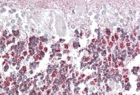 IHC (Immunohiostchemistry)
((3.8ug/ml) staining of paraffin embedded Human Cerebellum. Steamed antigen retrieval with citrate buffer pH 6, AP-staining.)
IHC (Immunohiostchemistry)
((3.8ug/ml) staining of paraffin embedded Human Cerebellum. Steamed antigen retrieval with citrate buffer pH 6, AP-staining.)
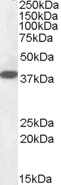
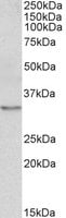
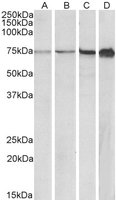
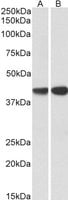
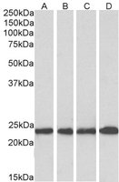
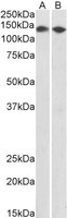
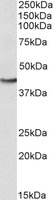
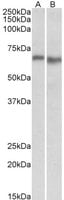
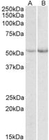
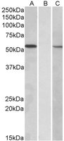
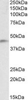
Laforin, Polyclonal Antibody (Cat# AAA61383)
 WB (Western Blot)
((0.3ug/ml) staining of Mouse Spleen (A), Mouse Thymus (B) and Rat Spleen (C) lysates (35ug protein in RIPA buffer). Primary incubation was 1 hour. Detected by chemiluminescence.)
WB (Western Blot)
((0.3ug/ml) staining of Mouse Spleen (A), Mouse Thymus (B) and Rat Spleen (C) lysates (35ug protein in RIPA buffer). Primary incubation was 1 hour. Detected by chemiluminescence.)
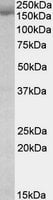
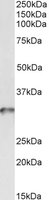
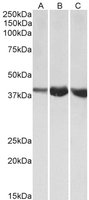
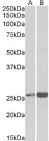
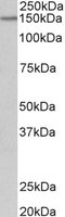
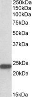
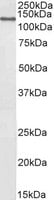
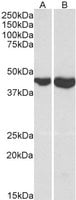
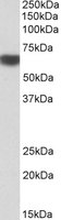
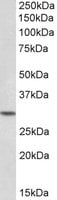
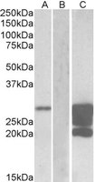
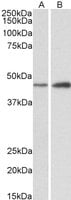
Stromal antigen 2/STAG2, Polyclonal Antibody (Cat# AAA61386)
 WB (Western Blot)
((0.3ug/ml) staining of Mouse Ovary (A) and Rat Uterus (B) lysate (35ug protein in RIPA buffer). Primary incubation was 1 hour. Detected by chemiluminescence.)
WB (Western Blot)
((0.3ug/ml) staining of Mouse Ovary (A) and Rat Uterus (B) lysate (35ug protein in RIPA buffer). Primary incubation was 1 hour. Detected by chemiluminescence.)
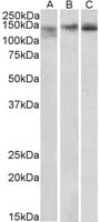
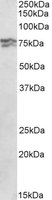
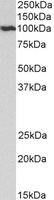
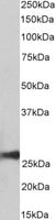
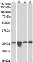
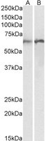
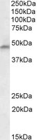
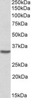
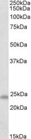
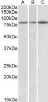
PRKCDBP, Polyclonal Antibody (Cat# AAA61392)
 WB (Western Blot)
((0.1ug/ml) staining of Mouse Brain lysate (35ug protein in RIPA buffer). Primary incubation was 1 hour. Detected by chemiluminescence.)
WB (Western Blot)
((0.1ug/ml) staining of Mouse Brain lysate (35ug protein in RIPA buffer). Primary incubation was 1 hour. Detected by chemiluminescence.)
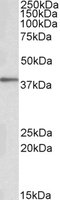
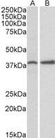
ATG16L1, Polyclonal Antibody (Cat# AAA61397)
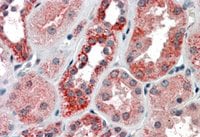 IHC (Immunohiostchemistry)
((3.8ug/ml) staining of paraffin embedded Human Kidney. Steamed antigen retrieval with citrate buffer pH 6, AP-staining.)
IHC (Immunohiostchemistry)
((3.8ug/ml) staining of paraffin embedded Human Kidney. Steamed antigen retrieval with citrate buffer pH 6, AP-staining.)
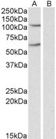
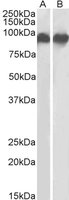
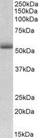
IREB2/IRP2, Polyclonal Antibody (Cat# AAA61399)
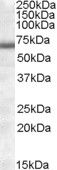 WB (Western Blot)
(AAA61420 (1ug/ml) staining of Human Heart lysate (35ug protein in RIPA buffer). Primary incubation was 1 hour. Detected by chemiluminescence.)
WB (Western Blot)
(AAA61420 (1ug/ml) staining of Human Heart lysate (35ug protein in RIPA buffer). Primary incubation was 1 hour. Detected by chemiluminescence.)
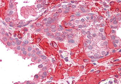
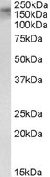
COL4A3BP, Polyclonal Antibody (Cat# AAA61420)
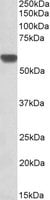
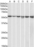 WB (Western Blot)
((0. 05ug/ml) staining of HeLa (A), A431 (B), A549 (C), MCF7 (D), Jurkat (E) and K562 (F) lysates (35ug protein in RIPA buffer). Primary incubation was 1 hour. Detected by chemiluminescence.)
WB (Western Blot)
((0. 05ug/ml) staining of HeLa (A), A431 (B), A549 (C), MCF7 (D), Jurkat (E) and K562 (F) lysates (35ug protein in RIPA buffer). Primary incubation was 1 hour. Detected by chemiluminescence.)
GPI/Neuroleukin, Polyclonal Antibody (Cat# AAA61429)
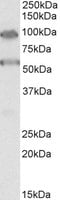
HWP1, Polyclonal Antibody (Cat# AAA61663)
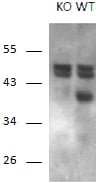 WB (Western Blot)
((1ug/ml) staining of Mouse Brain and KO Mouse Brain lysates (35ug protein in RIPA buffer). Primary incubation was 1 hour. Detected by chemiluminescence.)
WB (Western Blot)
((1ug/ml) staining of Mouse Brain and KO Mouse Brain lysates (35ug protein in RIPA buffer). Primary incubation was 1 hour. Detected by chemiluminescence.)
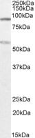
Calcipressin-1, Polyclonal Antibody (Cat# AAA61668)
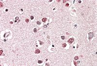 IHC (Immunohiostchemistry)
((5ug/ml) staining of paraffin embedded Human Cerebral Cortex. Steamed antigen retrieval with citrate buffer pH 6, AP-staining.)
IHC (Immunohiostchemistry)
((5ug/ml) staining of paraffin embedded Human Cerebral Cortex. Steamed antigen retrieval with citrate buffer pH 6, AP-staining.)
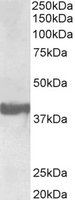
endophilin-A1/SH3GL2, Polyclonal Antibody (Cat# AAA61677)
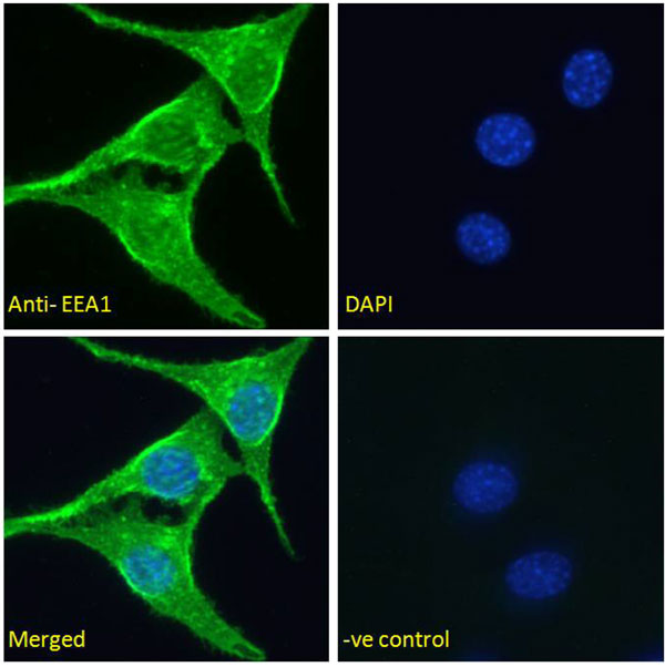 IF (Immunofluorescence)
(Immunofluorescence analysis of paraformaldehyde fixed NIH3T3 cells, permeabilized with 0.15% Triton. Primary incubation 1hr (5ug/ml) followed by Alexa Fluor 488 secondary antibody (2ug/ml), showing vesicle/cytoplasmic staining. The nuclear stain is DAPI (blue). Negative control: Unimmunized goat IgG (5ug/ml))
IF (Immunofluorescence)
(Immunofluorescence analysis of paraformaldehyde fixed NIH3T3 cells, permeabilized with 0.15% Triton. Primary incubation 1hr (5ug/ml) followed by Alexa Fluor 488 secondary antibody (2ug/ml), showing vesicle/cytoplasmic staining. The nuclear stain is DAPI (blue). Negative control: Unimmunized goat IgG (5ug/ml))
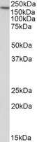
EEA1, Polyclonal Antibody (Cat# AAA61702)
Expected from sequence similarity: Human
 IHC (Immunohiostchemistry)
((5ug/ml) staining of paraffin embedded Human Testis. Steamed antigen retrieval with citrate buffer pH 6, AP-staining.)
IHC (Immunohiostchemistry)
((5ug/ml) staining of paraffin embedded Human Testis. Steamed antigen retrieval with citrate buffer pH 6, AP-staining.)
HOXA10, Polyclonal Antibody (Cat# AAA61707)
 IHC (Immunohistochemistry)
(AAA61710 (2.5ug/ml) staining of paraffin embedded Human Prostate. Steamed antigen retrieval with citrate buffer pH 6, AP-staining.)
IHC (Immunohistochemistry)
(AAA61710 (2.5ug/ml) staining of paraffin embedded Human Prostate. Steamed antigen retrieval with citrate buffer pH 6, AP-staining.)
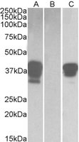
tropomyosin 4/TPM4, Polyclonal Antibody (Cat# AAA61710)
 WB (Western Blot)
((0.5ug/ml) staining of NIH3T3 (A), 3T3L1 (B), Mouse Lymph Node (C) and NSO (D) lysates (35ug protein in RIPA buffer). Primary incubation was 1 hour. Detected by chemiluminescence.)
WB (Western Blot)
((0.5ug/ml) staining of NIH3T3 (A), 3T3L1 (B), Mouse Lymph Node (C) and NSO (D) lysates (35ug protein in RIPA buffer). Primary incubation was 1 hour. Detected by chemiluminescence.)
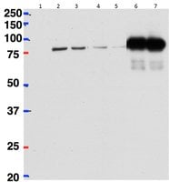
RPA1/RPA70, Polyclonal Antibody (Cat# AAA61712)
 WB (Western Blot)
((2ug) used to pull down betaCstf-64 from Mouse Brain (lane 5) lysate (500ug protein) while it cannot pull down Cstf-64 from Mouse Liver (lane 6) lysate (500ug) protein) using Protein G-coated magnetic beads. A 1/40th part of the lysates were loaded in lanes 1 (Brain) and 2 (Liver) before IP and in lanes 3 (Brain) and 4 (Liver) after IP. Western blot probed with mouse anti-betaCstf-64/Cstf-64 antibody and detected by chemiluminescence. Data obtained from Dr P Grozdanov, Texas Tech University Health Sciences Center, USA.)
WB (Western Blot)
((2ug) used to pull down betaCstf-64 from Mouse Brain (lane 5) lysate (500ug protein) while it cannot pull down Cstf-64 from Mouse Liver (lane 6) lysate (500ug) protein) using Protein G-coated magnetic beads. A 1/40th part of the lysates were loaded in lanes 1 (Brain) and 2 (Liver) before IP and in lanes 3 (Brain) and 4 (Liver) after IP. Western blot probed with mouse anti-betaCstf-64/Cstf-64 antibody and detected by chemiluminescence. Data obtained from Dr P Grozdanov, Texas Tech University Health Sciences Center, USA.)
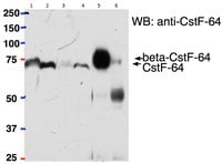
betaCstF-64 variant 1/ betaCstF-64 variant 3, Polyclonal Antibody (Cat# AAA61718)
 WB (Western Blot)
((0.3ug/ml) staining of Periheral Blood Lymphocytes ( lane A), lane B) lysates (35ug protein in RIPA buffer). Primary incubation was 1 hour. Detected by chemiluminescence.)
WB (Western Blot)
((0.3ug/ml) staining of Periheral Blood Lymphocytes ( lane A), lane B) lysates (35ug protein in RIPA buffer). Primary incubation was 1 hour. Detected by chemiluminescence.)
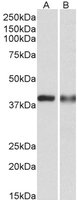
CAPG, Polyclonal Antibody (Cat# AAA61720)
 WB (Western Blot)
((1ug/ml) staining of Human (A), Mouse (B) and Rat (C) Skeletal Muscle lysates (35ug protein in RIPA buffer). Primary incubation was 1 hour. Detected by chemiluminescence.)
WB (Western Blot)
((1ug/ml) staining of Human (A), Mouse (B) and Rat (C) Skeletal Muscle lysates (35ug protein in RIPA buffer). Primary incubation was 1 hour. Detected by chemiluminescence.)
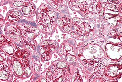
ALDOA, Polyclonal Antibody (Cat# AAA61725)
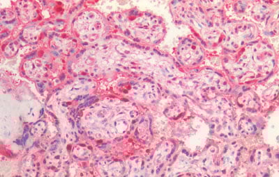 IHC (Immunohiostchemistry)
(AAA61730 (5ug/ml) staining of paraffin embedded Human Placenta. Steamed antigen retrieval with citrate buffer pH 6, AP-staining.)
IHC (Immunohiostchemistry)
(AAA61730 (5ug/ml) staining of paraffin embedded Human Placenta. Steamed antigen retrieval with citrate buffer pH 6, AP-staining.)
CDC48/YDL126C, Polyclonal Antibody (Cat# AAA61730)
Expected from sequence similarity: Saccharomyces cerevisiae S288c, Human, Mouse, Rat, Dog, Cow
 WB (Western Blot)
((1ug/ml) staining of Pig Brain lysate (35ug protein in RIPA buffer). Primary incubation was 1 hour. Detected by chemiluminescence.)
WB (Western Blot)
((1ug/ml) staining of Pig Brain lysate (35ug protein in RIPA buffer). Primary incubation was 1 hour. Detected by chemiluminescence.)
TBC1D9, Polyclonal Antibody (Cat# AAA61736)
 WB (Western Blot)
((0.1ug/ml) staining of Pig Liver lysate (35ug protein in RIPA buffer). Primary incubation was 1 hour. Detected by chemiluminescence.)
WB (Western Blot)
((0.1ug/ml) staining of Pig Liver lysate (35ug protein in RIPA buffer). Primary incubation was 1 hour. Detected by chemiluminescence.)
peroxiredoxin 6, Polyclonal Antibody (Cat# AAA61738)
 IHC (Immunohiostchemistry)
((3.8ug/ml) staining of paraffin embedded Human Breast. Steamed antigen retrieval with citrate buffer pH 6, AP-staining.)
IHC (Immunohiostchemistry)
((3.8ug/ml) staining of paraffin embedded Human Breast. Steamed antigen retrieval with citrate buffer pH 6, AP-staining.)
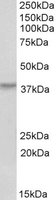
STS2/TULA, Polyclonal Antibody (Cat# AAA61589)
 WB (Western Blot)
((0.2ug/ml) staining of Mouse fetal Brain lysate (35ug protein in RIPA buffer). Primary incubation was 1 hour. Detected by chemiluminescence.)
WB (Western Blot)
((0.2ug/ml) staining of Mouse fetal Brain lysate (35ug protein in RIPA buffer). Primary incubation was 1 hour. Detected by chemiluminescence.)
PCDH17, Polyclonal Antibody (Cat# AAA61606)
 WB (Western Blot)
((0.1ug/ml) staining of Mouse Spinal Cord lysate (35ug protein in RIPA buffer). Primary incubation was 1 hour. Detected by chemiluminescence.)
WB (Western Blot)
((0.1ug/ml) staining of Mouse Spinal Cord lysate (35ug protein in RIPA buffer). Primary incubation was 1 hour. Detected by chemiluminescence.)
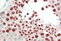
MNSOD, Polyclonal Antibody (Cat# AAA61608)
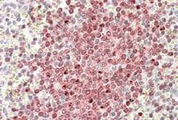 IHC (Immunohiostchemistry)
(In paraffin embedded Human Spleen shows nuclear staining in the germinal center with stronger nucleoli staining in some of the B cells. Recommended concentration, 3-5ug/ml.)
IHC (Immunohiostchemistry)
(In paraffin embedded Human Spleen shows nuclear staining in the germinal center with stronger nucleoli staining in some of the B cells. Recommended concentration, 3-5ug/ml.)
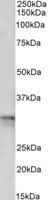
POU2AF1, Polyclonal Antibody (Cat# AAA61627)
 WB (Western Blot)
((0.3ug/ml) staining of Daudi lysate (35ug protein in RIPA buffer). Primary incubation was 1 hour. Detected by chemiluminescence.)
WB (Western Blot)
((0.3ug/ml) staining of Daudi lysate (35ug protein in RIPA buffer). Primary incubation was 1 hour. Detected by chemiluminescence.)
POU2AF1, Polyclonal Antibody (Cat# AAA61630)
 WB (Western Blot)
((1ug/ml) staining of A549 lysate (35ug protein in RIPA buffer). Primary incubation was 1 hour. Detected by chemiluminescence.)
WB (Western Blot)
((1ug/ml) staining of A549 lysate (35ug protein in RIPA buffer). Primary incubation was 1 hour. Detected by chemiluminescence.)
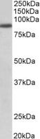
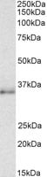
FGFR1, Polyclonal Antibody (Cat# AAA61633)
 WB (Western Blot)
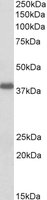
(AAA61634 (0.5ug/ml) staining of S. cerevisiae S288c lysate (35ug protein in RIPA buffer). Primary incubation was 1 hour. Detected by chemiluminescence. The blocking buffer contained non-animal derived protein. Data kindly provided by Dr. F Reggiori, University of Utrecht, Netherlands)
WB (Western Blot)
(AAA61634 (0.5ug/ml) staining of S. cerevisiae S288c lysate (35ug protein in RIPA buffer). Primary incubation was 1 hour. Detected by chemiluminescence. The blocking buffer contained non-animal derived protein. Data kindly provided by Dr. F Reggiori, University of Utrecht, Netherlands)
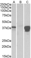
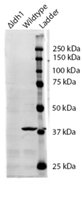
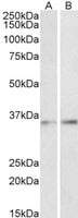
IDH1, Polyclonal Antibody (Cat# AAA61634)
 WB (Western Blot)
((0.3ug/ml) staining of Mouse Lung (A) and Rat Lung (B) lysates (35ug protein in RIPA buffer). Primary incubation was 1 hour. Detected by chemiluminescence.)
WB (Western Blot)
((0.3ug/ml) staining of Mouse Lung (A) and Rat Lung (B) lysates (35ug protein in RIPA buffer). Primary incubation was 1 hour. Detected by chemiluminescence.)
CDH1, Polyclonal Antibody (Cat# AAA61635)
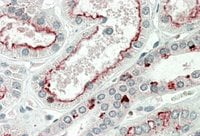 IHC (Immunohistochemisry)
((5ug/ml) staining of paraffin embedded Human Kidney. Steamed antigen retrieval with citrate buffer pH 6, AP-staining.)
IHC (Immunohistochemisry)
((5ug/ml) staining of paraffin embedded Human Kidney. Steamed antigen retrieval with citrate buffer pH 6, AP-staining.)
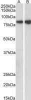
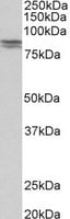
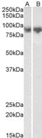
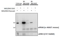
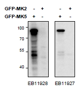
MK5/MAPKAPK5, Polyclonal Antibody (Cat# AAA61646)
 WB (Western Blot)
((1ug/ml) staining of NIH3T3 lysate (35ug protein in RIPA buffer). Primary incubation was 1 hour. Detected by chemiluminescence.)
WB (Western Blot)
((1ug/ml) staining of NIH3T3 lysate (35ug protein in RIPA buffer). Primary incubation was 1 hour. Detected by chemiluminescence.)
CEBPB, Polyclonal Antibody (Cat# AAA61741)
 WB (Western Blot)
((2ug/ml) staining of NIH3T3 nuclear lysates (35ug protein in RIPA buffer). Primary incubation was 1 hour. Detected by chemiluminescence.)
WB (Western Blot)
((2ug/ml) staining of NIH3T3 nuclear lysates (35ug protein in RIPA buffer). Primary incubation was 1 hour. Detected by chemiluminescence.)
CCAR2/DBC1, Polyclonal Antibody (Cat# AAA61743)
 WB (Western Blot)
((0.3ug/ml) staining of K562 (A) and U937 (B) lysates (35ug protein in RIPA buffer). Primary incubation was 1 hour. Detected by chemiluminescence.)
WB (Western Blot)
((0.3ug/ml) staining of K562 (A) and U937 (B) lysates (35ug protein in RIPA buffer). Primary incubation was 1 hour. Detected by chemiluminescence.)
ITGB2/LFA-1, Polyclonal Antibody (Cat# AAA61746)
 WB (Western Blot)
((0.3ug/ml) staining of HeLa nuclear lysate (35ug protein in RIPA buffer). Primary incubation was 1 hour. Detected by chemiluminescence.)
WB (Western Blot)
((0.3ug/ml) staining of HeLa nuclear lysate (35ug protein in RIPA buffer). Primary incubation was 1 hour. Detected by chemiluminescence.)
ERCC6, Polyclonal Antibody (Cat# AAA61749)
 WB (Western Blot)
((0.1ug/ml) staining of Mouse Brain (A) and Heart (B) lysates (35ug protein in RIPA buffer). Primary incubation was 1 hour. Detected by chemiluminescence.)
WB (Western Blot)
((0.1ug/ml) staining of Mouse Brain (A) and Heart (B) lysates (35ug protein in RIPA buffer). Primary incubation was 1 hour. Detected by chemiluminescence.)
GOT2, Polyclonal Antibody (Cat# AAA61754)
 WB (Western Blot)
((0. 01ug/ml) staining of Human (A) and Rat (B) Lung lysates (35ug protein in RIPA buffer). Primary incubation was 1 hour. Detected by chemiluminescence.)
WB (Western Blot)
((0. 01ug/ml) staining of Human (A) and Rat (B) Lung lysates (35ug protein in RIPA buffer). Primary incubation was 1 hour. Detected by chemiluminescence.)
Calreticulin, Polyclonal Antibody (Cat# AAA61760)
Expected from sequence similarity: Human, Mouse, Rat, Dog, Pig, Cow
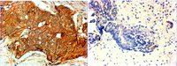 IHC (Immunohistochemistry)
((4ug/ml) staining of paraffin embedded Human breast cancer (Her+ left, triple negative right). Steamed antigen retrieval with citrate buffer pH 6, HRP-staining.)
IHC (Immunohistochemistry)
((4ug/ml) staining of paraffin embedded Human breast cancer (Her+ left, triple negative right). Steamed antigen retrieval with citrate buffer pH 6, HRP-staining.)
ERBB2/HER2, Polyclonal Antibody (Cat# AAA61762)
 WB (Western Blot)
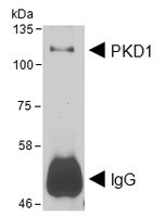
(HEK293 lysate overexpressing Human DYKDDDDK-tagged PKD1 was used to immunoprecipitate PKD1 with 2ug The precipitate was subsequently probed in Western blot using at 1ug/ml. The secondary anti-goat picks up the heavy chain of used for the immunoprecipitation (annotated as IgG). Data kindly obtained from Dr Peter Storz, Mayo Clinic, USA)
WB (Western Blot)
(HEK293 lysate overexpressing Human DYKDDDDK-tagged PKD1 was used to immunoprecipitate PKD1 with 2ug The precipitate was subsequently probed in Western blot using at 1ug/ml. The secondary anti-goat picks up the heavy chain of used for the immunoprecipitation (annotated as IgG). Data kindly obtained from Dr Peter Storz, Mayo Clinic, USA)
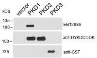
PRKD1, Polyclonal Antibody (Cat# AAA61795)
Tested: Human
Expected from sequence similarity: Human
 IF (Immunofluorescence)
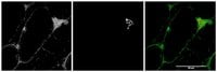
((1ug/ml) staining of Mouse Skeletal Muscle (first panel, and in green in third panel). Alpha-bungaratoxin staining in middle panel and in red in third panel. Detected by Fluorescence. Data kindly provided by Dr. Rdiger Rudolf, Karlsruhe, Germany)
IF (Immunofluorescence)
((1ug/ml) staining of Mouse Skeletal Muscle (first panel, and in green in third panel). Alpha-bungaratoxin staining in middle panel and in red in third panel. Detected by Fluorescence. Data kindly provided by Dr. Rdiger Rudolf, Karlsruhe, Germany)
MYO5A, Polyclonal Antibody (Cat# AAA61517)
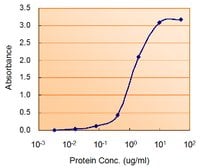 Application Data
((1.5ug/ml) as the reporter with as the capture rabbit antibody (2.5ug/ml).)
Application Data
((1.5ug/ml) as the reporter with as the capture rabbit antibody (2.5ug/ml).)
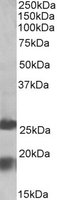
PRKAA2, Polyclonal Antibody (Cat# AAA61518)
 WB (Western Blot)
((0.1ug/ml) staining of Mouse (A) and Rat (B) Liver lysate (35ug protein in RIPA buffer). Primary incubation was 1 hour. Detected by chemiluminescence.)
WB (Western Blot)
((0.1ug/ml) staining of Mouse (A) and Rat (B) Liver lysate (35ug protein in RIPA buffer). Primary incubation was 1 hour. Detected by chemiluminescence.)
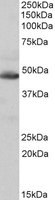
Phenylalanine Hydroxylase, Polyclonal Antibody (Cat# AAA61521)
 WB (Western Blot)
((0.1ug/ml) staining of Mouse Spinal Cord lysate (35ug protein in RIPA buffer). Primary incubation was 1 hour. Detected by chemiluminescence.)
WB (Western Blot)
((0.1ug/ml) staining of Mouse Spinal Cord lysate (35ug protein in RIPA buffer). Primary incubation was 1 hour. Detected by chemiluminescence.)
HOXC8, Polyclonal Antibody (Cat# AAA61522)
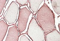 IHC (Immunohiostchemistry)
((3.8ug/ml) staining of paraffin embedded Human Skeletal Muscle. Steamed antigen retrieval with citrate buffer pH 6, AP-staining.)
IHC (Immunohiostchemistry)
((3.8ug/ml) staining of paraffin embedded Human Skeletal Muscle. Steamed antigen retrieval with citrate buffer pH 6, AP-staining.)
TRIM72, Polyclonal Antibody (Cat# AAA61529)
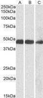 WB (Western Blot)
((0.1ug/ml) staining of Mouse (A), Rat (B) and Pig (C) Heart lysates (35ug protein in RIPA buffer). Primary incubation was 1 hour. Detected by chemiluminescence.)
WB (Western Blot)
((0.1ug/ml) staining of Mouse (A), Rat (B) and Pig (C) Heart lysates (35ug protein in RIPA buffer). Primary incubation was 1 hour. Detected by chemiluminescence.)
IDH2, Polyclonal Antibody (Cat# AAA61530)
 IHC (Immunohiostchemistry)
((5ug/ml) staining of paraffin embedded Human Liver. Steamed antigen retrieval with citrate buffer pH 6, AP-staining.)
IHC (Immunohiostchemistry)
((5ug/ml) staining of paraffin embedded Human Liver. Steamed antigen retrieval with citrate buffer pH 6, AP-staining.)
MPV17, Polyclonal Antibody (Cat# AAA61531)
ZDHHC1, Polyclonal Antibody (Cat# AAA61532)
 WB (Western Blot)
(HEK293 lysate (10ug protein in RIPA buffer) over expressing Human MGAT1 with C-terminal MYC tag probed with AAA61541 (1ug/ml) in Lane A and probed with anti-MYC Tag (1/1000) in lane C. Mock-transfected HEK293 probed with (1mg/ml) in Lane B. Primary incubations were for 1 hour. Detected by chemiluminescence.)
WB (Western Blot)
(HEK293 lysate (10ug protein in RIPA buffer) over expressing Human MGAT1 with C-terminal MYC tag probed with AAA61541 (1ug/ml) in Lane A and probed with anti-MYC Tag (1/1000) in lane C. Mock-transfected HEK293 probed with (1mg/ml) in Lane B. Primary incubations were for 1 hour. Detected by chemiluminescence.)
MGAT1, Polyclonal Antibody (Cat# AAA61541)
Expected from sequence similarity: Human, Mouse, Rat, Dog, Cow
 WB (Western Blot)
((0.3ug/ml) staining of Rat Kidney lysate (35ug protein in RIPA buffer). Primary incubation was 1 hour. Detected by chemiluminescence.)
WB (Western Blot)
((0.3ug/ml) staining of Rat Kidney lysate (35ug protein in RIPA buffer). Primary incubation was 1 hour. Detected by chemiluminescence.)
GM2A, Polyclonal Antibody (Cat# AAA61547)
 WB (Western Blot)
((0.3ug/ml) staining of Mouse (A) and Rat (B) Brain lysates (35ug protein in RIPA buffer). Primary incubation was 1 hour. Detected by chemiluminescence.)
WB (Western Blot)
((0.3ug/ml) staining of Mouse (A) and Rat (B) Brain lysates (35ug protein in RIPA buffer). Primary incubation was 1 hour. Detected by chemiluminescence.)
NDRG2, Polyclonal Antibody (Cat# AAA61549)
 WB (Western Blot)
((0.3ug/ml) staining of Human Cerebral Cortex (A), Human Frontal Cortex (B) and Mouse Brain lysates (35ug protein in RIPA buffer). Primary incubation was 1 hour. Detected by chemiluminescence.)
WB (Western Blot)
((0.3ug/ml) staining of Human Cerebral Cortex (A), Human Frontal Cortex (B) and Mouse Brain lysates (35ug protein in RIPA buffer). Primary incubation was 1 hour. Detected by chemiluminescence.)
PAPD5, Polyclonal Antibody (Cat# AAA61578)
Expected from sequence similarity: Human, Mouse
What are Polyclonal Antibodies?
Polyclonal antibodies are antibodies that come from multiple B cell clones of a host animal. The typical hosts used for the majority of polyclonal antibody production are rabbits, goats, sheep, and donkeys. These polyclonal antibodies, once having identified their target, will bind to different epitopes located at different regions or sequences on the same protein/antigen. This ability to bind multiple epitopes is what makes polyclonal antibodies highly sensitive, as explained in our detailed guide on polyclonal antibodies and why they matter.
As a result, they are ideal at locating and binding to the target, even if the target is in very low concentrations (due to many different antibodies being able to bind to the same target molecule, which allows for significant amplification of a downstream signal).
Polyclonal antibodies are typically produced by injecting an antigen into a host animal, which causes the animal’s immune system to attack the foreign antigen by mass generating antibodies against it. After a period of time, serum is collected from the animal and purified using physicochemical fractionation, class-specific affinity purification, and/or antigen-affinity purification.
Key Uses of Polyclonal Antibodies
- Western Blotting: This method is used to find specific proteins in biological samples after separating them by size.
- Immunohistochemistry: IHC helps visualize the location of proteins in tissue sections using various staining techniques.
- ELISA: (Enzyme-Linked Immunosorbent Assay) is typically used to identify specific protein quantities in a sample. ELISAs can be either “Quantitative” or “Qualitative”.
- Flow Cytometry: technique that identifies and measures the specific protein on the surface or inside the cells in a fluid suspension.
- Immunoprecipitation: IP isolates and studies a specific protein from a complex mixture using antibodies.
Why Buy Polyclonal Antibodies from AAA Biotech?
1. Ideal for Various Applications
Our antibodies are generally going to be validated for use in multiple types of assays, including ELISA, Western Blotting, Immunohistochemistry, Immunoprecipitation, amongst others. They are ideal for a wide range of research applications.
2. Rigorous Quality Control
All of the antibodies in our catalog undergo strict quality testing to ensure specificity, sensitivity, and consistent performance. We are confident in the ability of our antibodies to provide you with accurate results.
3. Wide Assortment of Antibodies
Antibodies in our catalog can be found for both common and exotic species, and these antibodies are also available in both conjugated and recombinant forms to suit many diverse experimental needs.
4. Highly Purified
Our antibodies are available in purified forms with over 85% purity, as confirmed by SDS-PAGE. They are also available with tags such as His, Flag, GST, or MBP. We cater to customers worldwide.
FAQ
1. How are polyclonal antibodies produced?
Traditionally, polyclonal antibodies are produced by injecting an antigen into a host animal (such as a rabbit or goat), which then triggers an immune response from the host animal. The animal’s B cells produce antibodies that will recognize different parts of the injected antigen. These antibodies are then collected from the animal’s blood and purified for use.
2. How do polyclonal antibodies differ from monoclonal antibodies?
Polyclonal antibodies are a mix of antibodies that bind to different locations (epitopes) of the same antigen, while monoclonal antibodies are identical and bind to just one specific epitope. This makes polyclonal antibodies more versatile and better at detecting proteins that may be present in low quantities or in altered/modified forms.
3. How should I store polyclonal antibodies?
Polyclonal antibodies should be stored at 4°C for short-term use (up to a few weeks) and at -20°C or -80°C for long-term storage. Avoid repeated freeze-thaw cycles by dividing them into small aliquots. Always check the datasheet for specific storage instructions.