Filters
▼Clonality
▼Type
▼Reactivity
▼Gene Name
▼Isotype
▼Host
▼Application
▼Clone
▼Polyclonal Antibodies
At AAA Biotech also known as AAA Bio or AAABio, we provide a broad range of purified polyclonal antibodies (pAbs) that are able to all be browsed online through our website. Due to their high specificity and strong binding affinity, these antibodies are ideal for wide swathes of research and experimental applications.
Our polyclonal antibodies can easily support your work, whether you use them for Western Blotting, Immunocytochemistry (with or without Immunofluorescence used in conjunction), Immunohistochemistry, Immunoprecipitation, and ELISA tests. We highly encourage you to browse our range of pAbs and choose the one that best suits your experimental model.
Viewing 1200-1250 of 118597 product results
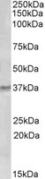
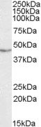
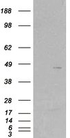
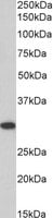
 WB (Western Blot)
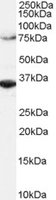
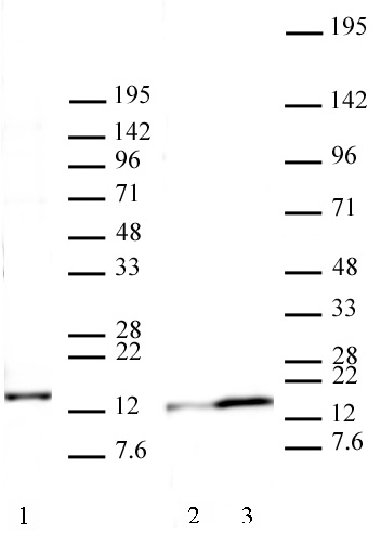
((0.3ug/ml) staining of nuclear NIH3T3 lysate (35ug protein in RIPA buffer). Primary incubation was 1 hour. Detected by chemiluminescence.)
WB (Western Blot)
((0.3ug/ml) staining of nuclear NIH3T3 lysate (35ug protein in RIPA buffer). Primary incubation was 1 hour. Detected by chemiluminescence.)
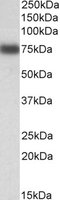
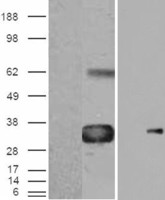
IRF2BP1, Polyclonal Antibody (Cat# AAA61555)
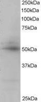
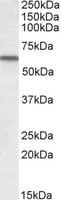
 WB (Western Blot)
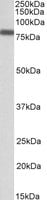
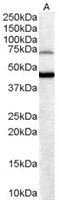
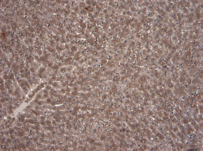
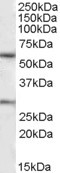
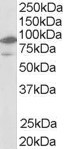
(AAA61558 (2ug/ml) staining of Mouse Liver lysate (35ug protein in RIPA buffer). Primary incubation was overnight at 4C. Detected by chemiluminescence.)
WB (Western Blot)
(AAA61558 (2ug/ml) staining of Mouse Liver lysate (35ug protein in RIPA buffer). Primary incubation was overnight at 4C. Detected by chemiluminescence.)
POLDIP2, Polyclonal Antibody (Cat# AAA61558)
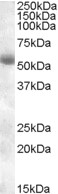
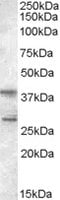
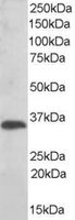
 WB (Western Blot)
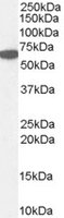
((0.3ug/ml) staining of Rat Kidney lysate (35ug protein in RIPA buffer). Primary incubation was 1 hour. Detected by chemiluminescence.)
WB (Western Blot)
((0.3ug/ml) staining of Rat Kidney lysate (35ug protein in RIPA buffer). Primary incubation was 1 hour. Detected by chemiluminescence.)
GM2A, Polyclonal Antibody (Cat# AAA61562)
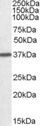
 WB (Western Blot)
((0. 03ug/ml) staining of Rat Liver lysate (35ug protein in RIPA buffer). Primary incubation was 1 hour. Detected by chemiluminescence.)
WB (Western Blot)
((0. 03ug/ml) staining of Rat Liver lysate (35ug protein in RIPA buffer). Primary incubation was 1 hour. Detected by chemiluminescence.)
Itih4, Polyclonal Antibody (Cat# AAA61571)
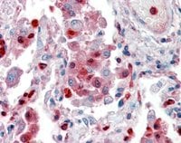
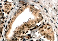
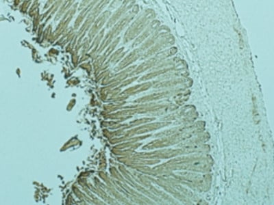
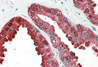 IHC (Immunohiostchemistry)
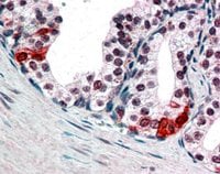
((3.8ug/ml) staining of paraffin embedded Human Prostate. Steamed antigen retrieval with citrate buffer pH 6, AP-staining.)
IHC (Immunohiostchemistry)
((3.8ug/ml) staining of paraffin embedded Human Prostate. Steamed antigen retrieval with citrate buffer pH 6, AP-staining.)
ACPP/PAP, Polyclonal Antibody (Cat# AAA61580)
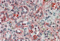
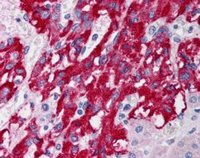 IHC (Immunohistochemistry)
((5ug/ml) staining of paraffin embedded Human Adrenal Gland. Steamed antigen retrieval with citrate buffer pH 6, AP-staining.)
IHC (Immunohistochemistry)
((5ug/ml) staining of paraffin embedded Human Adrenal Gland. Steamed antigen retrieval with citrate buffer pH 6, AP-staining.)
DLC1, Polyclonal Antibody (Cat# AAA61276)
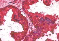 IHC (Immunohiostchemistry)
((2.5ug/ml) staining of paraffin embedded Human Prostate. Steamed antigen retrieval with citrate buffer pH 6, AP-staining.)
IHC (Immunohiostchemistry)
((2.5ug/ml) staining of paraffin embedded Human Prostate. Steamed antigen retrieval with citrate buffer pH 6, AP-staining.)
AIBZIP/CREB3L4, Polyclonal Antibody (Cat# AAA61283)
 IHC (Immunohiostchemistry)
((2ug/ml) staining of paraffin embedded Human Liver. Steamed antigen retrieval with citrate buffer pH 6, AP-staining.)
IHC (Immunohiostchemistry)
((2ug/ml) staining of paraffin embedded Human Liver. Steamed antigen retrieval with citrate buffer pH 6, AP-staining.)
Catalase/CAT, Polyclonal Antibody (Cat# AAA61285)
 IHC (Immunohiostchemistry)
((1ug/ml) staining of paraffin embedded Human Testis. Microwaved antigen retrieval with Tris/EDTA buffer pH9, HRP-staining.)
IHC (Immunohiostchemistry)
((1ug/ml) staining of paraffin embedded Human Testis. Microwaved antigen retrieval with Tris/EDTA buffer pH9, HRP-staining.)
RACGAP1/MgcRacGAP, Polyclonal Antibody (Cat# AAA61293)
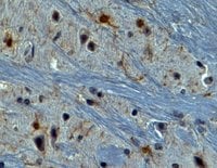 IHC (Immunohiostchemistry)
((2ug/ml) staining of paraffin embedded Mouse Brain. Steamed antigen retrieval with Tris/EDTA buffer pH 9, HRP-staining.)
IHC (Immunohiostchemistry)
((2ug/ml) staining of paraffin embedded Mouse Brain. Steamed antigen retrieval with Tris/EDTA buffer pH 9, HRP-staining.)
AIF1/IBA1, Polyclonal Antibody (Cat# AAA61296)
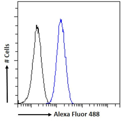 FCM/FACS (Flow Cytometry)
(Flow cytometric analysis of paraformaldehyde fixed U251 cells (blue line), permeabilized with 0.5% Triton. Primary incubation 1hr (10ug/ml) followed by Alexa Fluor 488 secondary antibody (1ug/ml). IgG control: Unimmunized goat IgG (black line) followed by Alexa Fluor 488 secondary antibody)
FCM/FACS (Flow Cytometry)
(Flow cytometric analysis of paraformaldehyde fixed U251 cells (blue line), permeabilized with 0.5% Triton. Primary incubation 1hr (10ug/ml) followed by Alexa Fluor 488 secondary antibody (1ug/ml). IgG control: Unimmunized goat IgG (black line) followed by Alexa Fluor 488 secondary antibody)
FOXG1/BF2, Polyclonal Antibody (Cat# AAA61298)
Expected from sequence similarity: Human, Mouse, Rat, Dog, Cow
 WB (Western Blot)
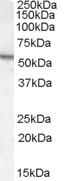
((0.5ug/ml) staining of Mouse Testis lysate (35ug protein in RIPA buffer). Primary incubation was 1 hour. Detected by chemiluminescence.)
WB (Western Blot)
((0.5ug/ml) staining of Mouse Testis lysate (35ug protein in RIPA buffer). Primary incubation was 1 hour. Detected by chemiluminescence.)
Arylsulfatase A, Polyclonal Antibody (Cat# AAA61300)
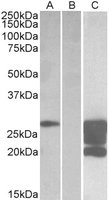
 WB (Western Blot)
(HEK293 lysate (10ug protein in RIPA buffer) overexpressing Human NRXN1 with DYKDDDDK tag probed in Lane A and probed with anti- DYKDDDDK Tag (1/3000) in lane C. Mock-transfected HEK293 probed in Lane B. Primary incubations were for 1 hour. Detected by chemiluminescence.)
WB (Western Blot)
(HEK293 lysate (10ug protein in RIPA buffer) overexpressing Human NRXN1 with DYKDDDDK tag probed in Lane A and probed with anti- DYKDDDDK Tag (1/3000) in lane C. Mock-transfected HEK293 probed in Lane B. Primary incubations were for 1 hour. Detected by chemiluminescence.)
Neurexin 1, Polyclonal Antibody (Cat# AAA61307)
 WB (Western Blot)
(HEK293 overexpressing GCNT3 (RC202007) and probed (mock transfection in first lane), tested by Origene.)
WB (Western Blot)
(HEK293 overexpressing GCNT3 (RC202007) and probed (mock transfection in first lane), tested by Origene.)
C2GnT-M, Polyclonal Antibody (Cat# AAA61319)
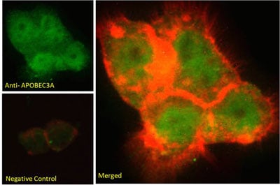
 IHC (Immunohistochemistry)
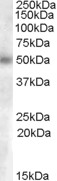
(AAA61320 (3.8ug/ml) staining of paraffin embedded Human Tonsil. Steamed antigen retrieval with citrate buffer pH 6, AP-staining.)
IHC (Immunohistochemistry)
(AAA61320 (3.8ug/ml) staining of paraffin embedded Human Tonsil. Steamed antigen retrieval with citrate buffer pH 6, AP-staining.)
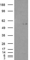
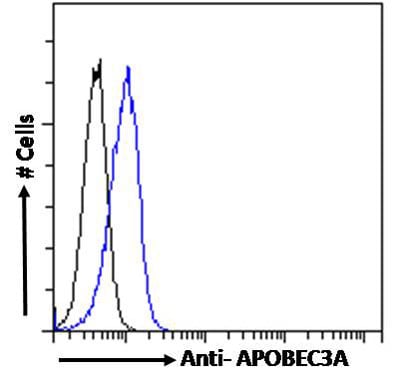
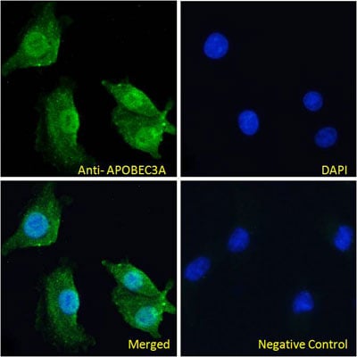
Phorbolin 1/APOBEC3A, Polyclonal Antibody (Cat# AAA61320)
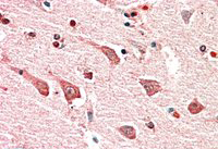 IHC (Immunohiostchemistry)
((5ug/ml) staining of paraffin embedded Human Cerebral Cortex. Steamed antigen retrieval with citrate buffer pH 6, AP-staining.)
IHC (Immunohiostchemistry)
((5ug/ml) staining of paraffin embedded Human Cerebral Cortex. Steamed antigen retrieval with citrate buffer pH 6, AP-staining.)
MYO1B, Polyclonal Antibody (Cat# AAA61584)
 WB (Western Blot)
((0.1ug/ml) staining of Mouse fetal Brain lysate (35ug protein in RIPA buffer). Primary incubation was 1 hour. Detected by chemiluminescence.)
WB (Western Blot)
((0.1ug/ml) staining of Mouse fetal Brain lysate (35ug protein in RIPA buffer). Primary incubation was 1 hour. Detected by chemiluminescence.)
Ptgds, Polyclonal Antibody (Cat# AAA61594)
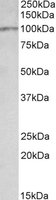
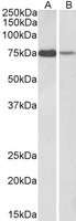
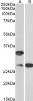 WB (Western Blot)
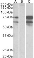
((0.1ug/ml) staining of Mouse (A) and Rat (B) Skin lysates (35ug protein in RIPA buffer). Primary incubation was 1 hour. Detected by chemiluminescence.)
WB (Western Blot)
((0.1ug/ml) staining of Mouse (A) and Rat (B) Skin lysates (35ug protein in RIPA buffer). Primary incubation was 1 hour. Detected by chemiluminescence.)
C16orf57, Polyclonal Antibody (Cat# AAA61609)
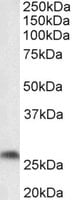
 WB (Western Blot)
((1ug/ml) staining of Mouse Kidney lysate (35ug protein in RIPA buffer). Primary incubation was 1 hour. Detected by chemiluminescence.)
WB (Western Blot)
((1ug/ml) staining of Mouse Kidney lysate (35ug protein in RIPA buffer). Primary incubation was 1 hour. Detected by chemiluminescence.)
Transglutaminase 2, Polyclonal Antibody (Cat# AAA61617)
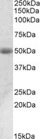
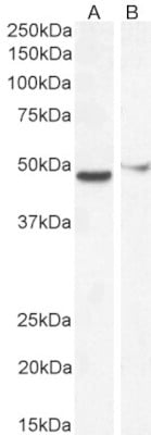
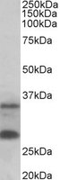
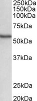 WB (Western Blot)
((0.5ug/ml) staining of Human Spleen lysate (35ug protein in RIPA buffer). Primary incubation was 1 hour. Detected by chemiluminescence.)
WB (Western Blot)
((0.5ug/ml) staining of Human Spleen lysate (35ug protein in RIPA buffer). Primary incubation was 1 hour. Detected by chemiluminescence.)
NDR1/STK38, Polyclonal Antibody (Cat# AAA61619)
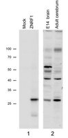
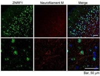 IF (Immunofluorescence)
((10ug/ml) staining of paraffin embedded Mouse Cerebral Cortex. Microwaved antigen retrieval with citrate buffer pH 6, streptavidfine-Alexa 488-staining after biotinylated anti-goat secondary. The Neurofilament M was labeled by Millipore AB1987 (1: 100).)
IF (Immunofluorescence)
((10ug/ml) staining of paraffin embedded Mouse Cerebral Cortex. Microwaved antigen retrieval with citrate buffer pH 6, streptavidfine-Alexa 488-staining after biotinylated anti-goat secondary. The Neurofilament M was labeled by Millipore AB1987 (1: 100).)
ZNRF1, Polyclonal Antibody (Cat# AAA61659)
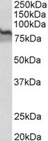
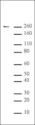
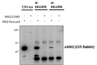 WB (Western Blot)
(And (1.5ug) immunoprecipitations from lysates of MK2/MK3 double knockout MEFs, with (third and fifth lanes) and without (fourth and sixth lanes) rescued MK2 expression through retroviral transduction. The corresponding lysates (first and second lane resp. ) were analyzed in parallel in this Western blot labelled with rabbit anti-MK2.)
WB (Western Blot)
(And (1.5ug) immunoprecipitations from lysates of MK2/MK3 double knockout MEFs, with (third and fifth lanes) and without (fourth and sixth lanes) rescued MK2 expression through retroviral transduction. The corresponding lysates (first and second lane resp. ) were analyzed in parallel in this Western blot labelled with rabbit anti-MK2.)
MK2/MAPKAPK2, Polyclonal Antibody (Cat# AAA61661)
 IHC (Immunohiostchemistry)
((2.5ug/ml) staining of paraffin embedded Human Lung. Steamed antigen retrieval with citrate buffer pH 6, AP-staining.)
IHC (Immunohiostchemistry)
((2.5ug/ml) staining of paraffin embedded Human Lung. Steamed antigen retrieval with citrate buffer pH 6, AP-staining.)
S100A9, Polyclonal Antibody (Cat# AAA61365)
 WB (Western Blot)
((0.5ug/ml) staining of Human Tonsil lysate (35ug protein in RIPA buffer). Primary incubation was 1 hour. Detected by chemiluminescence.)
WB (Western Blot)
((0.5ug/ml) staining of Human Tonsil lysate (35ug protein in RIPA buffer). Primary incubation was 1 hour. Detected by chemiluminescence.)
GCH1, Polyclonal Antibody (Cat# AAA61366)
GABRB2, Polyclonal Antibody (Cat# AAA61368)
 WB (Western Blot)
((1ug/ml) staining of Human Lung lysate (35ug protein in RIPA buffer). Primary incubation was 1 hour. Detected by chemiluminescence.)
WB (Western Blot)
((1ug/ml) staining of Human Lung lysate (35ug protein in RIPA buffer). Primary incubation was 1 hour. Detected by chemiluminescence.)
VCBP/GC, Polyclonal Antibody (Cat# AAA61378)
 WB (Western Blot)
(AAA61381(0.5ug/ml) staining of Human Pancreas Lysate (35ug protein in RIPA buffer). Detected by chemiluminescence.)
WB (Western Blot)
(AAA61381(0.5ug/ml) staining of Human Pancreas Lysate (35ug protein in RIPA buffer). Detected by chemiluminescence.)
TMPRSS2, Polyclonal Antibody (Cat# AAA61381)
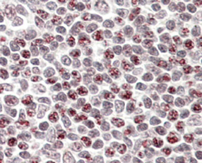
 Application Data
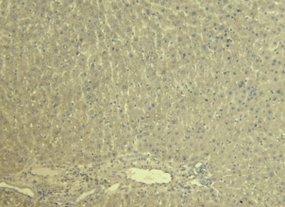
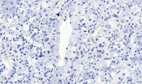
(AAA61393 Negative Control showing staining of paraffin embedded Rat Liver, with no primary antibody)
Application Data
(AAA61393 Negative Control showing staining of paraffin embedded Rat Liver, with no primary antibody)
KCNQ1, Polyclonal Antibody (Cat# AAA61393)
Expected from sequence similarity: Human, Mouse, Rat, Dog, Cow
Ascl1a, Polyclonal Antibody (Cat# AAA61394)
 WB (Western Blot)
((0. 01ug/ml) staining of Human Lung lysate (35ug protein in RIPA buffer). Primary incubation was 1 hour. Detected by chemiluminescence.)
WB (Western Blot)
((0. 01ug/ml) staining of Human Lung lysate (35ug protein in RIPA buffer). Primary incubation was 1 hour. Detected by chemiluminescence.)
Lactoperoxidase, Polyclonal Antibody (Cat# AAA61406)
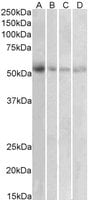
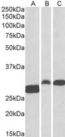
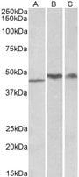 WB (Western Blot)
((1ug/ml) staining of Human Cerebellum (A), Frontal Cotex (B) and Hippocampus (C) lysates (35ug protein in RIPA buffer). Primary incubation was 1 hour. Detected by chemiluminescence.)
WB (Western Blot)
((1ug/ml) staining of Human Cerebellum (A), Frontal Cotex (B) and Hippocampus (C) lysates (35ug protein in RIPA buffer). Primary incubation was 1 hour. Detected by chemiluminescence.)
TDP-43, Polyclonal Antibody (Cat# AAA61411)
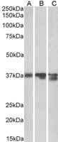 WB (Western Blot)
((0. 03ug/ml) staining of Pig (A), Mouse (B) and Rat (C) Liver lysates (35ug protein in RIPA buffer). Primary incubation was 1 hour. Detected by chemiluminescence.)
WB (Western Blot)
((0. 03ug/ml) staining of Pig (A), Mouse (B) and Rat (C) Liver lysates (35ug protein in RIPA buffer). Primary incubation was 1 hour. Detected by chemiluminescence.)
Arginase I, Polyclonal Antibody (Cat# AAA61413)
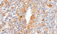
 IHC (Immunohistochemistry)
(AAA61225 Negative Control showing staining of paraffin embedded Human Adrenal Gland, with no primary antibody. This data is from a previous batch, not on sale.)
IHC (Immunohistochemistry)
(AAA61225 Negative Control showing staining of paraffin embedded Human Adrenal Gland, with no primary antibody. This data is from a previous batch, not on sale.)
VMAT2/SLC18A2, Polyclonal Antibody (Cat# AAA61225)
Expected from sequence similarity: Human, Mouse, Rat, Dog, Pig, Cow
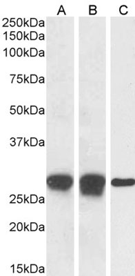
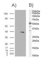
 WB (Western Blot)
(HEK293 overexpressing WISP1 (RC214390) with C-terminal tag (DYKDDDDK) and probed with anti-DYKDDDDK in the left panel and in the right panel (mock transfection in first lane in each panel), tested by Origene.)
WB (Western Blot)
(HEK293 overexpressing WISP1 (RC214390) with C-terminal tag (DYKDDDDK) and probed with anti-DYKDDDDK in the left panel and in the right panel (mock transfection in first lane in each panel), tested by Origene.)
WISP1, Polyclonal Antibody (Cat# AAA61230)
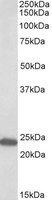
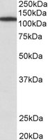
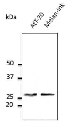
 WB (Western Blot)
((0.5ug/ml) staining of Jurkat lysate (35ug protein in RIPA buffer). Primary incubation was 1 hour. Detected by chemiluminescence.)
WB (Western Blot)
((0.5ug/ml) staining of Jurkat lysate (35ug protein in RIPA buffer). Primary incubation was 1 hour. Detected by chemiluminescence.)
SCAP2/PRAP, Polyclonal Antibody (Cat# AAA61243)
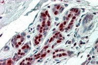
 IHC (Immunohiostchemistry)
(In paraffin embedded Human Prostate shows nuclear staining in secretory epithelial cells. Recommended concentration, 3-5ug/ml.)
IHC (Immunohiostchemistry)
(In paraffin embedded Human Prostate shows nuclear staining in secretory epithelial cells. Recommended concentration, 3-5ug/ml.)
BAF57/SMARCE1, Polyclonal Antibody (Cat# AAA61244)
 WB (Western Blot)
((2ug/ml) staining of Hela lysate (RIPA buffer, 1.4E5 cells per lane). Detected by western blot using chemiluminescence.)
WB (Western Blot)
((2ug/ml) staining of Hela lysate (RIPA buffer, 1.4E5 cells per lane). Detected by western blot using chemiluminescence.)
ORC3L, Polyclonal Antibody (Cat# AAA61245)
 IHC (Immunohiostchemistry)
((3ug/ml) staining of paraffin embedded Human Spleen. Steamed antigen retrieval with citrate buffer pH 6, AP-staining.)
IHC (Immunohiostchemistry)
((3ug/ml) staining of paraffin embedded Human Spleen. Steamed antigen retrieval with citrate buffer pH 6, AP-staining.)
LXR beta/NR1H2, Polyclonal Antibody (Cat# AAA61246)
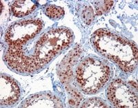
 IHC (Immunohistochemistry)
((4ug/ml) staining of paraffin embedded Human Prostate. Steamed antigen retrieval with Tris/EDTA buffer pH 9, HRP-staining.)
IHC (Immunohistochemistry)
((4ug/ml) staining of paraffin embedded Human Prostate. Steamed antigen retrieval with Tris/EDTA buffer pH 9, HRP-staining.)
ERG, Polyclonal Antibody (Cat# AAA61203)
 IHC (Immunohistochemisry)
((3.8ug/ml) staining of paraffin embedded Human Breast. Steamed antigen retrieval with citrate buffer pH 6, AP-staining.)
IHC (Immunohistochemisry)
((3.8ug/ml) staining of paraffin embedded Human Breast. Steamed antigen retrieval with citrate buffer pH 6, AP-staining.)
SSP29/ANP32B, Polyclonal Antibody (Cat# AAA61211)
 IHC (Immunohiostchemistry)
((3.8ug/ml) staining of paraffin embedded Human Spleen Steamed antigen retrieval with citrate buffer pH 6, AP-staining.)
IHC (Immunohiostchemistry)
((3.8ug/ml) staining of paraffin embedded Human Spleen Steamed antigen retrieval with citrate buffer pH 6, AP-staining.)
TXNDC1/TMX, Polyclonal Antibody (Cat# AAA61212)
D-3 Dopamine receptor IgG fraction, Polyclonal Antibody (Cat# AAA58829)
IGF-1 (Human), Polyclonal Antibody (Cat# AAA58830)
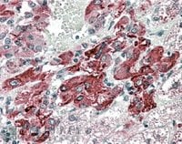
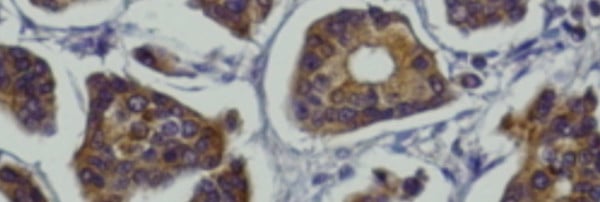 IHC (Immunohistochemistry)
(Anti-Rab27a antibody IHC staining of mammary tissue; immunohistochemistry of formalin-fixed, paraffin-embedded tissue after heat-induced antigen retrieval; antibody concentration 1/200)
IHC (Immunohistochemistry)
(Anti-Rab27a antibody IHC staining of mammary tissue; immunohistochemistry of formalin-fixed, paraffin-embedded tissue after heat-induced antigen retrieval; antibody concentration 1/200)
Rab27a, Polyclonal Antibody (Cat# AAA63064)
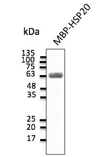 Application Data
(Anti-HSP20 Ab at 1:2,000 dilution; 30 ng of protein per lane; chicken polyclonal to goat IgG conjugated to HRP (AB1125) at 1/10,000 dilution;)
Application Data
(Anti-HSP20 Ab at 1:2,000 dilution; 30 ng of protein per lane; chicken polyclonal to goat IgG conjugated to HRP (AB1125) at 1/10,000 dilution;)
HSP20, Polyclonal Antibody (Cat# AAA63171)
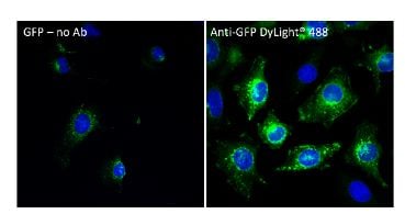
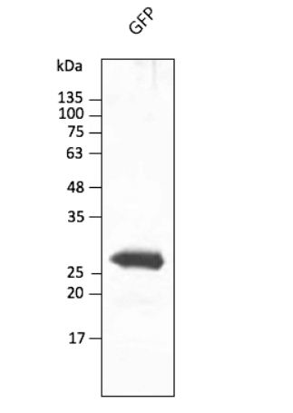
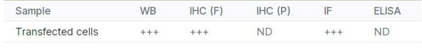
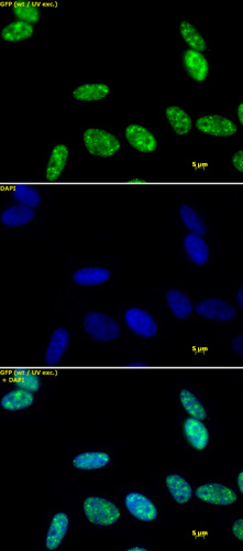
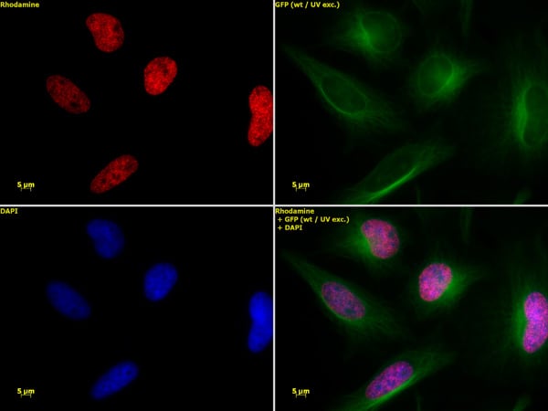
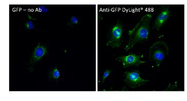 IF (Immunofluorescence)
(Immunofluorescence - anti-GFP Ab conjugated to DyLight 488 using hCEC cells transduced with GFP-Rab1a (signal amplification); cells were fixed with methanol and anti-GFP at 1/250;)
IF (Immunofluorescence)
(Immunofluorescence - anti-GFP Ab conjugated to DyLight 488 using hCEC cells transduced with GFP-Rab1a (signal amplification); cells were fixed with methanol and anti-GFP at 1/250;)
GFP, Polyclonal Antibody (Cat# AAA63247)
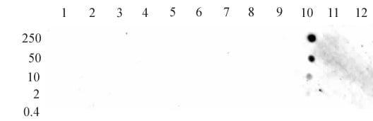
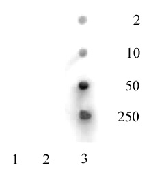
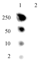 DB (Dot Blot)
(Dot blot of Histone H2A.J pAb. Dot blot analysis was used to confirm the specificity of Histone H2A.J antibody. Lane 1: H2A.J peptide. Lane 2: H2A peptide comprised of a.a. 114-126 of human Histone H2A.)
DB (Dot Blot)
(Dot blot of Histone H2A.J pAb. Dot blot analysis was used to confirm the specificity of Histone H2A.J antibody. Lane 1: H2A.J peptide. Lane 2: H2A peptide comprised of a.a. 114-126 of human Histone H2A.)
Histone H2A.J, Polyclonal Antibody (Cat# AAA60088)
 DB (Dot Blot)
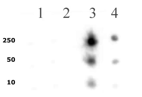
(RNA pol II CTD phospho Ser5 pAb tested by dot blot analysis. Dot blot analysis was used to confirm the specificity of RNA pol II CTD phospho Ser5 antibody for phospho-Ser5 of the RNA Pol II C-terminal domain heptad repeat. Modified and unmodified peptides were spotted onto PVDF and probed with the antibody at a dilution of 1:1,000. Decreasing amounts of peptide were spotted in each row. Lane 1: Peptide phosphorylated at CTD repeat serine 2. Lane 2: Unmodified CTD repeat serine 2 peptide. Lane 3: Peptide phosphorylated at CTD repeat serine 5. Lane 4: Unmodified CTD repeat serine 5 peptide.)
DB (Dot Blot)
(RNA pol II CTD phospho Ser5 pAb tested by dot blot analysis. Dot blot analysis was used to confirm the specificity of RNA pol II CTD phospho Ser5 antibody for phospho-Ser5 of the RNA Pol II C-terminal domain heptad repeat. Modified and unmodified peptides were spotted onto PVDF and probed with the antibody at a dilution of 1:1,000. Decreasing amounts of peptide were spotted in each row. Lane 1: Peptide phosphorylated at CTD repeat serine 2. Lane 2: Unmodified CTD repeat serine 2 peptide. Lane 3: Peptide phosphorylated at CTD repeat serine 5. Lane 4: Unmodified CTD repeat serine 5 peptide.)
RNA pol II CTD phospho Ser5, Polyclonal Antibody (Cat# AAA59848)
 DB (Dot Blot)
(Histone H3 monomethyl Lys27 pAb tested by dot blot analysis. Dot blot analysis was used to confirm the specificity of Histone H3 monomethyl Lys27 pAb for monomethyl Lys27 histone H3. Methylated peptides corresponding to the immunogen and related sequences derived from histone H3 were spotted onto PVDF and probed with the antibody at 1:1,000. The amount of peptide (picomoles) spotted is indicated next to each row. Lane 1: unmodified Lys4 peptide. Lane 2: monomethyl Lys4. Lane 3: dimethyl Lys4; Lane 4: trimethyl Lys4. Lane 5: monomethyl Lys9. Lane 6: unmodified Lys9. Lane 7: dimethyl Lys9. Lane 8: trimethyl Lys9. Lane 9: unmodified Lys27. Lane 10: monomethyl Lys27. Lane 11: dimethyl Lys27. Lane 12: trimethyl Lys27.)
DB (Dot Blot)
(Histone H3 monomethyl Lys27 pAb tested by dot blot analysis. Dot blot analysis was used to confirm the specificity of Histone H3 monomethyl Lys27 pAb for monomethyl Lys27 histone H3. Methylated peptides corresponding to the immunogen and related sequences derived from histone H3 were spotted onto PVDF and probed with the antibody at 1:1,000. The amount of peptide (picomoles) spotted is indicated next to each row. Lane 1: unmodified Lys4 peptide. Lane 2: monomethyl Lys4. Lane 3: dimethyl Lys4; Lane 4: trimethyl Lys4. Lane 5: monomethyl Lys9. Lane 6: unmodified Lys9. Lane 7: dimethyl Lys9. Lane 8: trimethyl Lys9. Lane 9: unmodified Lys27. Lane 10: monomethyl Lys27. Lane 11: dimethyl Lys27. Lane 12: trimethyl Lys27.)
Histone H3K27me1, Polyclonal Antibody (Cat# AAA59865)
 DB (Dot Blot)
(Histone H3 acetyl Lys36 antibody tested by dot blot analysis. Dot blot analysis was used to confirm the specificity of Histone H3 acetyl Lys36 antibody for acetyl Lys36 histone H3. Acetylated peptides corresponding to the immunogen and related peptides were spotted onto PVDF and probed with the antibody at 1:1,000. The amount of peptide (picomoles) spotted is indicated next to each row. Lane 1: acetyl Lys37 peptide. Lane 2: unmodified Lys36 peptide. Lane 3: acetyl Lys36 peptide. Not shown: Dot blot analysis was also performed against peptides acetylated at lysines 4, 9, 14, 18, 23 and 27 of histone H3 with no detection.)
DB (Dot Blot)
(Histone H3 acetyl Lys36 antibody tested by dot blot analysis. Dot blot analysis was used to confirm the specificity of Histone H3 acetyl Lys36 antibody for acetyl Lys36 histone H3. Acetylated peptides corresponding to the immunogen and related peptides were spotted onto PVDF and probed with the antibody at 1:1,000. The amount of peptide (picomoles) spotted is indicated next to each row. Lane 1: acetyl Lys37 peptide. Lane 2: unmodified Lys36 peptide. Lane 3: acetyl Lys36 peptide. Not shown: Dot blot analysis was also performed against peptides acetylated at lysines 4, 9, 14, 18, 23 and 27 of histone H3 with no detection.)
Histone H3K36ac, Polyclonal Antibody (Cat# AAA59866)
What are Polyclonal Antibodies?
Polyclonal antibodies are antibodies that come from multiple B cell clones of a host animal. The typical hosts used for the majority of polyclonal antibody production are rabbits, goats, sheep, and donkeys. These polyclonal antibodies, once having identified their target, will bind to different epitopes located at different regions or sequences on the same protein/antigen. This ability to bind multiple epitopes is what makes polyclonal antibodies highly sensitive, as explained in our detailed guide on polyclonal antibodies and why they matter.
As a result, they are ideal at locating and binding to the target, even if the target is in very low concentrations (due to many different antibodies being able to bind to the same target molecule, which allows for significant amplification of a downstream signal).
Polyclonal antibodies are typically produced by injecting an antigen into a host animal, which causes the animal’s immune system to attack the foreign antigen by mass generating antibodies against it. After a period of time, serum is collected from the animal and purified using physicochemical fractionation, class-specific affinity purification, and/or antigen-affinity purification.
Key Uses of Polyclonal Antibodies
- Western Blotting: This method is used to find specific proteins in biological samples after separating them by size.
- Immunohistochemistry: IHC helps visualize the location of proteins in tissue sections using various staining techniques.
- ELISA: (Enzyme-Linked Immunosorbent Assay) is typically used to identify specific protein quantities in a sample. ELISAs can be either “Quantitative” or “Qualitative”.
- Flow Cytometry: technique that identifies and measures the specific protein on the surface or inside the cells in a fluid suspension.
- Immunoprecipitation: IP isolates and studies a specific protein from a complex mixture using antibodies.
Why Buy Polyclonal Antibodies from AAA Biotech?
1. Ideal for Various Applications
Our antibodies are generally going to be validated for use in multiple types of assays, including ELISA, Western Blotting, Immunohistochemistry, Immunoprecipitation, amongst others. They are ideal for a wide range of research applications.
2. Rigorous Quality Control
All of the antibodies in our catalog undergo strict quality testing to ensure specificity, sensitivity, and consistent performance. We are confident in the ability of our antibodies to provide you with accurate results.
3. Wide Assortment of Antibodies
Antibodies in our catalog can be found for both common and exotic species, and these antibodies are also available in both conjugated and recombinant forms to suit many diverse experimental needs.
4. Highly Purified
Our antibodies are available in purified forms with over 85% purity, as confirmed by SDS-PAGE. They are also available with tags such as His, Flag, GST, or MBP. We cater to customers worldwide.
FAQ
1. How are polyclonal antibodies produced?
Traditionally, polyclonal antibodies are produced by injecting an antigen into a host animal (such as a rabbit or goat), which then triggers an immune response from the host animal. The animal’s B cells produce antibodies that will recognize different parts of the injected antigen. These antibodies are then collected from the animal’s blood and purified for use.
2. How do polyclonal antibodies differ from monoclonal antibodies?
Polyclonal antibodies are a mix of antibodies that bind to different locations (epitopes) of the same antigen, while monoclonal antibodies are identical and bind to just one specific epitope. This makes polyclonal antibodies more versatile and better at detecting proteins that may be present in low quantities or in altered/modified forms.
3. How should I store polyclonal antibodies?
Polyclonal antibodies should be stored at 4°C for short-term use (up to a few weeks) and at -20°C or -80°C for long-term storage. Avoid repeated freeze-thaw cycles by dividing them into small aliquots. Always check the datasheet for specific storage instructions.