
Filters
Clonality
Type
Reactivity
Gene Name
Isotype
Host
Application
Clone
10000 results for "Recombinant Proteins" - showing 2800-2850
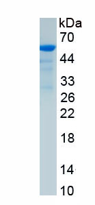
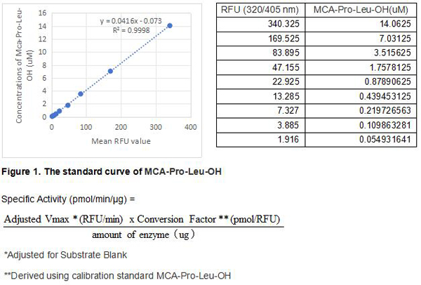 Bioactivity
(Coagulation Factor IX, also known as Christmas Factor, is a secreted by the liver and plays a key role in the activation of the intrinsic clotting cascade. Factor IX consists of a Gla domain, two tandem EGF-like domains, an activation peptide, and an S1 serine protease domain. Mature human Factor IX shares approximately 81% amino acid sequence identity with mouse and rat Factor IX. Alternative splicing generates an additional isoform that lacks the first EGF-like domain. The activity of recombinant rabbit Coagulation Factor IX is measured by its ability to cleave a fluorogenic peptide substrate Mca-RPKPVE-Nval-WRK(Dnp)-NH2 in the assay buffer 100 mM Tris, 150 mM NaCl, 10 mM CaCl2, 0.05% (w/v) Brij-35, pH 8.0. The Coagulation Factor IX is diluted to 100 ug/ml in assay buffer, then activated with a final concentration of 10 ug/ml Thermolysin at 37 degree C for 90min. Adding a final concentration of 10 mM 1,10 Phenanthroline to stop the activation. The activated Coagulation Factor IX is diluted to 6 ug/mL in assay buffer. Loading into a black well plate 50 uL of 6 ug/mL Coagulation Factor IX and start the reaction by adding 50 uL of 20 uM substrate, with a substrate blank containing 50 uL assay buffer, 50 uL substrate, and no Coagulation Factor IX. Then read at 320/405 nm in kinetic mode for 5 minutes. The specific activity of recombinant rabbit Coagulation Factor IX is > 20 pmol/min/ug.)
Bioactivity
(Coagulation Factor IX, also known as Christmas Factor, is a secreted by the liver and plays a key role in the activation of the intrinsic clotting cascade. Factor IX consists of a Gla domain, two tandem EGF-like domains, an activation peptide, and an S1 serine protease domain. Mature human Factor IX shares approximately 81% amino acid sequence identity with mouse and rat Factor IX. Alternative splicing generates an additional isoform that lacks the first EGF-like domain. The activity of recombinant rabbit Coagulation Factor IX is measured by its ability to cleave a fluorogenic peptide substrate Mca-RPKPVE-Nval-WRK(Dnp)-NH2 in the assay buffer 100 mM Tris, 150 mM NaCl, 10 mM CaCl2, 0.05% (w/v) Brij-35, pH 8.0. The Coagulation Factor IX is diluted to 100 ug/ml in assay buffer, then activated with a final concentration of 10 ug/ml Thermolysin at 37 degree C for 90min. Adding a final concentration of 10 mM 1,10 Phenanthroline to stop the activation. The activated Coagulation Factor IX is diluted to 6 ug/mL in assay buffer. Loading into a black well plate 50 uL of 6 ug/mL Coagulation Factor IX and start the reaction by adding 50 uL of 20 uM substrate, with a substrate blank containing 50 uL assay buffer, 50 uL substrate, and no Coagulation Factor IX. Then read at 320/405 nm in kinetic mode for 5 minutes. The specific activity of recombinant rabbit Coagulation Factor IX is > 20 pmol/min/ug.)
Coagulation Factor IX (F9), Active Protein (Cat# AAA161786)
Full Name
Active Coagulation Factor IX (F9)
Applications
Activity Assay, Cell Culture
Purity
Greater than 80% by SDS-PAGE
Pricing
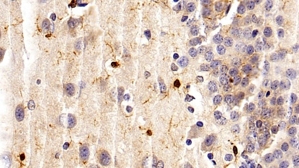
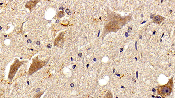
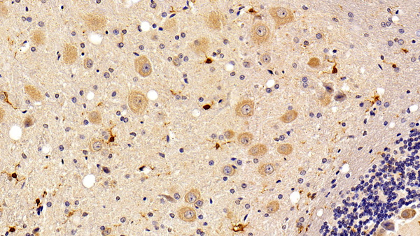 IHC (Immunohistochemisry)
(DAB staining on IHCP;Sample: Rat Cerebellum Tissue; Primary Ab: 10ug/ml Mouse AntiRat IBA1 AntibodySecond Ab: 2ug/mL HRPLinked Caprine AntiMouse IgG Polyclonal Antibody(Catalog: SAA544Mu19))
IHC (Immunohistochemisry)
(DAB staining on IHCP;Sample: Rat Cerebellum Tissue; Primary Ab: 10ug/ml Mouse AntiRat IBA1 AntibodySecond Ab: 2ug/mL HRPLinked Caprine AntiMouse IgG Polyclonal Antibody(Catalog: SAA544Mu19))
Ionized Calciumbinding Adapter Molecule 1 (IBA1), Monoclonal Antibody (Cat# AAA151799)
Full Name
Monoclonal Antibody to Ionized Calciumbinding Adapter Molecule 1 (IBA1)
Gene Names
Aif1; iba1; Bart1; mrf-1; BART-1
Reactivity
Rat
Applications
Immunoprecipitation, Immunocytochemistry, Immunohistochemistry, Western Blot
Purity
Protein A + Protein G affinity chromatography
Pricing
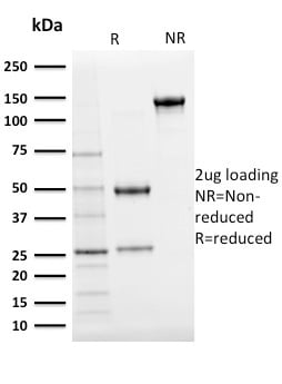
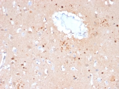
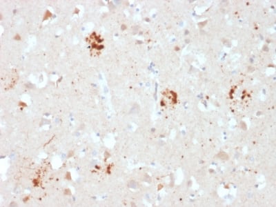
 Application Data
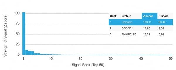
(Analysis of Protein Array containing more than 19, 000 full-length human proteins using Ubiquitin Mouse Monoclonal Antibody (UBB/2122)Z- and S- Score: The Z-score represents the strength of a signal that a monoclonal antibody (MAb) (in combination with a fluorescently-tagged anti-IgG secondary antibody) produces when binding to a particular protein on the HuProtTM array. Z-scores are described in units of standard deviations (SD's) above the mean value of all signals generated on that array. If targets on HuProtTM are arranged in descending order of the Z-score, the S-score is the difference (also in units of SD's) between the Z-score. S-score therefore represents the relative target specificity of a MAb to its intended target. A MAb is considered to specific to its intended target, if the MAb has an S-score of at least 2.5. For example, if a MAb binds to protein X with a Z-score of 43 and to protein Y with a Z-score of 14, then the S-score for the binding of that MAb to protein X is equal to 29.)
Application Data
(Analysis of Protein Array containing more than 19, 000 full-length human proteins using Ubiquitin Mouse Monoclonal Antibody (UBB/2122)Z- and S- Score: The Z-score represents the strength of a signal that a monoclonal antibody (MAb) (in combination with a fluorescently-tagged anti-IgG secondary antibody) produces when binding to a particular protein on the HuProtTM array. Z-scores are described in units of standard deviations (SD's) above the mean value of all signals generated on that array. If targets on HuProtTM are arranged in descending order of the Z-score, the S-score is the difference (also in units of SD's) between the Z-score. S-score therefore represents the relative target specificity of a MAb to its intended target. A MAb is considered to specific to its intended target, if the MAb has an S-score of at least 2.5. For example, if a MAb binds to protein X with a Z-score of 43 and to protein Y with a Z-score of 14, then the S-score for the binding of that MAb to protein X is equal to 29.)
Ubiquitin, Monoclonal Antibody (Cat# AAA214706)
Full Name
Ubiquitin (Autophagy Marker)
Gene Names
UBB; HEL-S-50
Reactivity
Human. Others not known.
Applications
Immunohistochemistry
Pricing
 Bioactivity
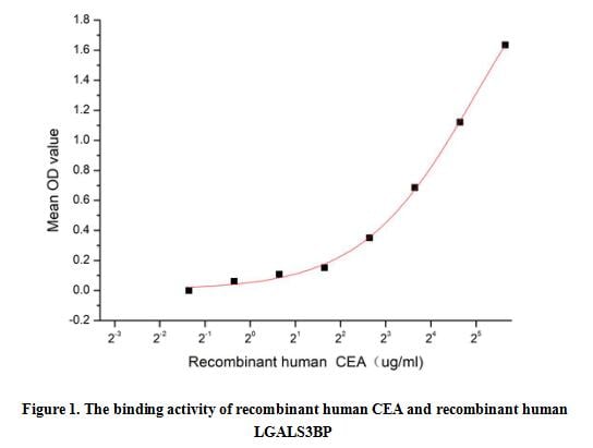
(Carcinoembryonic antigen (CEA), as one of the common tumor markers, is a human glycoprotein involved in cell adhesion and is expressed during human fetal development. Since the birth of human, CEA expression is largely inhibited, with only low levels in the plasma of healthy adults. Generally, CEA will overexpressed in many cancers, including gastric, breast, ovarian, lung, and pancreatic cancers, especially colorectal cancer. The lectin galactoside-binding soluble 3 binding protein (LGALS3BP) is also a secreted, which was first identified as cancer and metastasis associated protein. LGALS3BP has been identified as an interactor of CEA, thus a functional binding ELISA assay was conducted to detect the interaction of recombinant human CEA and recombinant human LGALS3BP. Briefly, biotin-linked CEA were diluted serially in PBS, with 0.01% BSA (pH 7.4). Duplicate samples of 100 ul were then transferred to LGALS3BP-coated microtiter wells and incubated for 1h at 37 degree C. Wells were washed with PBST 3 times and incubation with Streptavidin-HRP for 30min, then wells were aspirated and washed 5 times. With the addition of substrate solution, wells were incubated 15-25 minutes at 37 degree C. Finally, add 50 ul stop solution to the wells and read at 450 nm immediately. The binding activity of CEA and LGALS3BP was shown in Figure 1, and this effect was in a dose dependent manner.)
Bioactivity
(Carcinoembryonic antigen (CEA), as one of the common tumor markers, is a human glycoprotein involved in cell adhesion and is expressed during human fetal development. Since the birth of human, CEA expression is largely inhibited, with only low levels in the plasma of healthy adults. Generally, CEA will overexpressed in many cancers, including gastric, breast, ovarian, lung, and pancreatic cancers, especially colorectal cancer. The lectin galactoside-binding soluble 3 binding protein (LGALS3BP) is also a secreted, which was first identified as cancer and metastasis associated protein. LGALS3BP has been identified as an interactor of CEA, thus a functional binding ELISA assay was conducted to detect the interaction of recombinant human CEA and recombinant human LGALS3BP. Briefly, biotin-linked CEA were diluted serially in PBS, with 0.01% BSA (pH 7.4). Duplicate samples of 100 ul were then transferred to LGALS3BP-coated microtiter wells and incubated for 1h at 37 degree C. Wells were washed with PBST 3 times and incubation with Streptavidin-HRP for 30min, then wells were aspirated and washed 5 times. With the addition of substrate solution, wells were incubated 15-25 minutes at 37 degree C. Finally, add 50 ul stop solution to the wells and read at 450 nm immediately. The binding activity of CEA and LGALS3BP was shown in Figure 1, and this effect was in a dose dependent manner.)
Carcinoembryonic Antigen (CEA), Active Protein (Cat# AAA161710)
Full Name
Active Carcinoembryonic Antigen (CEA)
Gene Names
CEACAM5; CEA; CD66e
Applications
Activity Assay, Cell Culture
Purity
Greater than 95% by SDS-PAGE
Pricing
 FCM/FACS (Flow Cytometry)
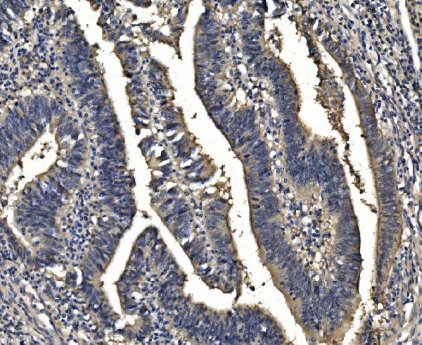
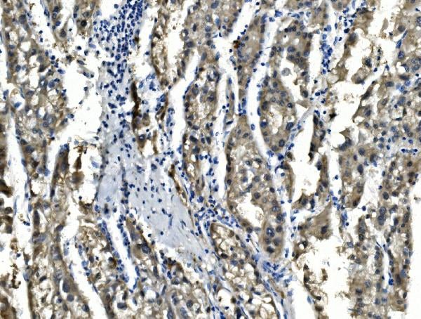
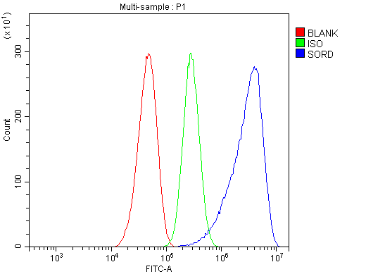
(Figure 5. Flow Cytometry analysis of CACO-2 cells using anti-Sorbitol Dehydrogenase/ SORD antibody (AAA125795).Overlay histogram showing CACO-2 cells stained with AAA125795 (Blue line). The cells were blocked with 10% normal goat serum. And then incubated with rabbit anti-Sorbitol Dehydrogenase/ SORD Antibody (AAA125795, 1μg/1x106 cells) for 30 min at 20 degree C. DyLight®488 conjugated goat anti-rabbit IgG (5-10μg/1x106 cells) was used as secondary antibody for 30 minutes at 20 degree C. Isotype control antibody (Green line) was rabbit IgG (1μg/1x106) used under the same conditions. Unlabelled sample (Red line) was also used as a control.)
FCM/FACS (Flow Cytometry)
(Figure 5. Flow Cytometry analysis of CACO-2 cells using anti-Sorbitol Dehydrogenase/ SORD antibody (AAA125795).Overlay histogram showing CACO-2 cells stained with AAA125795 (Blue line). The cells were blocked with 10% normal goat serum. And then incubated with rabbit anti-Sorbitol Dehydrogenase/ SORD Antibody (AAA125795, 1μg/1x106 cells) for 30 min at 20 degree C. DyLight®488 conjugated goat anti-rabbit IgG (5-10μg/1x106 cells) was used as secondary antibody for 30 minutes at 20 degree C. Isotype control antibody (Green line) was rabbit IgG (1μg/1x106) used under the same conditions. Unlabelled sample (Red line) was also used as a control.)
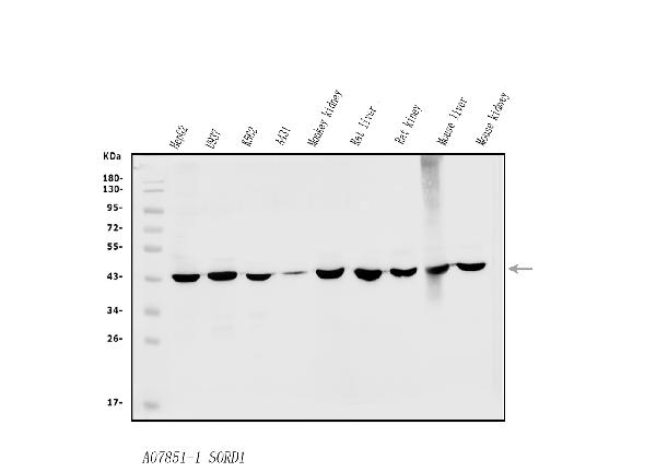
Sorbitol Dehydrogenase/SORD, Polyclonal Antibody (Cat# AAA125795)
Full Name
Anti-Sorbitol Dehydrogenase/SORD Antibody
Gene Names
SORD; SORD1; HEL-S-95n
Reactivity
Human, Mouse, Rat, Monkey
Applications
Direct ELISA, Flow Cytometry, Immunofluorescence, Immunocytochemistry, Immunohistochemistry, Western Blot
Purity
Immunogen affinity purified.
Pricing
 Bioactivity
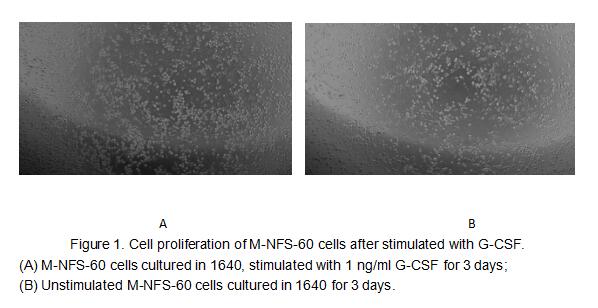
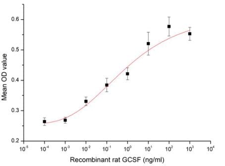
(Figure 2. Cell proliferation of M-NFS-60 cells after stimulated with G-CSF.)
Bioactivity
(Figure 2. Cell proliferation of M-NFS-60 cells after stimulated with G-CSF.)
Colony Stimulating Factor 3, Granulocyte (GCSF), Active Protein (Cat# AAA161666)
Full Name
Active Colony Stimulating Factor 3, Granulocyte (GCSF)
Applications
Activity Assay, Cell Culture
Purity
Greater than 90% by SDS-PAGE
Pricing
 Bioactivity
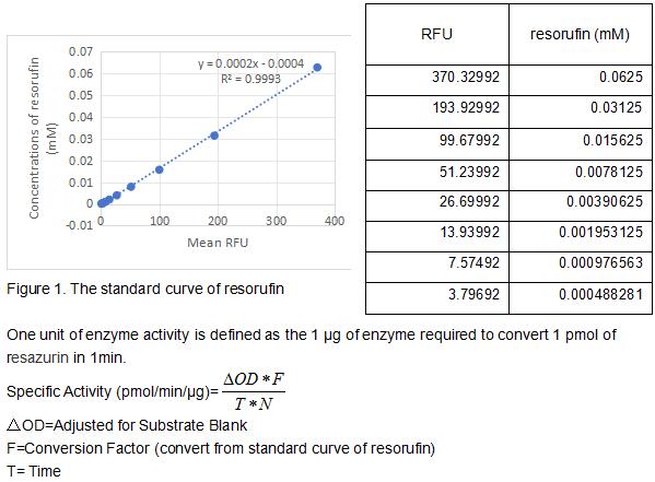
(NAD(P)H:quinone acceptor oxidoreductase 1 (NQO1), also known as DT-diaphorase, is a widely-distributed FAD-dependent flavoprotein that promotes 2-electron reductions of quinones, quinoneimines, nitroaromatics, and azo dyes. As a result it prevents the one electron reduction of quinones that results in the production of radical species. NQO1 is a highly-inducible enzyme that is regulated by the Keap1/Nrf2/ARE pathway. The increase and decrease of NQO1 levels are associated with decreased and increased susceptibilities to oxidative stress, respectively. Thus, NQO1 is a marker cytoprotective enzyme in oxidative stress. Independently of its catalytic function, NQO1 plays a role in regulating the proteosomal degradation of p53, p73a, and p33. NQO1 physically interacts with p53 and p73 in an NADH-dependent manner and protects them from 20S proteasomal degradation in a ubiquitin independent pathway. The activity assay of recombinant human NQO1 was measured by its ability to oxidize the substrate resazurin to resorufin. The rhNQO1 was diluted to 100 ug/ml in the assay buffer 50 mM HEPES, 0.2 M NaCl, 5 uM FAD, 0.05% Tween 20, pH 7.5. 50 ul 100 ug/ml rhNQO1 was added into the microplate and start the reaction by adding 50 ul substrate mixture of 400 uM beta-NADH and 20 uM resazurin which was diluted in assay buffer. Read at excitation and emission wavelengths of 540 nm and 585 nm (top read), respectively, in kinetic mode for 5 minutes. The specific activity of recombinant human NQO1 is >18 pmol/min/ug.)
Bioactivity
(NAD(P)H:quinone acceptor oxidoreductase 1 (NQO1), also known as DT-diaphorase, is a widely-distributed FAD-dependent flavoprotein that promotes 2-electron reductions of quinones, quinoneimines, nitroaromatics, and azo dyes. As a result it prevents the one electron reduction of quinones that results in the production of radical species. NQO1 is a highly-inducible enzyme that is regulated by the Keap1/Nrf2/ARE pathway. The increase and decrease of NQO1 levels are associated with decreased and increased susceptibilities to oxidative stress, respectively. Thus, NQO1 is a marker cytoprotective enzyme in oxidative stress. Independently of its catalytic function, NQO1 plays a role in regulating the proteosomal degradation of p53, p73a, and p33. NQO1 physically interacts with p53 and p73 in an NADH-dependent manner and protects them from 20S proteasomal degradation in a ubiquitin independent pathway. The activity assay of recombinant human NQO1 was measured by its ability to oxidize the substrate resazurin to resorufin. The rhNQO1 was diluted to 100 ug/ml in the assay buffer 50 mM HEPES, 0.2 M NaCl, 5 uM FAD, 0.05% Tween 20, pH 7.5. 50 ul 100 ug/ml rhNQO1 was added into the microplate and start the reaction by adding 50 ul substrate mixture of 400 uM beta-NADH and 20 uM resazurin which was diluted in assay buffer. Read at excitation and emission wavelengths of 540 nm and 585 nm (top read), respectively, in kinetic mode for 5 minutes. The specific activity of recombinant human NQO1 is >18 pmol/min/ug.)
NADH Dehydrogenase, Quinone 1 (NQO1), Active Protein (Cat# AAA161938)
Full Name
Active NADH Dehydrogenase, Quinone 1 (NQO1)
Gene Names
NQO1; DTD; QR1; DHQU; DIA4; NMOR1; NMORI
Applications
Activity Assay, Cell Culture
Purity
Greater than 95% by SDS-PAGE
Pricing
 Structure
 FCM/FACS (Flow Cytometry)
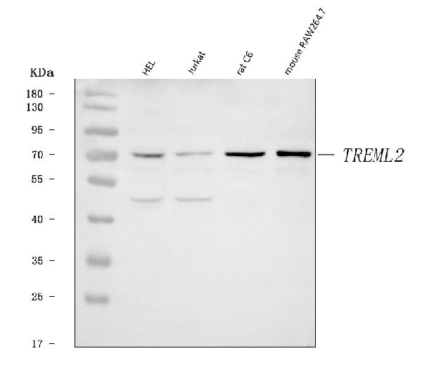
(Figure 2. Flow Cytometry analysis of HEL cells using anti-TREML2 antibody (AAA126538).Overlay histogram showing HEL cells stained with AAA126538 (Blue line). The cells were blocked with 10% normal goat serum. And then incubated with rabbit anti-TREML2 Antibody (AAA126538, 1 ug/1x10^6 cells) for 30 min at 20 degree C. DyLight488 conjugated goat anti-rabbit IgG was used as secondary antibody for 30 minutes at 20 degree C. Isotype control antibody (Green line) was rabbit IgG (1 ug/1x10^6) used under the same conditions. Unlabelled sample (Red line) was also used as a control.)
FCM/FACS (Flow Cytometry)
(Figure 2. Flow Cytometry analysis of HEL cells using anti-TREML2 antibody (AAA126538).Overlay histogram showing HEL cells stained with AAA126538 (Blue line). The cells were blocked with 10% normal goat serum. And then incubated with rabbit anti-TREML2 Antibody (AAA126538, 1 ug/1x10^6 cells) for 30 min at 20 degree C. DyLight488 conjugated goat anti-rabbit IgG was used as secondary antibody for 30 minutes at 20 degree C. Isotype control antibody (Green line) was rabbit IgG (1 ug/1x10^6) used under the same conditions. Unlabelled sample (Red line) was also used as a control.)
TREML2, Polyclonal Antibody (Cat# AAA126538)
Full Name
Anti-TREML2 Antibody Picoband
Gene Names
TREML2; TLT2; TLT-2; C6orf76; FLJ13693; MGC149715; MGC149716; dJ238O23.1
Reactivity
Human
Applications
Flow Cytometry, Western Blot
Purity
Immunogen affinity purified.
Pricing
 FCM/FACS (Flow Cytometry)
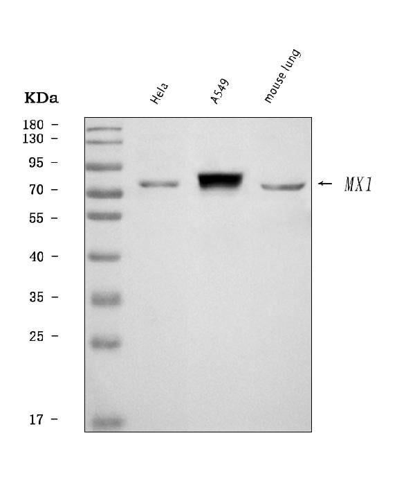
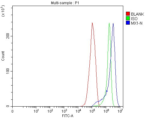
(Figure 2. Flow Cytometry analysis of RT4 cells using anti-MX1 antibody (AAA126088).Overlay histogram showing RT4 cells stained with AAA126088 (Blue line). The cells were blocked with 10% normal goat serum. And then incubated with rabbit anti-MX1 Antibody (AAA126088, 1 ug/1x10^6 cells) for 30 min at 20 degree C. DyLight488 conjugated goat anti-rabbit IgG was used as secondary antibody for 30 minutes at 20 degree C. Isotype control antibody (Green line) was rabbit IgG (1 ug/1x10^6) used under the same conditions. Unlabelled sample (Red line) was also used as a control.)
FCM/FACS (Flow Cytometry)
(Figure 2. Flow Cytometry analysis of RT4 cells using anti-MX1 antibody (AAA126088).Overlay histogram showing RT4 cells stained with AAA126088 (Blue line). The cells were blocked with 10% normal goat serum. And then incubated with rabbit anti-MX1 Antibody (AAA126088, 1 ug/1x10^6 cells) for 30 min at 20 degree C. DyLight488 conjugated goat anti-rabbit IgG was used as secondary antibody for 30 minutes at 20 degree C. Isotype control antibody (Green line) was rabbit IgG (1 ug/1x10^6) used under the same conditions. Unlabelled sample (Red line) was also used as a control.)
MX1, Polyclonal Antibody (Cat# AAA126088)
Full Name
Anti-MX1 Antibody Picoband
Gene Names
MX1; MX; MxA; IFI78; IFI-78K
Reactivity
Human, Mouse
Applications
Flow Cytometry, Western Blot
Purity
Immunogen affinity purified.
Pricing
 FCM/FACS (Flow Cytometry)
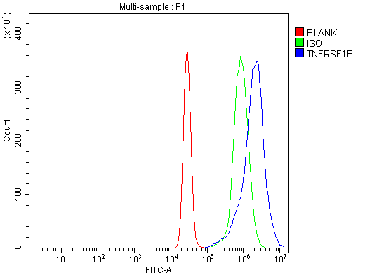
(Figure 2. Flow Cytometry analysis of K562 cells using anti-TNF Receptor II/TNFRSF1B antibody (AAA126142).Overlay histogram showing K562 cells stained with AAA126142 (Blue line). The cells were blocked with 10% normal goat serum. And then incubated with rabbit anti-TNF Receptor II/TNFRSF1B Antibody (AAA126142, 1 ug/1x10^6 cells) for 30 min at 20 degree C. DyLight488 conjugated goat anti-rabbit IgG was used as secondary antibody for 30 minutes at 20 degree C. Isotype control antibody (Green line) was rabbit IgG (1 ug/1x10^6) used under the same conditions. Unlabelled sample (Red line) was also used as a control.)
FCM/FACS (Flow Cytometry)
(Figure 2. Flow Cytometry analysis of K562 cells using anti-TNF Receptor II/TNFRSF1B antibody (AAA126142).Overlay histogram showing K562 cells stained with AAA126142 (Blue line). The cells were blocked with 10% normal goat serum. And then incubated with rabbit anti-TNF Receptor II/TNFRSF1B Antibody (AAA126142, 1 ug/1x10^6 cells) for 30 min at 20 degree C. DyLight488 conjugated goat anti-rabbit IgG was used as secondary antibody for 30 minutes at 20 degree C. Isotype control antibody (Green line) was rabbit IgG (1 ug/1x10^6) used under the same conditions. Unlabelled sample (Red line) was also used as a control.)
TNF Receptor II/TNFRSF1B, Polyclonal Antibody (Cat# AAA126142)
Full Name
Anti-TNF Receptor II/TNFRSF1B Antibody Picoband
Gene Names
TNFRSF1B; p75; TBPII; TNFBR; TNFR2; CD120b; TNFR1B; TNFR80; TNF-R75; p75TNFR; TNF-R-II
Reactivity
Human, Mouse, Rat
Applications
Flow Cytometry, Western Blot
Purity
Immunogen affinity purified.
Pricing
 IF (Immunofluorescence)
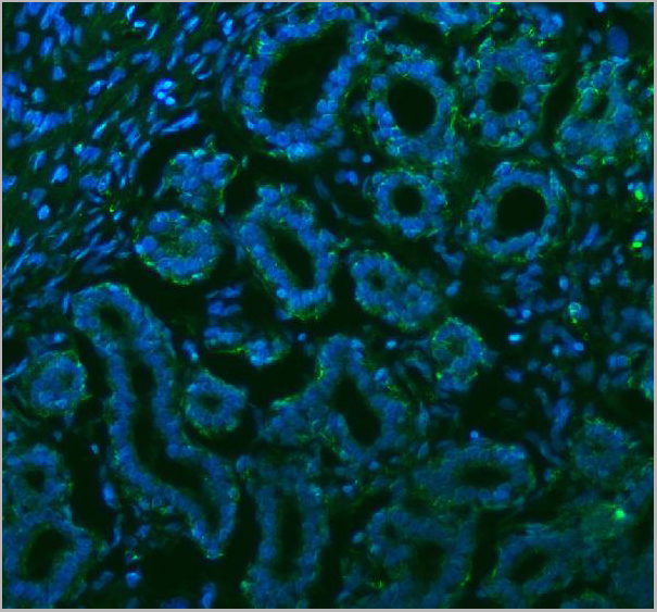
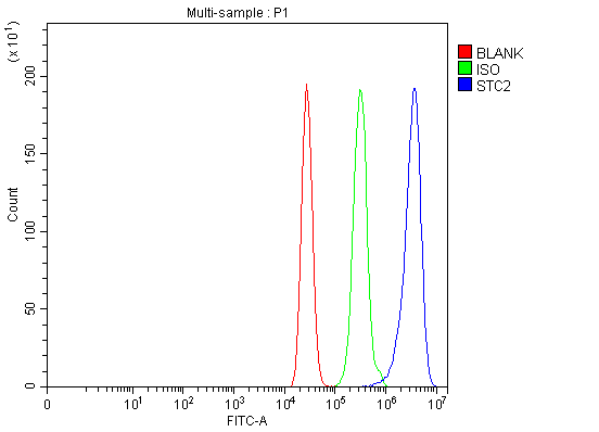
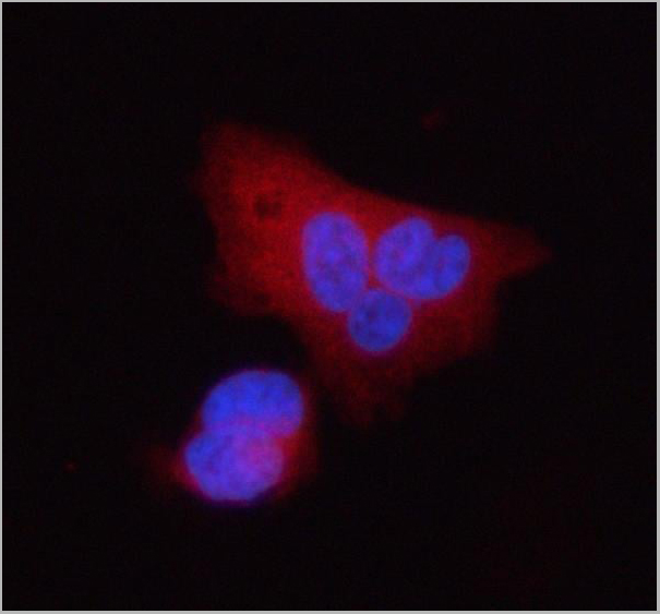
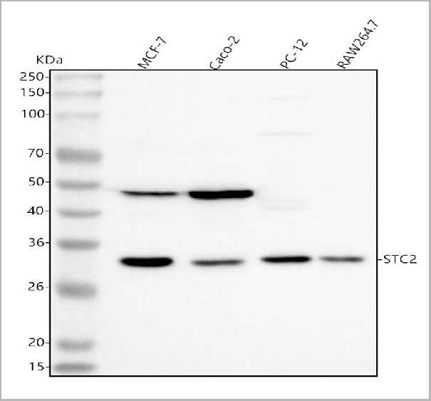
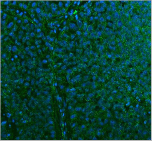
(Figure 15. IF analysis of Stanniocalcin 2/STC2 using anti-Stanniocalcin 2/STC2 antibody (AAA19831).Stanniocalcin 2/STC2 was detected in a paraffin-embedded section of human lung cancer tissue. Heat mediated antigen retrieval was performed in EDTA buffer (pH 8.0, epitope retrieval solution). The tissue section was blocked with 10% goat serum. The tissue section was then incubated with 5ug/mL rabbit anti-Stanniocalcin 2/STC2 Antibody (AAA19831) overnight at 4 degree C. DyLight®488 Conjugated Goat Anti-Rabbit IgG (BA1127) was used as secondary antibody at 1:500 dilution and incubated for 30 minutes at 37 degree C. The section was counterstained with DAPI. Visualize using a fluorescence microscope and filter sets appropriate for the label used.)
IF (Immunofluorescence)
(Figure 15. IF analysis of Stanniocalcin 2/STC2 using anti-Stanniocalcin 2/STC2 antibody (AAA19831).Stanniocalcin 2/STC2 was detected in a paraffin-embedded section of human lung cancer tissue. Heat mediated antigen retrieval was performed in EDTA buffer (pH 8.0, epitope retrieval solution). The tissue section was blocked with 10% goat serum. The tissue section was then incubated with 5ug/mL rabbit anti-Stanniocalcin 2/STC2 Antibody (AAA19831) overnight at 4 degree C. DyLight®488 Conjugated Goat Anti-Rabbit IgG (BA1127) was used as secondary antibody at 1:500 dilution and incubated for 30 minutes at 37 degree C. The section was counterstained with DAPI. Visualize using a fluorescence microscope and filter sets appropriate for the label used.)
Stanniocalcin 2/STC2, Polyclonal Antibody (Cat# AAA19831)
Full Name
Anti-Stanniocalcin 2/STC2 Antibody Picoband
Gene Names
STC2; STC-2; STCRP
Reactivity
Human, Mouse, Rat
Applications
Immunofluorescence, Immunohistochemistry, Immunocytochemistry, Western Blot, Flow Cytometry
Purity
Immunogen affinity purified.
Pricing
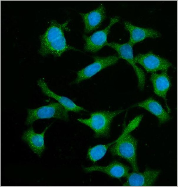
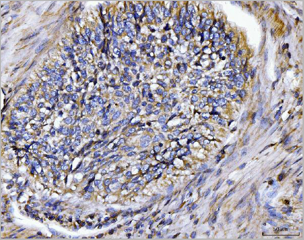
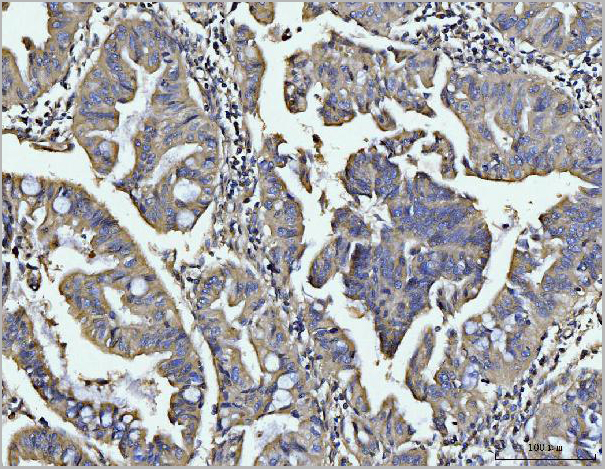
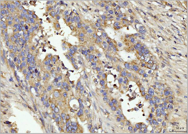
 FCM (Flow Cytometry)
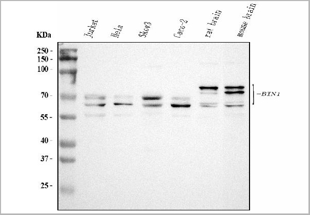
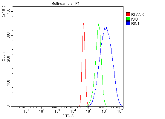
(Figure 6. Flow Cytometry analysis of A431 cells using anti-BIN1 antibody (AAA19424).Overlay histogram showing A431 cells stained with AAA19424 (Blue line). The cells were blocked with 10% normal goat serum. And then incubated with rabbit anti-BIN1 Antibody (AAA19424, 1 ug/1x10^6 cells) for 30 min at 20 degree C. DyLight488 conjugated goat anti-rabbit IgG was used as secondary antibody for 30 minutes at 20 degree C. Isotype control antibody (Green line) was rabbit IgG (1 ug/1x10^6) used under the same conditions. Unlabelled sample (Red line) was also used as a control.)
FCM (Flow Cytometry)
(Figure 6. Flow Cytometry analysis of A431 cells using anti-BIN1 antibody (AAA19424).Overlay histogram showing A431 cells stained with AAA19424 (Blue line). The cells were blocked with 10% normal goat serum. And then incubated with rabbit anti-BIN1 Antibody (AAA19424, 1 ug/1x10^6 cells) for 30 min at 20 degree C. DyLight488 conjugated goat anti-rabbit IgG was used as secondary antibody for 30 minutes at 20 degree C. Isotype control antibody (Green line) was rabbit IgG (1 ug/1x10^6) used under the same conditions. Unlabelled sample (Red line) was also used as a control.)
BIN1, Polyclonal Antibody (Cat# AAA19424)
Full Name
Anti-BIN1 Antibody Picoband
Gene Names
BIN1; AMPH2; AMPHL; SH3P9
Reactivity
Human, Mouse, Rat
Applications
Flow Cytometry, Immunofluorescence, Immunohistochemistry, Immunocytochemistry, Western Blot
Purity
Immunogen affinity purified.
Pricing
 FCM (Flow Cytometry)
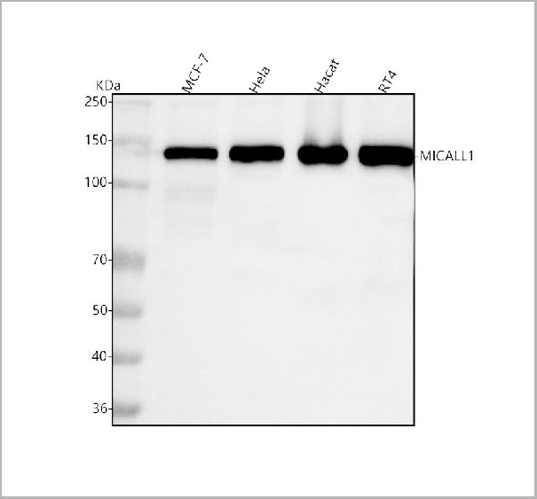
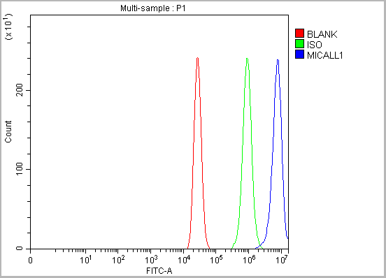
(Figure 14. Flow Cytometry analysis of MCF-7 cells using anti-MICALL1 antibody (AAA19933).Overlay histogram showing MCF-7 cells stained with AAA19933 (Blue line). To facilitate intracellular staining, cells were fixed with 4% paraformaldehyde and permeabilized with permeabilization buffer. The cells were blocked with 10% normal goat serum. And then incubated with rabbit anti-MICALL1 Antibody (AAA19933, 1ug/1x106 cells) for 30 min at 20 degree C. DyLight®488 conjugated goat anti-rabbit IgG (BA1127, 5-10ug/1x106 cells) was used as secondary antibody for 30 minutes at 20 degree C. Isotype control antibody (Green line) was rabbit IgG (1ug/1x106) used under the same conditions. Unlabelled sample (Red line) was also used as a control.)
FCM (Flow Cytometry)
(Figure 14. Flow Cytometry analysis of MCF-7 cells using anti-MICALL1 antibody (AAA19933).Overlay histogram showing MCF-7 cells stained with AAA19933 (Blue line). To facilitate intracellular staining, cells were fixed with 4% paraformaldehyde and permeabilized with permeabilization buffer. The cells were blocked with 10% normal goat serum. And then incubated with rabbit anti-MICALL1 Antibody (AAA19933, 1ug/1x106 cells) for 30 min at 20 degree C. DyLight®488 conjugated goat anti-rabbit IgG (BA1127, 5-10ug/1x106 cells) was used as secondary antibody for 30 minutes at 20 degree C. Isotype control antibody (Green line) was rabbit IgG (1ug/1x106) used under the same conditions. Unlabelled sample (Red line) was also used as a control.)
MICALL1, Polyclonal Antibody (Cat# AAA19933)
Full Name
Anti-MICALL1 Antibody Picoband
Gene Names
MICALL1; MIRAB13; MICAL-L1
Reactivity
Human, Mouse, Rat
Applications
Western Blot, Immunohistochemistry, Flow Cytometry
Purity
Immunogen affinity purified.
Pricing
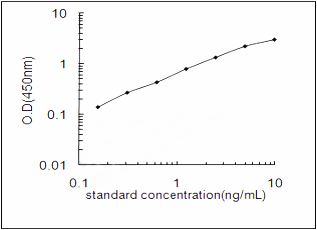 Standard Curve (Sample)
Microtubule-associated proteins 1A/1B light chain 3B, ELISA Kit (Cat# AAA193090)
Full Name
Human Microtubule-associated proteins 1A/1B light chain 3B ELISA Kit
Gene Names
MAP1LC3B; LC3B; ATG8F; MAP1LC3B-a; MAP1A/1BLC3
Reactivity
Human
Pricing
 SDS-PAGE
(The purity of MGMT was determined to be >95% by densitometry)
SDS-PAGE
(The purity of MGMT was determined to be >95% by densitometry)
MGMT, Recombinant Protein (Cat# AAA72923)
Full Name
MGMT recombinant protein
Applications
Western Blot
Purity
>95%
Pricing
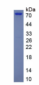
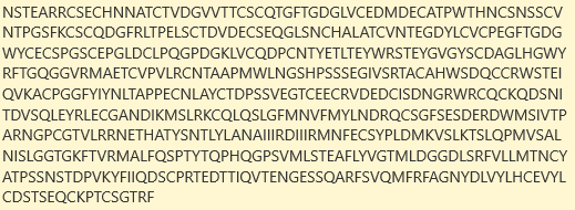 Amino Acid Info
Uromodulin (UMOD), Recombinant Protein (Cat# AAA153660)
Full Name
Recombinant Uromodulin (UMOD)
Gene Names
Umod; THP; Urehd1; urehr4
Applications
Western Blot
Purity
> 90%
Pricing
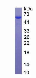
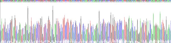
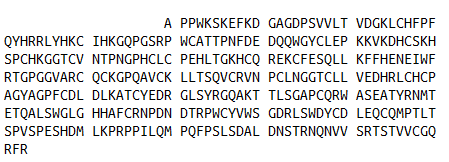 Amino Acid Info
(Amino Acid info)
Amino Acid Info
(Amino Acid info)
Coagulation Factor XII (F12), Recombinant Protein (Cat# AAA153383)
Full Name
Recombinant Coagulation Factor XII (F12)
Gene Names
F12; HAF
Applications
Western Blot
Purity
Greater than 90% by SDS-PAGE
Pricing
 Application Data
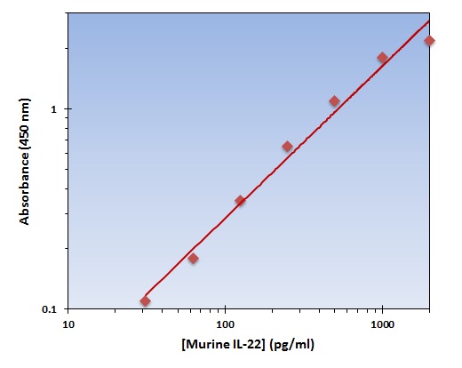
(The OmniKine Murine IL-22 ELISA Kit allows for the detection and quantification of endogenous levels of natural and/or recombinant Murine IL-22 proteins within the range of 32-2000 pg/ml.)
Application Data
(The OmniKine Murine IL-22 ELISA Kit allows for the detection and quantification of endogenous levels of natural and/or recombinant Murine IL-22 proteins within the range of 32-2000 pg/ml.)
IL-22, ELISA Kit (Cat# AAA315771)
Full Name
IL-22 (Murine) OmniKine ELISA Kit
Gene Names
Il22; IL-22; Iltif; IL-22a; ILTIFa
Reactivity
Mouse
Pricing
 Amino Acid Info
(Amino Acid info)
Amino Acid Info
(Amino Acid info)
Growth Differentiation Factor 15 (GDF15), Recombinant Protein (Cat# AAA153509)
Full Name
Recombinant Growth Differentiation Factor 15 (GDF15)
Applications
Western Blot
Purity
Greater than 95% by SDS-PAGE
Pricing


 SDS-PAGE
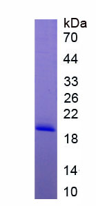
(Figure. SDS-PAGE)
SDS-PAGE
(Figure. SDS-PAGE)
Adiponectin Receptor 1 (ADIPOR1), Recombinant Protein (Cat# AAA150661)
Full Name
Recombinant Adiponectin Receptor 1 (ADIPOR1)
Applications
Western Blot
Purity
>97%
Pricing
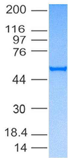
 Application Data
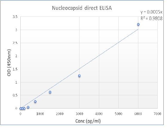
(Figure-2: Wells of a 96-microtiter plate were coated with 4ug/ml of SARS-Cov-2/nCov/COVID-19 Nucleocapsid recombinant protein. The binding was detected by addition of different dilution of polyclonal antibody. The reactivity was detected by a HRP-conjugated goat-anti-rabbit polyclonal antibody.)
Application Data
(Figure-2: Wells of a 96-microtiter plate were coated with 4ug/ml of SARS-Cov-2/nCov/COVID-19 Nucleocapsid recombinant protein. The binding was detected by addition of different dilution of polyclonal antibody. The reactivity was detected by a HRP-conjugated goat-anti-rabbit polyclonal antibody.)
COVID 19 Nucleocapsid (NP) Coronavirus, Recombinant Protein (Cat# AAA78527)
Full Name
SARS-CoV-2/COVID-19 Nucleocapsid Protein His Tag
Purity
>95% by SDS-PAGE.
Pricing
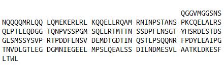
 SDS-PAGE
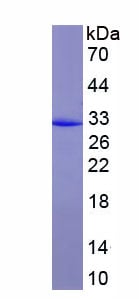
(SDS-PAGE)
SDS-PAGE
(SDS-PAGE)
Yes Associated Protein 1 (YAP1), Recombinant Protein (Cat# AAA152020)
Full Name
Recombinant Yes Associated Protein 1 (YAP1)
Applications
Western Blot
Purity
Greater than 97% by SDS-PAGE
Pricing
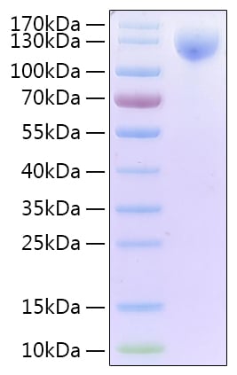
 Application Data
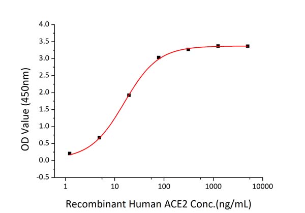
(Fig 2: Immobilized Recombinant 2019-nCoV Spike S1-His at 2ug/mL (100 uL/well) can bind Recombinant Human ACE2 with a linear range of 1.5-15 ng/mL.)
Application Data
(Fig 2: Immobilized Recombinant 2019-nCoV Spike S1-His at 2ug/mL (100 uL/well) can bind Recombinant Human ACE2 with a linear range of 1.5-15 ng/mL.)
COVID 19 Spike S1 Coronavirus, Active Protein (Cat# AAA78533)
Full Name
Recombinant SARS-CoV-2 Spike S1 Protein with His tag and Avi
Applications
Functional Assay
Purity
>95% by SDS-PAGE.
Pricing
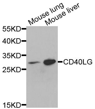
 IF (Immunofluorescence)
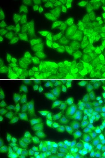
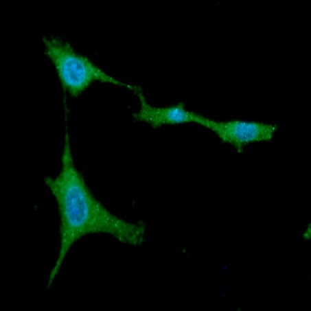
(Immunofluorescence analysis of U2OS cells using CD40LG antibody. Blue: DAPI for nuclear staining.)
IF (Immunofluorescence)
(Immunofluorescence analysis of U2OS cells using CD40LG antibody. Blue: DAPI for nuclear staining.)
CD40LG, Polyclonal Antibody (Cat# AAA37438)
Full Name
CD40LG Polyclonal Antibody
Gene Names
CD40LG; IGM; IMD3; TRAP; gp39; CD154; CD40L; HIGM1; T-BAM; TNFSF5; hCD40L
Reactivity
Human, Mouse
Applications
Immunofluorescence, Immunohistochemistry, Western Blot
Purity
Affinity Purification
Pricing
 WB (Western Blot)
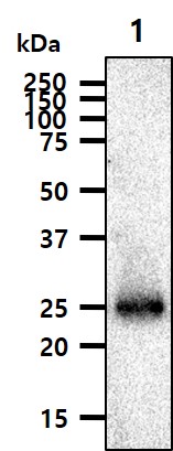
(The mouse tissue lysate(40ug) was resolved by SDS-PAGE, transferred to PVDF membrane and probed with anti-human Rab5a (1:500). Proteins were visualized using a goat anti-mouse secondary antibody conjugated to HRP and an ECL detection system.;;Lane 1.: Mouse brain tissue lysate)
WB (Western Blot)
(The mouse tissue lysate(40ug) was resolved by SDS-PAGE, transferred to PVDF membrane and probed with anti-human Rab5a (1:500). Proteins were visualized using a goat anti-mouse secondary antibody conjugated to HRP and an ECL detection system.;;Lane 1.: Mouse brain tissue lysate)
Rab5a, Monoclonal Antibody (Cat# AAA48282)
Full Name
Rab5a
Gene Names
RAB5A; RAB5
Reactivity
Human
Applications
Immunofluorescence, Immunocytochemistry, Western Blot
Purity
Protein-A Affinity Chromatography
Pricing

 Amino Acid Info
(Amino Acid info)
Amino Acid Info
(Amino Acid info)
Ubiquitin Conjugating Enzyme E2A (UBE2A), Recombinant Protein (Cat# AAA153425)
Full Name
Recombinant Ubiquitin Conjugating Enzyme E2A (UBE2A)
Gene Names
Ube2a; HR6A; HHR6A; Mhr6a
Applications
Western Blot
Purity
Greater than 90% by SDS-PAGE
Pricing
 Application Data
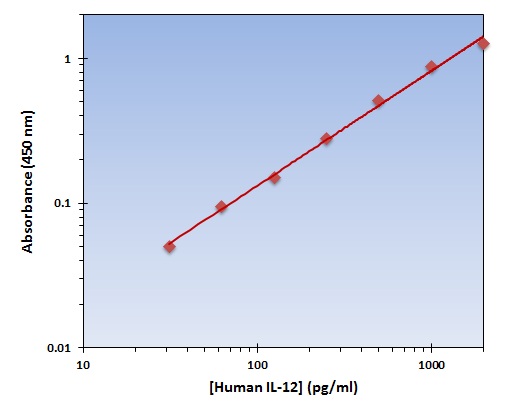
(The OmniKine Human IL-12 ELISA Kit allows for the detection and quantification of endogenous levels of natural and/or recombinant Human IL-12 proteins within the range of 32-2000 pg/ml.)
Application Data
(The OmniKine Human IL-12 ELISA Kit allows for the detection and quantification of endogenous levels of natural and/or recombinant Human IL-12 proteins within the range of 32-2000 pg/ml.)
IL-12, ELISA Kit (Cat# AAA315617)
Full Name
IL-12 (Human) OmniKine ELISA Kit
Gene Names
IL12A; P35; CLMF; NFSK; NKSF1; IL-12A
Reactivity
Human
Pricing
 Bioactivity
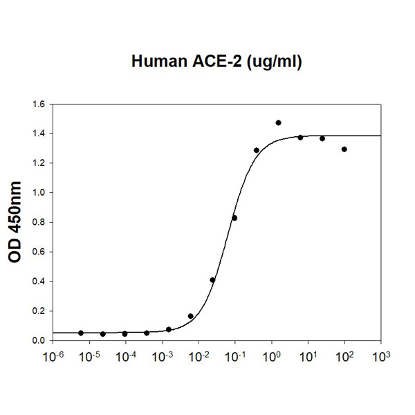
(SARS-CoV-2 Spike RBD is coated at 5ug/ml (100 ul/well) can bind ACE-2 (CAT# ATGP3963) in a Functional ELISA assay.)
Bioactivity
(SARS-CoV-2 Spike RBD is coated at 5ug/ml (100 ul/well) can bind ACE-2 (CAT# ATGP3963) in a Functional ELISA assay.)
COVID 19 Spike RBD Coronavirus, Active Protein (Cat# AAA48376)
Full Name
Recombinant SARS-CoV-2 (2019-nCoV) Spike RBD protein
Applications
Bioactivity, SDS-PAGE
Purity
>95% by SDS-PAGE
Pricing
 FCM/FACS (Flow Cytometry)
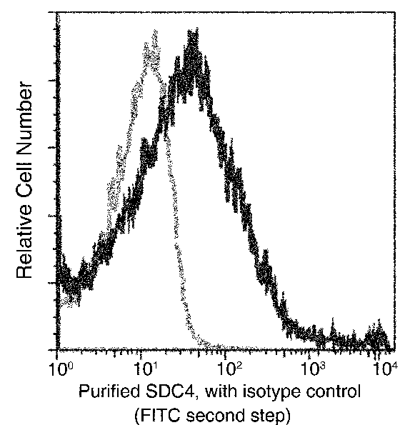
(Flow cytometric analysis of Mouse SDC4 expression on BABL/c splenocytes. Cells were stained with purified anti-Mouse SDC4, then a FITC-conjugated second step antibody. The fluorescence histograms were derived from gated events with the forward and side light-scatter characteristics of intact cells.)
FCM/FACS (Flow Cytometry)
(Flow cytometric analysis of Mouse SDC4 expression on BABL/c splenocytes. Cells were stained with purified anti-Mouse SDC4, then a FITC-conjugated second step antibody. The fluorescence histograms were derived from gated events with the forward and side light-scatter characteristics of intact cells.)
Syndecan-4, Monoclonal Antibody (Cat# AAA257219)
Full Name
Recombinant Anti-Syndecan-4 Antibody, Rabbit Monoclonal
Gene Names
Sdc4; Synd4; AA959608; AW108331; ryudocan; syndecan-4
Reactivity
Mouse
Applications
Flow Cytometry, Western Blot
Purity
Protein A
Pricing
 Bioactivity
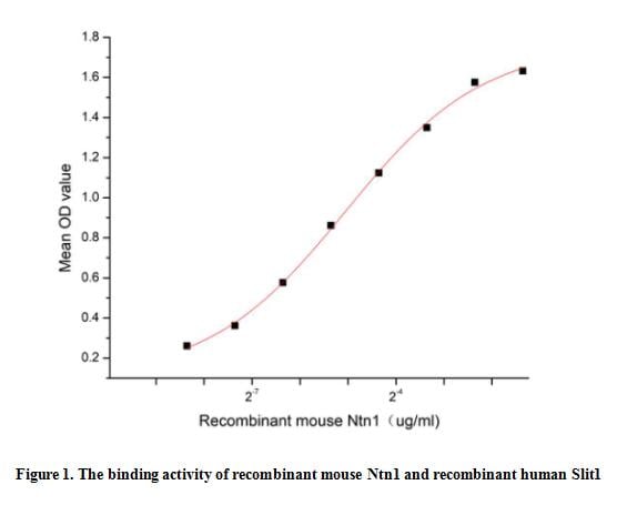
(Netrin 1 (Ntn1) is included in a family of laminin-related secreted proteins. Mouse Ntn1 is a 66-76 kDa glycoprotein that is well known for its involvement in axonal guidance during embryonic development and as an enhancer of cancer cell metastasis. Ntn1 is also involved in outgrowth and migration orientation in the developing CNS and plays a significant role in the morphogenesis of endothelial cells and vascular smooth-muscle cells. It is also involved in the processes of cytoskeleton reorganization, angiogenesis, epithelial cell adhesion, and cell migration in the lungs, mammary gland, and pancreas. Slit Homolog 1 (Slit1) can acts as a potent promoter of both Netrin-1 attractive and repulsive activities on distinct neuronal cell types, thereby opening novel perspectives on the role of combinations of cues in brain wiring. Thus a functional binding ELISA assay was conducted to detect the interaction of recombinant mouse Ntn1 and recombinant human Slit1. Briefly, Ntn1 was diluted serially in PBS with 0.01% BSA (pH 7.4). Duplicate samples of 100 ul were then transferred to Slit1-coated microtiter wells and incubated for 1h at 37 degree C. Wells were washed with PBST and incubated for 1h with anti-Ntn1 pAb, then aspirated and washed 3 times. After incubation with HRP labelled secondary antibody for 1h at 37 degree C, wells were aspirated and washed 5 times. With the addition of substrate solution, wells were incubated 15-25 minutes at 37 degree C. Finally, add 50 uL stop solution to the wells and read at 450/630 nm immediately. The binding activity of recombinant mouse Ntn1 and recombinant human Slit1 was shown in Figure 1, the EC50 for this effect is 0.03 ug/mL.)
Bioactivity
(Netrin 1 (Ntn1) is included in a family of laminin-related secreted proteins. Mouse Ntn1 is a 66-76 kDa glycoprotein that is well known for its involvement in axonal guidance during embryonic development and as an enhancer of cancer cell metastasis. Ntn1 is also involved in outgrowth and migration orientation in the developing CNS and plays a significant role in the morphogenesis of endothelial cells and vascular smooth-muscle cells. It is also involved in the processes of cytoskeleton reorganization, angiogenesis, epithelial cell adhesion, and cell migration in the lungs, mammary gland, and pancreas. Slit Homolog 1 (Slit1) can acts as a potent promoter of both Netrin-1 attractive and repulsive activities on distinct neuronal cell types, thereby opening novel perspectives on the role of combinations of cues in brain wiring. Thus a functional binding ELISA assay was conducted to detect the interaction of recombinant mouse Ntn1 and recombinant human Slit1. Briefly, Ntn1 was diluted serially in PBS with 0.01% BSA (pH 7.4). Duplicate samples of 100 ul were then transferred to Slit1-coated microtiter wells and incubated for 1h at 37 degree C. Wells were washed with PBST and incubated for 1h with anti-Ntn1 pAb, then aspirated and washed 3 times. After incubation with HRP labelled secondary antibody for 1h at 37 degree C, wells were aspirated and washed 5 times. With the addition of substrate solution, wells were incubated 15-25 minutes at 37 degree C. Finally, add 50 uL stop solution to the wells and read at 450/630 nm immediately. The binding activity of recombinant mouse Ntn1 and recombinant human Slit1 was shown in Figure 1, the EC50 for this effect is 0.03 ug/mL.)
Netrin 1 (Ntn1), Active Protein (Cat# AAA161863)
Full Name
Active Netrin 1 (Ntn1)
Gene Names
Ntn1; AI561871; BC019633; Netrin-1
Applications
Activity Assay, Cell Culture
Purity
Greater than 90% by SDS-PAGE
Pricing
 Application Data
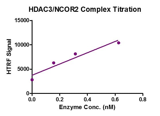
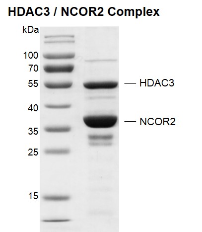
(HTRF assay for HDAC3 / NCOR2 Complex, His-tag activity 3 uM histone H3K9ac peptide was incubated with different concentrations of HDAC3 / NCOR2 Complex, His-tag in reaction buffer for 30 minutes at 37 degree C. H3K9me0-Eu antibody was used to detect the reaction products.)
Application Data
(HTRF assay for HDAC3 / NCOR2 Complex, His-tag activity 3 uM histone H3K9ac peptide was incubated with different concentrations of HDAC3 / NCOR2 Complex, His-tag in reaction buffer for 30 minutes at 37 degree C. H3K9me0-Eu antibody was used to detect the reaction products.)
HDAC3/NCOR2, Recombinant Protein (Cat# AAA59781)
Full Name
Recombinant HDAC3/NCOR2 complex, His-Tag
Gene Names
HDAC3; HD3; RPD3; RPD3-2
Pricing
 Application Data
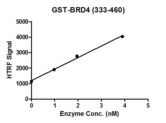
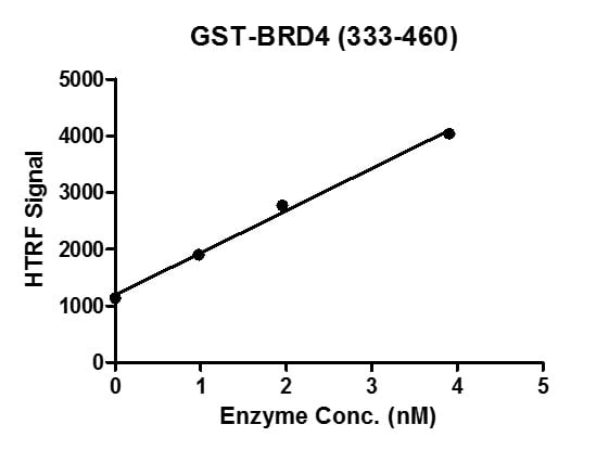
(HTRF assay for BRD4 (333-460), GST-Tag activity 1 uM H4K5/8/12/16(ac4) peptide was incubated with different concentrations of BRD4 (333-460), GST-Tag protein in a 10 ul binding system containing 50 mM HEPES-NaOH pH 7.4, 0.1% BSA for 1 hour, then 10 ul GST antibody and SA-XL665 mixture (each 1:100 dilution in Binding Buffer) were added to each reaction system and incubated for 30 min. All the operations and reactions were performed at room temperature. HTRF assay was used for detection.)
Application Data
(HTRF assay for BRD4 (333-460), GST-Tag activity 1 uM H4K5/8/12/16(ac4) peptide was incubated with different concentrations of BRD4 (333-460), GST-Tag protein in a 10 ul binding system containing 50 mM HEPES-NaOH pH 7.4, 0.1% BSA for 1 hour, then 10 ul GST antibody and SA-XL665 mixture (each 1:100 dilution in Binding Buffer) were added to each reaction system and incubated for 30 min. All the operations and reactions were performed at room temperature. HTRF assay was used for detection.)
BRD4, Recombinant Protein (Cat# AAA60163)
Full Name
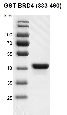
Recombinant BRD4 (333-460) protein, GST-Tag
Gene Names
BRD4; CAP; MCAP; HUNK1; HUNKI
Pricing
 Bioactivity
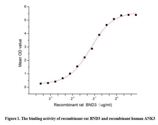
(Band 3 (BND3), also known as Anion Exchanger 1 and SLC4A1, is a 90-100 kDa polytropic membrane protein that is part of the anion exchanger (AE) family and is expressed in the erythrocyte plasma membrane, where it mediates the exchange of the cellular HCO3- with CI- in plasma. BND3 is required for normal flexibility and stability of the erythrocyte membrane and for normal erythrocyte shape via the interactions of its cytoplasmic domain with cytoskeletal proteins, glycolytic enzymes, and hemoglobin. Scaffold protein Ankyrin 3, Node Of Ranvier (ANK3) can Interacts with BND3, thus a functional binding ELISA assay was conducted to detect the interaction of recombinant rat BND3 and recombinant human ANK3. Briefly, biotin-linked BND3 were diluted serially in PBS, with 0.01% BSA (pH 7.4). Duplicate samples of 100 ul were then transferred to ANK3-coated microtiter wells and incubated for 1h at 37 degree C. Wells were washed with PBST 3 times and incubation with Streptavidin-HRP for 30min, then wells were aspirated and washed 5 times. With the addition of substrate solution, wells were incubated 15-25 minutes at 37 degree C. Finally, add 50 ul stop solution to the wells and read at 450 nm immediately. The binding activity of recombinant rat BND3 and recombinant human ANK3 was shown in Figure 1, the EC50 for this effect is 0.35 ug/mL.)
Bioactivity
(Band 3 (BND3), also known as Anion Exchanger 1 and SLC4A1, is a 90-100 kDa polytropic membrane protein that is part of the anion exchanger (AE) family and is expressed in the erythrocyte plasma membrane, where it mediates the exchange of the cellular HCO3- with CI- in plasma. BND3 is required for normal flexibility and stability of the erythrocyte membrane and for normal erythrocyte shape via the interactions of its cytoplasmic domain with cytoskeletal proteins, glycolytic enzymes, and hemoglobin. Scaffold protein Ankyrin 3, Node Of Ranvier (ANK3) can Interacts with BND3, thus a functional binding ELISA assay was conducted to detect the interaction of recombinant rat BND3 and recombinant human ANK3. Briefly, biotin-linked BND3 were diluted serially in PBS, with 0.01% BSA (pH 7.4). Duplicate samples of 100 ul were then transferred to ANK3-coated microtiter wells and incubated for 1h at 37 degree C. Wells were washed with PBST 3 times and incubation with Streptavidin-HRP for 30min, then wells were aspirated and washed 5 times. With the addition of substrate solution, wells were incubated 15-25 minutes at 37 degree C. Finally, add 50 ul stop solution to the wells and read at 450 nm immediately. The binding activity of recombinant rat BND3 and recombinant human ANK3 was shown in Figure 1, the EC50 for this effect is 0.35 ug/mL.)
Band 3 (BND3), Active Protein (Cat# AAA161856)
Full Name
Active Band 3 (BND3)
Applications
Activity Assay, Cell Culture
Purity
Greater than 95% by SDS-PAGE
Pricing
 IF (Immunofluorescence)
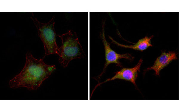
(Immunofluorescence analysis of A549 (left) and SK-BR-3 (right) cells using CTNNB1 antibody (green). Red: Actin filaments have been labeled with DY-554 phalloidin. Blue: DRAQ5 fluorescent DNA dye.)
IF (Immunofluorescence)
(Immunofluorescence analysis of A549 (left) and SK-BR-3 (right) cells using CTNNB1 antibody (green). Red: Actin filaments have been labeled with DY-554 phalloidin. Blue: DRAQ5 fluorescent DNA dye.)
CTNNB1, Monoclonal Antibody (Cat# AAA74903)
Full Name
CTNNB1 antibody
Gene Names
CTNNB1; CTNNB; MRD19; armadillo
Applications
Western Blot, Immunofluorescence, Flow Cytometry
Pricing
 WB (Western Blot)
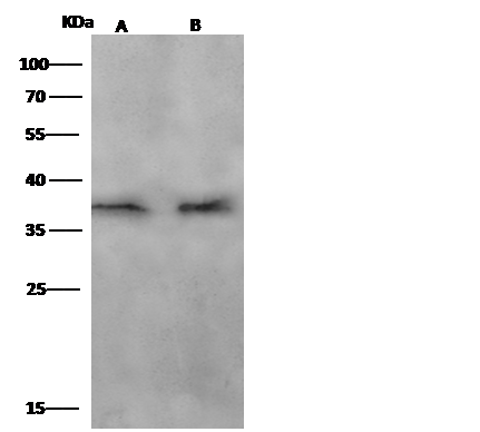
(Anti-CCNA1 mouse monoclonal antibody at 1:500 dilutionLane A: THP1 Whole Cell LysateLane B: Hela Whole Cell LysateLysates/proteins at 30 ug per lane.SecondaryRabbit Anti-Mouse IgG F(ab)2/HRP at 1/10000 dilution.Developed using the ECL technique.Performed under reducing conditions.Predicted band size:52 kDaObserved band size:37 kDa)
WB (Western Blot)
(Anti-CCNA1 mouse monoclonal antibody at 1:500 dilutionLane A: THP1 Whole Cell LysateLane B: Hela Whole Cell LysateLysates/proteins at 30 ug per lane.SecondaryRabbit Anti-Mouse IgG F(ab)2/HRP at 1/10000 dilution.Developed using the ECL technique.Performed under reducing conditions.Predicted band size:52 kDaObserved band size:37 kDa)
Cyclin A1, Monoclonal Antibody (Cat# AAA255288)
Full Name
Anti-Cyclin A1 Antibody, Mouse Monoclonal
Gene Names
CCNA1; CT146
Reactivity
Human
Applications
Immunofluorescence, Immunocytochemistry, Western Blot
Purity
Protein A
Pricing
 SDS-PAGE
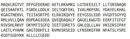
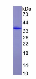
(SDS-PAGE)
SDS-PAGE
(SDS-PAGE)
Annexin V (ANXA5), Recombinant Protein (Cat# AAA152066)
Full Name
Eukaryotic Annexin V (ANXA5)
Gene Names
ANXA5; PP4; ANX5; ENX2; RPRGL3; HEL-S-7
Applications
Western Blot
Purity
Greater than 97% by SDS-PAGE
Pricing
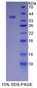 SDS-PAGE
Actinin Alpha 4, Recombinant Protein (Cat# AAA144188)
Full Name
Recombinant Actinin Alpha 4 (ACTN4)
Gene Names
ACTN4; FSGS; FSGS1; ACTININ-4
Applications
Western Blot
Purity
> 95%
Pricing
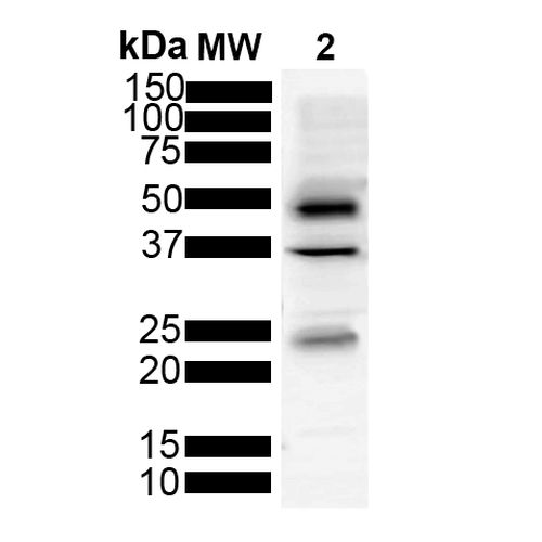
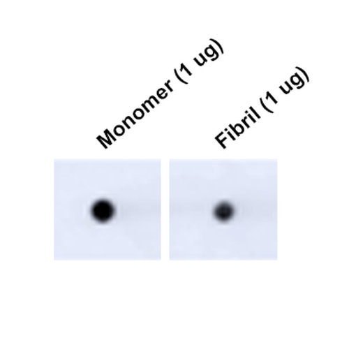 DB (Dot Blot)
(Dot Blot analysis using Mouse Anti-Tau Monoclonal Antibody, Clone 3D4 (SMC-608). Tissue: Recombinant Protein. Species: Human. Primary Antibody: Mouse Anti-Tau Monoclonal Antibody (SMC-608) at 1:1000 for 2 hours at RT with shaking. Secondary Antibody: Goat anti-mouse IgG:HRP at 1:5000 for 1 hour at RT with shaking.)
DB (Dot Blot)
(Dot Blot analysis using Mouse Anti-Tau Monoclonal Antibody, Clone 3D4 (SMC-608). Tissue: Recombinant Protein. Species: Human. Primary Antibody: Mouse Anti-Tau Monoclonal Antibody (SMC-608) at 1:1000 for 2 hours at RT with shaking. Secondary Antibody: Goat anti-mouse IgG:HRP at 1:5000 for 1 hour at RT with shaking.)
Tau, Monoclonal Antibody (Cat# AAA253990)
Full Name
Tau Antibody, Clone 3D4: ATTO 390
Gene Names
MAPT; TAU; MSTD; PPND; DDPAC; MAPTL; MTBT1; MTBT2; FTDP-17; PPP1R103
Reactivity
Human, Mouse, Rat
Applications
Immunofluorescence, Immunocytochemistry, Dot Blot, Western Blot
Purity
Protein G Purified
Pricing
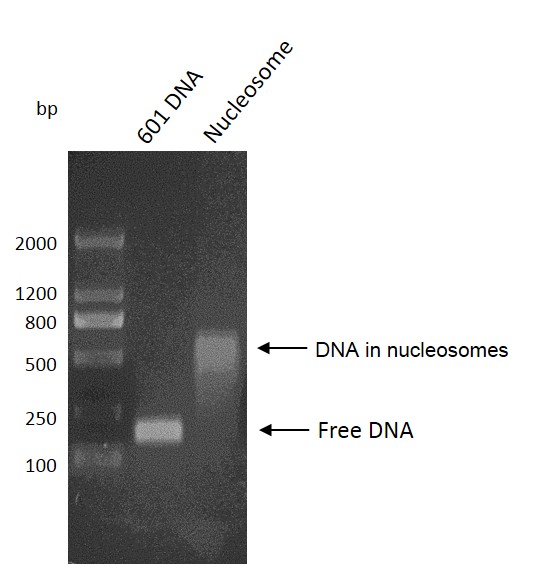

 WB (Western Blot)
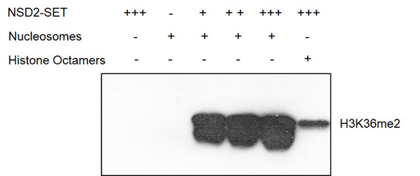
(Recombinant Mononucleosomes (H3.3) - biotinylated activity. 2 ?g Mononucleosomes were incubated with 0 ?g, 0.25 ?g, 0.5 ?g, 1 ?g NSD2-SET in reaction buffer for 3 h at room temperature, respectively. Western blot was used for detecting the generation of reaction products (H3K36me2). Recombinant Histone Octamers was used as a control substrate. The Western blot result shows that mononucleosomes are a more suitable substrate for NSD2-SET than histone octamers.)
WB (Western Blot)
(Recombinant Mononucleosomes (H3.3) - biotinylated activity. 2 ?g Mononucleosomes were incubated with 0 ?g, 0.25 ?g, 0.5 ?g, 1 ?g NSD2-SET in reaction buffer for 3 h at room temperature, respectively. Western blot was used for detecting the generation of reaction products (H3K36me2). Recombinant Histone Octamers was used as a control substrate. The Western blot result shows that mononucleosomes are a more suitable substrate for NSD2-SET than histone octamers.)
Mononucleosomes, Recombinant Protein (Cat# AAA59703)
Full Name
Recombinant Mononucleosomes (H3.3)-biotinylated
Purity
The recombinant protein is >95% pure by SDS-PAGE.
Pricing
 Bioactivity
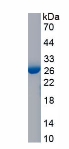
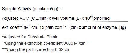
(GSTa3 (Glutathione S-transferase a3) is an enzyme that plays an important role in detoxification by catalyzing the conjugation of many hydrophobic and electrophilic compounds with reduced glutathione. This subfamily of enzymes has a particular role in protecting cells from Reactive Oxygen Species and the products of peroxidation. Polymorphisms in this gene influence the ability of individuals to metabolize different drugs. GSTa3 catalyze the endogenous glutathione conjugation 1-Chloro-2,4-dinitrobenzene (CDNB), which can increase in the absorbance at 340 nm. The reaction was performed in adding 10 ul 200 mM glutathione (reduced) and 10 ul 100 mM CDNB in 980 ul 100 mM NaH2PO4 (pH7.0), rapidly mixed. Then add 50 ul mixed substrates to 50 ul different concentrations of recombinant mouse GSTa3, mix gently. Incubated at 37 degree C for 5min, then read at a wavelength of 340 nm. The specific activity of recombinant mouse GSTa3 is >60,000 pmol/min/ug.)
Bioactivity
(GSTa3 (Glutathione S-transferase a3) is an enzyme that plays an important role in detoxification by catalyzing the conjugation of many hydrophobic and electrophilic compounds with reduced glutathione. This subfamily of enzymes has a particular role in protecting cells from Reactive Oxygen Species and the products of peroxidation. Polymorphisms in this gene influence the ability of individuals to metabolize different drugs. GSTa3 catalyze the endogenous glutathione conjugation 1-Chloro-2,4-dinitrobenzene (CDNB), which can increase in the absorbance at 340 nm. The reaction was performed in adding 10 ul 200 mM glutathione (reduced) and 10 ul 100 mM CDNB in 980 ul 100 mM NaH2PO4 (pH7.0), rapidly mixed. Then add 50 ul mixed substrates to 50 ul different concentrations of recombinant mouse GSTa3, mix gently. Incubated at 37 degree C for 5min, then read at a wavelength of 340 nm. The specific activity of recombinant mouse GSTa3 is >60,000 pmol/min/ug.)
Glutathione S Transferase Alpha 3 (GSTa3), Active Protein (Cat# AAA161767)
Full Name
Active Glutathione S Transferase Alpha 3 (GSTa3)
Gene Names
Gsta3; Gst2-3
Applications
Activity Assay, Cell Culture
Purity
Greater than 90% by SDS-PAGE
Pricing
 Bioactivity
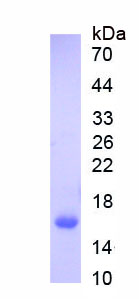
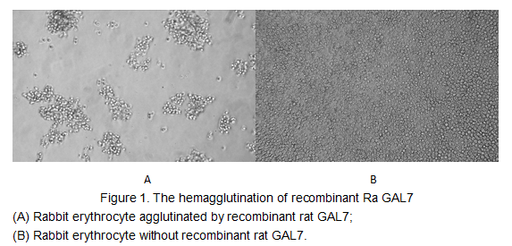
(The galectins constitute a large family of carbohydrate-binding proteins with specificity for N-acetyl-lactosamine-containing glycoproteins. At least 14 mammalian galectins, which share structural similarities in their carbohydrate recognition domains (CRD), have been identified. The galectins have been classified into the prototype galectins (-1, -2, -5, -7, -10, -11, -13, -14), which contain one CRD and exist either as a monomer or a noncovalent homodimer; the chimera galectins (Galectin-3) containing one CRD linked to a nonlectin domain; and the tandem-repeat galectins (-4, -6, -8, -9, -12) consisting of two CRDs joined by a linker peptide. Galectin-7 may also be involved in cell-cell and cell-matrix interactions and exogenous galectin has been found to accelerate the re-epithelialization of woundsIt can agglutinate red blood.In this case, we chose rabbit erythrocyte (RaE) to assay its ability of agglutination. A general procedure for hemagglutination assay (or haemagglutination assay; HA) is as follows, two-fold dilute the recombinant Ra GAL7 with 0.9% sodium chloride injection, add 50uL a serial dilution of GAL7 to each well of a U or V-bottom shaped 96-well microtiter plate. The final well serves as a negative control with no GAL7, replace with 50uL 0.9% sodium chloride injection. Then add 50uL 1% rabbit erythrocyte to each well and mixed gently. The plate is incubated for 3 hours at room temperature. The results are shown in Figure 1. It was obvious that the minimal effective concentration of GAL7 is 25 ug/mL.)
Bioactivity
(The galectins constitute a large family of carbohydrate-binding proteins with specificity for N-acetyl-lactosamine-containing glycoproteins. At least 14 mammalian galectins, which share structural similarities in their carbohydrate recognition domains (CRD), have been identified. The galectins have been classified into the prototype galectins (-1, -2, -5, -7, -10, -11, -13, -14), which contain one CRD and exist either as a monomer or a noncovalent homodimer; the chimera galectins (Galectin-3) containing one CRD linked to a nonlectin domain; and the tandem-repeat galectins (-4, -6, -8, -9, -12) consisting of two CRDs joined by a linker peptide. Galectin-7 may also be involved in cell-cell and cell-matrix interactions and exogenous galectin has been found to accelerate the re-epithelialization of woundsIt can agglutinate red blood.In this case, we chose rabbit erythrocyte (RaE) to assay its ability of agglutination. A general procedure for hemagglutination assay (or haemagglutination assay; HA) is as follows, two-fold dilute the recombinant Ra GAL7 with 0.9% sodium chloride injection, add 50uL a serial dilution of GAL7 to each well of a U or V-bottom shaped 96-well microtiter plate. The final well serves as a negative control with no GAL7, replace with 50uL 0.9% sodium chloride injection. Then add 50uL 1% rabbit erythrocyte to each well and mixed gently. The plate is incubated for 3 hours at room temperature. The results are shown in Figure 1. It was obvious that the minimal effective concentration of GAL7 is 25 ug/mL.)
Galectin 7 (GAL7), Active Protein (Cat# AAA152173)
Full Name
Active Galectin 7 (GAL7)
Applications
Activity Assay, Cell Culture
Purity
Greater than 95% by SDS-PAGE
Pricing

 Bioactivity
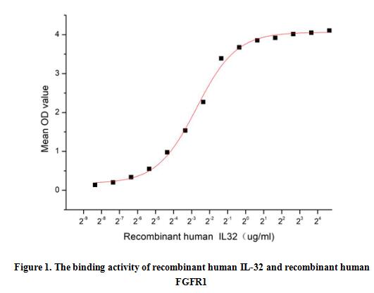
(Interleukin-32 (IL-32) is an interleukin cytokine usually linked to inflammation. The IL-32 transcript is expressed in various human tissues and organs such as the spleen, thymus, leukocyte, lung, small intestine, colon, prostate, heart, placenta, liver, muscle, kidney, pancreas, and brain. Cytokines are critical components of cell signaling pathways that are involved in the regulation of cell growth, metabolism, hormone signaling, immune regulation and a variety of other physiological functions. FGFR1 has been identified as an interactor of IL-32, thus a functional binding ELISA assay was conducted to detect the interaction of recombinant human IL-32 and recombinant human FGFR1. Briefly, IL-32 was diluted serially in PBS with 0.01% BSA (pH 7.4). Duplicate samples of 100 ul were then transferred to FGFR1-coated microtiter wells and incubated for 1h at 37 degree C. Wells were washed with PBST and incubated for 1h with anti-IL-32 pAb, then aspirated and washed 3 times. After incubation with HRP labelled secondary antibody for 1h at 37 degree C, wells were aspirated and washed 5 times. With the addition of substrate solution, wells were incubated 15-25 minutes at 37 degree C. Finally, add 50 uL stop solution to the wells and read at 450/630nm immediately. The binding activity of recombinant human IL-32 and recombinant human FGFR1 was shown in Figure 1, the EC50 for this effect is 0.15 ug/mL.)
Bioactivity
(Interleukin-32 (IL-32) is an interleukin cytokine usually linked to inflammation. The IL-32 transcript is expressed in various human tissues and organs such as the spleen, thymus, leukocyte, lung, small intestine, colon, prostate, heart, placenta, liver, muscle, kidney, pancreas, and brain. Cytokines are critical components of cell signaling pathways that are involved in the regulation of cell growth, metabolism, hormone signaling, immune regulation and a variety of other physiological functions. FGFR1 has been identified as an interactor of IL-32, thus a functional binding ELISA assay was conducted to detect the interaction of recombinant human IL-32 and recombinant human FGFR1. Briefly, IL-32 was diluted serially in PBS with 0.01% BSA (pH 7.4). Duplicate samples of 100 ul were then transferred to FGFR1-coated microtiter wells and incubated for 1h at 37 degree C. Wells were washed with PBST and incubated for 1h with anti-IL-32 pAb, then aspirated and washed 3 times. After incubation with HRP labelled secondary antibody for 1h at 37 degree C, wells were aspirated and washed 5 times. With the addition of substrate solution, wells were incubated 15-25 minutes at 37 degree C. Finally, add 50 uL stop solution to the wells and read at 450/630nm immediately. The binding activity of recombinant human IL-32 and recombinant human FGFR1 was shown in Figure 1, the EC50 for this effect is 0.15 ug/mL.)
Interleukin 32 (IL32), Active Protein (Cat# AAA161861)
Full Name
Active Interleukin 32 (IL32)
Gene Names
IL32; NK4; TAIF; TAIFa; TAIFb; TAIFc; TAIFd; IL-32beta; IL-32alpha; IL-32delta; IL-32gamma
Applications
Activity Assay, Cell Culture
Purity
Greater than 80% by SDS-PAGE
Pricing
 Bioactivity
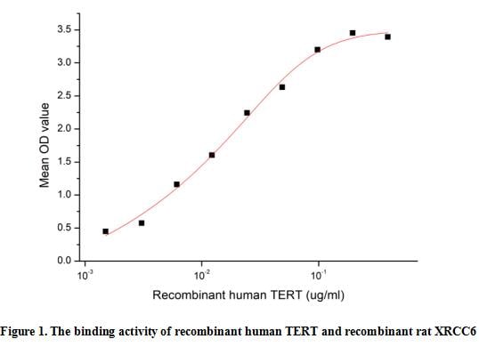
(Telomerase reverse transcriptase (TERT) is a subunit of the enzyme complex telomerase, which adds nucleotides to the ends of telomeres as they become shortened during cell division .Telomerase complex plays a key role in cancer formation by telomere dependent or independent mechanisms. According to statistics, human telomerase reverse transcriptase (h-TERT) is overexpressed in more than 85% of tumors with diverse histologies, with little expression in normal tissues. Expression of h-TERT correlates with activity of telomerase, which is required for the capacity for limitless replication, a hallmark of cancer. The X-Ray Repair Cross Complementing 6 (XRCC6) is high affinity receptor for TERT, thus a functional binding ELISA assay was conducted to detect the interaction of recombinant human TERT and recombinant rat XRCC6. Briefly, TERT was diluted serially in PBS with 0.01% BSA (pH 7.4). Duplicate samples of 100 ul were then transferred to XRCC6-coated microtiter wells and incubated for 1h at 37 degree C. Wells were washed with PBST and incubated for 1h with anti-TERT pAb, then aspirated and washed 3 times. After incubation with HRP labelled secondary antibody for 1h at 37 degree C, wells were aspirated and washed 5 times. With the addition of substrate solution, wells were incubated 15-25 minutes at 37 degree C. Finally, add 50 uL stop solution to the wells and read at 450/630 nm immediately. The binding activity of recombinant TERT and recombinant rat XRCC6 was shown in Figure 1, the EC50 for this effect is 0.009 ug/mL.)
Bioactivity
(Telomerase reverse transcriptase (TERT) is a subunit of the enzyme complex telomerase, which adds nucleotides to the ends of telomeres as they become shortened during cell division .Telomerase complex plays a key role in cancer formation by telomere dependent or independent mechanisms. According to statistics, human telomerase reverse transcriptase (h-TERT) is overexpressed in more than 85% of tumors with diverse histologies, with little expression in normal tissues. Expression of h-TERT correlates with activity of telomerase, which is required for the capacity for limitless replication, a hallmark of cancer. The X-Ray Repair Cross Complementing 6 (XRCC6) is high affinity receptor for TERT, thus a functional binding ELISA assay was conducted to detect the interaction of recombinant human TERT and recombinant rat XRCC6. Briefly, TERT was diluted serially in PBS with 0.01% BSA (pH 7.4). Duplicate samples of 100 ul were then transferred to XRCC6-coated microtiter wells and incubated for 1h at 37 degree C. Wells were washed with PBST and incubated for 1h with anti-TERT pAb, then aspirated and washed 3 times. After incubation with HRP labelled secondary antibody for 1h at 37 degree C, wells were aspirated and washed 5 times. With the addition of substrate solution, wells were incubated 15-25 minutes at 37 degree C. Finally, add 50 uL stop solution to the wells and read at 450/630 nm immediately. The binding activity of recombinant TERT and recombinant rat XRCC6 was shown in Figure 1, the EC50 for this effect is 0.009 ug/mL.)
Telomerase Reverse Transcriptase (TERT), Active Protein (Cat# AAA161880)
Full Name
Active Telomerase Reverse Transcriptase (TERT)
Gene Names
TERT; TP2; TRT; CMM9; EST2; TCS1; hTRT; DKCA2; DKCB4; hEST2; PFBMFT1
Applications
Activity Assay, Cell Culture
Purity
Greater than 90% by SDS-PAGE
Pricing
 Application Data
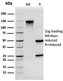
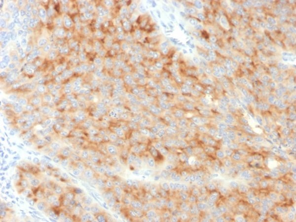
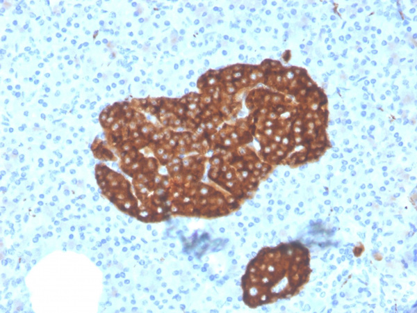
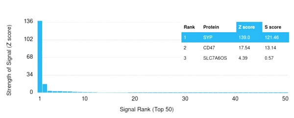
(Analysis of Protein Array containing more than 19,000 full-length human proteins using Synaptophysin-Monospecific Mouse Monoclonal Antibody (SYP/3551). Z- and S- Score: The Z-score represents the strength of a signal that a monoclonal antibody (MAb) (in combination with a fluorescently-tagged anti-IgG secondary antibody) produces when binding to a particular protein on the HuProtTM array. Z-scores are described in units of standard deviations (SD’s) above the mean value of all signals generated on that array. If targets on HuProtTM are arranged in descending order of the Z-score, the S-score is the difference (also in units of SD’s) between the Z-score. S-score therefore represents the relative target specificity of a MAb to its intended target. A MAb is considered to specific to its intended target, if the MAb has an S-score of at least 2.5. For example, if a MAb binds to protein X with a Z-score of 43 and to protein Y with a Z-score of 14, then the S-score for the binding of that MAb to protein X is equal to 29.)
Application Data
(Analysis of Protein Array containing more than 19,000 full-length human proteins using Synaptophysin-Monospecific Mouse Monoclonal Antibody (SYP/3551). Z- and S- Score: The Z-score represents the strength of a signal that a monoclonal antibody (MAb) (in combination with a fluorescently-tagged anti-IgG secondary antibody) produces when binding to a particular protein on the HuProtTM array. Z-scores are described in units of standard deviations (SD’s) above the mean value of all signals generated on that array. If targets on HuProtTM are arranged in descending order of the Z-score, the S-score is the difference (also in units of SD’s) between the Z-score. S-score therefore represents the relative target specificity of a MAb to its intended target. A MAb is considered to specific to its intended target, if the MAb has an S-score of at least 2.5. For example, if a MAb binds to protein X with a Z-score of 43 and to protein Y with a Z-score of 14, then the S-score for the binding of that MAb to protein X is equal to 29.)
Synaptophysin, Monoclonal Antibody (Cat# AAA215212)
Full Name
Synaptophysin (Neuroendocrine Marker)
Gene Names
SYP; MRX96; MRXSYP
Reactivity
Human
Applications
Immunohistochemistry
Purity
Purified Ab with BSA and Azide at 200ug/ml OR Purified Ab WITHOUT BSA and Azide at 1.0mg/ml
Pricing
 Application Data
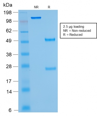
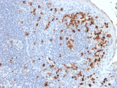
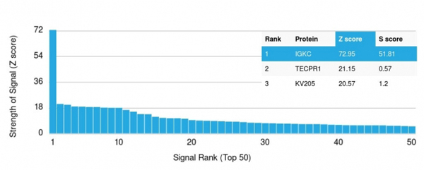
(Analysis of Protein Array containing >19, 000 full-length human proteins using Kappa Light Chain Mouse Monoclonal Antibody (rKLC/264) Z- and S- Score: The Z-score represents the strength of a signal that a monoclonal antibody (MAb) (in combination with a fluorescently-tagged anti-IgG secondary antibody) produces when binding to a particular protein on the HuProtTM array. Z-scores are described in units of standard deviations (SD's) above the mean value of all signals generated on that array. If targets on HuProtTM are arranged in descending order of the Z-score, the S-score is the difference (also in units of SD's) between the Z-score. S-score therefore represents the relative target specificity of a MAb to its intended target. A MAb is considered to specific to its intended target, if the MAb has an S-score of at least 2.5. For example, if a MAb binds to protein X with a Z-score of 43 and to protein Y with a Z-score of 14, then the S-score for the binding of that MAb to protein X is equal to 29.)
Application Data
(Analysis of Protein Array containing >19, 000 full-length human proteins using Kappa Light Chain Mouse Monoclonal Antibody (rKLC/264) Z- and S- Score: The Z-score represents the strength of a signal that a monoclonal antibody (MAb) (in combination with a fluorescently-tagged anti-IgG secondary antibody) produces when binding to a particular protein on the HuProtTM array. Z-scores are described in units of standard deviations (SD's) above the mean value of all signals generated on that array. If targets on HuProtTM are arranged in descending order of the Z-score, the S-score is the difference (also in units of SD's) between the Z-score. S-score therefore represents the relative target specificity of a MAb to its intended target. A MAb is considered to specific to its intended target, if the MAb has an S-score of at least 2.5. For example, if a MAb binds to protein X with a Z-score of 43 and to protein Y with a Z-score of 14, then the S-score for the binding of that MAb to protein X is equal to 29.)
Kappa Light Chain, Monoclonal Antibody (Cat# AAA214398)
Full Name
Kappa Light Chain (B-Cell Marker)
Reactivity
Human. Others not known.
Applications
Immunohistochemistry, Western Blot, Immunofluorescence, Flow Cytometry
Pricing
 WB (Western Blot)
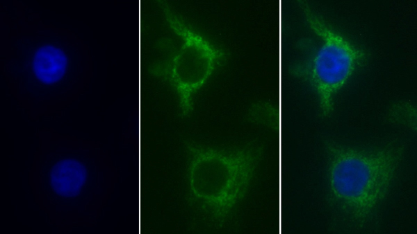
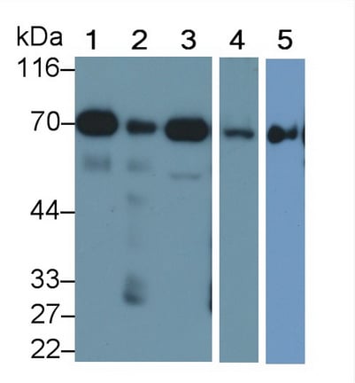
(Western Blot; Sample: Lane1: Mouse Heart lysate; Lane2: Mouse Liver lysate; Lane3: Mouse Cerebrum lysate; Lane4: Mouse Kidney lysate; Lane5: HepG2 cell lysate Primary Ab: 0.1ug/ml Mouse AntiMouse SDHA Antibody Second Ab: 0.2ug/mL HRPLinked Caprine AntiMouse IgG Polyclonal Antibody (Catalog: SAA54)
WB (Western Blot)
(Western Blot; Sample: Lane1: Mouse Heart lysate; Lane2: Mouse Liver lysate; Lane3: Mouse Cerebrum lysate; Lane4: Mouse Kidney lysate; Lane5: HepG2 cell lysate Primary Ab: 0.1ug/ml Mouse AntiMouse SDHA Antibody Second Ab: 0.2ug/mL HRPLinked Caprine AntiMouse IgG Polyclonal Antibody (Catalog: SAA54)
Succinate Dehydrogenase Complex Subunit A (SDHA), Monoclonal Antibody (Cat# AAA151898)
Full Name
Monoclonal Antibody to Succinate Dehydrogenase Complex Subunit A (SDHA)
Gene Names
Sdha; FP; SDH2; SDHF; C81073; 4921513A11; 1500032O14Rik; 2310034D06Rik
Reactivity
Human, Mouse
Applications
Immunoprecipitation, Immunocytochemistry, Immunohistochemistry, Western Blot
Purity
Protein A + Protein G affinity chromatography
Pricing
 Application Data
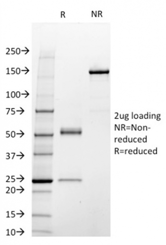
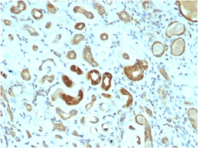
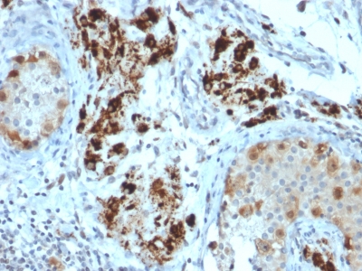
(Analysis of Protein Array containing more than 19, 000 full-length human proteins using STAT3 Mouse Monoclonal Antibody (STAT3/2409). Z- and S- Score: The Z-score represents the strength of a signal that a monoclonal antibody (MAb) (in combination with a fluorescently-tagged anti-IgG secondary antibody) produces when binding to a particular protein on the HuProtTM array. Z-scores are described in units of standard deviations (SD's) above the mean value of all signals generated on that array. If targets on HuProtTM are arranged in descending order of the Z-score, the S-score is the difference (also in units of SD's) between the Z-score. S-score therefore represents the relative target specificity of a MAb to its intended target. A MAb is considered to specific to its intended target, if the MAb has an S-score of at least 2.5. For example, if a MAb binds to protein X with a Z-score of 43 and to protein Y with a Z-score of 14, then the S-score for the binding of that MAb to protein X is equal to 29.)
Application Data
(Analysis of Protein Array containing more than 19, 000 full-length human proteins using STAT3 Mouse Monoclonal Antibody (STAT3/2409). Z- and S- Score: The Z-score represents the strength of a signal that a monoclonal antibody (MAb) (in combination with a fluorescently-tagged anti-IgG secondary antibody) produces when binding to a particular protein on the HuProtTM array. Z-scores are described in units of standard deviations (SD's) above the mean value of all signals generated on that array. If targets on HuProtTM are arranged in descending order of the Z-score, the S-score is the difference (also in units of SD's) between the Z-score. S-score therefore represents the relative target specificity of a MAb to its intended target. A MAb is considered to specific to its intended target, if the MAb has an S-score of at least 2.5. For example, if a MAb binds to protein X with a Z-score of 43 and to protein Y with a Z-score of 14, then the S-score for the binding of that MAb to protein X is equal to 29.)
STAT3/Signal Transducer and Activator of Transcription 3, Monoclonal Antibody (Cat# AAA214694)
Full Name
STAT3/Signal Transducer and Activator of Transcription 3
Gene Names
STAT3; APRF; HIES; ADMIO; ADMIO1
Reactivity
Human. Others not tested.
Applications
Immunohistochemistry
Pricing
 Application Data
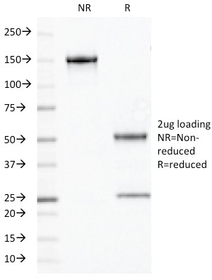
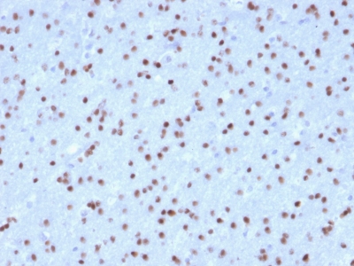
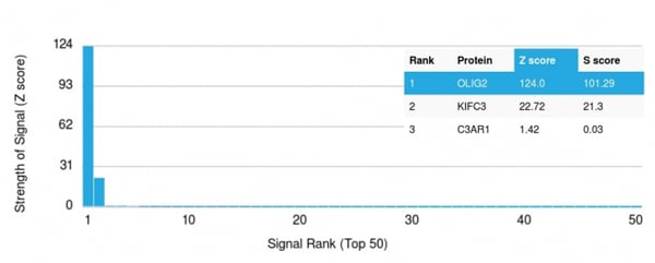
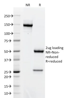
(Analysis of Protein Array containing >19, 000 full-length human proteins using OLIG2 Mouse Monoclonal Antibody (OLIG2/2400) Z- and S- Score: The Z-score represents the strength of a signal that a monoclonal antibody (MAb) (in combination with a fluorescently-tagged anti-IgG secondary antibody) produces when binding to a particular protein on the HuProtTM array. Z-scores are described in units of standard deviations (SD's) above the mean value of all signals generated on that array. If targets on HuProtTM are arranged in descending order of the Z-score, the S-score is the difference (also in units of SD's) between the Z-score. S-score therefore represents the relative target specificity of a MAb to its intended target. A MAb is considered to specific to its intended target, if the MAb has an S-score of at least 2.5. For example, if a MAb binds to protein X with a Z-score of 43 and to protein Y with a Z-score of 14, then the S-score for the binding of that MAb to protein X is equal to 29.)
Application Data
(Analysis of Protein Array containing >19, 000 full-length human proteins using OLIG2 Mouse Monoclonal Antibody (OLIG2/2400) Z- and S- Score: The Z-score represents the strength of a signal that a monoclonal antibody (MAb) (in combination with a fluorescently-tagged anti-IgG secondary antibody) produces when binding to a particular protein on the HuProtTM array. Z-scores are described in units of standard deviations (SD's) above the mean value of all signals generated on that array. If targets on HuProtTM are arranged in descending order of the Z-score, the S-score is the difference (also in units of SD's) between the Z-score. S-score therefore represents the relative target specificity of a MAb to its intended target. A MAb is considered to specific to its intended target, if the MAb has an S-score of at least 2.5. For example, if a MAb binds to protein X with a Z-score of 43 and to protein Y with a Z-score of 14, then the S-score for the binding of that MAb to protein X is equal to 29.)
OLIG2, Monoclonal Antibody (Cat# AAA214640)
Full Name
OLIG2 (Marker of Glial Brain Tumors)
Gene Names
OLIG2; BHLHB1; OLIGO2; RACK17; PRKCBP2; bHLHe19
Reactivity
Human. Others not known.
Applications
Immunohistochemistry
Pricing
 Application Data
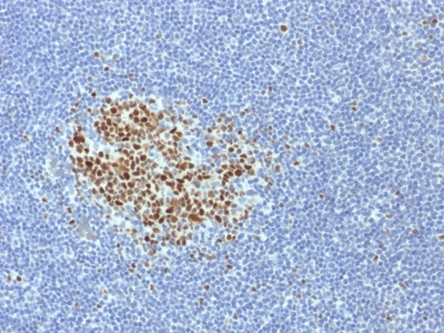
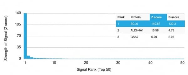
(Analysis of Protein Array containing more than 19, 000 full-length human proteins using BCL-6 Mouse Monoclonal Antibody (BCL6/1718).Z- and S- Score: The Z-score represents the strength of a signal that a monoclonal antibody (MAb) (in combination with a fluorescently-tagged anti-IgG secondary antibody) produces when binding to a particular protein on the HuProtTM array. Z-scores are described in units of standard deviations (SD's) above the mean value of all signals generated on that array. If targets on HuProtTM are arranged in descending order of the Z-score, the S-score is the difference (also in units of SD's) between the Z-score. S-score therefore represents the relative target specificity of a MAb to its intended target. A MAb is considered to specific to its intended target, if the MAb has an S-score of at least 2.5. For example, if a MAb binds to protein X with a Z-score of 43 and to protein Y with a Z-score of 14, then the S-score for the binding of that MAb to protein X is equal to 29.)
Application Data
(Analysis of Protein Array containing more than 19, 000 full-length human proteins using BCL-6 Mouse Monoclonal Antibody (BCL6/1718).Z- and S- Score: The Z-score represents the strength of a signal that a monoclonal antibody (MAb) (in combination with a fluorescently-tagged anti-IgG secondary antibody) produces when binding to a particular protein on the HuProtTM array. Z-scores are described in units of standard deviations (SD's) above the mean value of all signals generated on that array. If targets on HuProtTM are arranged in descending order of the Z-score, the S-score is the difference (also in units of SD's) between the Z-score. S-score therefore represents the relative target specificity of a MAb to its intended target. A MAb is considered to specific to its intended target, if the MAb has an S-score of at least 2.5. For example, if a MAb binds to protein X with a Z-score of 43 and to protein Y with a Z-score of 14, then the S-score for the binding of that MAb to protein X is equal to 29.)
Bcl-6, Monoclonal Antibody (Cat# AAA214507)
Full Name
Bcl-6 (Follicular Lymphoma Marker)
Gene Names
BCL6; BCL5; LAZ3; BCL6A; ZNF51; ZBTB27
Reactivity
Human. Others not known.
Applications
Immunohistochemistry
Pricing