
Filters
Clonality
Type
Reactivity
Gene Name
Isotype
Host
Application
Clone
10000 results for "Recombinant Proteins" - showing 2750-2800
 Bioactivity
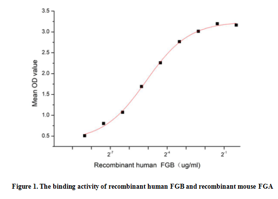
(Fibrinogen Beta Chain (FGB) is the beta subunit of the coagulation factor fibrinogen, which is a component of the blood clot. Following vascular injury, fibrinogen is cleaved by thrombin to form fibrin which is the most abundant component of blood clots. Fibrinogen Alpha Chain (FGA) has been identified as an interactor of FGB. Thus a functional binding ELISA assay was conducted to detect the interaction of recombinant human FGB and recombinant mouse FGA. Briefly, FGB was diluted serially in PBS with 0.01% BSA (pH 7.4). Duplicate samples of 100 ul were then transferred to FGA-coated microtiter wells and incubated for 1h at 37 degree C. Wells were washed with PBST and incubated for 1h with anti-FGB pAb, then aspirated and washed 3 times. After incubation with HRP labelled secondary antibody for 1h at 37 degree C, wells were aspirated and washed 5 times. With the addition of substrate solution, wells were incubated 15-25 minutes at 37 degree C. Finally, add 50 uL stop solution to the wells and read at 450/630 nm immediately. The binding activity of recombinant human FGB and recombinant mouse FGA was shown in Figure 1, the EC50 for this effect is 0.03 ug/mL.)
Bioactivity
(Fibrinogen Beta Chain (FGB) is the beta subunit of the coagulation factor fibrinogen, which is a component of the blood clot. Following vascular injury, fibrinogen is cleaved by thrombin to form fibrin which is the most abundant component of blood clots. Fibrinogen Alpha Chain (FGA) has been identified as an interactor of FGB. Thus a functional binding ELISA assay was conducted to detect the interaction of recombinant human FGB and recombinant mouse FGA. Briefly, FGB was diluted serially in PBS with 0.01% BSA (pH 7.4). Duplicate samples of 100 ul were then transferred to FGA-coated microtiter wells and incubated for 1h at 37 degree C. Wells were washed with PBST and incubated for 1h with anti-FGB pAb, then aspirated and washed 3 times. After incubation with HRP labelled secondary antibody for 1h at 37 degree C, wells were aspirated and washed 5 times. With the addition of substrate solution, wells were incubated 15-25 minutes at 37 degree C. Finally, add 50 uL stop solution to the wells and read at 450/630 nm immediately. The binding activity of recombinant human FGB and recombinant mouse FGA was shown in Figure 1, the EC50 for this effect is 0.03 ug/mL.)
Fibrinogen Beta Chain (FGB), Active Protein (Cat# AAA161833)
Full Name
Active Fibrinogen Beta Chain (FGB)
Applications
Activity Assay, Cell Culture
Purity
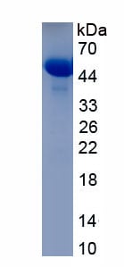
Greater than 90% by SDS-PAGE
Pricing

 Bioactivity
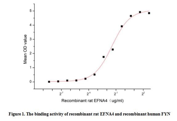
(Ephrin-A4, also known as EFNA4 and EFL-4, is a member of the ligand of the EPH family. It is mainly expressed in the spleen, lymph nodes, ovary, small intestine and colon of adults, as well as in the heart, lungs, liver, and kidneys of the fetus. It is involved in the development of neurons, blood vessels, and epithelium by regulating cell migration, rejection, and adhesion. Ephrin-A4 has been shown to bind FYN,EphA2, EphA3, EphA4, EphA5, EphA6, EphA7, and EphB1. Thus a functional binding ELISA assay was conducted to detect the interaction of recombinant rat EFNA4 and recombinant human FYN. Briefly, EFNA4 was diluted serially in PBS with 0.01% BSA (pH 7.4). Duplicate samples of 100 ul were then transferred to FYN-coated microtiter wells and incubated for 1h at 37 degree C. Wells were washed with PBST and incubated for 1h with anti-EFNA4 pAb, then aspirated and washed 3 times. After incubation with HRP labelled secondary antibody for 1h at 37 degree C, wells were aspirated and washed 5 times. With the addition of substrate solution, wells were incubated 15-25 minutes at 37 degree C. Finally, add 50 uL stop solution to the wells and read at 450/630 nm immediately. The binding activity of recombinant rat EFNA4 and recombinant human FYN was shown in Figure 1, the EC50 for this effect is 0.37 ug/mL.)
Bioactivity
(Ephrin-A4, also known as EFNA4 and EFL-4, is a member of the ligand of the EPH family. It is mainly expressed in the spleen, lymph nodes, ovary, small intestine and colon of adults, as well as in the heart, lungs, liver, and kidneys of the fetus. It is involved in the development of neurons, blood vessels, and epithelium by regulating cell migration, rejection, and adhesion. Ephrin-A4 has been shown to bind FYN,EphA2, EphA3, EphA4, EphA5, EphA6, EphA7, and EphB1. Thus a functional binding ELISA assay was conducted to detect the interaction of recombinant rat EFNA4 and recombinant human FYN. Briefly, EFNA4 was diluted serially in PBS with 0.01% BSA (pH 7.4). Duplicate samples of 100 ul were then transferred to FYN-coated microtiter wells and incubated for 1h at 37 degree C. Wells were washed with PBST and incubated for 1h with anti-EFNA4 pAb, then aspirated and washed 3 times. After incubation with HRP labelled secondary antibody for 1h at 37 degree C, wells were aspirated and washed 5 times. With the addition of substrate solution, wells were incubated 15-25 minutes at 37 degree C. Finally, add 50 uL stop solution to the wells and read at 450/630 nm immediately. The binding activity of recombinant rat EFNA4 and recombinant human FYN was shown in Figure 1, the EC50 for this effect is 0.37 ug/mL.)
Ephrin A4 (EFNA4), Active Protein (Cat# AAA161918)
Full Name
Active Ephrin A4 (EFNA4)
Applications
Activity Assay, Cell Culture
Purity
Greater than 90% by SDS-PAGE
Pricing
 Amino Acid Info
Eukaryotic Permeability Glycoprotein (Pgp), Protein (Cat# AAA153213)
Full Name
Eukaryotic Permeability Glycoprotein (Pgp)
Gene Names
ABCB1; CLCS; MDR1; P-GP; PGY1; ABC20; CD243; GP170
Applications
Western Blot
Purity
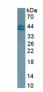
Greater than 90% by SDS-PAGE
Pricing
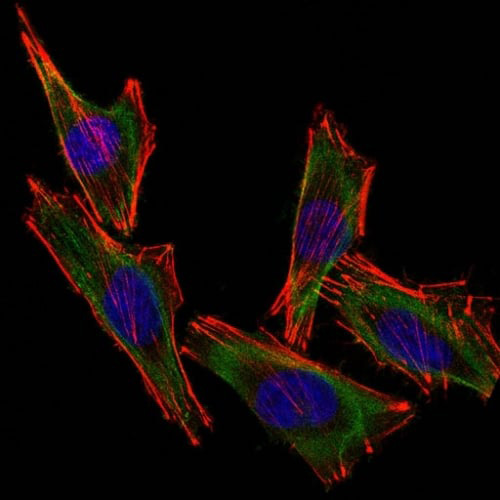
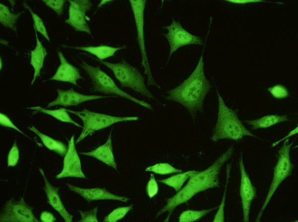
 IF (Immunofluorescence)
(Confocal Immunofluorescent analysis of A2058 cells using CF488-labeled Isotype Control Monoclonal Antibody (IgG2a) (Green). F-actin filaments were labeled with Dylight 554 Phalloidin (red). DAPI was used to stain the cell nuclei (blue). (Negative Control))
IF (Immunofluorescence)
(Confocal Immunofluorescent analysis of A2058 cells using CF488-labeled Isotype Control Monoclonal Antibody (IgG2a) (Green). F-actin filaments were labeled with Dylight 554 Phalloidin (red). DAPI was used to stain the cell nuclei (blue). (Negative Control))
S100B, Monoclonal Antibody (Cat# AAA215189)
Full Name
S100B (Astrocyte and Melanoma Marker)
Gene Names
S100B; NEF; S100; S100-B; S100beta
Reactivity
Human, Mouse, Rat, Cow
Applications
Immunofluorescence, Flow Cytometry
Purity
Purified Ab conjugated to CF488
Pricing
 Bioactivity
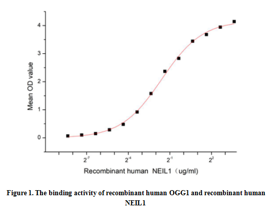
(Oxoguanine Glycosylase 1 (OGG1), a prominent member of the BER enzyme family, is an essential DNA repair enzyme. OGG1 is highly conserved across eukaryotic organisms and is expressed in various cell types. Its activity is regulated by several factors, including cellular redox status, DNA damage, and cell cycle progression. In addition to its role in repairing oxidative DNA damage, OGG1 has also been implicated in other cellular processes, such as transcription, replication, and apoptosis. In addition, the combination of OGG1 and NEIL1 can contribute to improve the repair efficiency of oxidative DNA damage, maintain genomic stability, and reduce the risk of cell mutation. Thus a functional binding ELISA assay was conducted to detect the interaction of recombinant human OGG1 and recombinant human NEIL1. Briefly, biotin-linked OGG1 were diluted serially in PBS, with 0.01% BSA (pH 7.4). Duplicate samples of 100 ul were then transferred to NEIL1-coated microtiter wells and incubated for 1h at 37 degree C. Wells were washed with PBST 3 times and incubation with Streptavidin-HRP for 30min, then wells were aspirated and washed 5 times. With the addition of substrate solution, wells were incubated 15-25 minutes at 37 degree C. Finally, add 50 ul stop solution to the wells and read at 450 nm immediately. The binding activity of recombinant human OGG1 and recombinant human NEIL1 was shown in Figure 1, the EC50 for this effect is 0.33 ug/mL.)
Bioactivity
(Oxoguanine Glycosylase 1 (OGG1), a prominent member of the BER enzyme family, is an essential DNA repair enzyme. OGG1 is highly conserved across eukaryotic organisms and is expressed in various cell types. Its activity is regulated by several factors, including cellular redox status, DNA damage, and cell cycle progression. In addition to its role in repairing oxidative DNA damage, OGG1 has also been implicated in other cellular processes, such as transcription, replication, and apoptosis. In addition, the combination of OGG1 and NEIL1 can contribute to improve the repair efficiency of oxidative DNA damage, maintain genomic stability, and reduce the risk of cell mutation. Thus a functional binding ELISA assay was conducted to detect the interaction of recombinant human OGG1 and recombinant human NEIL1. Briefly, biotin-linked OGG1 were diluted serially in PBS, with 0.01% BSA (pH 7.4). Duplicate samples of 100 ul were then transferred to NEIL1-coated microtiter wells and incubated for 1h at 37 degree C. Wells were washed with PBST 3 times and incubation with Streptavidin-HRP for 30min, then wells were aspirated and washed 5 times. With the addition of substrate solution, wells were incubated 15-25 minutes at 37 degree C. Finally, add 50 ul stop solution to the wells and read at 450 nm immediately. The binding activity of recombinant human OGG1 and recombinant human NEIL1 was shown in Figure 1, the EC50 for this effect is 0.33 ug/mL.)
Oxoguanine Glycosylase 1 (OGG1), Active Protein (Cat# AAA161893)
Full Name
Active Oxoguanine Glycosylase 1 (OGG1)
Gene Names
OGG1; HMMH; MUTM; OGH1; HOGG1
Applications
Activity Assay, Cell Culture
Purity
Greater than 90% by SDS-PAGE
Pricing
 Bioactivity
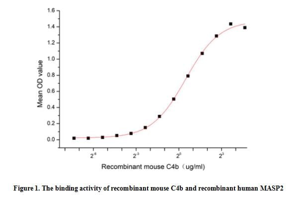
(Complement Component 4b (C4b) is a component of the complement system, which is activated by the recognition of foreign pathogens, such as bacteria and viruses, or altered self-cells. In the classical activation pathway of complement, C4b is produced by the proteolytic cleavage of the precursor protein C4 by the activated enzyme C1s, and it can bind to C2a to form C3 invertase (C4b2a) which is responsible for the cleavage of C3. Besides, the binding of MASP2 to C4b is an important step in the lectin pathway of the complement system, thus a functional binding ELISA assay was conducted to detect the interaction of recombinant mouse C4b and recombinant human MASP2. Briefly, C4b was diluted serially in PBS with 0.01% BSA (pH 7.4). Duplicate samples of 100 ul were then transferred to MASP2-coated microtiter wells and incubated for 1h at 37 degree C. Wells were washed with PBST and incubated for 1h with anti-C4b pAb, then aspirated and washed 3 times. After incubation with HRP labelled secondary antibody for 1h at 37 degree C, wells were aspirated and washed 5 times. With the addition of substrate solution, wells were incubated 15-25 minutes at 37 degree C. Finally, add 50 uL stop solution to the wells and read at 450/630 nm immediately. The binding activity of recombinant mouse C4b and recombinant human MASP2 was shown in Figure 1, the EC50 for this effect is 1.39 ug/mL.)
Bioactivity
(Complement Component 4b (C4b) is a component of the complement system, which is activated by the recognition of foreign pathogens, such as bacteria and viruses, or altered self-cells. In the classical activation pathway of complement, C4b is produced by the proteolytic cleavage of the precursor protein C4 by the activated enzyme C1s, and it can bind to C2a to form C3 invertase (C4b2a) which is responsible for the cleavage of C3. Besides, the binding of MASP2 to C4b is an important step in the lectin pathway of the complement system, thus a functional binding ELISA assay was conducted to detect the interaction of recombinant mouse C4b and recombinant human MASP2. Briefly, C4b was diluted serially in PBS with 0.01% BSA (pH 7.4). Duplicate samples of 100 ul were then transferred to MASP2-coated microtiter wells and incubated for 1h at 37 degree C. Wells were washed with PBST and incubated for 1h with anti-C4b pAb, then aspirated and washed 3 times. After incubation with HRP labelled secondary antibody for 1h at 37 degree C, wells were aspirated and washed 5 times. With the addition of substrate solution, wells were incubated 15-25 minutes at 37 degree C. Finally, add 50 uL stop solution to the wells and read at 450/630 nm immediately. The binding activity of recombinant mouse C4b and recombinant human MASP2 was shown in Figure 1, the EC50 for this effect is 1.39 ug/mL.)
Complement C4-B (C4B), Active Protein (Cat# AAA161840)
Full Name
Active Complement C4-B (C4B)
Gene Names
C4b; C4; Ss
Applications
Activity Assay, Cell Culture
Purity
Greater than 95% by SDS-PAGE
Pricing
 Bioactivity
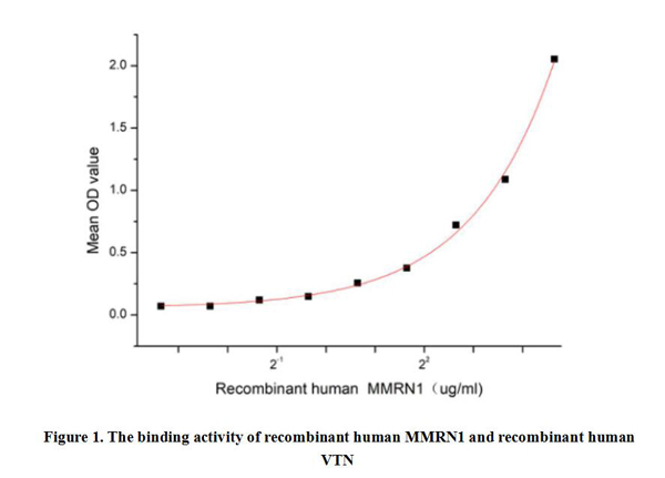
(Multimerin 1 (MMRN1) is a secreted glycoprotein that consists of multiple subunits, forming a complex with a molecular weight of approximately 150 kDa. MMRN1 is a member of the multimerin family and it is mainly expressed in platelets and endothelial cells. MMRN1 plays an important role in maintaining vascular integrity, regulating clotting and inflammatory responses, among others. In addition, the binding of MMRN1 to Vitronectin (VTN) plays an important role in a variety of physiological and pathological processes, including cell adhesion, migration, blood coagulation, and cell signaling. Thus a functional binding ELISA assay was conducted to detect the interaction of recombinant human MMRN1 and recombinant human VTN. Briefly, MMRN1 was diluted serially in PBS with 0.01% BSA (pH 7.4). Duplicate samples of 100 ul were then transferred to VTN-coated microtiter wells and incubated for 1h at 37 degree C. Wells were washed with PBST and incubated for 1h with anti-MMRN1 pAb, then aspirated and washed 3 times. After incubation with HRP labelled secondary antibody for 1h at 37 degree C, wells were aspirated and washed 5 times. With the addition of substrate solution, wells were incubated 15-25 minutes at 37 degree C. Finally, add 50 uL stop solution to the wells and read at 450/630 nm immediately. The binding activity of recombinant human MMRN1 and recombinant human VTN was shown in Figure 1, and this effect was in a dose dependent manner.)
Bioactivity
(Multimerin 1 (MMRN1) is a secreted glycoprotein that consists of multiple subunits, forming a complex with a molecular weight of approximately 150 kDa. MMRN1 is a member of the multimerin family and it is mainly expressed in platelets and endothelial cells. MMRN1 plays an important role in maintaining vascular integrity, regulating clotting and inflammatory responses, among others. In addition, the binding of MMRN1 to Vitronectin (VTN) plays an important role in a variety of physiological and pathological processes, including cell adhesion, migration, blood coagulation, and cell signaling. Thus a functional binding ELISA assay was conducted to detect the interaction of recombinant human MMRN1 and recombinant human VTN. Briefly, MMRN1 was diluted serially in PBS with 0.01% BSA (pH 7.4). Duplicate samples of 100 ul were then transferred to VTN-coated microtiter wells and incubated for 1h at 37 degree C. Wells were washed with PBST and incubated for 1h with anti-MMRN1 pAb, then aspirated and washed 3 times. After incubation with HRP labelled secondary antibody for 1h at 37 degree C, wells were aspirated and washed 5 times. With the addition of substrate solution, wells were incubated 15-25 minutes at 37 degree C. Finally, add 50 uL stop solution to the wells and read at 450/630 nm immediately. The binding activity of recombinant human MMRN1 and recombinant human VTN was shown in Figure 1, and this effect was in a dose dependent manner.)
Multimerin 1 (MMRN1), Active Protein (Cat# AAA161892)
Full Name
Active Multimerin 1 (MMRN1)
Gene Names
MMRN1; ECM; MMRN; GPIa*; EMILIN4
Applications
Activity Assay, Cell Culture
Purity
Greater than 95% by SDS-PAGE
Pricing



 WB (Western Blot)
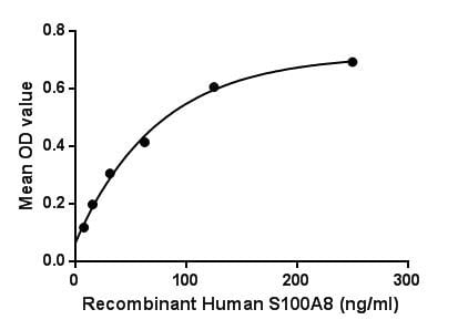
(Sample: Recombinant S100A8, Human;Antibody: Rabbit Anti-Human S100A8 Ab)
WB (Western Blot)
(Sample: Recombinant S100A8, Human;Antibody: Rabbit Anti-Human S100A8 Ab)
S100 Calcium Binding Protein A8 (S100A8), Active Protein (Cat# AAA149250)
Full Name
Active S100 Calcium Binding Protein A8 (S100A8)
Gene Names
S100A8; P8; MIF; NIF; CAGA; CFAG; CGLA; L1Ag; MRP8; CP-10; MA387; 60B8AG
Applications
Activity Assay, Cell Culture
Purity
>95%
Pricing
 Application Data
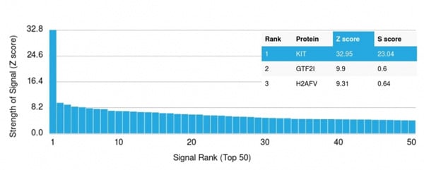
(Analysis of Protein Array containing more than 19,000 full-length human proteins using CD117 Mouse Monoclonal Antibody (KIT/2669). Z- and S- Score: The Z-score represents the strength of a signal that a monoclonal antibody (MAb) (in combination with a fluorescently-tagged anti-IgG secondary antibody) produces when binding to a particular protein on the HuProtTM array. Z-scores are described in units of standard deviations (SD's) above the mean value of all signals generated on that array. If targets on HuProtTM are arranged in descending order of the Z-score, the S-score is the difference (also in units of SD's) between the Z-score. S-score therefore represents the relative target specificity of a MAb to its intended target. A MAb is considered to specific to its intended target, if the MAb has an S-score of at least 2.5. For example, if a MAb binds to protein X with a Z-score of 43 and to protein Y with a Z-score of 14, then the S-score for the binding of that MAb to protein X is equal to 29.)
Application Data
(Analysis of Protein Array containing more than 19,000 full-length human proteins using CD117 Mouse Monoclonal Antibody (KIT/2669). Z- and S- Score: The Z-score represents the strength of a signal that a monoclonal antibody (MAb) (in combination with a fluorescently-tagged anti-IgG secondary antibody) produces when binding to a particular protein on the HuProtTM array. Z-scores are described in units of standard deviations (SD's) above the mean value of all signals generated on that array. If targets on HuProtTM are arranged in descending order of the Z-score, the S-score is the difference (also in units of SD's) between the Z-score. S-score therefore represents the relative target specificity of a MAb to its intended target. A MAb is considered to specific to its intended target, if the MAb has an S-score of at least 2.5. For example, if a MAb binds to protein X with a Z-score of 43 and to protein Y with a Z-score of 14, then the S-score for the binding of that MAb to protein X is equal to 29.)
CD117/c-Kit, Monoclonal Antibody (Cat# AAA215052)
Full Name
CD117/c-Kit (Marker for Gastrointestinal Stromal Tumors)
Gene Names
KIT; PBT; SCFR; C-Kit; CD117; MASTC
Reactivity
Human
Applications
ELISA
Purity
Purified Ab with BSA and Azide at 200ug/ml OR Purified Ab WITHOUT BSA and Azide at 1.0mg/ml
Pricing
 IP (Immunoprecipitation)
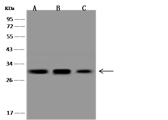
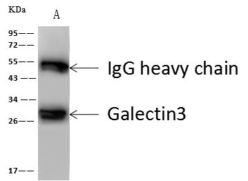
(NKX3-1 was immunoprecipitated using:Lane A:0.5 mg mouse pancreas Whole Cell Lysate4 uL anti-NKX3-1 rabbit polyclonal antibody and 60 ug of Immunomagnetic beads Protein A/G.Primary antibody:Anti-NKX3-1 rabbit polyclonal antibody,at 1:100 dilutionSecondary antibody:Goat Anti-Rabbit IgG (H+L)/HRP at 1/10000 dilutionDeveloped using the ECL technique.Performed under reducing conditions.Predicted band size: 27 kDaObserved band size :30 kDa)
IP (Immunoprecipitation)
(NKX3-1 was immunoprecipitated using:Lane A:0.5 mg mouse pancreas Whole Cell Lysate4 uL anti-NKX3-1 rabbit polyclonal antibody and 60 ug of Immunomagnetic beads Protein A/G.Primary antibody:Anti-NKX3-1 rabbit polyclonal antibody,at 1:100 dilutionSecondary antibody:Goat Anti-Rabbit IgG (H+L)/HRP at 1/10000 dilutionDeveloped using the ECL technique.Performed under reducing conditions.Predicted band size: 27 kDaObserved band size :30 kDa)
Galectin 3/LGALS3, Monoclonal Antibody (Cat# AAA254796)
Full Name
Recombinant Anti-Galectin 3/LGALS3 Antibody, Rabbit Monoclonal
Gene Names
LGALS3; L31; GAL3; MAC2; CBP35; GALBP; GALIG; LGALS2
Reactivity
Human
Applications
Immunoprecipitation, Immunofluorescence, Immunocytochemistry, Flow Cytometry, Western Blot
Purity
Protein A
Pricing
 WB (Western Blot)
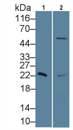
(Western Blot Sample: Lane1: Human Serum; Lane2: Porcine Liver lysate Primary Ab: 1ug/ml Mouse AntiHuman RBP4 Antibody Second Ab: 0.2ug/mL HRPLinked Caprine AntiMouse IgG Polyclonal Antibody (Catalog: SAA544Mu19))
WB (Western Blot)
(Western Blot Sample: Lane1: Human Serum; Lane2: Porcine Liver lysate Primary Ab: 1ug/ml Mouse AntiHuman RBP4 Antibody Second Ab: 0.2ug/mL HRPLinked Caprine AntiMouse IgG Polyclonal Antibody (Catalog: SAA544Mu19))
Retinol Binding Protein 4 (RBP4), Monoclonal Antibody (Cat# AAA151656)
Full Name
Monoclonal Antibody to Retinol Binding Protein 4 (RBP4)
Gene Names
RBP4; RDCCAS
Reactivity
Human, Pig
Applications
Immunoprecipitation, Immunocytochemistry, Immunohistochemistry, Western Blot
Purity
Protein A + Protein G affinity chromatography
Pricing
 Bioactivity
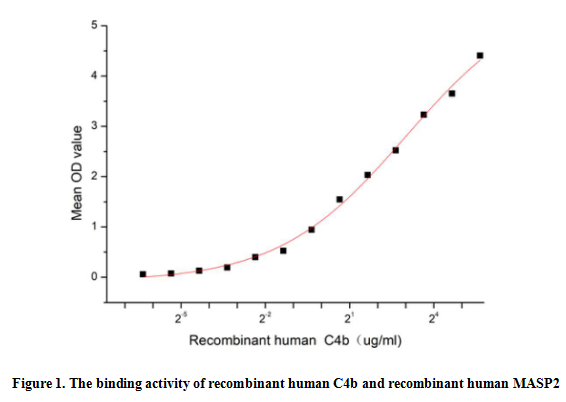
(Complement Component 4b (C4b) is a component of the complement system, which is activated by the recognition of foreign pathogens, such as bacteria and viruses, or altered self-cells. In the classical activation pathway of complement, C4b is produced by the proteolytic cleavage of the precursor protein C4 by the activated enzyme C1s, and it can bind to C2a to form C3 invertase (C4b2a) which is responsible for the cleavage of C3. Besides, the binding of MASP2 to C4b is an important step in the lectin pathway of the complement system, thus a functional binding ELISA assay was conducted to detect the interaction of recombinant human C4b and recombinant human MASP2. Briefly, C4b was diluted serially in PBS with 0.01% BSA (pH 7.4). Duplicate samples of 100 ul were then transferred to MASP2-coated microtiter wells and incubated for 1h at 37 degree C. Wells were washed with PBST and incubated for 1h with anti-C4b pAb, then aspirated and washed 3 times. After incubation with HRP labelled secondary antibody for 1h at 37 degree C, wells were aspirated and washed 5 times. With the addition of substrate solution, wells were incubated 15-25 minutes at 37 degree C. Finally, add 50 uL stop solution to the wells and read at 450/630 nm immediately. The binding activity of recombinant human C4b and recombinant human MASP2 was shown in Figure 1, the EC50 for this effect is 1.39 ug/mL.)
Bioactivity
(Complement Component 4b (C4b) is a component of the complement system, which is activated by the recognition of foreign pathogens, such as bacteria and viruses, or altered self-cells. In the classical activation pathway of complement, C4b is produced by the proteolytic cleavage of the precursor protein C4 by the activated enzyme C1s, and it can bind to C2a to form C3 invertase (C4b2a) which is responsible for the cleavage of C3. Besides, the binding of MASP2 to C4b is an important step in the lectin pathway of the complement system, thus a functional binding ELISA assay was conducted to detect the interaction of recombinant human C4b and recombinant human MASP2. Briefly, C4b was diluted serially in PBS with 0.01% BSA (pH 7.4). Duplicate samples of 100 ul were then transferred to MASP2-coated microtiter wells and incubated for 1h at 37 degree C. Wells were washed with PBST and incubated for 1h with anti-C4b pAb, then aspirated and washed 3 times. After incubation with HRP labelled secondary antibody for 1h at 37 degree C, wells were aspirated and washed 5 times. With the addition of substrate solution, wells were incubated 15-25 minutes at 37 degree C. Finally, add 50 uL stop solution to the wells and read at 450/630 nm immediately. The binding activity of recombinant human C4b and recombinant human MASP2 was shown in Figure 1, the EC50 for this effect is 1.39 ug/mL.)
Complement C4-B (C4B), Active Protein (Cat# AAA161839)
Full Name
Active Complement C4-B (C4B)
Gene Names
C4B; CH; C4F; CO4; C4B1; C4B2; C4B3; C4B5; C4BD; C4B12; C4B_2; CPAMD3
Applications
Activity Assay, Cell Culture
Purity
Greater than 90% by SDS-PAGE
Pricing
 Application Data
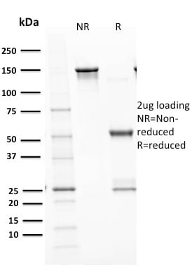
(Analysis of Protein Array containing more than 19, 000 full-length human proteins using p53 Recombinant Rabbit Monoclonal Antibody (TP53/1799R)Z- and S- Score: The Z-score represents the strength of a signal that a monoclonal antibody (MAb) (in combination with a fluorescently-tagged anti-IgG secondary antibody) produces when binding to a particular protein on the HuProtTM array. Z-scores are described in units of standard deviations (SD's) above the mean value of all signals generated on that array. If targets on HuProtTM are arranged in descending order of the Z-score, the S-score is the difference (also in units of SD's) between the Z-score. S-score therefore represents the relative target specificity of a MAb to its intended target. A MAb is considered to specific to its intended target, if the MAb has an S-score of at least 2.5. For example, if a MAb binds to protein X with a Z-score of 43 and to protein Y with a Z-score of 14, then the S-score for the binding of that MAb to protein X is equal to 29.)
Application Data
(Analysis of Protein Array containing more than 19, 000 full-length human proteins using p53 Recombinant Rabbit Monoclonal Antibody (TP53/1799R)Z- and S- Score: The Z-score represents the strength of a signal that a monoclonal antibody (MAb) (in combination with a fluorescently-tagged anti-IgG secondary antibody) produces when binding to a particular protein on the HuProtTM array. Z-scores are described in units of standard deviations (SD's) above the mean value of all signals generated on that array. If targets on HuProtTM are arranged in descending order of the Z-score, the S-score is the difference (also in units of SD's) between the Z-score. S-score therefore represents the relative target specificity of a MAb to its intended target. A MAb is considered to specific to its intended target, if the MAb has an S-score of at least 2.5. For example, if a MAb binds to protein X with a Z-score of 43 and to protein Y with a Z-score of 14, then the S-score for the binding of that MAb to protein X is equal to 29.)
p53 Tumor Suppressor Protein, Monoclonal Antibody (Cat# AAA214544)
Full Name
p53 Tumor Suppressor Protein
Gene Names
TP53; P53; BCC7; LFS1; TRP53
Reactivity
Human. Others not known
Applications
Immunohistochemistry, Western Blot, Immunofluorescence, Flow Cytometry
Pricing
 Application Data
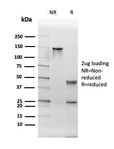
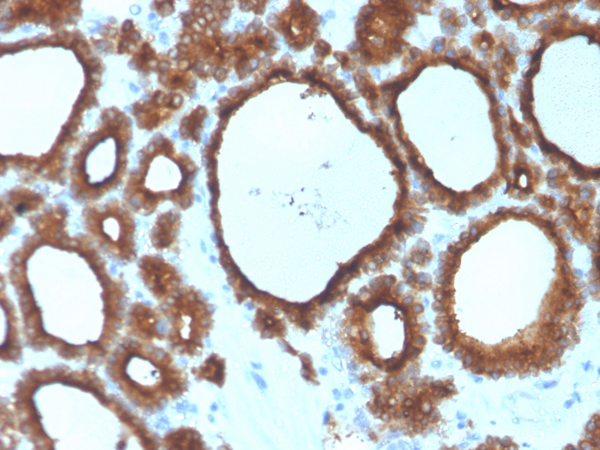
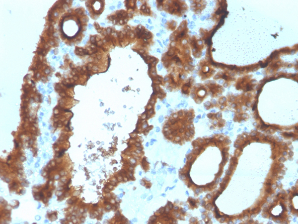
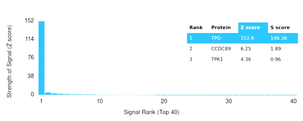
(Analysis of Protein Array containing more than 19,000 full-length human proteins using Thyroid Peroxidase Mouse Monoclonal Antibody (TPO/3700). Z- and S- Score: The Z-score represents the strength of a signal that a monoclonal antibody (MAb) (in combination with a fluorescently-tagged anti-IgG secondary antibody) produces when binding to a particular protein on the HuProtTM array. Z-scores are described in units of standard deviations (SD's) above the mean value of all signals generated on that array. If targets on HuProtTM are arranged in descending order of the Z-score, the S-score is the difference (also in units of SD's) between the Z-score. S-score therefore represents the relative target specificity of a MAb to its intended target. A MAb is considered to specific to its intended target, if the MAb has an S-score of at least 2.5. For example, if a MAb binds to protein X with a Z-score of 43 and to protein Y with a Z-score of 14, then the S-score for the binding of that MAb to protein X is equal to 29.)
Application Data
(Analysis of Protein Array containing more than 19,000 full-length human proteins using Thyroid Peroxidase Mouse Monoclonal Antibody (TPO/3700). Z- and S- Score: The Z-score represents the strength of a signal that a monoclonal antibody (MAb) (in combination with a fluorescently-tagged anti-IgG secondary antibody) produces when binding to a particular protein on the HuProtTM array. Z-scores are described in units of standard deviations (SD's) above the mean value of all signals generated on that array. If targets on HuProtTM are arranged in descending order of the Z-score, the S-score is the difference (also in units of SD's) between the Z-score. S-score therefore represents the relative target specificity of a MAb to its intended target. A MAb is considered to specific to its intended target, if the MAb has an S-score of at least 2.5. For example, if a MAb binds to protein X with a Z-score of 43 and to protein Y with a Z-score of 14, then the S-score for the binding of that MAb to protein X is equal to 29.)
TPO (Thyroid Peroxidase), Monoclonal Antibody (Cat# AAA215464)
Full Name
TPO (Thyroid Peroxidase) (Thyroid Marker)
Gene Names
TPO; MSA; TPX; TDH2A
Reactivity
Human
Applications
Immunohistochemistry
Purity
Purified Ab with BSA and Azide at 200ug/ml or Purified Ab with BSA and Azide at 200ug/ml or Purified Ab WITHOUT BSA and Azide at 1.0mg/ml
Pricing
 WB (Western Blot)
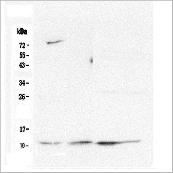
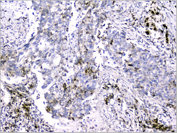
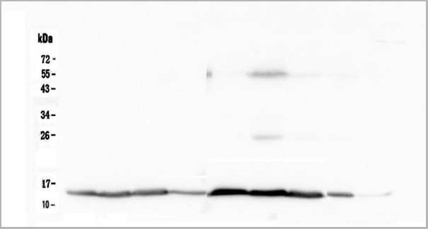
(Figure 8. Western blot analysis of Cytochrome C using anti-Cytochrome C antibody (M03529-5).Electrophoresis was performed on a 12% SDS-PAGE gel at 70V (Stacking gel)/90V (Resolving gel) for 2-3 hours. The sample well of each lane was loaded with 50ug of sample under reducing conditions.Lane 1: rat brain tissue lysate,Lane 2: rat heart tissue lysate,Lane 3: rat kidney tissue lysate,Lane 4: rat testis tissue lysate,Lane 5: mouse brain tissue lysate,Lane 6: mouse heart tissue lysate,Lane 7: mouse kidney tissue lysate,Lane 8: mouse testis tissue lysate,Lane 9: mouse Neuro-2a whole cell lysate.After Electrophoresis, proteins were transferred to a Nitrocellulose membrane at 150mA for 50-90 minutes. Blocked the membrane with 5% Non-fat Milk/TBS for 1.5 hour at RT. The membrane was incubated with rabbit anti-Cytochrome C antigen affinity purified polyclonal antibody at 0.5 ug/ml overnight at 4 degree C, then washed with TBS-0.1%Tween 3 times with 5 minutes each and probed with a goat anti-rabbit IgG-HRP secondary antibody at a dilution of 1:10000 for 1.5 hour at RT. The signal is developed using an Enhanced Chemiluminescent detection (ECL) kit with Tanon 5200 system.)
WB (Western Blot)
(Figure 8. Western blot analysis of Cytochrome C using anti-Cytochrome C antibody (M03529-5).Electrophoresis was performed on a 12% SDS-PAGE gel at 70V (Stacking gel)/90V (Resolving gel) for 2-3 hours. The sample well of each lane was loaded with 50ug of sample under reducing conditions.Lane 1: rat brain tissue lysate,Lane 2: rat heart tissue lysate,Lane 3: rat kidney tissue lysate,Lane 4: rat testis tissue lysate,Lane 5: mouse brain tissue lysate,Lane 6: mouse heart tissue lysate,Lane 7: mouse kidney tissue lysate,Lane 8: mouse testis tissue lysate,Lane 9: mouse Neuro-2a whole cell lysate.After Electrophoresis, proteins were transferred to a Nitrocellulose membrane at 150mA for 50-90 minutes. Blocked the membrane with 5% Non-fat Milk/TBS for 1.5 hour at RT. The membrane was incubated with rabbit anti-Cytochrome C antigen affinity purified polyclonal antibody at 0.5 ug/ml overnight at 4 degree C, then washed with TBS-0.1%Tween 3 times with 5 minutes each and probed with a goat anti-rabbit IgG-HRP secondary antibody at a dilution of 1:10000 for 1.5 hour at RT. The signal is developed using an Enhanced Chemiluminescent detection (ECL) kit with Tanon 5200 system.)
Cytochrome C, Monoclonal Antibody (Cat# AAA19187)
Full Name
Anti-Cytochrome C Antibody (monoclonal, 15F10)
Gene Names
CYCS; CYC; HCS; THC4
Reactivity
Human, Mouse, Rat
Applications
Western Blot, Immunohistochemistry, Immunocytochemistry, Flow Cytometry
Purity
Immunogen Affinity Purified
Pricing
 FCM/FACS (Flow Cytometry)
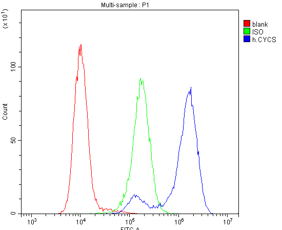
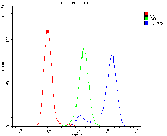
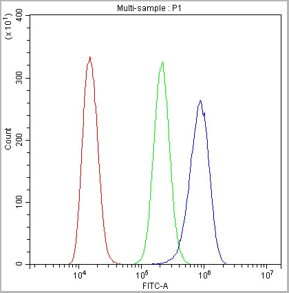
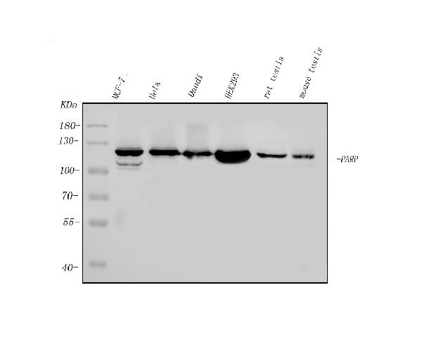
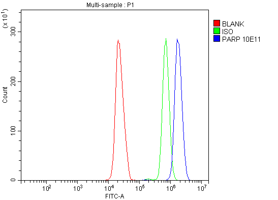
(Figure 5. Flow Cytometry analysis of HL-60 cells using anti-PARP antibody (AAA125899).Overlay histogram showing HL-60 cells stained with AAA125899 (Blue line). The cells were blocked with 10% normal goat serum. And then incubated with mouse anti- PARP Antibody (AAA125899, 1μg/1x106 cells) for 30 min at 20 degree C. DyLight®488 conjugated goat anti-mouse IgG (BA1126, 5-10μg/1x106 cells) was used as secondary antibody for 30 minutes at 20 degree C. Isotype control antibody (Green line) was mouse IgG (1μg/1x106) used under the same conditions. Unlabelled sample (Red line) was also used as a control.)
FCM/FACS (Flow Cytometry)
(Figure 5. Flow Cytometry analysis of HL-60 cells using anti-PARP antibody (AAA125899).Overlay histogram showing HL-60 cells stained with AAA125899 (Blue line). The cells were blocked with 10% normal goat serum. And then incubated with mouse anti- PARP Antibody (AAA125899, 1μg/1x106 cells) for 30 min at 20 degree C. DyLight®488 conjugated goat anti-mouse IgG (BA1126, 5-10μg/1x106 cells) was used as secondary antibody for 30 minutes at 20 degree C. Isotype control antibody (Green line) was mouse IgG (1μg/1x106) used under the same conditions. Unlabelled sample (Red line) was also used as a control.)
PARP, Monoclonal Antibody (Cat# AAA125899)
Full Name
Anti-PARP Antibody (monoclonal, 10E11)
Gene Names
PARP1; PARP; PPOL; ADPRT; ARTD1; ADPRT1; PARP-1; ADPRT 1; pADPRT-1
Reactivity
Human, Mouse, Rat
Applications
Flow Cytometry, Immunofluorescence, Immunocytochemistry, Immunohistochemistry, Western Blot
Purity
Immunogen affinity purified.
Pricing
 FCM/FACS (Flow Cytometry)
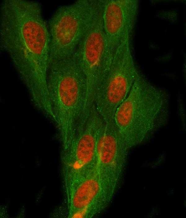
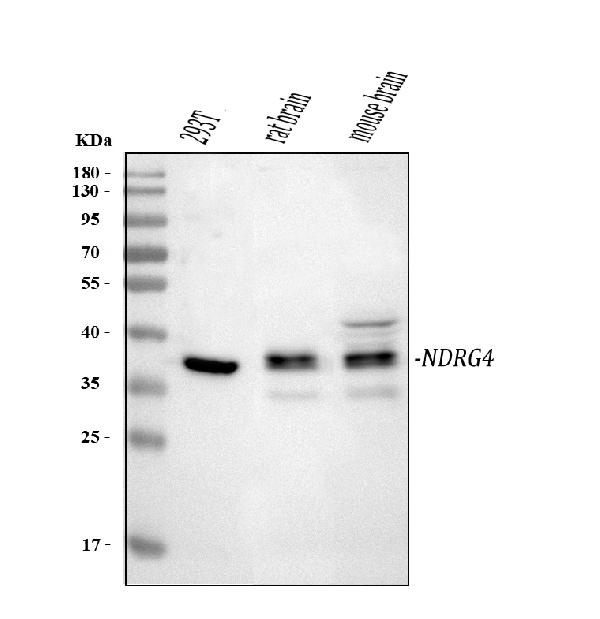
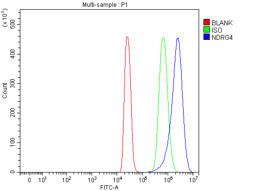
(Figure 3. Flow Cytometry analysis of 293T cells using anti-NDRG4 antibody (AAA127269).Overlay histogram showing 293T cells stained with AAA127269 (Blue line). To facilitate intracellular staining, cells were fixed with 4% paraformaldehyde and permeabilized with permeabilization buffer. The cells were blocked with 10% normal goat serum. And then incubated with rabbit anti-NDRG4 Antibody (AAA127269, 1ug/1x106 cells) for 30 min at 20 degree C. DyLight488 conjugated goat anti-rabbit IgG was used as secondary antibody for 30 minutes at 20 degree C. Isotype control antibody (Green line) was rabbit IgG (1ug/1x106) used under the same conditions. Unlabelled sample (Red line) was also used as a control.)
FCM/FACS (Flow Cytometry)
(Figure 3. Flow Cytometry analysis of 293T cells using anti-NDRG4 antibody (AAA127269).Overlay histogram showing 293T cells stained with AAA127269 (Blue line). To facilitate intracellular staining, cells were fixed with 4% paraformaldehyde and permeabilized with permeabilization buffer. The cells were blocked with 10% normal goat serum. And then incubated with rabbit anti-NDRG4 Antibody (AAA127269, 1ug/1x106 cells) for 30 min at 20 degree C. DyLight488 conjugated goat anti-rabbit IgG was used as secondary antibody for 30 minutes at 20 degree C. Isotype control antibody (Green line) was rabbit IgG (1ug/1x106) used under the same conditions. Unlabelled sample (Red line) was also used as a control.)
NDRG4, Polyclonal Antibody (Cat# AAA127269)
Full Name
Anti-NDRG4 Antibody Picoband
Gene Names
NDRG4; BDM1; SMAP8; SMAP-8
Reactivity
Human, Mouse, Rat
Applications
Flow Cytometry, Immunofluorescence, Immunocytochemistry, Western Blot
Purity
Immunogen affinity purified.
Pricing
 Bioactivity
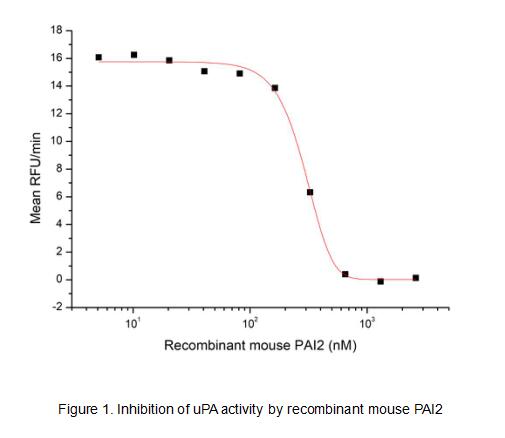
(Serpin B2, also known as PAI-2, is an approximately 60 kDa serine protease inhibitor. It is primarily secreted by macrophages and monocytes and can form disulfide-linked multimers. Serpin B2 inhibits both the urokinase-type and tissue-type plasminogen activators (uPA and tPA). Serpin B2 also promotes the clearance of uPA by enhancing its binding and uptake by LRP. It limits fibril formation by Huntington protein (HTT) and beta-Amyloid peptides. It promotes Th2 biased immune responses and is important for intestinal CCL2 production, monocyte recruitment, and nematode clearance. A non-glycosylated form of Serpin B2 is retained intracellularly where it interferes with TNF-a induced apoptosis by protecting the Retinoblastoma protein (RB1) from calpain digestion. It also inhibits proteasome activity in activated endothelial cells. The activity of recombinant mouse PAI-2 was measured by its ability to inhibit uPA cleavage of a peptide substrate, N-carbobenzyloxy-Gly-Gly-Arg-7-amido-4-methylcoumarin (Z-GGR-AMC) in the assay buffer 50 mM Tris, 0.01% Tween 20, pH 8.5. The 50 ul different concentrations of rmPAI-2 (MW: 47.9 KD) was incubated with 50ul 2ug/ml rhuPA (EPA140Mu61) at room temperature for 15 minutes. Loading 50 uL of the incubated mixtures into empty wells of a plate, and start the reaction by adding 50 uL of 200 uM substrate (Z-GGR-AMC). Include a substrate blank containing 50 uL of assay buffer and 50 uL of 200 uM substrate. Then read at excitiation and emission wavelengths of 380 nm and 460 nm, respectively, in kinetic mode for 5 minutes. The result was shown in Figure 1 and it was obvious that PAI2 significantly decreased uPA activity. The inhibition IC50 was )
Bioactivity
(Serpin B2, also known as PAI-2, is an approximately 60 kDa serine protease inhibitor. It is primarily secreted by macrophages and monocytes and can form disulfide-linked multimers. Serpin B2 inhibits both the urokinase-type and tissue-type plasminogen activators (uPA and tPA). Serpin B2 also promotes the clearance of uPA by enhancing its binding and uptake by LRP. It limits fibril formation by Huntington protein (HTT) and beta-Amyloid peptides. It promotes Th2 biased immune responses and is important for intestinal CCL2 production, monocyte recruitment, and nematode clearance. A non-glycosylated form of Serpin B2 is retained intracellularly where it interferes with TNF-a induced apoptosis by protecting the Retinoblastoma protein (RB1) from calpain digestion. It also inhibits proteasome activity in activated endothelial cells. The activity of recombinant mouse PAI-2 was measured by its ability to inhibit uPA cleavage of a peptide substrate, N-carbobenzyloxy-Gly-Gly-Arg-7-amido-4-methylcoumarin (Z-GGR-AMC) in the assay buffer 50 mM Tris, 0.01% Tween 20, pH 8.5. The 50 ul different concentrations of rmPAI-2 (MW: 47.9 KD) was incubated with 50ul 2ug/ml rhuPA (EPA140Mu61) at room temperature for 15 minutes. Loading 50 uL of the incubated mixtures into empty wells of a plate, and start the reaction by adding 50 uL of 200 uM substrate (Z-GGR-AMC). Include a substrate blank containing 50 uL of assay buffer and 50 uL of 200 uM substrate. Then read at excitiation and emission wavelengths of 380 nm and 460 nm, respectively, in kinetic mode for 5 minutes. The result was shown in Figure 1 and it was obvious that PAI2 significantly decreased uPA activity. The inhibition IC50 was )
Plasminogen Activator Inhibitor 2 (PAI2), Active Protein (Cat# AAA161740)
Full Name
Active Plasminogen Activator Inhibitor 2 (PAI2)
Gene Names
Serpinb2; PAI-2; Planh2; ovalbumin
Applications
Activity Assay, Cell Culture
Purity
Greater than 95% by SDS-PAGE
Pricing
 FCM (Flow Cytometry)
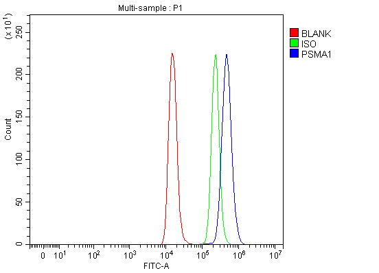
(Figure 7. Flow Cytometry analysis of Raji cells using anti-PSMA1 antibody (AAA19890).Overlay histogram showing Raji cells stained with AAA19890 (Blue line). To facilitate intracellular staining, cells were fixed with 4% paraformaldehyde and permeabilized with permeabilization buffer. The cells were blocked with 10% normal goat serum. And then incubated with rabbit anti-PSMA1 Antibody (AAA19890, 1ug/1x106 cells) for 30 min at 20 degree C. DyLight488 conjugated goat anti-rabbit IgG was used as secondary antibody for 30 minutes at 20 degree C. Isotype control antibody (Green line) was rabbit IgG (1ug/1x106) used under the same conditions. Unlabelled sample (Red line) was also used as a control.)
FCM (Flow Cytometry)
(Figure 7. Flow Cytometry analysis of Raji cells using anti-PSMA1 antibody (AAA19890).Overlay histogram showing Raji cells stained with AAA19890 (Blue line). To facilitate intracellular staining, cells were fixed with 4% paraformaldehyde and permeabilized with permeabilization buffer. The cells were blocked with 10% normal goat serum. And then incubated with rabbit anti-PSMA1 Antibody (AAA19890, 1ug/1x106 cells) for 30 min at 20 degree C. DyLight488 conjugated goat anti-rabbit IgG was used as secondary antibody for 30 minutes at 20 degree C. Isotype control antibody (Green line) was rabbit IgG (1ug/1x106) used under the same conditions. Unlabelled sample (Red line) was also used as a control.)
PSMA1, Polyclonal Antibody (Cat# AAA19890)
Full Name
Anti-PSMA1 Antibody Picoband
Gene Names
PSMA1; NU; HC2; PROS30; HEL-S-275
Reactivity
Human, Mouse, Rat
Applications
Western Blot, Immunohistochemistry, Flow Cytometry
Purity
Immunogen affinity purified.
Pricing
 FCM (Flow Cytometry)
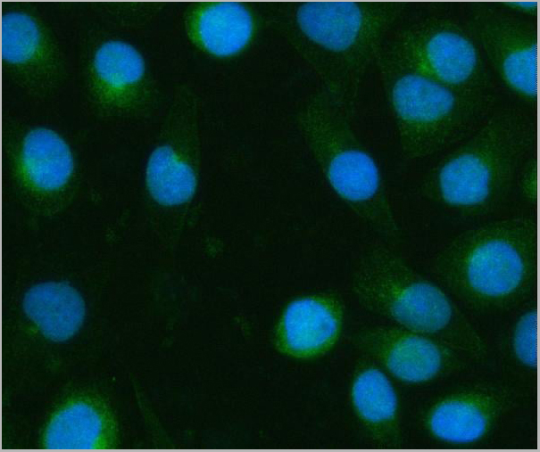
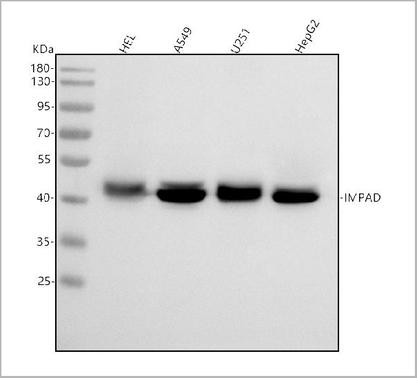
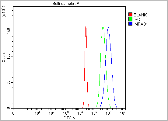
(Figure 14. Flow Cytometry analysis of HepG2 cells using anti-BPNT2 antibody (AAA19959).Overlay histogram showing HepG2 cells stained with AAA19959 (Blue line). To facilitate intracellular staining, cells were fixed with 4% paraformaldehyde and permeabilized with permeabilization buffer. The cells were blocked with 10% normal goat serum. And then incubated with rabbit anti-BPNT2 Antibody (AAA19959, 1ug/1x106 cells) for 30 min at 20 degree C. DyLight®488 conjugated goat anti-rabbit IgG (BA1127, 5-10ug/1x106 cells) was used as secondary antibody for 30 minutes at 20 degree C. Isotype control antibody (Green line) was rabbit IgG (1ug/1x106) used under the same conditions. Unlabelled sample (Red line) was also used as a control.)
FCM (Flow Cytometry)
(Figure 14. Flow Cytometry analysis of HepG2 cells using anti-BPNT2 antibody (AAA19959).Overlay histogram showing HepG2 cells stained with AAA19959 (Blue line). To facilitate intracellular staining, cells were fixed with 4% paraformaldehyde and permeabilized with permeabilization buffer. The cells were blocked with 10% normal goat serum. And then incubated with rabbit anti-BPNT2 Antibody (AAA19959, 1ug/1x106 cells) for 30 min at 20 degree C. DyLight®488 conjugated goat anti-rabbit IgG (BA1127, 5-10ug/1x106 cells) was used as secondary antibody for 30 minutes at 20 degree C. Isotype control antibody (Green line) was rabbit IgG (1ug/1x106) used under the same conditions. Unlabelled sample (Red line) was also used as a control.)
BPNT2, Polyclonal Antibody (Cat# AAA19959)
Full Name
Anti-BPNT2 Antibody Picoband
Gene Names
IMPAD1; GPAPP; IMP 3; IMP-3; IMPA3
Reactivity
Human
Applications
Western Blot, Immunohistochemistry, Immunocytochemistry, Immunofluorescence, Flow Cytometry
Purity
Immunogen affinity purified.
Pricing
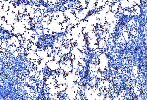
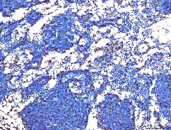
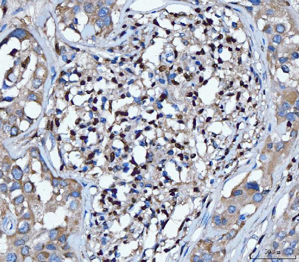
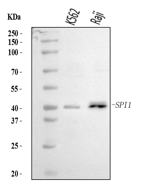
 FCM/FACS (Flow Cytometry)
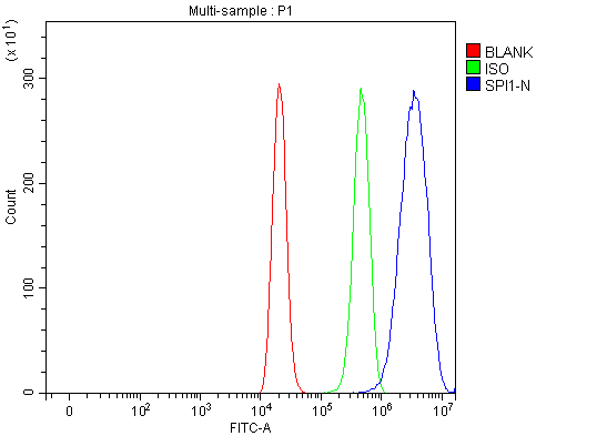
(Figure 5. Flow Cytometry analysis of THP-1 cells using anti-PU.1/SPI1 antibody (AAA126110).Overlay histogram showing THP-1 cells stained with AAA126110 (Blue line). The cells were blocked with 10% normal goat serum. And then incubated with rabbit anti-PU.1/SPI1 Antibody (AAA126110, 1 ug/1x10^6 cells) for 30 min at 20 degree C. DyLight488 conjugated goat anti-rabbit IgG was used as secondary antibody for 30 minutes at 20 degree C. Isotype control antibody (Green line) was rabbit IgG (1 ug/1x10^6) used under the same conditions. Unlabelled sample (Red line) was also used as a control.)
FCM/FACS (Flow Cytometry)
(Figure 5. Flow Cytometry analysis of THP-1 cells using anti-PU.1/SPI1 antibody (AAA126110).Overlay histogram showing THP-1 cells stained with AAA126110 (Blue line). The cells were blocked with 10% normal goat serum. And then incubated with rabbit anti-PU.1/SPI1 Antibody (AAA126110, 1 ug/1x10^6 cells) for 30 min at 20 degree C. DyLight488 conjugated goat anti-rabbit IgG was used as secondary antibody for 30 minutes at 20 degree C. Isotype control antibody (Green line) was rabbit IgG (1 ug/1x10^6) used under the same conditions. Unlabelled sample (Red line) was also used as a control.)
PU.1/SPI1, Polyclonal Antibody (Cat# AAA126110)
Full Name
Anti-PU.1/SPI1 Antibody Picoband
Gene Names
SPI1; OF; PU.1; SFPI1; SPI-1; SPI-A
Reactivity
Human, Mouse, Rat
Applications
Flow Cytometry, Immunohistochemistry, Western Blot
Purity
Immunogen affinity purified.
Pricing
 SDS-PAGE
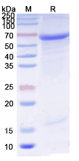
(SDS-PAGE for Recombinant Feline calicivirus (FCV) Capsid protein VP1 protein)
SDS-PAGE
(SDS-PAGE for Recombinant Feline calicivirus (FCV) Capsid protein VP1 protein)
Capsid protein VP1, Recombinant Protein (Cat# AAA120780)
Full Name
Recombinant FCV Capsid protein VP1 Protein, N-His
Applications
Western Blot
Purity
>90% as determined by SDS-PAGE.
Pricing
 Bioactivity
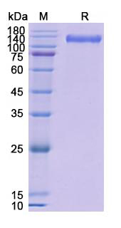
(Detects Pepinemab in indirect ELISAs.)
Bioactivity
(Detects Pepinemab in indirect ELISAs.)
CD100/SEMA4D, Recombinant Protein (Cat# AAA120543)
Full Name
Recombinant Human CD100/SEMA4D, C-His
Applications
Western Blot
Purity
>90% as determined by SDS-PAGE
Pricing
 Application Data
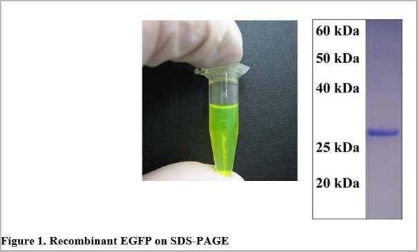
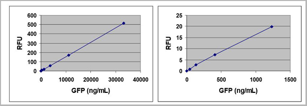
(The following figures demonstrate typical fluorescence rEGFP as tested in the GFP Fluorometric Quantitation Kit . Fluorescence measurement was performed on SpectraMax Gemini XS Fluorometer (Molecular Devices) with a 485/538 nm filter set and 530 nm cutoff. One should use the data below for reference only. This data should not be used to interpret actual results.)
Application Data
(The following figures demonstrate typical fluorescence rEGFP as tested in the GFP Fluorometric Quantitation Kit . Fluorescence measurement was performed on SpectraMax Gemini XS Fluorometer (Molecular Devices) with a 485/538 nm filter set and 530 nm cutoff. One should use the data below for reference only. This data should not be used to interpret actual results.)
 Standard Curve (Sample)
Galectin 3, ELISA Kit (Cat# AAA105667)
Full Name
Mouse Galectin 3 ELISA Kit
Gene Names
Lgals3; GBP; L-34; gal3; Mac-2
Reactivity
Mouse
Applications
Sandwich ELISA
Pricing
 Standard Curve (Sample)
PI3, ELISA Kit (Cat# AAA105715)
Full Name
Human PI3 ELISA Kit
Gene Names
PI3; ESI; WAP3; SKALP; WFDC14; cementoin
Reactivity
Human
Applications
Sandwich ELISA
Pricing

 SDS-PAGE
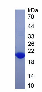
(Figure. SDS-PAGE)
SDS-PAGE
(Figure. SDS-PAGE)
Mannose Receptor C Type 1 (MRC1), Recombinant Protein (Cat# AAA150689)
Full Name
Recombinant Mannose Receptor C Type 1 (MRC1)
Gene Names
MRC1; MMR; CD206; MRC1L1; CLEC13D; CLEC13DL; bA541I19.1
Reactivity
Homo sapiens (Human)
Applications
Western Blot
Purity
> 90%
Pricing
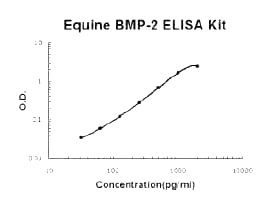 Standard Curve (Sample)
BMP-2, ELISA Kit (Cat# AAA124765)
Full Name
Horse equine BMP-2 PicoKine ELISA Kit
Gene Names
BMP2; BDA2; BMP2A
Reactivity
Equine
There is no detectable cross-reactivity with other relevant proteins.
There is no detectable cross-reactivity with other relevant proteins.
Applications
ELISA
Pricing
 Application Data
(The OmniKine Human Cathepsin V ELISA Kit allows for the detection and quantification of endogenous levels of natural and/or recombinant Human Cathepsin V proteins within the range of 63-4000 pg/ml.)
Application Data
(The OmniKine Human Cathepsin V ELISA Kit allows for the detection and quantification of endogenous levels of natural and/or recombinant Human Cathepsin V proteins within the range of 63-4000 pg/ml.)
Cathepsin V, ELISA Kit (Cat# AAA315787)
Full Name
Cathepsin V (Human) OmniKine ELISA Kit
Gene Names
CTSV; CTSU; CATL2; CTSL2
Reactivity
Human
Pricing
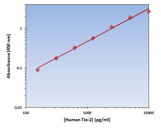 Application Data
(The OmniKine Human Tie-2 ELISA Kit allows for the detection and quantification of endogenous levels of natural and/or recombinant Human Tie-2 proteins within the range of 157-10000 pg/ml.)
Application Data
(The OmniKine Human Tie-2 ELISA Kit allows for the detection and quantification of endogenous levels of natural and/or recombinant Human Tie-2 proteins within the range of 157-10000 pg/ml.)
Tie-2, ELISA Kit (Cat# AAA315839)
Full Name
Tie-2 (Human) OmniKine ELISA Kit
Gene Names
TEK; TIE2; VMCM; GLC3E; TIE-2; VMCM1; CD202B
Reactivity
Human
Pricing
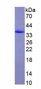 SDS-PAGE
(Figure. SDS-PAGE)
SDS-PAGE
(Figure. SDS-PAGE)
Major Histocompatibility Complex Class I A (MHCA), Recombinant Protein (Cat# AAA150702)
Full Name
Recombinant Major Histocompatibility Complex Class I A (MHCA)
Applications
Western Blot
Purity
>90%
Pricing
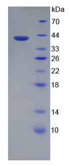
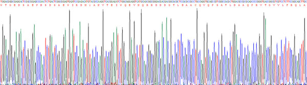
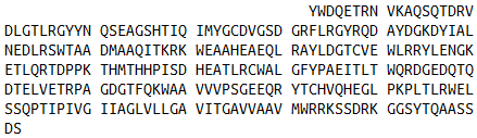
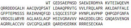 Amino Acid Info
(Amino Acid info)
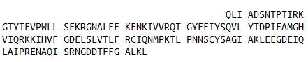
Amino Acid Info
(Amino Acid info)
Superoxide Dismutase 3, Extracellular (SOD3), Recombinant Protein (Cat# AAA153279)
Full Name
Recombinant Superoxide Dismutase 3, Extracellular (SOD3)
Gene Names
SOD3; EC-SOD
Applications
Western Blot
Purity
Greater than 90% by SDS-PAGE
Pricing
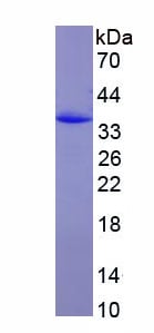 SDS-PAGE
(SDS-PAGE)
SDS-PAGE
(SDS-PAGE)
B-Cell Activating Factor (BAFF), Recombinant Protein (Cat# AAA152343)
Full Name
Recombinant B-Cell Activating Factor (BAFF)
Applications
Western Blot
Purity
Greater than 95% by SDS-PAGE
Pricing
 ELISA
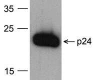
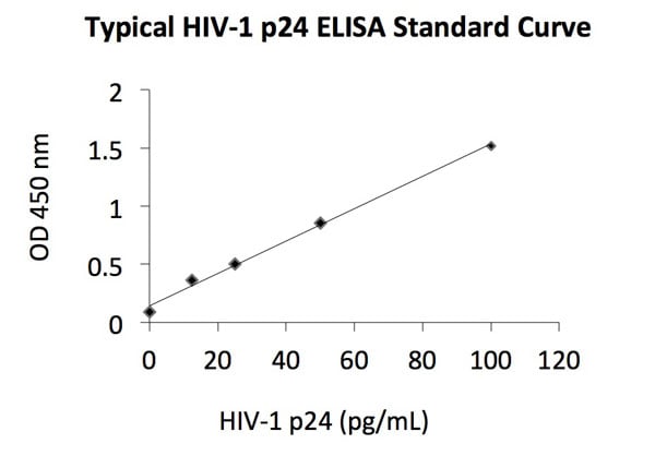
(Sandwich ELISA analysis of recombinant p24 proteins using capture antibody [7F4] and biotin labeled detection antibody [8G9]. (Assay sensitivity = 2.5 pg of recombinant p24 protein).)
ELISA
(Sandwich ELISA analysis of recombinant p24 proteins using capture antibody [7F4] and biotin labeled detection antibody [8G9]. (Assay sensitivity = 2.5 pg of recombinant p24 protein).)
HIV-1 p24, Monoclonal Antibody (Cat# AAA40478)
Full Name
HIV-1 p24 Antibody [8G9]
Reactivity
Virus
Applications
Western Blot
Purity
HIV-1 p24 Monoclonal Antibody is immunoaffinity chromotography purified IgG.
Pricing
 WB (Western Blot)
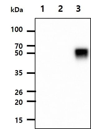
(The recombinant proteins (100ng) were resolved by SDS-PAGE, transferred to PVDF membrane and probed with anti-Influenza A H3N2/HA antibody (1:1000). Proteins were visualized using a goat anti-mouse secondary antibody conjugated to HRP and an ECL detection system.;;Lane 1.: Recombinant Influenza A H1N1/HA protein;;Lane 2.: Recombinant Influenza A H5N2/HA protein;;Lane 3.: Recombinant Influenza A H3N2/HA protein;;)
WB (Western Blot)
(The recombinant proteins (100ng) were resolved by SDS-PAGE, transferred to PVDF membrane and probed with anti-Influenza A H3N2/HA antibody (1:1000). Proteins were visualized using a goat anti-mouse secondary antibody conjugated to HRP and an ECL detection system.;;Lane 1.: Recombinant Influenza A H1N1/HA protein;;Lane 2.: Recombinant Influenza A H5N2/HA protein;;Lane 3.: Recombinant Influenza A H3N2/HA protein;;)
H3N2/HA, Monoclonal Antibody (Cat# AAA48292)
Full Name
H3N2/HA
Reactivity
Influenza A
Applications
Western Blot
Purity
By protein-G affinity chromatography
Pricing
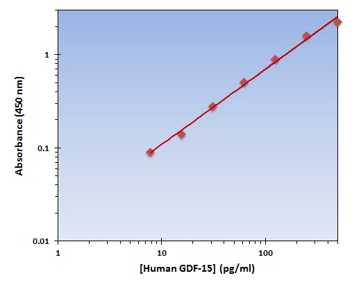 Application Data
(The OmniKine Human GDF-15 ELISA Kit allows for the detection and quantification of endogenous levels of natural and/or recombinant Human GDF-15 proteins within the range of 8-500 pg/ml.)
Application Data
(The OmniKine Human GDF-15 ELISA Kit allows for the detection and quantification of endogenous levels of natural and/or recombinant Human GDF-15 proteins within the range of 8-500 pg/ml.)
GDF-15, ELISA Kit (Cat# AAA315812)
Full Name
GDF-15 (Human) OmniKine ELISA Kit
Gene Names
GDF15; PDF; MIC1; PLAB; MIC-1; NAG-1; PTGFB; GDF-15
Reactivity
Human
Pricing
 Amino Acid Info
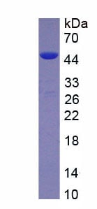
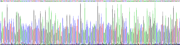
(Amino Acid info)
Amino Acid Info
(Amino Acid info)
Fibronectin Type III Domain Containing Protein 5 (FNDC5), Recombinant Protein (Cat# AAA153717)
Full Name
Recombinant Fibronectin Type III Domain Containing Protein 5 (FNDC5)
Gene Names
FNDC5; FRCP2; irisin
Applications
Western Blot
Purity
Greater than 80% by SDS-PAGE
Pricing
 Application Data
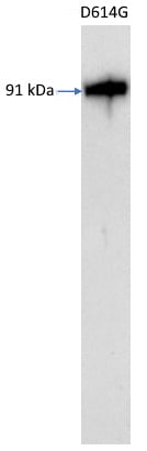
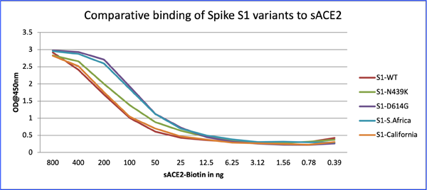
(Figure-3: Comparative binding of Spike S1 variants to sACE2: Wells of a 96-well microtiter plate were coated with 100 ng in duplicates each of S1-WT, S1-N439K, S1-D614G, S1-South Africa, and S1-Southern California. Binding to sACE2 was determined by adding different concentrations of biotinylated-sACE2.)
Application Data
(Figure-3: Comparative binding of Spike S1 variants to sACE2: Wells of a 96-well microtiter plate were coated with 100 ng in duplicates each of S1-WT, S1-N439K, S1-D614G, S1-South Africa, and S1-Southern California. Binding to sACE2 was determined by adding different concentrations of biotinylated-sACE2.)
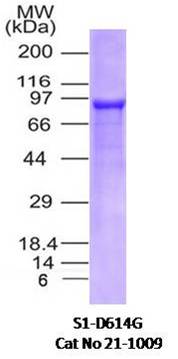
SARS-CoV-2 (2019-nCoV) Spike S1 (D614G), Recombinant Protein (Cat# AAA78596)
Full Name
SARS-CoV-2 (2019-nCoV) Spike S1 (D614G) His Tag Protein
Applications
Western Blot
Purity
>95% by SDS-PAGE.
Pricing
 IF (Immunofluorescence)
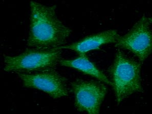
(ICC/IF analysis of FLIP in HeLa cells line, stained with DAPI (Blue) for nucleus staining and monoclonal anti-human FLIP antibody (1:100) with goat anti-mouse IgG-Alexa fluor 488 conjugate (Green))
IF (Immunofluorescence)
(ICC/IF analysis of FLIP in HeLa cells line, stained with DAPI (Blue) for nucleus staining and monoclonal anti-human FLIP antibody (1:100) with goat anti-mouse IgG-Alexa fluor 488 conjugate (Green))
FLIP, Monoclonal Antibody (Cat# AAA48041)
Full Name
FLIP antibody
Gene Names
CFLAR; CASH; FLIP; MRIT; CLARP; FLAME; Casper; FLAME1; c-FLIP; FLAME-1; I-FLICE; c-FLIPL; c-FLIPR; c-FLIPS; CASP8AP1
Reactivity
Human
Applications
Immunofluorescence, Immunocytochemistry, Western Blot
Purity
By protein-G affinity chromatography
Pricing
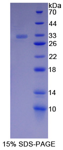 SDS-PAGE
Catenin Alpha 1, Recombinant Protein (Cat# AAA144202)
Full Name
Recombinant Catenin Alpha 1 (CTNNa1)
Gene Names
CTNNA1; CAP102
Applications
Western Blot
Purity
> 97%
Pricing
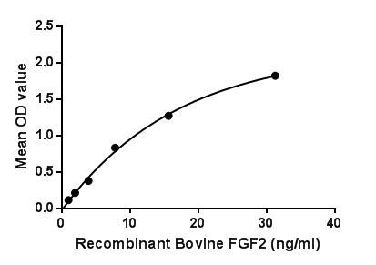
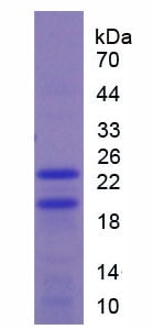 SDS-PAGE
Fibroblast Growth Factor 2 (FGF2), Active Protein (Cat# AAA149236)
Full Name
Active Fibroblast Growth Factor 2, Basic (FGF2)
Gene Names
FGF2; BFGF; FGF-2; HBGF-2
Applications
Activity Assay, Cell Culture
Purity
> 97%
Pricing
 Bioactivity
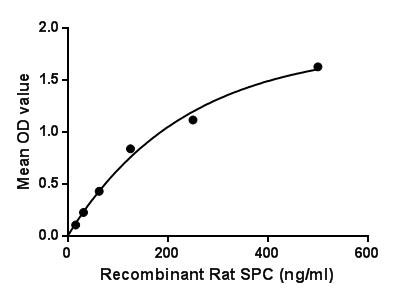
(Surfactant Associated Protein C (SPC) is one of the pulmonary surfactant proteins. It is a membrane protein which manufactures surfactant. The propeptide of pulmonary surfactant C has an N-terminal alpha-helical segment whose suggested function was stabilization of the protein structure, since the latter can irreversibly transform from its native alpha-helical structure to beta-sheet aggregates and form amyloid fibrils. Besides, Monokine Induced By Interferon Gamma (MIg) has been identified as an interactor of SPC, thus a binding ELISA assay was conducted to detect the interaction of recombinant rat SPC and recombinant rat MIg. Briefly, SPC were diluted serially in PBS, with 0.01% BSA (pH 7.4). Duplicate samples of 100L were then transferred to MIg-coated microtiter wells and incubated for 2h at 37. Wells were washed with PBST and incubated for 1h with anti-SPC pAb, then aspirated and washed 3 times. After incubation with HRP labelled secondary antibody, wells were aspirated and washed 3 times. With the addition of substrate solution, wells were incubated 15-25 minutes at 37. Finally, add 50uL stop solution to the wells and read at 450nm immediately. The binding activity of SPC and MIg was shown in Figure 1, and this effect was in a dose dependent manner.Figure. The binding activity of SPC with MIg.)
Bioactivity
(Surfactant Associated Protein C (SPC) is one of the pulmonary surfactant proteins. It is a membrane protein which manufactures surfactant. The propeptide of pulmonary surfactant C has an N-terminal alpha-helical segment whose suggested function was stabilization of the protein structure, since the latter can irreversibly transform from its native alpha-helical structure to beta-sheet aggregates and form amyloid fibrils. Besides, Monokine Induced By Interferon Gamma (MIg) has been identified as an interactor of SPC, thus a binding ELISA assay was conducted to detect the interaction of recombinant rat SPC and recombinant rat MIg. Briefly, SPC were diluted serially in PBS, with 0.01% BSA (pH 7.4). Duplicate samples of 100L were then transferred to MIg-coated microtiter wells and incubated for 2h at 37. Wells were washed with PBST and incubated for 1h with anti-SPC pAb, then aspirated and washed 3 times. After incubation with HRP labelled secondary antibody, wells were aspirated and washed 3 times. With the addition of substrate solution, wells were incubated 15-25 minutes at 37. Finally, add 50uL stop solution to the wells and read at 450nm immediately. The binding activity of SPC and MIg was shown in Figure 1, and this effect was in a dose dependent manner.Figure. The binding activity of SPC with MIg.)
Surfactant Protein C, Active Protein (Cat# AAA150111)
Full Name
Active Surfactant Protein C (SP-C)
Gene Names
Sftpc; SP-C
Applications
Activity Assay, Cell Culture
Purity
>95%
Pricing
 Bioactivity
(Reactive protein (CRP) is an annular (ring-shaped), pentameric protein found in blood plasma, whose levels rise in response to inflammation. It is an acute-phase protein of hepatic origin that increases following interleukin-6 secretion by macrophages and T cells. Its physiological role is to bind to lysophosphatidylcholine expressed on the surface of dead or dying cells (and some types of bacteria) in order to activate the complement system via C1q. Besides, Coagulation Factor II (F2) has been identified as an interactor of CRP, thus a functional binding ELISA assay was conducted to detect the interaction of recombinant rabit CRP and recombinant rat F2. Briefly, CRP was diluted serially in PBS with 0.01% BSA (pH 7.4). Duplicate samples of 100 ul were then transferred to F2-coated microtiter wells and incubated for 1h at 37 degree C. Wells were washed with PBST and incubated for 1h with anti-CRP pAb, then aspirated and washed 3 times. After incubation with HRP labelled secondary antibody for 1h at 37 degree C, wells were aspirated and washed 5 times. With the addition of substrate solution, wells were incubated 15-25 minutes at 37 degree C. Finally, add 50 uL stop solution to the wells and read at 450/630 nm immediately. The binding activity of recombinant rabbit CRP and recombinant rat F2 was shown in Figure 1, the EC50 for this effect is 0.02 ug/mL.)
Bioactivity
(Reactive protein (CRP) is an annular (ring-shaped), pentameric protein found in blood plasma, whose levels rise in response to inflammation. It is an acute-phase protein of hepatic origin that increases following interleukin-6 secretion by macrophages and T cells. Its physiological role is to bind to lysophosphatidylcholine expressed on the surface of dead or dying cells (and some types of bacteria) in order to activate the complement system via C1q. Besides, Coagulation Factor II (F2) has been identified as an interactor of CRP, thus a functional binding ELISA assay was conducted to detect the interaction of recombinant rabit CRP and recombinant rat F2. Briefly, CRP was diluted serially in PBS with 0.01% BSA (pH 7.4). Duplicate samples of 100 ul were then transferred to F2-coated microtiter wells and incubated for 1h at 37 degree C. Wells were washed with PBST and incubated for 1h with anti-CRP pAb, then aspirated and washed 3 times. After incubation with HRP labelled secondary antibody for 1h at 37 degree C, wells were aspirated and washed 5 times. With the addition of substrate solution, wells were incubated 15-25 minutes at 37 degree C. Finally, add 50 uL stop solution to the wells and read at 450/630 nm immediately. The binding activity of recombinant rabbit CRP and recombinant rat F2 was shown in Figure 1, the EC50 for this effect is 0.02 ug/mL.)
C ReProtein (CRP), Active Protein (Cat# AAA161785)
Full Name
Active C Reactive Protein (CRP)
Gene Names
CRP; PTX1
Applications
Activity Assay, Cell Culture
Purity
Greater than 97% by SDS-PAGE
Pricing
 Bioactivity
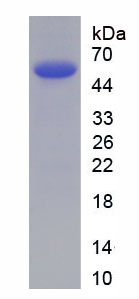
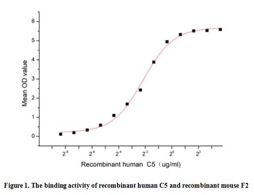
(Complement Component 5 (C5), which forms part of the later stages of the complement pathway, is cleaved into C5a and C5b. C5a is a chemotactic agent for neutrophils, while C5b forms part of the complement membrane attack complex. Coagulation Factor II (F2) has been identified as an interactor of C5, thus a functional binding ELISA assay was conducted to detect the interaction of recombinant human C5 and recombinant mouse F2. Briefly, C5 was diluted serially in PBS with 0.01% BSA (pH 7.4). Duplicate samples of 100 ul were then transferred to F2-coated microtiter wells and incubated for 1h at 37 degree C. Wells were washed with PBST and incubated for 1h with anti-C5 pAb, then aspirated and washed 3 times. After incubation with HRP labelled secondary antibody for 1h at 37 degree C, wells were aspirated and washed 5 times. With the addition of substrate solution, wells were incubated 15-25 minutes at 37 degree C. Finally, add 50 uL stop solution to the wells and read at 450/630 nm immediately. The binding activity of recombinant human C5 and recombinant mouse F2 was shown in Figure 1, the EC50 for this effect is 0.22 ug/mL.)
Bioactivity
(Complement Component 5 (C5), which forms part of the later stages of the complement pathway, is cleaved into C5a and C5b. C5a is a chemotactic agent for neutrophils, while C5b forms part of the complement membrane attack complex. Coagulation Factor II (F2) has been identified as an interactor of C5, thus a functional binding ELISA assay was conducted to detect the interaction of recombinant human C5 and recombinant mouse F2. Briefly, C5 was diluted serially in PBS with 0.01% BSA (pH 7.4). Duplicate samples of 100 ul were then transferred to F2-coated microtiter wells and incubated for 1h at 37 degree C. Wells were washed with PBST and incubated for 1h with anti-C5 pAb, then aspirated and washed 3 times. After incubation with HRP labelled secondary antibody for 1h at 37 degree C, wells were aspirated and washed 5 times. With the addition of substrate solution, wells were incubated 15-25 minutes at 37 degree C. Finally, add 50 uL stop solution to the wells and read at 450/630 nm immediately. The binding activity of recombinant human C5 and recombinant mouse F2 was shown in Figure 1, the EC50 for this effect is 0.22 ug/mL.)
Complement Component 5 (C5), Active Protein (Cat# AAA161871)
Full Name
Active Complement Component 5 (C5)
Gene Names
C5; C5a; C5b; CPAMD4
Applications
Activity Assay, Cell Culture
Purity
Greater than 95% by SDS-PAGE
Pricing
 Bioactivity
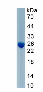
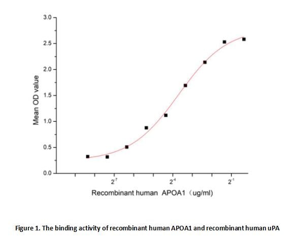
(Apolipoprotein A1 (APOA1) is the major protein component of HDL particles in plasma. It is a cofactor for lecithin cholesterolacyltransferase (LCAT) which is responsible for the formation of most plasma cholesteryl esters. ApoA1 was also isolated as a prostacyclin (PGI2) stabilizing factor, and thus may have an anticlotting effect. ApoA1 is often used as a biomarker for prediction of cardiovascular diseases. Besides, Plasminogen Activator, Urokinase (uPA) has been identified as an interactor of APOA1, thus a functional binding ELISA assay was conducted to detect the interaction of recombinant human APOA1 and recombinant human uPA. Briefly, biotin-linked APOA1 were diluted serially in PBS, with 0.01% BSA (pH 7.4). Duplicate samples of 100 ul were then transferred to uPA-coated microtiter wells and incubated for 1h at 37 degree C. Wells were washed with PBST 3 times and incubation with Streptavidin-HRP for 30min, then wells were aspirated and washed 5 times. With the addition of substrate solution, wells were incubated 15-25 minutes at 37 degree C. Finally, add 50 ul stop solution to the wells and read at 450/630 nm immediately. The binding activity of recombinant human uPA and recombinant human APOA1 was shown in Figure 1, the EC50 for this effect is 0.08 ug/mL.)
Bioactivity
(Apolipoprotein A1 (APOA1) is the major protein component of HDL particles in plasma. It is a cofactor for lecithin cholesterolacyltransferase (LCAT) which is responsible for the formation of most plasma cholesteryl esters. ApoA1 was also isolated as a prostacyclin (PGI2) stabilizing factor, and thus may have an anticlotting effect. ApoA1 is often used as a biomarker for prediction of cardiovascular diseases. Besides, Plasminogen Activator, Urokinase (uPA) has been identified as an interactor of APOA1, thus a functional binding ELISA assay was conducted to detect the interaction of recombinant human APOA1 and recombinant human uPA. Briefly, biotin-linked APOA1 were diluted serially in PBS, with 0.01% BSA (pH 7.4). Duplicate samples of 100 ul were then transferred to uPA-coated microtiter wells and incubated for 1h at 37 degree C. Wells were washed with PBST 3 times and incubation with Streptavidin-HRP for 30min, then wells were aspirated and washed 5 times. With the addition of substrate solution, wells were incubated 15-25 minutes at 37 degree C. Finally, add 50 ul stop solution to the wells and read at 450/630 nm immediately. The binding activity of recombinant human uPA and recombinant human APOA1 was shown in Figure 1, the EC50 for this effect is 0.08 ug/mL.)
Apolipoprotein A1 (APOA1), Active Protein (Cat# AAA161738)
Full Name
Active Apolipoprotein A1 (APOA1)
Applications
Activity Assay, Cell Culture
Purity
Greater than 90% by SDS-PAGE
Pricing
 Application Data
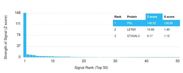
(Analysis of Protein Array containing more than 19,000 full-length human proteins using Prolactin Mouse Monoclonal Antibody (PRL/2643).Z- and S- Score: The Z-score represents the strength of a signal that a monoclonal antibody (Monoclonal Antibody) (in combination with a fluorescently-tagged anti-IgG secondary antibody) produces when binding to a particular protein on the HuProtTM array. Z-scores are described in units of standard deviations (SD’s) above the mean value of all signals generated on that array. If targets on HuProtTM are arranged in descending order of the Z-score, the S-score is the difference (also in units of SD’s) between the Z-score. S-score therefore represents the relative target specificity of a Monoclonal Antibody to its intended target. A Monoclonal Antibody is considered to specific to its intended target, if the Monoclonal Antibody has an S-score of at least 2.5. For example, if a Monoclonal Antibody binds to protein X with a Z-score of 43 and to protein Y with a Z-score of 14, then the S-score for the binding of that Monoclonal Antibody to protein X is equal to 29.)
Application Data
(Analysis of Protein Array containing more than 19,000 full-length human proteins using Prolactin Mouse Monoclonal Antibody (PRL/2643).Z- and S- Score: The Z-score represents the strength of a signal that a monoclonal antibody (Monoclonal Antibody) (in combination with a fluorescently-tagged anti-IgG secondary antibody) produces when binding to a particular protein on the HuProtTM array. Z-scores are described in units of standard deviations (SD’s) above the mean value of all signals generated on that array. If targets on HuProtTM are arranged in descending order of the Z-score, the S-score is the difference (also in units of SD’s) between the Z-score. S-score therefore represents the relative target specificity of a Monoclonal Antibody to its intended target. A Monoclonal Antibody is considered to specific to its intended target, if the Monoclonal Antibody has an S-score of at least 2.5. For example, if a Monoclonal Antibody binds to protein X with a Z-score of 43 and to protein Y with a Z-score of 14, then the S-score for the binding of that Monoclonal Antibody to protein X is equal to 29.)
Prolactin, Monoclonal Antibody (Cat# AAA215162)
Full Name
Prolactin (Pituitary Tumor Marker)
Gene Names
PRL; GHA1
Reactivity
Human
Applications
Immunohistochemistry
Purity
Purified Ab with BSA and Azide at 200ug/ml OR Purified Ab WITHOUT BSA and Azide at 1.0mg/ml
Pricing

 FCM/FACS (Flow Cytometry)
(Figure 3. Flow Cytometry analysis of THP-1 cells using anti-AIRE antibody (AAA46634).Overlay histogram showing THP-1 cells stained with AAA46634 (Blue line).The cells were blocked with 10% normal goat serum. And then incubated with rabbit anti-AIRE Antibody (AAA46634,1ug/1x10^6 cells) for 30 min at 20 degree C. DyLight®488 conjugated goat anti-rabbit IgG (5-10ug/1x10^6 cells) was used as secondary antibody for 30 minutes at 20 degree C. Isotype control antibody (Green line) was rabbit IgG (1ug/1x106) used under the same conditions. Unlabelled sample (Red line) was also used as a control.)
FCM/FACS (Flow Cytometry)
(Figure 3. Flow Cytometry analysis of THP-1 cells using anti-AIRE antibody (AAA46634).Overlay histogram showing THP-1 cells stained with AAA46634 (Blue line).The cells were blocked with 10% normal goat serum. And then incubated with rabbit anti-AIRE Antibody (AAA46634,1ug/1x10^6 cells) for 30 min at 20 degree C. DyLight®488 conjugated goat anti-rabbit IgG (5-10ug/1x10^6 cells) was used as secondary antibody for 30 minutes at 20 degree C. Isotype control antibody (Green line) was rabbit IgG (1ug/1x106) used under the same conditions. Unlabelled sample (Red line) was also used as a control.)
AIRE, Polyclonal Antibody (Cat# AAA46634)
Full Name
Anti-AIRE Antibody
Gene Names
AIRE; APS1; APSI; PGA1; AIRE1; APECED
Reactivity
Human, Rat
No cross reactivity with other proteins.
No cross reactivity with other proteins.
Applications
Western Blot
Purity
Immunogen affinity purified.
Pricing
 Application Data
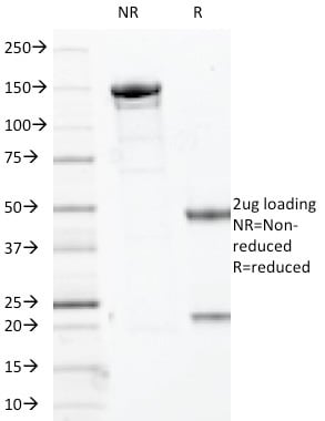
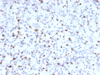
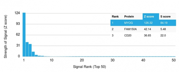
(Analysis of Protein Array containing more than 19, 000 full-length human proteins using Myogenin Mouse Monoclonal Antibody (MYOG/2660). Z- and S- Score: The Z-score represents the strength of a signal that a monoclonal antibody (MAb) (in combination with a fluorescently-tagged anti-IgG secondary antibody) produces when binding to a particular protein on the HuProtTM array. Z-scores are described in units of standard deviations (SD's) above the mean value of all signals generated on that array. If targets on HuProtTM are arranged in descending order of the Z-score, the S-score is the difference (also in units of SD's) between the Z-score. S-score therefore represents the relative target specificity of a MAb to its intended target. A MAb is considered to specific to its intended target, if the MAb has an S-score of at least 2.5. For example, if a MAb binds to protein X with a Z-score of 43 and to protein Y with a Z-score of 14, then the S-score for the binding of that MAb to protein X is equal to 29.)
Application Data
(Analysis of Protein Array containing more than 19, 000 full-length human proteins using Myogenin Mouse Monoclonal Antibody (MYOG/2660). Z- and S- Score: The Z-score represents the strength of a signal that a monoclonal antibody (MAb) (in combination with a fluorescently-tagged anti-IgG secondary antibody) produces when binding to a particular protein on the HuProtTM array. Z-scores are described in units of standard deviations (SD's) above the mean value of all signals generated on that array. If targets on HuProtTM are arranged in descending order of the Z-score, the S-score is the difference (also in units of SD's) between the Z-score. S-score therefore represents the relative target specificity of a MAb to its intended target. A MAb is considered to specific to its intended target, if the MAb has an S-score of at least 2.5. For example, if a MAb binds to protein X with a Z-score of 43 and to protein Y with a Z-score of 14, then the S-score for the binding of that MAb to protein X is equal to 29.)
Myogenin, Monoclonal Antibody (Cat# AAA214670)
Full Name
Myogenin (Skeletal Muscle Marker)
Gene Names
MYOG; MYF4; myf-4; bHLHc3
Reactivity
Human. Others not known.
Applications
Immunohistochemistry
Pricing
 Application Data
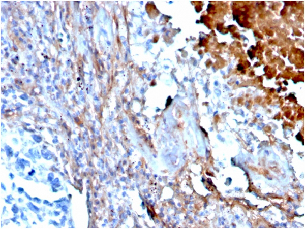
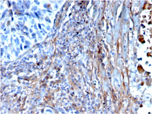
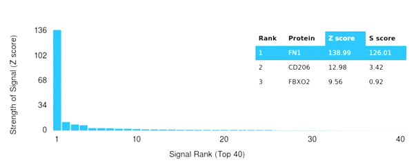
(Analysis of Protein Array containing more than 19,000 full-length human proteins using Fibronectin Mouse Monoclonal Antibody (FN1/3568). Z- and S- Score: The Z-score represents the strength of a signal that a monoclonal antibody (MAb) (in combination with a fluorescently-tagged anti-IgG secondary antibody) produces when binding to a particular protein on the HuProtTM array. Z-scores are described in units of standard deviations (SD's) above the mean value of all signals generated on that array. If targets on HuProtTM are arranged in descending order of the Z-score, the S-score is the difference (also in units of SD's) between the Z-score. S-score therefore represents the relative target specificity of a MAb to its intended target. A MAb is considered to specific to its intended target, if the MAb has an S-score of at least 2.5. For example, if a MAb binds to protein X with a Z-score of 43 and to protein Y with a Z-score of 14, then the S-score for the binding of that MAb to protein X is equal to 29.)
Application Data
(Analysis of Protein Array containing more than 19,000 full-length human proteins using Fibronectin Mouse Monoclonal Antibody (FN1/3568). Z- and S- Score: The Z-score represents the strength of a signal that a monoclonal antibody (MAb) (in combination with a fluorescently-tagged anti-IgG secondary antibody) produces when binding to a particular protein on the HuProtTM array. Z-scores are described in units of standard deviations (SD's) above the mean value of all signals generated on that array. If targets on HuProtTM are arranged in descending order of the Z-score, the S-score is the difference (also in units of SD's) between the Z-score. S-score therefore represents the relative target specificity of a MAb to its intended target. A MAb is considered to specific to its intended target, if the MAb has an S-score of at least 2.5. For example, if a MAb binds to protein X with a Z-score of 43 and to protein Y with a Z-score of 14, then the S-score for the binding of that MAb to protein X is equal to 29.)
Fibronectin, Monoclonal Antibody (Cat# AAA215453)
Full Name
Fibronectin
Gene Names
FN1; FN; CIG; FNZ; MSF; ED-B; FINC; GFND; LETS; GFND2; SMDCF
Reactivity
Human
Applications
Immunohistochemistry
Purity
Purified Ab with BSA and Azide at 200ug/ml or Purified Ab with BSA and Azide at 200ug/ml or Purified Ab WITHOUT BSA and Azide at 1.0mg/ml
Pricing
 SDS-PAGE
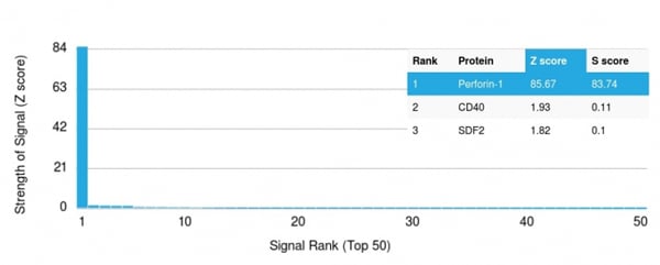
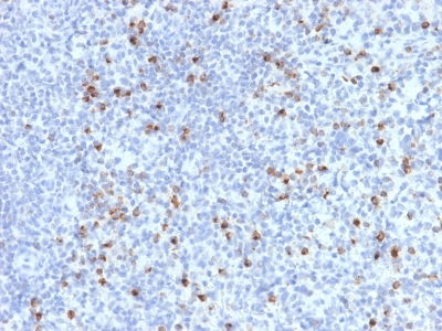
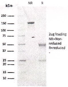
(SDS-PAGE Analysis Purified Perforin-1 Mouse Monoclonal Antibody (PRF1/2470).(Confirmation of Integrity and Purity of Antibody)
SDS-PAGE
(SDS-PAGE Analysis Purified Perforin-1 Mouse Monoclonal Antibody (PRF1/2470).(Confirmation of Integrity and Purity of Antibody)
Perforin-1, Monoclonal Antibody (Cat# AAA214800)
Full Name
Perforin-1 (Pore Forming Protein) (Apoptosis Marker)
Gene Names
PRF1; P1; PFP; HPLH2
Reactivity
Human. Others not known.
Applications
Immunohistochemistry
Purity
Purified Ab with BSA and Azide at 200ug/ml OR Purified Ab WITHOUT BSA and Azide at 1.0mg/ml
Pricing