
Filters
Clonality
Type
Reactivity
Gene Name
Isotype
Host
Application
Clone
10000 results for "Recombinant Proteins" - showing 2650-2700
 Bioactivity
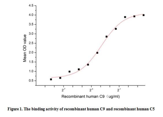
(Complement Component 9 (C9), a component of the Membrane attack Complex (MAC) plays a key role in the innate and adaptive immune response by forming pores in the plasma membrane of target cells. During MAC assembly, multiple copies of C9 are sequentially recruited to membrane associated C5b8 to form a pore. Thus a functional binding ELISA assay was conducted to detect the interaction of recombinant human C9 and recombinant human C5. Briefly, biotin-linked C9 were diluted serially in PBS, with 0.01% BSA (pH 7.4). Duplicate samples of 100 ul were then transferred to C5-coated microtiter wells and incubated for 1h at 37 degree C. Wells were washed with PBST 3 times and incubation with Streptavidin-HRP for 30min, then wells were aspirated and washed 5 times. With the addition of substrate solution, wells were incubated 15-25 minutes at 37 degree C. Finally, add 50 ul stop solution to the wells and read at 450 nm immediately. The binding activity of recombinant human C9 and recombinant human C5 was shown in Figure 1, the EC50 for this effect is 0.13 ug/mL.)
Bioactivity
(Complement Component 9 (C9), a component of the Membrane attack Complex (MAC) plays a key role in the innate and adaptive immune response by forming pores in the plasma membrane of target cells. During MAC assembly, multiple copies of C9 are sequentially recruited to membrane associated C5b8 to form a pore. Thus a functional binding ELISA assay was conducted to detect the interaction of recombinant human C9 and recombinant human C5. Briefly, biotin-linked C9 were diluted serially in PBS, with 0.01% BSA (pH 7.4). Duplicate samples of 100 ul were then transferred to C5-coated microtiter wells and incubated for 1h at 37 degree C. Wells were washed with PBST 3 times and incubation with Streptavidin-HRP for 30min, then wells were aspirated and washed 5 times. With the addition of substrate solution, wells were incubated 15-25 minutes at 37 degree C. Finally, add 50 ul stop solution to the wells and read at 450 nm immediately. The binding activity of recombinant human C9 and recombinant human C5 was shown in Figure 1, the EC50 for this effect is 0.13 ug/mL.)
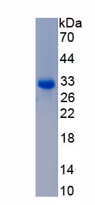
Complement Component 9 (C9), Active Protein (Cat# AAA161862)
Full Name
Active Complement Component 9 (C9)
Applications
Activity Assay, Cell Culture
Purity
Greater than 95% by SDS-PAGE
Pricing
 Bioactivity
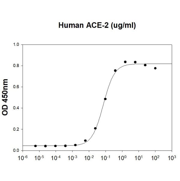
(SARS-CoV S1 Subunit is coated at 5 ug/ml (100 ul/well) can bind Human ACE-2 (CAT# ATGP3963) in a Functional ELISA assay.)
Bioactivity
(SARS-CoV S1 Subunit is coated at 5 ug/ml (100 ul/well) can bind Human ACE-2 (CAT# ATGP3963) in a Functional ELISA assay.)
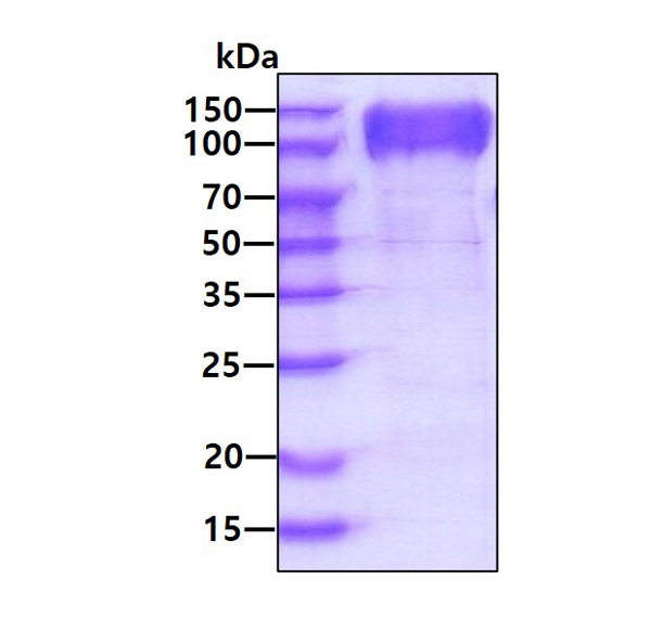
COVID 19 Spike S1 Subunit Coronavirus, Active Protein (Cat# AAA48390)
Full Name
Recombinant SARS-CoV S1 Subunit protein
Applications
Bioactivity, SDS-PAGE
Purity
>90% by SDS-PAGE
Pricing
 ELISA
(Biotinylated Human Semaphorin 4D/SEMA4D/CD100 Protein ELISA DataImmobilized Anti-Semaphorin 4D Antibody, hFc Tag at 0.5?g/ml (100?l/Well) on the plate. Dose response curve for Biotinylated Human Semaphorin 4D, His Tag with the EC50 of 44.8ng/ml determined by ELISA.)
ELISA
(Biotinylated Human Semaphorin 4D/SEMA4D/CD100 Protein ELISA DataImmobilized Anti-Semaphorin 4D Antibody, hFc Tag at 0.5?g/ml (100?l/Well) on the plate. Dose response curve for Biotinylated Human Semaphorin 4D, His Tag with the EC50 of 44.8ng/ml determined by ELISA.)
Semaphorin 4D/SEMA4D/CD100, Recombinant Protein (Cat# AAA76152)
Full Name
Biotinylated Human Semaphorin 4D/SEMA4D/CD100 Protein Recombinant
Gene Names
SEMA4D; CD100; SEMAJ; coll-4; C9orf164; M-sema-G
Purity
>95%, as determined by SDS-PAGE and HPLC
Pricing
 Bioactivity
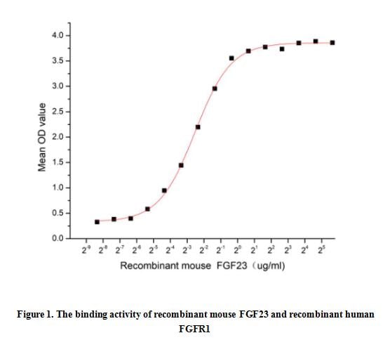
(Fibroblast growth factor 23 or FGF23 is a member of the fibroblast growth factor (FGF) family which is responsible for phosphate and vitamin D metabolism. The main function of FGF23 seems to be regulation of phosphate concentration in plasma. FGF23 decreases the reabsorption and increases excretion of phosphate and suppress 1-alpha-hydroxylase, reducing its ability to activate vitamin D and subsequently impairing calcium absorption. Besides, Fibroblast Growth Factor Receptor 1 (FGFR1) has been identified as an interactor of FGF23, thus a functional binding ELISA assay was conducted to detect the interaction of recombinant mouse FGF23 and recombinant human FGFR1. Briefly, FGF23 were diluted serially in PBS, with 0.01% BSA (pH 7.4). Duplicate samples of 100 ul were then transferred to FGFR1-coated microtiter wells and incubated for 1h at 37 degree C. Wells were washed with PBST and incubated for 1h with anti-FGF23 pAb, then aspirated and washed 3 times. After incubation with HRP labelled secondary antibody, wells were aspirated and washed 5 times. With the addition of substrate solution, wells were incubated 15-25 minutes at 37 degree C. Finally, add 50 uL stop solution to the wells and read at 450 nm immediately. The binding activity of recombinant mouse FGF23 and recombinant human FGFR1 was shown in Figure 1, the EC50 for this effect is 0.17 ug/mL.)
Bioactivity
(Fibroblast growth factor 23 or FGF23 is a member of the fibroblast growth factor (FGF) family which is responsible for phosphate and vitamin D metabolism. The main function of FGF23 seems to be regulation of phosphate concentration in plasma. FGF23 decreases the reabsorption and increases excretion of phosphate and suppress 1-alpha-hydroxylase, reducing its ability to activate vitamin D and subsequently impairing calcium absorption. Besides, Fibroblast Growth Factor Receptor 1 (FGFR1) has been identified as an interactor of FGF23, thus a functional binding ELISA assay was conducted to detect the interaction of recombinant mouse FGF23 and recombinant human FGFR1. Briefly, FGF23 were diluted serially in PBS, with 0.01% BSA (pH 7.4). Duplicate samples of 100 ul were then transferred to FGFR1-coated microtiter wells and incubated for 1h at 37 degree C. Wells were washed with PBST and incubated for 1h with anti-FGF23 pAb, then aspirated and washed 3 times. After incubation with HRP labelled secondary antibody, wells were aspirated and washed 5 times. With the addition of substrate solution, wells were incubated 15-25 minutes at 37 degree C. Finally, add 50 uL stop solution to the wells and read at 450 nm immediately. The binding activity of recombinant mouse FGF23 and recombinant human FGFR1 was shown in Figure 1, the EC50 for this effect is 0.17 ug/mL.)
Fibroblast Growth Factor 23 (FGF23), Active Protein (Cat# AAA161772)
Full Name
Active Fibroblast Growth Factor 23 (FGF23)
Applications
Activity Assay, Cell Culture
Purity
Greater than 90% by SDS-PAGE
Pricing
 Bioactivity
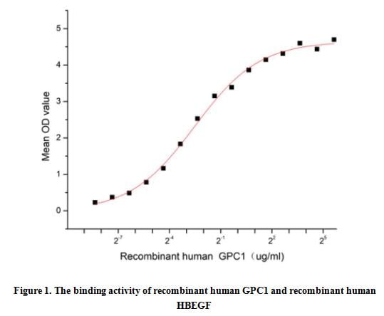
(Glypican 1 (GPC1) is one of the six glycosylphosphatidylinositol-anchored, cell surface heparan sulfate proteoglycans that acts as a growth factor signaling. Cells known to express GPC-1 include neurons, smooth and skeletal muscle cells, keratinocytes, osteoblasts, Schwann cells, immature dendritic cells, and tumor, plus tumor-associated vascular endothelial cells. The function of GPC-1 is complex and varied, it can modulate various signaling pathways including Heparin Binding Epidermal Growth Factor Like Growth Factor (HBEGF), fibroblast growth factors (FGF), vascular endothelial growth factor-A (VEGF-A), transforming growth factor-beta (TGF-beta), Wnt, Hedgehog (Hh), and bone morphogenic protein (BMP) through specific interactions with pathway ligands and receptors. Thus a functional binding ELISA assay was conducted to detect the interaction of recombinant human GPC1 and recombinant human HBEGF. Briefly, GPC1 was diluted serially in PBS with 0.01% BSA (pH 7.4). Duplicate samples of 100 ul were then transferred to HBEGF-coated microtiter wells and incubated for 1h at 37 degree C. Wells were washed with PBST and incubated for 1h with anti-GPC1 pAb, then aspirated and washed 3 times. After incubation with HRP labelled secondary antibody for 1h at 37 degree C, wells were aspirated and washed 5 times. With the addition of substrate solution, wells were incubated 15-25 minutes at 37 degree C. Finally, add 50 uL stop solution to the wells and read at 450/630 nm immediately. The binding activity of recombinant human GPC1 and recombinant human HBEGF was shown in Figure 1, the EC50 for this effect is 0.17 ug/mL.)
Bioactivity
(Glypican 1 (GPC1) is one of the six glycosylphosphatidylinositol-anchored, cell surface heparan sulfate proteoglycans that acts as a growth factor signaling. Cells known to express GPC-1 include neurons, smooth and skeletal muscle cells, keratinocytes, osteoblasts, Schwann cells, immature dendritic cells, and tumor, plus tumor-associated vascular endothelial cells. The function of GPC-1 is complex and varied, it can modulate various signaling pathways including Heparin Binding Epidermal Growth Factor Like Growth Factor (HBEGF), fibroblast growth factors (FGF), vascular endothelial growth factor-A (VEGF-A), transforming growth factor-beta (TGF-beta), Wnt, Hedgehog (Hh), and bone morphogenic protein (BMP) through specific interactions with pathway ligands and receptors. Thus a functional binding ELISA assay was conducted to detect the interaction of recombinant human GPC1 and recombinant human HBEGF. Briefly, GPC1 was diluted serially in PBS with 0.01% BSA (pH 7.4). Duplicate samples of 100 ul were then transferred to HBEGF-coated microtiter wells and incubated for 1h at 37 degree C. Wells were washed with PBST and incubated for 1h with anti-GPC1 pAb, then aspirated and washed 3 times. After incubation with HRP labelled secondary antibody for 1h at 37 degree C, wells were aspirated and washed 5 times. With the addition of substrate solution, wells were incubated 15-25 minutes at 37 degree C. Finally, add 50 uL stop solution to the wells and read at 450/630 nm immediately. The binding activity of recombinant human GPC1 and recombinant human HBEGF was shown in Figure 1, the EC50 for this effect is 0.17 ug/mL.)
Glypican 1 (GPC1), Active Protein (Cat# AAA161824)
Full Name
Active Glypican 1 (GPC1)
Gene Names
GPC1; glypican
Applications
Activity Assay, Cell Culture
Purity
Greater than 95% by SDS-PAGE
Pricing
 WB (Western Blot)
(Western Blot; Sample: Human Urine Primary Ab: 2ug/ml Mouse AntiHuman PGA Antibody Second Ab: 0.2ug/mL HRPLinked Caprine AntiMouse IgG Polyclonal Antibody (Catalog: SAA544Mu19))
WB (Western Blot)
(Western Blot; Sample: Human Urine Primary Ab: 2ug/ml Mouse AntiHuman PGA Antibody Second Ab: 0.2ug/mL HRPLinked Caprine AntiMouse IgG Polyclonal Antibody (Catalog: SAA544Mu19))
Pepsinogen A (PGA), Monoclonal Antibody (Cat# AAA151502)
Full Name
Monoclonal Antibody to Pepsinogen A (PGA)
Gene Names
PGA5; Pg5
Reactivity
Human
Applications
Immunoprecipitation, Immunocytochemistry, Immunohistochemistry, Western Blot
Purity
Protein A + Protein G affinity chromatography
Pricing
 Bioactivity
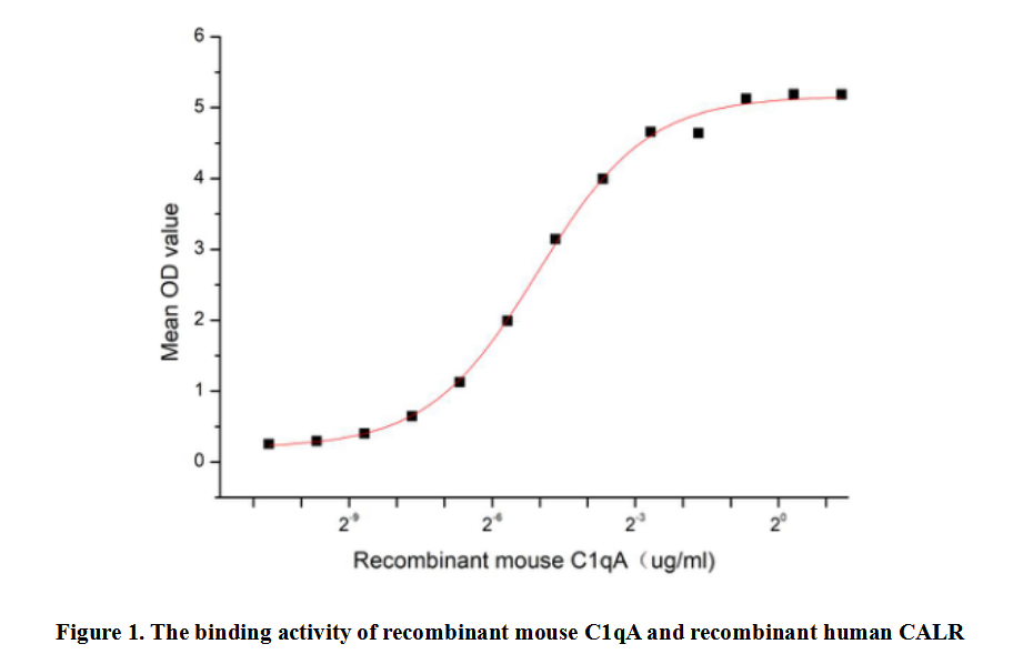
(Complement Component 1, Q Subcomponent A (C1qA) is a protein that plays a crucial role in the innate immune system. It is part of the C1 complex, which is the first component of the classical complement pathway. The C1 complex is composed of one molecule each of C1q, C1r, and C1s. C1q is the recognition subcomponent of the complex and and initiating the complement cascade. C1qA plays a key role in eliminating pathogens, regulating inflammatory response and maintaining immune tolerance. It is reported that C1qA can bind to the C-terminal of CALR through its gelatin-as-region, and this interaction may play an important role in regulating complement activation and cell signaling. Thus a functional binding ELISA assay was conducted to detect the interaction of recombinant mouse C1qA and recombinant human CALR. Briefly, C1qA was diluted serially in PBS with 0.01% BSA (pH 7.4). Duplicate samples of 100 ul were then transferred to CALR-coated microtiter wells and incubated for 1h at 37 degree C. Wells were washed with PBST and incubated for 1h with anti-C1qA pAb, then aspirated and washed 3 times. After incubation with HRP labelled secondary antibody for 1h at 37 degree C, wells were aspirated and washed 5 times. With the addition of substrate solution, wells were incubated 15-25 minutes at 37 degree C. Finally, add 50 uL stop solution to the wells and read at 450/630 nm immediately. The binding activity of recombinant mouse C1qA and recombinant human CALR was shown in Figure 1, the EC50 for this effect is 0.03 ug/mL.)
Bioactivity
(Complement Component 1, Q Subcomponent A (C1qA) is a protein that plays a crucial role in the innate immune system. It is part of the C1 complex, which is the first component of the classical complement pathway. The C1 complex is composed of one molecule each of C1q, C1r, and C1s. C1q is the recognition subcomponent of the complex and and initiating the complement cascade. C1qA plays a key role in eliminating pathogens, regulating inflammatory response and maintaining immune tolerance. It is reported that C1qA can bind to the C-terminal of CALR through its gelatin-as-region, and this interaction may play an important role in regulating complement activation and cell signaling. Thus a functional binding ELISA assay was conducted to detect the interaction of recombinant mouse C1qA and recombinant human CALR. Briefly, C1qA was diluted serially in PBS with 0.01% BSA (pH 7.4). Duplicate samples of 100 ul were then transferred to CALR-coated microtiter wells and incubated for 1h at 37 degree C. Wells were washed with PBST and incubated for 1h with anti-C1qA pAb, then aspirated and washed 3 times. After incubation with HRP labelled secondary antibody for 1h at 37 degree C, wells were aspirated and washed 5 times. With the addition of substrate solution, wells were incubated 15-25 minutes at 37 degree C. Finally, add 50 uL stop solution to the wells and read at 450/630 nm immediately. The binding activity of recombinant mouse C1qA and recombinant human CALR was shown in Figure 1, the EC50 for this effect is 0.03 ug/mL.)
Complement Component 1, Q Subcomponent A (C1qA), Active Protein (Cat# AAA161911)
Full Name
Active Complement Component 1, Q Subcomponent A (C1qA)
Gene Names
C1qa; C1q; AI255395
Applications
Activity Assay, Cell Culture
Purity
Greater than 95% by SDS-PAGE
Pricing
 WB (Western Blot)
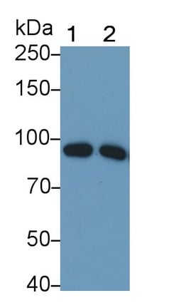
(Western Blot; Sample: Lane1: Human Plasma; Lane2: Human Serum Primary Ab: 0.05ug/ml Mouse AntiHuman C1s Antibody Second Ab: 0.2ug/mL HRPLinked Caprine AntiMouse IgG Polyclonal Antibody (Catalog: SAA544Mu19))
WB (Western Blot)
(Western Blot; Sample: Lane1: Human Plasma; Lane2: Human Serum Primary Ab: 0.05ug/ml Mouse AntiHuman C1s Antibody Second Ab: 0.2ug/mL HRPLinked Caprine AntiMouse IgG Polyclonal Antibody (Catalog: SAA544Mu19))
Complement Component 1, S Subcomponent (C1s), Monoclonal Antibody (Cat# AAA151730)
Full Name
Monoclonal Antibody to Complement Component 1, S Subcomponent (C1s)
Reactivity
Human
Applications
Immunoprecipitation, Immunocytochemistry, Immunohistochemistry, Western Blot
Purity
Protein A + Protein G affinity chromatography
Pricing

 Bioactivity
(CD146 (cluster of differentiation 146), also known as MUC18 or MCAM, is a putative adhesion molecule that belongs to the immunoglobulin superfamily (IgSF). It is an approximately 113-kDa type1 transmembrane glycoprotein that is expressed at the intercellular junction of endothelial cells. CD146 was originally identified as a tumor marker for melanoma (MCAM) due to its existence only in melanoma but not in the corresponding normal counterpart. However CD146 is not just a CAM for the inter-cellular and cell-matrix adhesion. Recent evidence indicates that CD146 is actively involved in miscellaneous processes, such as development, signaling transduction, cell migration, mesenchymal stem cells differentiation, angiogenesis and immune response. CD146 and PECAM-1 are both members of immunoglobulin superfamily of cell adhesion molecules. PECAM-1 has been identified as an interactor of CD146, thus a functional binding ELISA assay was conducted to detect the interaction of recombinant human CD146 and recombinant rat PECAM-1. Briefly, CD146 was diluted serially in PBS with 0.01% BSA (pH 7.4). Duplicate samples of 100 ul were then transferred to PECAM-1-coated microtiter wells and incubated for 1h at 37 degree C. Wells were washed with PBST and incubated for 1h with anti-CD146 pAb, then aspirated and washed 3 times. After incubation with HRP labelled secondary antibody for 1h at 37 degree C, wells were aspirated and washed 5 times. With the addition of substrate solution, wells were incubated 15-25 minutes at 37 degree C. Finally, add 50 ul stop solution to the wells and read at 450/630 nm immediately. The binding activity of recombinant human CD146 and recombinant rat PECAM-1 was shown in Figure 1, the EC50 for this effect is 3.6 ug/mL.)
Bioactivity
(CD146 (cluster of differentiation 146), also known as MUC18 or MCAM, is a putative adhesion molecule that belongs to the immunoglobulin superfamily (IgSF). It is an approximately 113-kDa type1 transmembrane glycoprotein that is expressed at the intercellular junction of endothelial cells. CD146 was originally identified as a tumor marker for melanoma (MCAM) due to its existence only in melanoma but not in the corresponding normal counterpart. However CD146 is not just a CAM for the inter-cellular and cell-matrix adhesion. Recent evidence indicates that CD146 is actively involved in miscellaneous processes, such as development, signaling transduction, cell migration, mesenchymal stem cells differentiation, angiogenesis and immune response. CD146 and PECAM-1 are both members of immunoglobulin superfamily of cell adhesion molecules. PECAM-1 has been identified as an interactor of CD146, thus a functional binding ELISA assay was conducted to detect the interaction of recombinant human CD146 and recombinant rat PECAM-1. Briefly, CD146 was diluted serially in PBS with 0.01% BSA (pH 7.4). Duplicate samples of 100 ul were then transferred to PECAM-1-coated microtiter wells and incubated for 1h at 37 degree C. Wells were washed with PBST and incubated for 1h with anti-CD146 pAb, then aspirated and washed 3 times. After incubation with HRP labelled secondary antibody for 1h at 37 degree C, wells were aspirated and washed 5 times. With the addition of substrate solution, wells were incubated 15-25 minutes at 37 degree C. Finally, add 50 ul stop solution to the wells and read at 450/630 nm immediately. The binding activity of recombinant human CD146 and recombinant rat PECAM-1 was shown in Figure 1, the EC50 for this effect is 3.6 ug/mL.)
Cluster Of Differentiation 146 (CD146), Active Protein (Cat# AAA161855)
Full Name
Active Cluster Of Differentiation 146 (CD146)
Gene Names
MCAM; CD146; MUC18
Applications
Activity Assay, Cell Culture
Purity
Greater than 90% by SDS-PAGE
Pricing
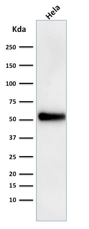
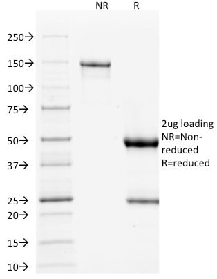
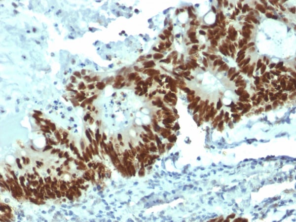
 Application Data
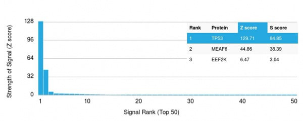
(Analysis of Protein Array containing more than 19,000 full-length human proteins using p53 Mouse Monoclonal Antibody (DO-1) Z- and S- Score: The Z-score represents the strength of a signal that a monoclonal antibody (MAb) (in combination with a fluorescently-tagged anti-IgG secondary antibody) produces when binding to a particular protein on the HuProtTM array. Z-scores are described in units of standard deviations (SD's) above the mean value of all signals generated on that array. If targets on HuProtTM are arranged in descending order of the Z-score, the S-score is the difference (also in units of SD's) between the Z-score. S-score therefore represents the relative target specificity of a MAb to its intended target. A MAb is considered to specific to its intended target, if the MAb has an S-score of at least 2.5. For example, if a MAb binds to protein X with a Z-score of 43 and to protein Y with a Z-score of 14, then the S-score for the binding of that MAb to protein X is equal to 29.)
Application Data
(Analysis of Protein Array containing more than 19,000 full-length human proteins using p53 Mouse Monoclonal Antibody (DO-1) Z- and S- Score: The Z-score represents the strength of a signal that a monoclonal antibody (MAb) (in combination with a fluorescently-tagged anti-IgG secondary antibody) produces when binding to a particular protein on the HuProtTM array. Z-scores are described in units of standard deviations (SD's) above the mean value of all signals generated on that array. If targets on HuProtTM are arranged in descending order of the Z-score, the S-score is the difference (also in units of SD's) between the Z-score. S-score therefore represents the relative target specificity of a MAb to its intended target. A MAb is considered to specific to its intended target, if the MAb has an S-score of at least 2.5. For example, if a MAb binds to protein X with a Z-score of 43 and to protein Y with a Z-score of 14, then the S-score for the binding of that MAb to protein X is equal to 29.)
p53 Tumor Suppressor Protein, Monoclonal Antibody (Cat# AAA215231)
Full Name
p53 Tumor Suppressor Protein
Gene Names
TP53; P53; BCC7; LFS1; BMFS5; TRP53
Reactivity
Human
Does not react with Monkey or Rat.
Does not react with Monkey or Rat.
Applications
Immunohistochemistry, Western Blot, Immunofluorescence, Flow Cytometry
Purity
Purified Ab with BSA and Azide at 200ug/ml OR Purified Ab WITHOUT BSA and Azide at 1.0mg/ml
Pricing
 Bioactivity
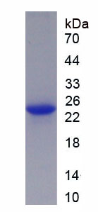
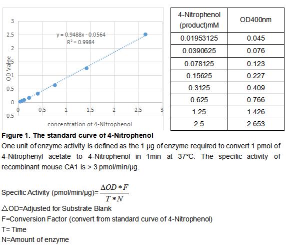
(Carbonic Anhydrase (CA) catalyzes the reversible reaction of CO2 H2O = HCO3- H, which is fundamental to many processes such as respiration, renal tubular acidification and bone resorption. CA1 is a cytosolic enzyme with the highest levels in erythrocytes and is a very early marker for erythroid differentiation. The activity of recombinant mouse CA1 was measured by its ability to hydrolyze 4-Nitrophenyl acetate (4-NPA) to 4-Nitrophenol. The reaction was performed in 12.5 mM Tris, 75 mM NaCl, pH 7.5 (assay buffer), initiated by addition 50 uL of various concentrations of CA1 (diluted by assay buffer) to 50 uL of 2 mM substrate 4-NPA (100 mM stock in Acetone, diluted by assay buffer). Incubated at 37 degree C for 5min, then read at a wavelength of 400 nm.)
Bioactivity
(Carbonic Anhydrase (CA) catalyzes the reversible reaction of CO2 H2O = HCO3- H, which is fundamental to many processes such as respiration, renal tubular acidification and bone resorption. CA1 is a cytosolic enzyme with the highest levels in erythrocytes and is a very early marker for erythroid differentiation. The activity of recombinant mouse CA1 was measured by its ability to hydrolyze 4-Nitrophenyl acetate (4-NPA) to 4-Nitrophenol. The reaction was performed in 12.5 mM Tris, 75 mM NaCl, pH 7.5 (assay buffer), initiated by addition 50 uL of various concentrations of CA1 (diluted by assay buffer) to 50 uL of 2 mM substrate 4-NPA (100 mM stock in Acetone, diluted by assay buffer). Incubated at 37 degree C for 5min, then read at a wavelength of 400 nm.)
Carbonic Anhydrase I (CA1), Active Protein (Cat# AAA161797)
Full Name
Active Carbonic Anhydrase I (CA1)
Gene Names
Car1; Ca1; Car-1; AW555628
Applications
Activity Assay, Cell Culture
Purity
Greater than 95% by SDS-PAGE
Pricing
 Application Data
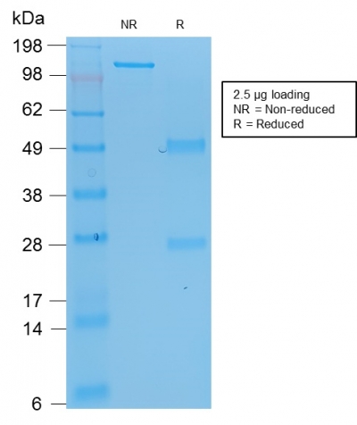
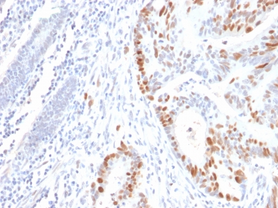
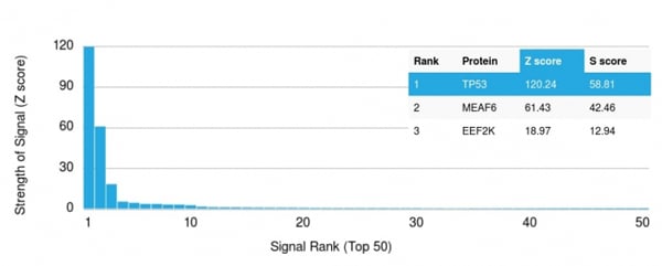
(Analysis of Protein Array containing >19, 000 full-length human proteins usingp53 Mouse Recombinant Monoclonal Antibody (rBP53-12)Z- and S- Score: The Z-score represents the strength of a signal that a monoclonal antibody (MAb) (in combination with a fluorescently-tagged anti-IgG secondary antibody) produces when binding to a particular protein on the HuProtTM array. Z-scores are described in units of standard deviations (SD's) above the mean value of all signals generated on that array. If targets on HuProtTM are arranged in descending order of the Z-score, the S-score is the difference (also in units of SD's) between the Z-score. S-score therefore represents the relative target specificity of a MAb to its intended target. A MAb is considered to specific to its intended target, if the MAb has an S-score of at least 2.5. For example, if a MAb binds to protein X with a Z-score of 43 and to protein Y with a Z-score of 14, then the S-score for the binding of that MAb to protein X is equal to 29.)
Application Data
(Analysis of Protein Array containing >19, 000 full-length human proteins usingp53 Mouse Recombinant Monoclonal Antibody (rBP53-12)Z- and S- Score: The Z-score represents the strength of a signal that a monoclonal antibody (MAb) (in combination with a fluorescently-tagged anti-IgG secondary antibody) produces when binding to a particular protein on the HuProtTM array. Z-scores are described in units of standard deviations (SD's) above the mean value of all signals generated on that array. If targets on HuProtTM are arranged in descending order of the Z-score, the S-score is the difference (also in units of SD's) between the Z-score. S-score therefore represents the relative target specificity of a MAb to its intended target. A MAb is considered to specific to its intended target, if the MAb has an S-score of at least 2.5. For example, if a MAb binds to protein X with a Z-score of 43 and to protein Y with a Z-score of 14, then the S-score for the binding of that MAb to protein X is equal to 29.)
p53 Tumor Suppressor Protein, Monoclonal Antibody (Cat# AAA214542)
Full Name
p53 Tumor Suppressor Protein
Reactivity
Human, Monkey, Dog, Hamster, Chicken.
Does not react with Mouse and Rat. Others not known
Does not react with Mouse and Rat. Others not known
Applications
Immunohistochemistry, Western Blot, Immunofluorescence, Flow Cytometry
Pricing
 Bioactivity
(Serpin B2, also known as PAI-2, is an approximately 60 kDa serine protease inhibitor. It is primarily secreted by macrophages and monocytes and can form disulfide-linked multimers. Serpin B2 inhibits both the urokinase-type and tissue-type plasminogen activators (uPA and tPA). Serpin B2 also promotes the clearance of uPA by enhancing its binding and uptake by LRP. It limits fibril formation by Huntington protein (HTT) and beta-Amyloid peptides. It promotes Th2 biased immune responses and is important for intestinal CCL2 production, monocyte recruitment, and nematode clearance. A non-glycosylated form of Serpin B2 is retained intracellularly where it interferes with TNF-a induced apoptosis by protecting the Retinoblastoma protein (RB1) from calpain digestion. It also inhibits proteasome activity in activated endothelial cells. The activity of recombinant human PAI-2 was measured by its ability to inhibit uPA cleavage of a peptide substrate, N-carbobenzyloxy-Gly-Gly-Arg-7-amido-4-methylcoumarin (Z-GGR-AMC) in the assay buffer 50 mM Tris, 0.01% Tween 20, pH 8.5. The 50 ul different concentrations of rhPAI-2 (MW: 48.2 KD) was incubated with 50ul 2ug/ml rhuPA (EPA140Mu61) at room temperature for 15 minutes. Loading 50 uL of the incubated mixtures into empty wells of a plate, and start the reaction by adding 50 uL of 200 uM substrate (Z-GGR-AMC). Include a substrate blank containing 50 uL of assay buffer and 50 uL of 200 uM substrate. Then read at excitiation and emission wavelengths of 380 nm and 460 nm, respectively, in kinetic mode for 5 minutes. The result was shown in Figure 1 and it was obvious that recombinant human PAI2 significantly decreased uPA activity. The inhibition IC50 was )
Bioactivity
(Serpin B2, also known as PAI-2, is an approximately 60 kDa serine protease inhibitor. It is primarily secreted by macrophages and monocytes and can form disulfide-linked multimers. Serpin B2 inhibits both the urokinase-type and tissue-type plasminogen activators (uPA and tPA). Serpin B2 also promotes the clearance of uPA by enhancing its binding and uptake by LRP. It limits fibril formation by Huntington protein (HTT) and beta-Amyloid peptides. It promotes Th2 biased immune responses and is important for intestinal CCL2 production, monocyte recruitment, and nematode clearance. A non-glycosylated form of Serpin B2 is retained intracellularly where it interferes with TNF-a induced apoptosis by protecting the Retinoblastoma protein (RB1) from calpain digestion. It also inhibits proteasome activity in activated endothelial cells. The activity of recombinant human PAI-2 was measured by its ability to inhibit uPA cleavage of a peptide substrate, N-carbobenzyloxy-Gly-Gly-Arg-7-amido-4-methylcoumarin (Z-GGR-AMC) in the assay buffer 50 mM Tris, 0.01% Tween 20, pH 8.5. The 50 ul different concentrations of rhPAI-2 (MW: 48.2 KD) was incubated with 50ul 2ug/ml rhuPA (EPA140Mu61) at room temperature for 15 minutes. Loading 50 uL of the incubated mixtures into empty wells of a plate, and start the reaction by adding 50 uL of 200 uM substrate (Z-GGR-AMC). Include a substrate blank containing 50 uL of assay buffer and 50 uL of 200 uM substrate. Then read at excitiation and emission wavelengths of 380 nm and 460 nm, respectively, in kinetic mode for 5 minutes. The result was shown in Figure 1 and it was obvious that recombinant human PAI2 significantly decreased uPA activity. The inhibition IC50 was )
Plasminogen Activator Inhibitor 2 (PAI2), Active Protein (Cat# AAA161739)
Full Name
Active Plasminogen Activator Inhibitor 2 (PAI2)
Gene Names
SERPINB2; PAI; PAI2; PAI-2; PLANH2; HsT1201
Applications
Activity Assay, Cell Culture
Purity
Greater than 80% by SDS-PAGE
Pricing

 FCM/FACS (Flow Cytometry)
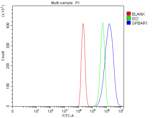
(Figure 2. Flow Cytometry analysis of THP-1 cells using anti-GPCR TGR5/GPBAR1 antibody (AAA126191).Overlay histogram showing THP-1 cells stained with AAA126191 (Blue line). The cells were blocked with 10% normal goat serum. And then incubated with rabbit anti-GPCR TGR5/GPBAR1 Antibody (AAA126191, 1 ug/1x10^6 cells) for 30 min at 20 degree C. DyLight488 conjugated goat anti-rabbit IgG was used as secondary antibody for 30 minutes at 20 degree C. Isotype control antibody (Green line) was rabbit IgG (1 ug/1x10^6) used under the same conditions. Unlabelled sample (Red line) was also used as a control.)
FCM/FACS (Flow Cytometry)
(Figure 2. Flow Cytometry analysis of THP-1 cells using anti-GPCR TGR5/GPBAR1 antibody (AAA126191).Overlay histogram showing THP-1 cells stained with AAA126191 (Blue line). The cells were blocked with 10% normal goat serum. And then incubated with rabbit anti-GPCR TGR5/GPBAR1 Antibody (AAA126191, 1 ug/1x10^6 cells) for 30 min at 20 degree C. DyLight488 conjugated goat anti-rabbit IgG was used as secondary antibody for 30 minutes at 20 degree C. Isotype control antibody (Green line) was rabbit IgG (1 ug/1x10^6) used under the same conditions. Unlabelled sample (Red line) was also used as a control.)
GPCR TGR5/GPBAR1, Polyclonal Antibody (Cat# AAA126191)
Full Name
Anti-GPCR TGR5/GPBAR1 Antibody Picoband
Gene Names
GPBAR1; BG37; TGR5; M-BAR; GPCR19; GPR131
Reactivity
Human, Mouse, Rat
Applications
Flow Cytometry, Western Blot
Purity
Immunogen affinity purified.
Pricing
 FCM (Flow Cytometry)
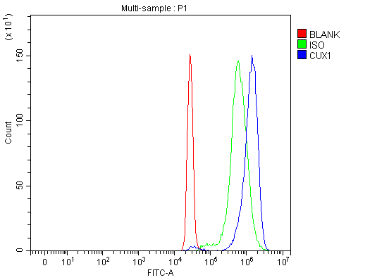
(Figure 6. Flow Cytometry analysis of HepG2 cells using anti-CUX1 antibody (AAA19427).Overlay histogram showing HepG2 cells stained with AAA19427 (Blue line). The cells were blocked with 10% normal goat serum. And then incubated with rabbit anti-CUX1 Antibody (AAA19427, 1 ug/1x10^6 cells) for 30 min at 20 degree C. DyLight488 conjugated goat anti-rabbit IgG was used as secondary antibody for 30 minutes at 20 degree C. Isotype control antibody (Green line) was rabbit IgG (1 ug/1x10^6) used under the same conditions. Unlabelled sample (Red line) was also used as a control.)
FCM (Flow Cytometry)
(Figure 6. Flow Cytometry analysis of HepG2 cells using anti-CUX1 antibody (AAA19427).Overlay histogram showing HepG2 cells stained with AAA19427 (Blue line). The cells were blocked with 10% normal goat serum. And then incubated with rabbit anti-CUX1 Antibody (AAA19427, 1 ug/1x10^6 cells) for 30 min at 20 degree C. DyLight488 conjugated goat anti-rabbit IgG was used as secondary antibody for 30 minutes at 20 degree C. Isotype control antibody (Green line) was rabbit IgG (1 ug/1x10^6) used under the same conditions. Unlabelled sample (Red line) was also used as a control.)
CUX1, Polyclonal Antibody (Cat# AAA19427)
Full Name
Anti-CUX1 Antibody Picoband
Gene Names
CUX1; CDP; CUX; p75; CASP; CDP1; COY1; Clox; p100; p110; p200; CUTL1; GOLIM6; CDP/Cut; Cux/CDP; Nbla10317
Reactivity
Human, Mouse
Applications
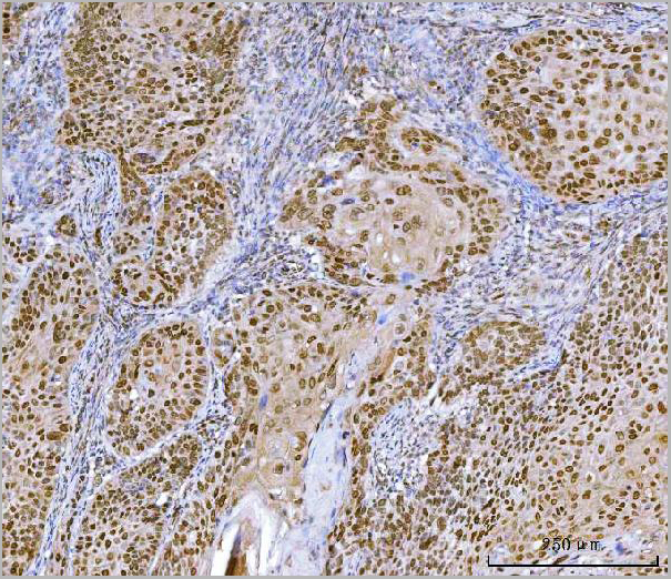
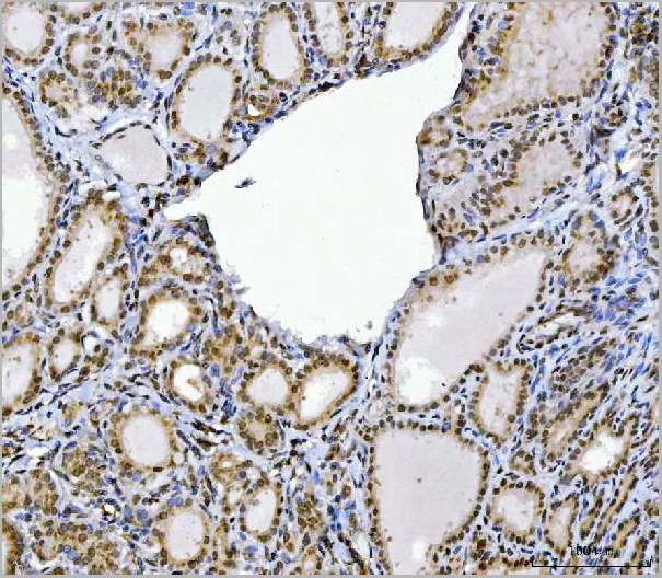
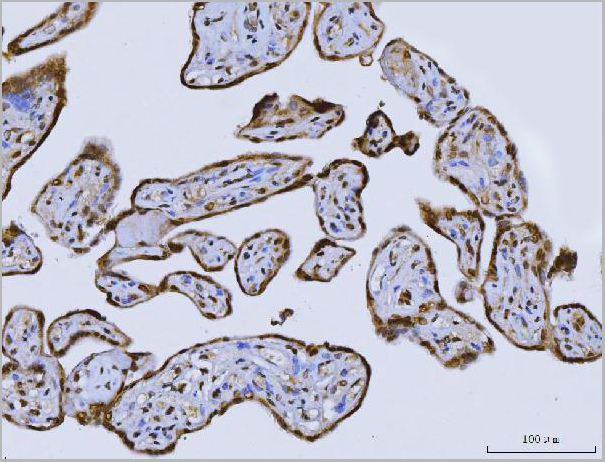
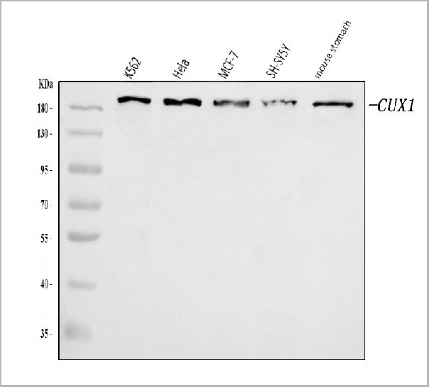
Flow Cytometry, Immunohistochemistry, Western Blot
Purity
Immunogen affinity purified.
Pricing
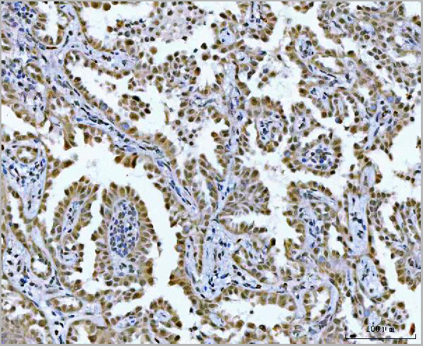
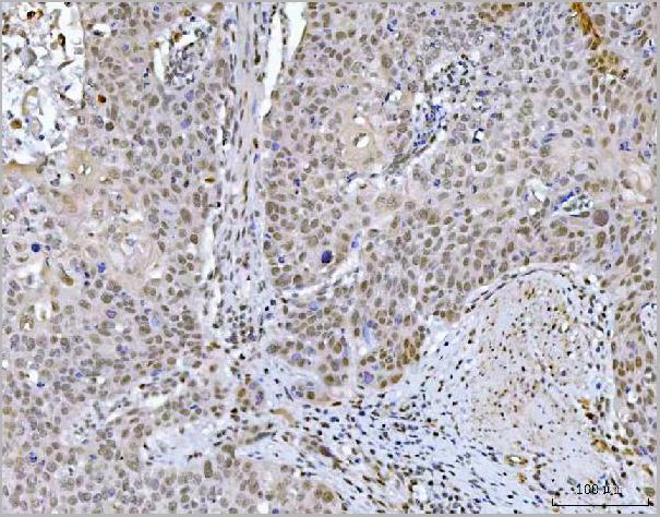
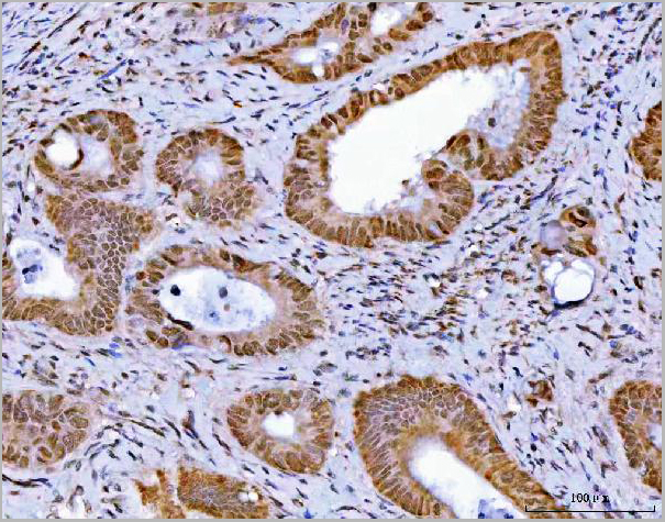
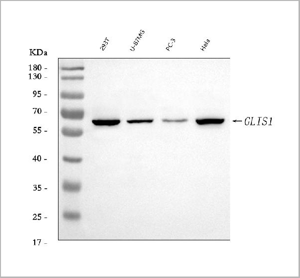
 FCM (Flow Cytometry)
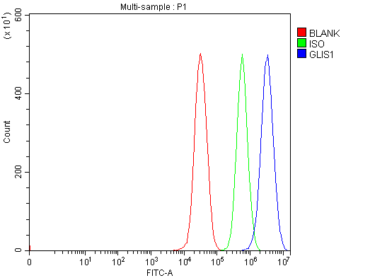
(Figure 8. Flow Cytometry analysis of 293T cells using anti-GLIS1 antibody (AAA19630).Overlay histogram showing 293T cells stained with AAA19630 (Blue line). The cells were blocked with 10% normal goat serum. And then incubated with rabbit anti-GLIS1 Antibody (AAA19630, 1 ug/1x10^6 cells) for 30 min at 20 degree C. DyLight488 conjugated goat anti-rabbit IgG was used as secondary antibody for 30 minutes at 20 degree C. Isotype control antibody (Green line) was rabbit IgG (1 ug/1x10^6) used under the same conditions. Unlabelled sample (Red line) was also used as a control.)
FCM (Flow Cytometry)
(Figure 8. Flow Cytometry analysis of 293T cells using anti-GLIS1 antibody (AAA19630).Overlay histogram showing 293T cells stained with AAA19630 (Blue line). The cells were blocked with 10% normal goat serum. And then incubated with rabbit anti-GLIS1 Antibody (AAA19630, 1 ug/1x10^6 cells) for 30 min at 20 degree C. DyLight488 conjugated goat anti-rabbit IgG was used as secondary antibody for 30 minutes at 20 degree C. Isotype control antibody (Green line) was rabbit IgG (1 ug/1x10^6) used under the same conditions. Unlabelled sample (Red line) was also used as a control.)
GLIS1, Polyclonal Antibody (Cat# AAA19630)
Full Name
Anti-GLIS1 Antibody Picoband
Reactivity
Human
Applications
Flow Cytometry, Immunofluorescence, Immunohistochemistry, Immunocytochemistry, Western Blot
Purity
Immunogen affinity purified.
Pricing
 Bioactivity
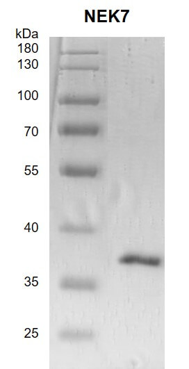
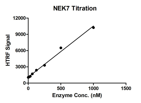
(HTRF assay for NEK7 activity 1 uM STK S3 substrate was incubated with different concentrations of NEK7 protein in a 10 ul reaction system containing 1×Enzymatic Buffer, 5 mM MgCl2, 1 mM DTT, 100 uM ATP for 1 hour. The 10 ul detection reagents containing anti-STK antibody (1:2) and SA-XL665 (1:100) diluted with 1× Detection Buffer were added and incubated with the reactions for 30 min. All the operations and reactions were performed at room temperature. HTRF assay was used for detection.)
Bioactivity
(HTRF assay for NEK7 activity 1 uM STK S3 substrate was incubated with different concentrations of NEK7 protein in a 10 ul reaction system containing 1×Enzymatic Buffer, 5 mM MgCl2, 1 mM DTT, 100 uM ATP for 1 hour. The 10 ul detection reagents containing anti-STK antibody (1:2) and SA-XL665 (1:100) diluted with 1× Detection Buffer were added and incubated with the reactions for 30 min. All the operations and reactions were performed at room temperature. HTRF assay was used for detection.)
 ELISA
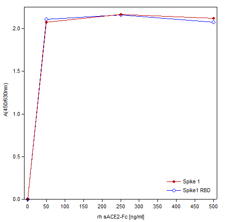
(Binding of sACE2-Fc to recombinant SARS-CoV2 Spike1 proteins in a functional ELISA. Recombinant SARS-CoV2 Spike1 and Spike1 RBD were coated with 1ug/ml in PBS. The sACE2-Fc [Cat# SFC-002] was added in increasing concentrations.)
ELISA
(Binding of sACE2-Fc to recombinant SARS-CoV2 Spike1 proteins in a functional ELISA. Recombinant SARS-CoV2 Spike1 and Spike1 RBD were coated with 1ug/ml in PBS. The sACE2-Fc [Cat# SFC-002] was added in increasing concentrations.)
COVID 19 Spike -1 RBD Coronavirus, Recombinant Protein (Cat# AAA79331)
Full Name
SARS-CoV-2 Spike-1 RBD
Purity
> 98% by SDS-PAGE & Coomassie
Pricing
 SDS-PAGE
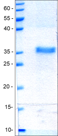
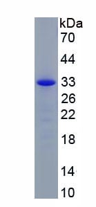
(Figure. SDS-PAGE)
SDS-PAGE
(Figure. SDS-PAGE)
Interleukin 4 Induced Protein 1 (IL4I1), Recombinant Protein (Cat# AAA150750)
Full Name
Recombinant Interleukin 4 Induced Protein 1 (IL4I1)
Gene Names
IL4I1; LAO; FIG1; LAAO
Applications
Western Blot
Purity
>95%
Pricing
 Application Data
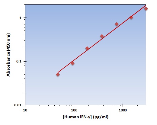
(The OmniKine Human IFN-gamma ELISA Kit allows for the detection and quantification of endogenous levels of natural and/or recombinant Human IFN-gamma proteins within the range of 46-3000 pg/ml.)
Application Data
(The OmniKine Human IFN-gamma ELISA Kit allows for the detection and quantification of endogenous levels of natural and/or recombinant Human IFN-gamma proteins within the range of 46-3000 pg/ml.)
IFN-gamma, ELISA Kit (Cat# AAA315615)
Full Name
IFN-gamma (Human) OmniKine ELISA Kit
Gene Names
IFNG; IFG; IFI
Reactivity
Human
Pricing

 SDS-PAGE
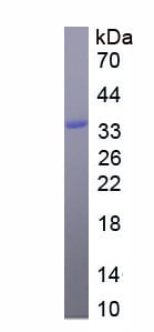
(SDS-PAGE)
SDS-PAGE
(SDS-PAGE)
Erythropoietin (EPO), Recombinant Protein (Cat# AAA152040)
Full Name
Eukaryotic Erythropoietin (EPO)
Applications
Western Blot
Purity
Greater than 90% by SDS-PAGE
Pricing
 IP (Immunoprecipitation)
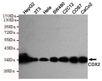
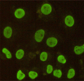
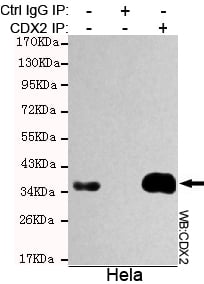
(Immunoprecipitation analysis of Hela cell lysate using CDX2 antibody.)
IP (Immunoprecipitation)
(Immunoprecipitation analysis of Hela cell lysate using CDX2 antibody.)
CDX2, Monoclonal Antibody (Cat# AAA300757)
Full Name
CDX2 Monoclonal Antibody
Gene Names
CDX2; CDX3; CDX-3; CDX2/AS
Reactivity
Human, Monkey, Mouse
Applications
Immunoprecipitation, Immunocytochemistry, Western Blot
Purity
Affinity purified
Pricing
 WB (Western Blot)
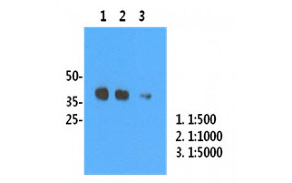
(H1N1/HA1 recombinant protein (50 ng) was resolved by SDS-PAGE, transferred to PVDF membrane and probed with H1N1/HA1 antibody (1:500, 1:1000, 1:5000). Proteins were visualized using a goat anti-mouse secondary antibody conjugated to HRP and an ECL detection system.)
WB (Western Blot)
(H1N1/HA1 recombinant protein (50 ng) was resolved by SDS-PAGE, transferred to PVDF membrane and probed with H1N1/HA1 antibody (1:500, 1:1000, 1:5000). Proteins were visualized using a goat anti-mouse secondary antibody conjugated to HRP and an ECL detection system.)
Influenza A H1N1 HA1, Monoclonal Antibody (Cat# AAA106743)
Full Name
Influenza A H1N1 HA1 antibody
Purity
Influenza A HA1 antibody was purified by protein G affinity chromatography
Pricing
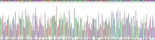
 Amino Acid Info
(Amino Acid info)
Amino Acid Info
(Amino Acid info)
Transforming Growth Factor Beta 2 (TGFb2), Recombinant Protein (Cat# AAA153306)
Full Name
Recombinant Transforming Growth Factor Beta 2 (TGFb2)
Gene Names
Tgfb2; TGF-B2
Applications
Western Blot
Purity
Greater than 90% by SDS-PAGE
Pricing
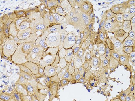
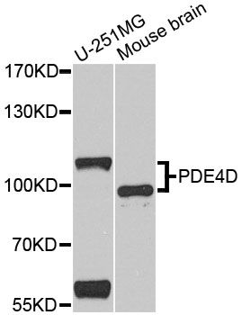
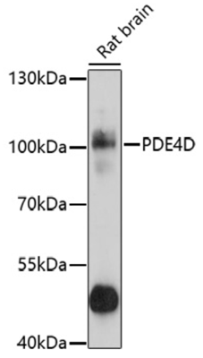 WB (Western Blot)
(Western blot analysis of extracts of rat brain, using PDE4D antibody (AAA37279) at 1:1000 dilution.Secondary antibody: HRP Goat Anti-Rabbit IgG (H+L) at 1:10000 dilution.Lysates/proteins: 25ug per lane.Blocking buffer: 3% nonfat dry milk in TBST.Detection: ECL Basic Kit (RM00020).Exposure time: 5s.)
WB (Western Blot)
(Western blot analysis of extracts of rat brain, using PDE4D antibody (AAA37279) at 1:1000 dilution.Secondary antibody: HRP Goat Anti-Rabbit IgG (H+L) at 1:10000 dilution.Lysates/proteins: 25ug per lane.Blocking buffer: 3% nonfat dry milk in TBST.Detection: ECL Basic Kit (RM00020).Exposure time: 5s.)
PDE4D, Polyclonal Antibody (Cat# AAA37279)
Full Name
PDE4D Polyclonal Antibody
Gene Names
PDE4D; DPDE3; PDE43; STRK1; ACRDYS2; HSPDE4D; PDE4DN2
Applications
Immunohistochemistry, Western Blot
Purity
Affinity Purification
Pricing
 IHC (Immunohiostchemistry)
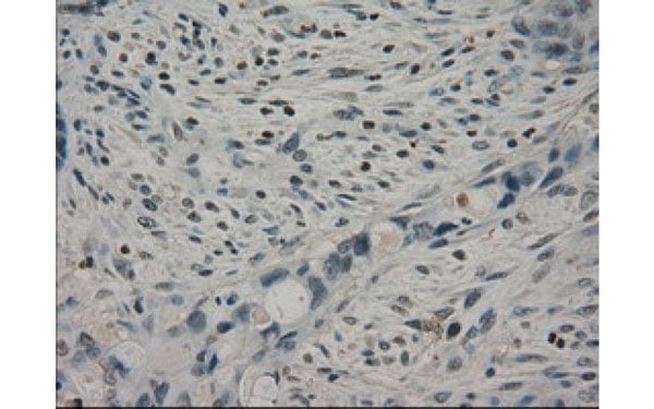
(Immunohistochemical analysis of CRYAB protein in paraffin embedded Adenocarcinoma of Human colon tissue using CRYAB antibody)
IHC (Immunohiostchemistry)
(Immunohistochemical analysis of CRYAB protein in paraffin embedded Adenocarcinoma of Human colon tissue using CRYAB antibody)
CRYAB, Monoclonal Antibody (Cat# AAA106324)
Full Name
CRYAB antibody
Applications
Western Blot, Immunohistochemistry
Purity
CRYAB antibody was purified by affinity chromatography.
Pricing
 Amino Acid Info
Eukaryotic Vascular Endothelial Growth Factor C (VEGFC), Protein (Cat# AAA153161)
Full Name
Eukaryotic Vascular Endothelial Growth Factor C (VEGFC)
Applications
Western Blot
Purity
Greater than 90% by SDS-PAGE
Pricing
 SDS-PAGE
(SDS-PAGE)
SDS-PAGE
(SDS-PAGE)
Tumor Necrosis Factor Related Apoptosis Inducing Ligand (TRAIL), Recombinant Protein (Cat# AAA152099)
Full Name
Recombinant Tumor Necrosis Factor Related Apoptosis Inducing Ligand (TRAIL)
Gene Names
TNFSF10; TL2; APO2L; CD253; TRAIL; Apo-2L
Applications
Western Blot
Purity
Greater than 95% by SDS-PAGE
Pricing
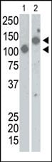 WB (Western Blot)
(The anti-SARS-Sn Pab is used in Western blot to detect recombinant Spike proteins, aa17-537 (Lane 1) and aa17-756 (Lane 2).)
WB (Western Blot)
(The anti-SARS-Sn Pab is used in Western blot to detect recombinant Spike proteins, aa17-537 (Lane 1) and aa17-756 (Lane 2).)
SARS virus Sn, Polyclonal Antibody (Cat# AAA285264)
Full Name
SARS virus Sn Antibody
Applications
Western Blot
Purity
Purified Rabbit Polyclonal Antibody (Pab)
Pricing
 IHC (Immunohistochemistry)
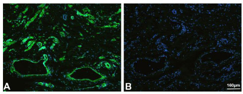
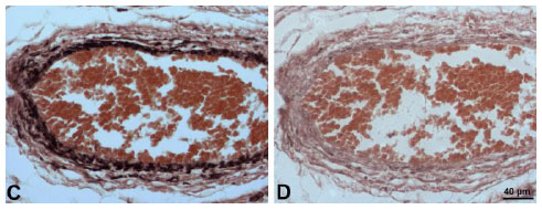
(Fig. 4: Immunoperoxidase staining of paraffin-embedded sections of human foreskin with mouse anti-human sFlt1-14 antibody. (D) Control; Fixation: 4% PFA (app. 25min); Dilution: 1:100-500. The staining in smooth muscle cells is visible.)
IHC (Immunohistochemistry)
(Fig. 4: Immunoperoxidase staining of paraffin-embedded sections of human foreskin with mouse anti-human sFlt1-14 antibody. (D) Control; Fixation: 4% PFA (app. 25min); Dilution: 1:100-500. The staining in smooth muscle cells is visible.)
sFlt1-14, Monoclonal Antibody (Cat# AAA79326)
Full Name
Anti-human sFlt1-14
Reactivity
Human
Applications
Immunohistochemistry, Immunofluorescence, Western Blot
Purity
Protein G purified
Pricing
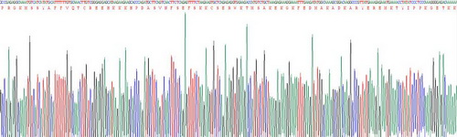 Application Data
High Mobility Group Protein 1 (HMG1), Active Protein (Cat# AAA148152)
Full Name
Active High Mobility Group Protein 1 (HMG1)
Gene Names
HMGB1; HMG1; HMG3; HMG-1; SBP-1
Reactivity
Human
Applications
Activity Assay, Cell Culture
Purity
> 95%
Pricing
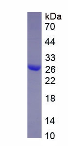 SDS-PAGE
Heat Shock 27kDa Protein 1 (HSPB1), Recombinant Protein (Cat# AAA134592)
Full Name
Recombinant Heat Shock 27kDa Protein 1 (HSPB1)
Gene Names
Hspb1; 27kDa; Hsp25
Applications
Western Blot
Purity
> 95%
Pricing
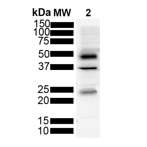
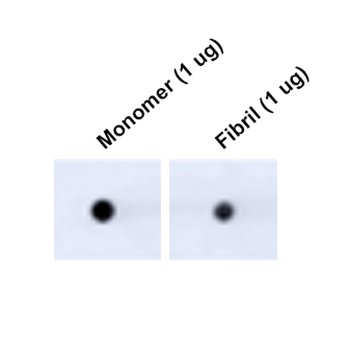 DB (Dot Blot)
(Dot Blot analysis using Mouse Anti-Tau Monoclonal Antibody, Clone 3D4 (SMC-608). Tissue: Recombinant Protein. Species: Human. Primary Antibody: Mouse Anti-Tau Monoclonal Antibody (SMC-608) at 1:1000 for 2 hours at RT with shaking. Secondary Antibody: Goat anti-mouse IgG:HRP at 1:5000 for 1 hour at RT with shaking.)
DB (Dot Blot)
(Dot Blot analysis using Mouse Anti-Tau Monoclonal Antibody, Clone 3D4 (SMC-608). Tissue: Recombinant Protein. Species: Human. Primary Antibody: Mouse Anti-Tau Monoclonal Antibody (SMC-608) at 1:1000 for 2 hours at RT with shaking. Secondary Antibody: Goat anti-mouse IgG:HRP at 1:5000 for 1 hour at RT with shaking.)
Tau, Monoclonal Antibody (Cat# AAA253989)
Full Name
Tau Antibody, Clone 3D4
Gene Names
MAPT; TAU; MSTD; PPND; DDPAC; MAPTL; MTBT1; MTBT2; FTDP-17; PPP1R103
Reactivity
Human, Mouse, Rat
Applications
Immunofluorescence, Immunocytochemistry, Dot Blot, Western Blot
Purity
Protein G Purified
Pricing
 Bioactivity
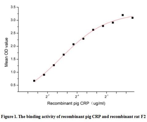
(Reactive protein (CRP) is an annular (ring-shaped), pentameric protein found in blood plasma, whose levels rise in response to inflammation. It is an acute-phase protein of hepatic origin that increases following interleukin-6 secretion by macrophages and T cells. Its physiological role is to bind to lysophosphatidylcholine expressed on the surface of dead or dying cells (and some types of bacteria) in order to activate the complement system via C1q. Besides, Coagulation Factor II (F2) has been identified as an interactor of CRP, thus a functional binding ELISA assay was conducted to detect the interaction of recombinant pig CRP and recombinant rat F2. Briefly, CRP was diluted serially in PBS with 0.01% BSA (pH 7.4). Duplicate samples of 100 ul were then transferred to F2-coated microtiter wells and incubated for 1h at 37 degree C. Wells were washed with PBST and incubated for 1h with anti-CRP pAb, then aspirated and washed 3 times. After incubation with HRP labelled secondary antibody for 1h at 37 degree C, wells were aspirated and washed 5 times. With the addition of substrate solution, wells were incubated 15-25 minutes at 37 degree C. Finally, add 50 uL stop solution to the wells and read at 450/630 nm immediately. The binding activity of recombinant pig CRP and recombinant rat F2 was shown in Figure 1, the EC50 for this effect is 0.02 ug/mL.)
Bioactivity
(Reactive protein (CRP) is an annular (ring-shaped), pentameric protein found in blood plasma, whose levels rise in response to inflammation. It is an acute-phase protein of hepatic origin that increases following interleukin-6 secretion by macrophages and T cells. Its physiological role is to bind to lysophosphatidylcholine expressed on the surface of dead or dying cells (and some types of bacteria) in order to activate the complement system via C1q. Besides, Coagulation Factor II (F2) has been identified as an interactor of CRP, thus a functional binding ELISA assay was conducted to detect the interaction of recombinant pig CRP and recombinant rat F2. Briefly, CRP was diluted serially in PBS with 0.01% BSA (pH 7.4). Duplicate samples of 100 ul were then transferred to F2-coated microtiter wells and incubated for 1h at 37 degree C. Wells were washed with PBST and incubated for 1h with anti-CRP pAb, then aspirated and washed 3 times. After incubation with HRP labelled secondary antibody for 1h at 37 degree C, wells were aspirated and washed 5 times. With the addition of substrate solution, wells were incubated 15-25 minutes at 37 degree C. Finally, add 50 uL stop solution to the wells and read at 450/630 nm immediately. The binding activity of recombinant pig CRP and recombinant rat F2 was shown in Figure 1, the EC50 for this effect is 0.02 ug/mL.)
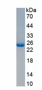
C ReProtein (CRP), Active Protein (Cat# AAA161784)
Full Name
Active C Reactive Protein (CRP)
Applications
Activity Assay, Cell Culture
Purity
Greater than 97% by SDS-PAGE
Pricing
 Bioactivity
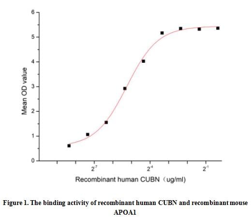
(Cubilin (CUBN) is a 460 kDa intrinsic factor-vitamin B12 receptor protein and it is expressed in kidney and small intestine. CUBN is a component of a multiligand endocytic receptor complex, which plays a role in lipoprotein, vitamin and iron metabolism by facilitating their uptake. Besides CUBN serves as important transporter in several absorptive epithelia, including intestine, renal proximal tubules and embryonic yolk sac and it plays an important role in the development of the peri-implantation embryo through internalization of APOA1 and cholesterol. Thus a functional binding ELISA assay was conducted to detect the interaction of recombinant human CUBN and recombinant mouse APOA1. Briefly, CUBN was diluted serially in PBS with 0.01% BSA (pH 7.4). Duplicate samples of 100 ul were then transferred to APOA1-coated microtiter wells and incubated for 1h at 37 degree C. Wells were washed with PBST and incubated for 1h with anti-CUBN pAb, then aspirated and washed 3 times. After incubation with HRP labelled secondary antibody for 1h at 37 degree C, wells were aspirated and washed 5 times. With the addition of substrate solution, wells were incubated 15-25 minutes at 37 degree C. Finally, add 50 uL stop solution to the wells and read at 450/630 nm immediately. The binding activity of recombinant human CUBN and recombinant mouse APOA1 was shown in Figure 1, the EC50 for this effect is 0.03 ug/mL.)
Bioactivity
(Cubilin (CUBN) is a 460 kDa intrinsic factor-vitamin B12 receptor protein and it is expressed in kidney and small intestine. CUBN is a component of a multiligand endocytic receptor complex, which plays a role in lipoprotein, vitamin and iron metabolism by facilitating their uptake. Besides CUBN serves as important transporter in several absorptive epithelia, including intestine, renal proximal tubules and embryonic yolk sac and it plays an important role in the development of the peri-implantation embryo through internalization of APOA1 and cholesterol. Thus a functional binding ELISA assay was conducted to detect the interaction of recombinant human CUBN and recombinant mouse APOA1. Briefly, CUBN was diluted serially in PBS with 0.01% BSA (pH 7.4). Duplicate samples of 100 ul were then transferred to APOA1-coated microtiter wells and incubated for 1h at 37 degree C. Wells were washed with PBST and incubated for 1h with anti-CUBN pAb, then aspirated and washed 3 times. After incubation with HRP labelled secondary antibody for 1h at 37 degree C, wells were aspirated and washed 5 times. With the addition of substrate solution, wells were incubated 15-25 minutes at 37 degree C. Finally, add 50 uL stop solution to the wells and read at 450/630 nm immediately. The binding activity of recombinant human CUBN and recombinant mouse APOA1 was shown in Figure 1, the EC50 for this effect is 0.03 ug/mL.)
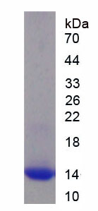
Cubilin (CUBN), Active Protein (Cat# AAA161888)
Full Name
Active Cubilin (CUBN)
Gene Names
CUBN; IFCR; MGA1; gp280
Applications
Activity Assay, Cell Culture
Purity
Greater than 95% by SDS-PAGE
Pricing

 Bioactivity
(Reactive protein (CRP) is an annular (ring-shaped), pentameric protein found in blood plasma, whose levels rise in response to inflammation. It is an acute-phase protein of hepatic origin that increases following interleukin-6 secretion by macrophages and T cells. Its physiological role is to bind to lysophosphatidylcholine expressed on the surface of dead or dying cells (and some types of bacteria) in order to activate the complement system via C1q. Besides, Coagulation Factor II (F2) has been identified as an interactor of CRP, thus a functional binding ELISA assay was conducted to detect the interaction of recombinant chicken CRP and recombinant rat F2. Briefly, CRP was diluted serially in PBS with 0.01% BSA (pH 7.4). Duplicate samples of 100 ul were then transferred to F2-coated microtiter wells and incubated for 1h at 37 degree C. Wells were washed with PBST and incubated for 1h with anti-CRP pAb, then aspirated and washed 3 times. After incubation with HRP labelled secondary antibody for 1h at 37 degree C, wells were aspirated and washed 5 times. With the addition of substrate solution, wells were incubated 15-25 minutes at 37 degree C. Finally, add 50 uL stop solution to the wells and read at 450/630 nm immediately. The binding activity of recombinant chicken CRP and recombinant rat F2 was shown in Figure 1, the EC50 for this effect is 0.82 ug/mL.)
Bioactivity
(Reactive protein (CRP) is an annular (ring-shaped), pentameric protein found in blood plasma, whose levels rise in response to inflammation. It is an acute-phase protein of hepatic origin that increases following interleukin-6 secretion by macrophages and T cells. Its physiological role is to bind to lysophosphatidylcholine expressed on the surface of dead or dying cells (and some types of bacteria) in order to activate the complement system via C1q. Besides, Coagulation Factor II (F2) has been identified as an interactor of CRP, thus a functional binding ELISA assay was conducted to detect the interaction of recombinant chicken CRP and recombinant rat F2. Briefly, CRP was diluted serially in PBS with 0.01% BSA (pH 7.4). Duplicate samples of 100 ul were then transferred to F2-coated microtiter wells and incubated for 1h at 37 degree C. Wells were washed with PBST and incubated for 1h with anti-CRP pAb, then aspirated and washed 3 times. After incubation with HRP labelled secondary antibody for 1h at 37 degree C, wells were aspirated and washed 5 times. With the addition of substrate solution, wells were incubated 15-25 minutes at 37 degree C. Finally, add 50 uL stop solution to the wells and read at 450/630 nm immediately. The binding activity of recombinant chicken CRP and recombinant rat F2 was shown in Figure 1, the EC50 for this effect is 0.82 ug/mL.)
C ReProtein (CRP), Active Protein (Cat# AAA161781)
Full Name
Active C Reactive Protein (CRP)
Gene Names
CRP; APCS
Applications
Activity Assay, Cell Culture
Purity
Greater than 95% by SDS-PAGE
Pricing
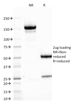
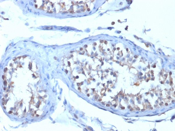
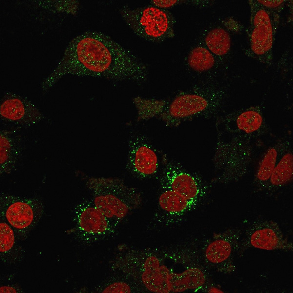
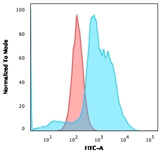
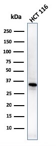 WB (Western Blot)
(Western Blot of HCT116 cell lysates using B7-H4 Mouse Monoclonal Antibody (B7H4/1788).)
WB (Western Blot)
(Western Blot of HCT116 cell lysates using B7-H4 Mouse Monoclonal Antibody (B7H4/1788).)
B7-H4, Monoclonal Antibody (Cat# AAA215264)
Full Name
B7-H4 (Immuno-Inhibitory Protein)
Gene Names
VTCN1; B7X; B7H4; B7S1; B7-H4; B7h.5; VCTN1; PRO1291
Reactivity
Human
Applications
Immunohistochemistry, Western Blot, Immunofluorescence, Flow Cytometry
Purity
Purified Ab with BSA and Azide at 200ug/ml OR Purified Ab WITHOUT BSA and Azide at 1.0mg/ml
Pricing
 Application Data
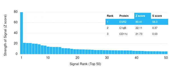
(Analysis of Protein Array containing more than 19,000 full-length human proteins using Estrogen Receptor beta-1 Mouse Monoclonal Antibody (ESR2/3005) Z- and S- Score: The Z-score represents the strength of a signal that a monoclonal antibody (Monoclonal Antibody) (in combination with a fluorescently-tagged anti-IgG secondary antibody) produces when binding to a particular protein on the HuProtTM array. Z-scores are described in units of standard deviations (SD's) above the mean value of all signals generated on that array. If targets on HuProtTM are arranged in descending order of the Z-score, the S-score is the difference (also in units of SD's) between the Z-score. S-score therefore represents the relative target specificity of a Monoclonal Antibody to its intended target. A Monoclonal Antibody is considered to specific to its intended target, if the Monoclonal Antibody has an S-score of at least 2.5. For example, if a Monoclonal Antibody binds to protein X with a Z-score of 43 and to protein Y with a Z-score of 14, then the S-score for the binding of that Monoclonal Antibody to protein X is equal to 29.)
Application Data
(Analysis of Protein Array containing more than 19,000 full-length human proteins using Estrogen Receptor beta-1 Mouse Monoclonal Antibody (ESR2/3005) Z- and S- Score: The Z-score represents the strength of a signal that a monoclonal antibody (Monoclonal Antibody) (in combination with a fluorescently-tagged anti-IgG secondary antibody) produces when binding to a particular protein on the HuProtTM array. Z-scores are described in units of standard deviations (SD's) above the mean value of all signals generated on that array. If targets on HuProtTM are arranged in descending order of the Z-score, the S-score is the difference (also in units of SD's) between the Z-score. S-score therefore represents the relative target specificity of a Monoclonal Antibody to its intended target. A Monoclonal Antibody is considered to specific to its intended target, if the Monoclonal Antibody has an S-score of at least 2.5. For example, if a Monoclonal Antibody binds to protein X with a Z-score of 43 and to protein Y with a Z-score of 14, then the S-score for the binding of that Monoclonal Antibody to protein X is equal to 29.)
ER-beta-1 (Estrogen Receptor beta-1), Monoclonal Antibody (Cat# AAA214958)
Full Name
ER-beta-1 (Estrogen Receptor beta-1)
Gene Names
ESR2; Erb; ESRB; ODG8; ESTRB; NR3A2; ER-BETA; ESR-BETA
Reactivity
Human
Applications
ELISA
Purity
Purified Ab with BSA and Azide at 200ug/ml OR Purified Ab WITHOUT BSA and Azide at 1.0mg/ml
Pricing
 Application Data
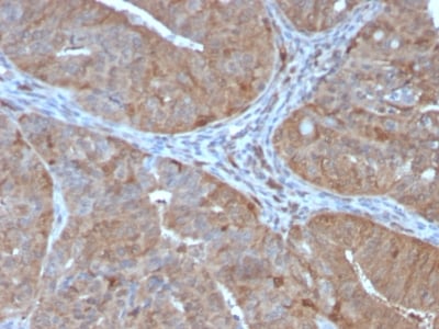
(Analysis of Protein Array containing more than 19,000 full-length human proteins using Aldo-keto Reductase Family 1 Member B1 Mouse Monoclonal Antibody (CPTC-AKR1B1-2).Z- and S- Score: The Z-score represents the strength of a signal that a monoclonal antibody (MAb) (in combination with a fluorescently-tagged anti-IgG secondary antibody) produces when binding to a particular protein on the HuProtTM array. Z-scores are described in units of standard deviations (SD's) above the mean value of all signals generated on that array. If targets on HuProtTM are arranged in descending order of the Z-score, the S-score is the difference (also in units of SD's) between the Z-score. S-score therefore represents the relative target specificity of a MAb to its intended target. A MAb is considered to specific to its intended target, if the MAb has an S-score of at least 2.5. For example, if a MAb binds to protein X with a Z-score of 43 and to protein Y with a Z-score of 14, then the S-score for the binding of that MAb to protein X is equal to 29.)
Application Data
(Analysis of Protein Array containing more than 19,000 full-length human proteins using Aldo-keto Reductase Family 1 Member B1 Mouse Monoclonal Antibody (CPTC-AKR1B1-2).Z- and S- Score: The Z-score represents the strength of a signal that a monoclonal antibody (MAb) (in combination with a fluorescently-tagged anti-IgG secondary antibody) produces when binding to a particular protein on the HuProtTM array. Z-scores are described in units of standard deviations (SD's) above the mean value of all signals generated on that array. If targets on HuProtTM are arranged in descending order of the Z-score, the S-score is the difference (also in units of SD's) between the Z-score. S-score therefore represents the relative target specificity of a MAb to its intended target. A MAb is considered to specific to its intended target, if the MAb has an S-score of at least 2.5. For example, if a MAb binds to protein X with a Z-score of 43 and to protein Y with a Z-score of 14, then the S-score for the binding of that MAb to protein X is equal to 29.)
Aldo-keto Reductase Family 1 Member B1, Monoclonal Antibody (Cat# AAA214739)
Full Name
Aldo-keto Reductase Family 1 Member B1 (Adrenal Marker)
Gene Names
AKR1B1; AR; ADR; ALR2; ALDR1
Reactivity
Human. Others not tested.
Applications
Immunohistochemistry, Western Blot
Purity
Purified Ab with BSA and Azide at 200ug/ml OR Purified Ab WITHOUT BSA and Azide at 1.0mg/ml
Pricing
 WB (Western Blot)
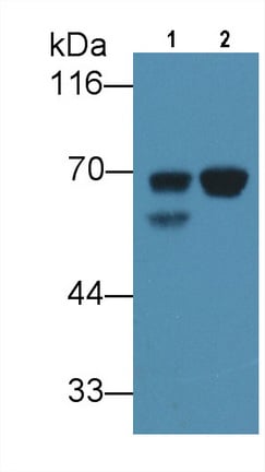
(Western Blot; Sample: Lane1: Human Liver lysate; Lane2: Human Lung lysate Primary Ab: 3ug/ml Mouse AntiHuman aFP Antibody Second Ab: 0.2ug/mL HRPLinked Caprine AntiMouse IgG Polyclonal Antibody (Catalog: SAA544Mu19))
WB (Western Blot)
(Western Blot; Sample: Lane1: Human Liver lysate; Lane2: Human Lung lysate Primary Ab: 3ug/ml Mouse AntiHuman aFP Antibody Second Ab: 0.2ug/mL HRPLinked Caprine AntiMouse IgG Polyclonal Antibody (Catalog: SAA544Mu19))
AlphaFetoprotein (AFP), Monoclonal Antibody (Cat# AAA151494)
Full Name
Monoclonal Antibody to AlphaFetoprotein (AFP)
Gene Names
AFP; FETA; HPAFP
Reactivity
Human
Applications
Immunoprecipitation, Immunocytochemistry, Immunohistochemistry, Western Blot
Purity
Protein A + Protein G affinity chromatography
Pricing
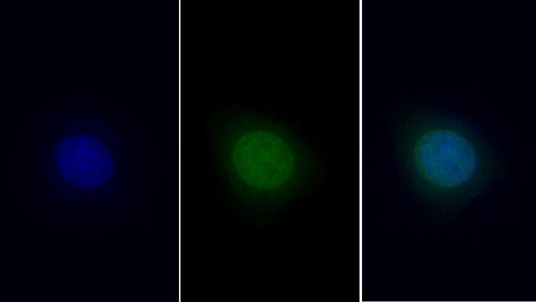
 IHC (Immunohiostchemistry)
(IHC Image)
IHC (Immunohiostchemistry)
(IHC Image)
High Mobility Group Protein 1 (HMGB1), Monoclonal Antibody (Cat# AAA151529)
Full Name
Monoclonal Antibody to High Mobility Group Protein 1 (HMGB1)
Gene Names
HMGB1; HMG1; HMG3; SBP-1
Reactivity
Human
Applications
Immunoprecipitation, Immunocytochemistry, Immunohistochemistry, Western Blot
Purity
Protein A + Protein G affinity chromatography
Pricing
 FCM (Flow Cytometry)
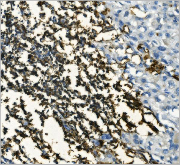
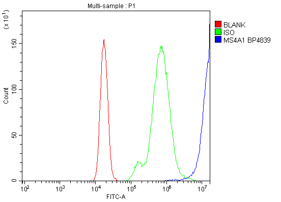
(Figure 9. Flow Cytometry analysis of Raji cells using anti-CD20/MS4A1 antibody (AAA19281).Overlay histogram showing Raji cells stained with AAA19281 (Blue line). The cells were blocked with 10% normal goat serum. And then incubated with rabbit anti-CD20/MS4A1 Antibody (AAA19281, 1μg/1x106 cells) for 30 min at 20 degree C. DyLight®488 conjugated goat anti-rabbit IgG (5-10μg/1x106 cells) was used as secondary antibody for 30 minutes at 20 degree C. Isotype control antibody (Green line) was rabbit IgG (1μg/1x106) used under the same conditions. Unlabelled sample (Red line) was also used as a control.)
FCM (Flow Cytometry)
(Figure 9. Flow Cytometry analysis of Raji cells using anti-CD20/MS4A1 antibody (AAA19281).Overlay histogram showing Raji cells stained with AAA19281 (Blue line). The cells were blocked with 10% normal goat serum. And then incubated with rabbit anti-CD20/MS4A1 Antibody (AAA19281, 1μg/1x106 cells) for 30 min at 20 degree C. DyLight®488 conjugated goat anti-rabbit IgG (5-10μg/1x106 cells) was used as secondary antibody for 30 minutes at 20 degree C. Isotype control antibody (Green line) was rabbit IgG (1μg/1x106) used under the same conditions. Unlabelled sample (Red line) was also used as a control.)
CD20/MS4A1, Polyclonal Antibody (Cat# AAA19281)
Full Name
Anti-CD20/MS4A1 Antibody
Gene Names
MS4A1; B1; S7; Bp35; CD20; CVID5; MS4A2; LEU-16
Reactivity
Human
Applications
Western Blot, Immunohistochemistry, Flow Cytometry, Direct ELISA
Purity
Immunogen affinity purified.
Pricing
 Bioactivity
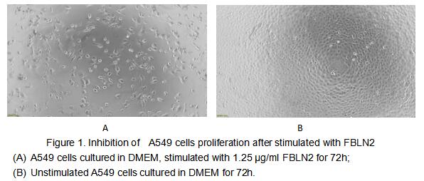
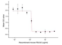
(Figure 2. Inhibition of A549 cells proliferation after stimulated with FBLN2.)
Bioactivity
(Figure 2. Inhibition of A549 cells proliferation after stimulated with FBLN2.)
Fibulin 2 (FBLN2), Active Protein (Cat# AAA161909)
Full Name
Active Fibulin 2 (FBLN2)
Gene Names
Fbln2; FIBL-2; 5730577E14Rik
Applications
Activity Assay, Cell Culture
Purity
Greater than 90% by SDS-PAGE
Pricing
 WB (Western Blot)
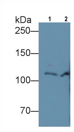
(Western Blot; Sample: Lane1: Human Serum; Lane2: Human Plasma Primary Ab: 4ug/ml Mouse AntiHuman C5a Antibody Second Ab: 0.2ug/mL HRPLinked Caprine AntiMouse IgG Polyclonal Antibody (Catalog: SAA544Mu19))
WB (Western Blot)
(Western Blot; Sample: Lane1: Human Serum; Lane2: Human Plasma Primary Ab: 4ug/ml Mouse AntiHuman C5a Antibody Second Ab: 0.2ug/mL HRPLinked Caprine AntiMouse IgG Polyclonal Antibody (Catalog: SAA544Mu19))
Complement Component 5a (C5a), Monoclonal Antibody (Cat# AAA151526)
Full Name
Monoclonal Antibody to Complement Component 5a (C5a)
Gene Names
C5; C5a; C5b; CPAMD4
Reactivity
Human, Pig
Applications
Immunoprecipitation, Immunocytochemistry, Immunohistochemistry, Western Blot
Purity
Protein A + Protein G affinity chromatography
Pricing

 IHC (Immunohistochemistry)
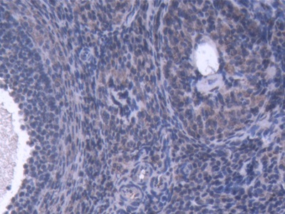
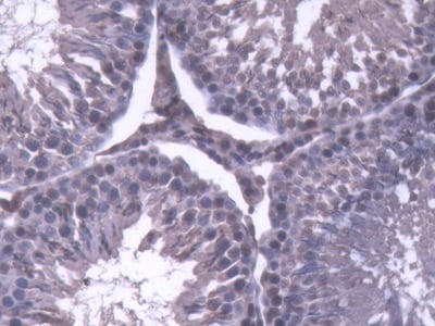
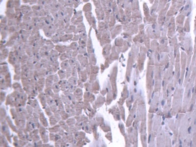
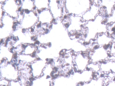
(DAB staining on IHCP;Sample: Rat Lung Tissue; Primary Ab: 20ug/ml Mouse AntiRat IL1b AntibodySecond Ab: 2ug/mL HRPLinked Caprine AntiMouse IgG Polyclonal Antibody(Catalog: SAA544Mu19))
IHC (Immunohistochemistry)
(DAB staining on IHCP;Sample: Rat Lung Tissue; Primary Ab: 20ug/ml Mouse AntiRat IL1b AntibodySecond Ab: 2ug/mL HRPLinked Caprine AntiMouse IgG Polyclonal Antibody(Catalog: SAA544Mu19))
Interleukin 1 Beta (IL1b), Monoclonal Antibody (Cat# AAA151568)
Full Name
Monoclonal Antibody to Interleukin 1 Beta (IL1b)
Reactivity
Rat
Applications
Immunoprecipitation, Immunocytochemistry, Immunohistochemistry, Western Blot
Purity
Protein A + Protein G affinity chromatography
Pricing
 WB (Western Blot)
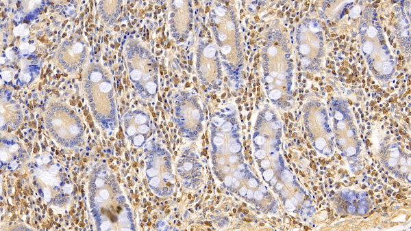
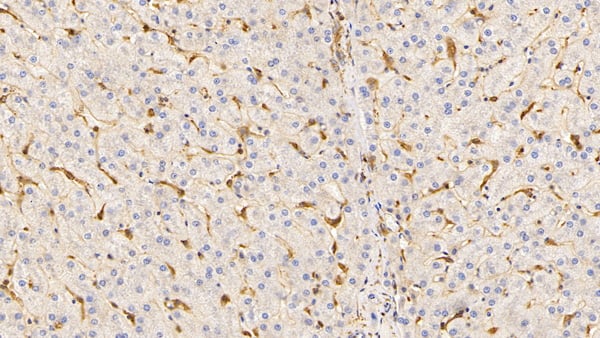
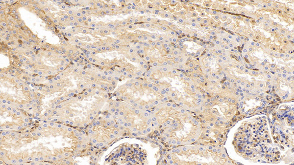
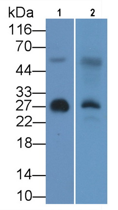
(Western Blot; Sample: Lane1: Human Serum; Lane2: Rat Cerebrum lysate Primary Ab: 3ug/ml Mouse AntiHuman APOA1 Antibody Second Ab: 0.2ug/mL HRPLinked Caprine AntiMouse IgG Polyclonal Antibody (Catalog: SAA544Mu19))
WB (Western Blot)
(Western Blot; Sample: Lane1: Human Serum; Lane2: Rat Cerebrum lysate Primary Ab: 3ug/ml Mouse AntiHuman APOA1 Antibody Second Ab: 0.2ug/mL HRPLinked Caprine AntiMouse IgG Polyclonal Antibody (Catalog: SAA544Mu19))
Apolipoprotein A1 (APOA1), Monoclonal Antibody (Cat# AAA151549)
Full Name
Monoclonal Antibody to Apolipoprotein A1 (APOA1)
Reactivity
Human, Rat, Pig
Applications
Immunoprecipitation, Immunocytochemistry, Immunohistochemistry, Western Blot
Purity
Protein A + Protein G affinity chromatography
Pricing

 Sequence
Galectin 8 (GAL8), Active Protein (Cat# AAA152175)
Full Name
Active Galectin 8 (GAL8)
Applications
Activity Assay, Cell Culture
Purity
>95%
Pricing
 FCM/FACS (Flow Cytometry)
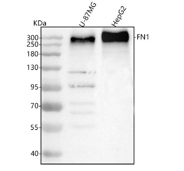
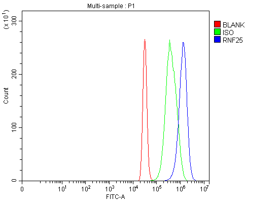
(Figure 2. Flow Cytometry analysis of HepG2 cells using anti-Fibronectin/FN1 antibody (AAA126997).Overlay histogram showing HepG2 cells stained with AAA126997 (Blue line). The cells were fixed with 4% paraformaldehyde and blocked with 10% normal goat serum. And then incubated with rabbit anti-Fibronectin/FN1 Antibody (AAA126997, 1ug/1x106 cells) for 30 min at 20 degree C. DyLight488 conjugated goat anti-rabbit IgG was used as secondary antibody for 30 minutes at 20 degree C. Isotype control antibody (Green line) was rabbit IgG (1ug/1x106) used under the same conditions. Unlabelled sample (Red line) was also used as a control.)
FCM/FACS (Flow Cytometry)
(Figure 2. Flow Cytometry analysis of HepG2 cells using anti-Fibronectin/FN1 antibody (AAA126997).Overlay histogram showing HepG2 cells stained with AAA126997 (Blue line). The cells were fixed with 4% paraformaldehyde and blocked with 10% normal goat serum. And then incubated with rabbit anti-Fibronectin/FN1 Antibody (AAA126997, 1ug/1x106 cells) for 30 min at 20 degree C. DyLight488 conjugated goat anti-rabbit IgG was used as secondary antibody for 30 minutes at 20 degree C. Isotype control antibody (Green line) was rabbit IgG (1ug/1x106) used under the same conditions. Unlabelled sample (Red line) was also used as a control.)
Fibronectin/FN1, Polyclonal Antibody (Cat# AAA126997)
Full Name
Anti-Fibronectin/FN1 Antibody Picoband
Gene Names
FN1; FN; CIG; FNZ; MSF; ED-B; FINC; GFND; LETS; GFND2
Reactivity
Human
Applications
Western Blot, Flow Cytometry
Purity
Immunogen affinity purified.
Pricing
 IF (Immunofluorescence)
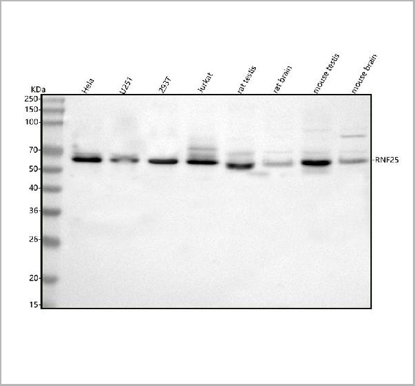
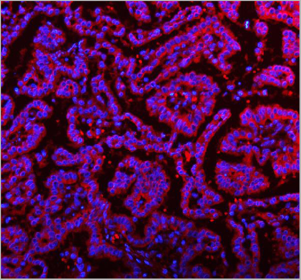
(Figure 13. IF analysis of RNF25 using anti-RNF25 antibody (AAA19946).RNF25 was detected in a paraffin-embedded section of human lung cancer tissue. Heat mediated antigen retrieval was performed in EDTA buffer (pH 8.0, epitope retrieval solution). The tissue section was blocked with 10% goat serum. The tissue section was then incubated with 5ug/mL rabbit anti-RNF25 Antibody (AAA19946) overnight at 4 degree C. DyLight®594 Conjugated Goat Anti-Rabbit IgG (BA1142) was used as secondary antibody at 1:500 dilution and incubated for 30 minutes at 37 degree C. The section was counterstained with DAPI. Visualize using a fluorescence microscope and filter sets appropriate for the label used.)
IF (Immunofluorescence)
(Figure 13. IF analysis of RNF25 using anti-RNF25 antibody (AAA19946).RNF25 was detected in a paraffin-embedded section of human lung cancer tissue. Heat mediated antigen retrieval was performed in EDTA buffer (pH 8.0, epitope retrieval solution). The tissue section was blocked with 10% goat serum. The tissue section was then incubated with 5ug/mL rabbit anti-RNF25 Antibody (AAA19946) overnight at 4 degree C. DyLight®594 Conjugated Goat Anti-Rabbit IgG (BA1142) was used as secondary antibody at 1:500 dilution and incubated for 30 minutes at 37 degree C. The section was counterstained with DAPI. Visualize using a fluorescence microscope and filter sets appropriate for the label used.)
RNF25, Polyclonal Antibody (Cat# AAA19946)
Full Name
Anti-RNF25 Antibody Picoband
Gene Names
RNF25; AO7
Reactivity
Human, Mouse, Rat
Applications
Flow Cytometry, Immunofluorescence, Immunohistochemistry, Western Blot
Purity
Immunogen affinity purified.
Pricing
 IF (Immunofluorescence)
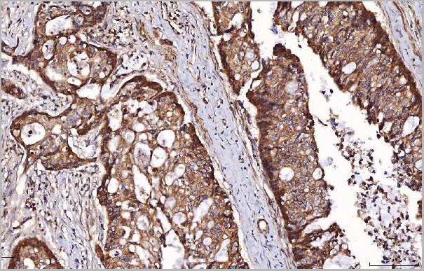
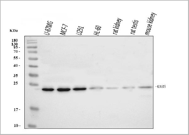
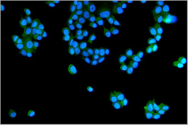
(Figure 7. IF analysis of RAB5A/B/C using anti-RAB5A/B/C antibody (AAA19438).RAB5A/B/C was detected in an immunocytochemical section of MCF-7 cells. Enzyme antigen retrieval was performed using IHC enzyme antigen retrieval reagent (AR0022) for 15 mins. The cells were blocked with 10% goat serum. And then incubated with 5 ug/mL rabbit anti-RAB5A/B/C Antibody (AAA19438) overnight at 4 degree C. DyLight488 Conjugated Goat Anti-Rabbit IgG was used as secondary antibody at 1:100 dilution and incubated for 30 minutes at 37 degree C. The section was counterstained with DAPI. Visualize using a fluorescence microscope and filter sets appropriate for the label used.)
IF (Immunofluorescence)
(Figure 7. IF analysis of RAB5A/B/C using anti-RAB5A/B/C antibody (AAA19438).RAB5A/B/C was detected in an immunocytochemical section of MCF-7 cells. Enzyme antigen retrieval was performed using IHC enzyme antigen retrieval reagent (AR0022) for 15 mins. The cells were blocked with 10% goat serum. And then incubated with 5 ug/mL rabbit anti-RAB5A/B/C Antibody (AAA19438) overnight at 4 degree C. DyLight488 Conjugated Goat Anti-Rabbit IgG was used as secondary antibody at 1:100 dilution and incubated for 30 minutes at 37 degree C. The section was counterstained with DAPI. Visualize using a fluorescence microscope and filter sets appropriate for the label used.)
RAB5A/B/C, Polyclonal Antibody (Cat# AAA19438)
Full Name
Anti-RAB5A/B/C Antibody Picoband
Gene Names
RAB5A; RAB5
Reactivity
Human, Mouse, Rat
Applications
Western Blot, Immunohistochemistry, Immunocytochemistry, Immunofluorescence
Purity
Immunogen affinity purified.
Pricing