
Filters
Clonality
Type
Reactivity
Gene Name
Isotype
Host
Application
Clone
10000 results for "Recombinant Proteins" - showing 2600-2650
Jagged-1:Fc, Active Protein (Cat# AAA14750)
Full Name
Jagged-1, Recombinant, Mouse:Fc Human(Protein Jagged-1, CD339, JAG1)
Purity
> 90%
Pricing
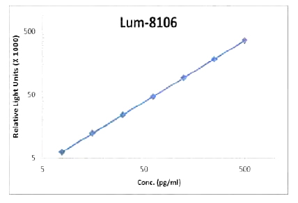 Standard Curve (Sample)
beta-NGF, CLIA Kit (Cat# AAA318522)
Full Name
beta-NGF (Human) LumiAb ELISA Kit
Gene Names
NGF; NGFB; HSAN5; Beta-NGF
Reactivity
Human
Pricing
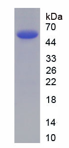
 Amino Acid Info
Insulin Receptor (INSR), Recombinant Protein (Cat# AAA153403)
Full Name
Recombinant Insulin Receptor (INSR)
Gene Names
INSR; HHF5; CD220
Applications
Western Blot
Purity
Greater than 90% by SDS-PAGE
Pricing
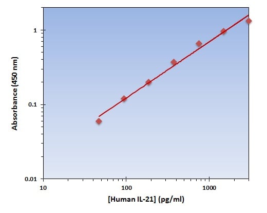 Application Data
(The OmniKine Human IL-21 ELISA Kit allows for the detection and quantification of endogenous levels of natural and/or recombinant Human IL-21 proteins within the range of 46-3000 pg/ml.)
Application Data
(The OmniKine Human IL-21 ELISA Kit allows for the detection and quantification of endogenous levels of natural and/or recombinant Human IL-21 proteins within the range of 46-3000 pg/ml.)
IL-21, ELISA Kit (Cat# AAA315626)
Full Name
IL-21 (Human) OmniKine ELISA Kit
Gene Names
IL21; Za11; IL-21; CVID11
Reactivity
Human
Pricing
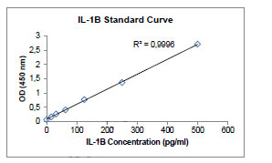 Standard Curve (Sample)
IL-1 beta, ELISA Kit (Cat# AAA58604)
Full Name
Human IL-1 beta ELISA Kit
Gene Names
IL1B; IL-1; IL1F2; IL1-BETA
Reactivity
Human. No cross reactivity with other human cytokines.
Pricing
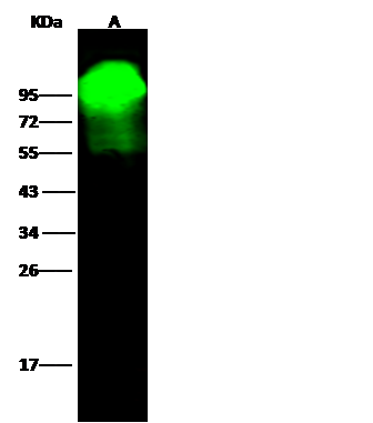
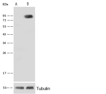 WB (Western Blot)
(Anti-ALPL rabbit monoclonal antibody at 1:500 dilutionLane A: ALPL konckout Hela Whole Cell LysateLane B: Hela Whole Cell LysateLysates/proteins at 20 ug per lane.SecondaryGoat Anti-Rabbit IgG (H+L)/HRP at 1/10000 dilution.Developed using the ECL technique.Performed under reducing conditions.Predicted band size:57 kDaObserved band size:95 kDa)
WB (Western Blot)
(Anti-ALPL rabbit monoclonal antibody at 1:500 dilutionLane A: ALPL konckout Hela Whole Cell LysateLane B: Hela Whole Cell LysateLysates/proteins at 20 ug per lane.SecondaryGoat Anti-Rabbit IgG (H+L)/HRP at 1/10000 dilution.Developed using the ECL technique.Performed under reducing conditions.Predicted band size:57 kDaObserved band size:95 kDa)
Alkaline Phosphatase/ALPL, Monoclonal Antibody (Cat# AAA254207)
Full Name
Recombinant Anti-Alkaline Phosphatase/ALPL Antibody, Rabbit Monoclonal
Gene Names
ALPL; HOPS; TNAP; APTNAP; TNSALP; AP-TNAP
Reactivity
Human
Applications
Western Blot
Purity
Protein A
Pricing
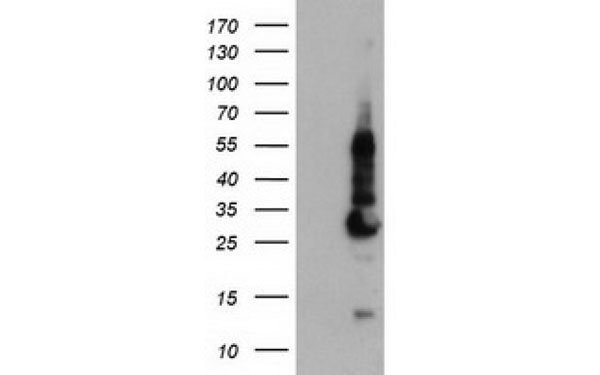
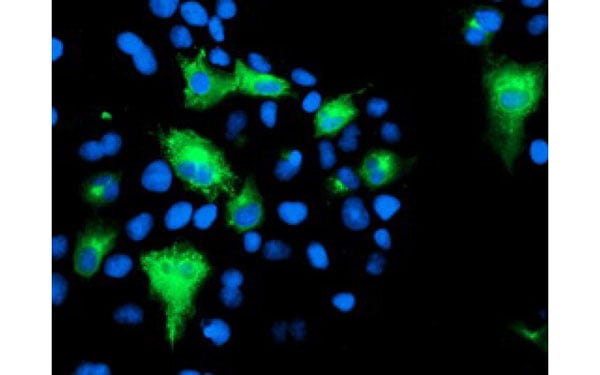 IF (Immunofluorescence)
(Immunofluorescent staining of COS7 cells transiently transfected with recombinant KCNJ3 protein using KCNJ3 antibody)
IF (Immunofluorescence)
(Immunofluorescent staining of COS7 cells transiently transfected with recombinant KCNJ3 protein using KCNJ3 antibody)
KCNJ3, Monoclonal Antibody (Cat# AAA107402)
Full Name
KCNJ3 antibody
Gene Names
KCNJ3; KGA; GIRK1; KIR3.1
Applications
Flow Cytometry, Immunofluorescence
Purity
KCNJ3 antibody was purified by affinity chromatography.
Pricing
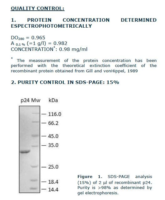
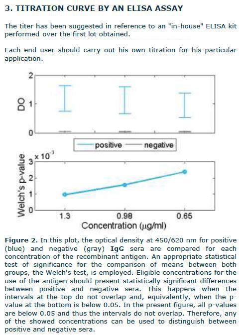 Application Data
gag gene, Recombinant Protein (Cat# AAA72852)
Full Name
gag gene recombinant
Applications
Western Blot
Purity
0.98
Pricing
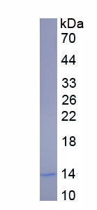
 Gene Sequence
Complement Component 3a (C3a), Protein (Cat# AAA150978)
Full Name
Eukaryotic Complement Component 3a (C3a)
Gene Names
C3; ASP; Plp; HSE-MSF; AI255234
Reactivity
Mus musculus (Mouse)
Applications
Western Blot
Purity
> 95%
Pricing
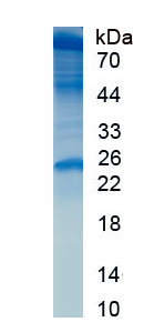
 Bioactivity
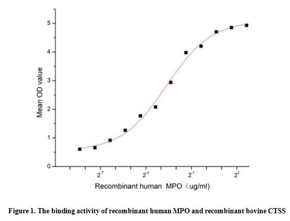
(Myeloperoxidase (MPO), a member of the XPO subfamily of peroxidases, is a 150 kDa heme protein produced mostly from polymorphonuclear neutrophils and monocytes. MPO is a part of the host defense system of polymorphonuclear leukocytes. It is responsible for microbicidal activity against a wide range of organisms. In the stimulated PMN, MPO catalyzes the production of hypohalous acids, primarily hypochlorous acid in physiologic situations, and other toxic intermediates that greatly enhance PMN microbicidal activity. Cathepsin S (CTSS) is one of targets of MPO, thus a functional binding ELISA assay was conducted to detect the interaction of recombinant human MPO and recombinant bovine CTSS. Briefly, biotin-linked MPO were diluted serially in PBS, with 0.01% BSA (pH 7.4). Duplicate samples of 100 ul were then transferred to CTSS-coated microtiter wells and incubated for 1h at 37 degree C. Wells were washed with PBST 3 times and incubation with Streptavidin-HRP for 30min, then wells were aspirated and washed 5 times. With the addition of substrate solution, wells were incubated 15-25 minutes at 37 degree C. Finally, add 50 ul stop solution to the wells and read at 450 nm immediately. The binding activity of recombinant human MPO and recombinant bovine CTSS was shown in Figure 1, the EC50 for this effect is 0.16 ug/mL.)
Bioactivity
(Myeloperoxidase (MPO), a member of the XPO subfamily of peroxidases, is a 150 kDa heme protein produced mostly from polymorphonuclear neutrophils and monocytes. MPO is a part of the host defense system of polymorphonuclear leukocytes. It is responsible for microbicidal activity against a wide range of organisms. In the stimulated PMN, MPO catalyzes the production of hypohalous acids, primarily hypochlorous acid in physiologic situations, and other toxic intermediates that greatly enhance PMN microbicidal activity. Cathepsin S (CTSS) is one of targets of MPO, thus a functional binding ELISA assay was conducted to detect the interaction of recombinant human MPO and recombinant bovine CTSS. Briefly, biotin-linked MPO were diluted serially in PBS, with 0.01% BSA (pH 7.4). Duplicate samples of 100 ul were then transferred to CTSS-coated microtiter wells and incubated for 1h at 37 degree C. Wells were washed with PBST 3 times and incubation with Streptavidin-HRP for 30min, then wells were aspirated and washed 5 times. With the addition of substrate solution, wells were incubated 15-25 minutes at 37 degree C. Finally, add 50 ul stop solution to the wells and read at 450 nm immediately. The binding activity of recombinant human MPO and recombinant bovine CTSS was shown in Figure 1, the EC50 for this effect is 0.16 ug/mL.)
Myeloperoxidase (MPO), Active Protein (Cat# AAA161752)
Full Name
Active Myeloperoxidase (MPO)
Applications
Activity Assay, Cell Culture
Purity
Greater than 80% by SDS-PAGE
Pricing
 Application Data
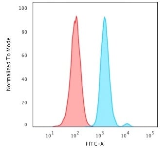
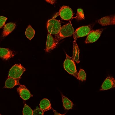
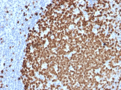
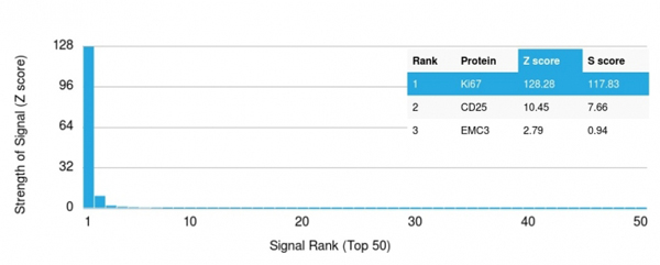
(Analysis of Protein Array containing more than 19,000 full-length human proteins using Ki67 Mouse Monoclonal Antibody (MKI67/2461). Z- and S- Score: The Z-score represents the strength of a signal that a monoclonal antibody (MAb) (in combination with a fluorescently-tagged anti-IgG secondary antibody) produces when binding to a particular protein on the HuProtTM array. Z-scores are described in units of standard deviations (SD's) above the mean value of all signals generated on that array. If targets on HuProtTM are arranged in descending order of the Z-score, the S-score is the difference (also in units of SD's) between the Z-score. S-score therefore represents the relative target specificity of a MAb to its intended target. A MAb is considered to specific to its intended target, if the MAb has an S-score of at least 2.5. For example, if a MAb binds to protein X with a Z-score of 43 and to protein Y with a Z-score of 14, then the S-score for the binding of that MAb to protein X is equal to 29.)
Application Data
(Analysis of Protein Array containing more than 19,000 full-length human proteins using Ki67 Mouse Monoclonal Antibody (MKI67/2461). Z- and S- Score: The Z-score represents the strength of a signal that a monoclonal antibody (MAb) (in combination with a fluorescently-tagged anti-IgG secondary antibody) produces when binding to a particular protein on the HuProtTM array. Z-scores are described in units of standard deviations (SD's) above the mean value of all signals generated on that array. If targets on HuProtTM are arranged in descending order of the Z-score, the S-score is the difference (also in units of SD's) between the Z-score. S-score therefore represents the relative target specificity of a MAb to its intended target. A MAb is considered to specific to its intended target, if the MAb has an S-score of at least 2.5. For example, if a MAb binds to protein X with a Z-score of 43 and to protein Y with a Z-score of 14, then the S-score for the binding of that MAb to protein X is equal to 29.)
Ki-67, Monoclonal Antibody (Cat# AAA214778)
Full Name
Ki-67 (Proliferating Cell Marker)
Gene Names
MKI67; KIA; MIB-; MIB-1; PPP1R105
Reactivity
Human. Others not known.
Applications
Immunohistochemistry, Immunofluorescence, Flow Cytometry
Purity
Purified Ab with BSA and Azide at 200ug/ml OR Purified Ab WITHOUT BSA and Azide at 1.0mg/ml
Pricing
 SDS-PAGE
High Mobility Group Protein 1 (HMGB1), Active Protein (Cat# AAA149232)
Full Name
Active High Mobility Group Protein 1 (HMGB1)
Gene Names
Hmgb1; p30; Hmg1; HMG-1; SBP-1
Applications
Activity Assay, Cell Culture
Purity
> 90%
Pricing
 Bioactivity
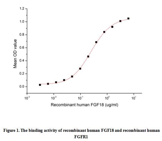
(Fibroblast Growth Factor 18 (FGF-18) is a 20 kDa protein that plays an important role in skeletal development and bone homeostasis. It is expressed in embryonic somites and the neural fold, adult lung, cerebellar and hippocampal neurons. FGF-18 binds to FGF R2c, FGF R3c as well as the Golgi protein GLG1 and induces the proliferation of astrocytes and microglia, vascular endothelial cells, dermal fibroblasts, papilla cells, and keratinocytes. FGF-18 is required for normal skeletal development. A functional binding ELISA assay was conducted to detect the interaction of recombinant human FGF18 and recombinant human FGFR1. Briefly, FGF18 was diluted serially in PBS with 0.01% BSA (pH 7.4). Duplicate samples of 100 ul were then transferred to FGFR1-coated microtiter wells and incubated for 1h at 37 degree C. Wells were washed with PBST and incubated for 1h with anti-FGF18 pAb, then aspirated and washed 3 times. After incubation with HRP labelled secondary antibody for 1h at 37 degree C, wells were aspirated and washed 5 times. With the addition of substrate solution, wells were incubated 15-25 minutes at 37 degree C. Finally, add 50 uL stop solution to the wells and read at 450/630nm immediately. The binding activity of recombinant human FGF18 and recombinant human FGFR1 was shown in Figure 1, the EC50 for this effect is 0.26 ug/mL.)
Bioactivity
(Fibroblast Growth Factor 18 (FGF-18) is a 20 kDa protein that plays an important role in skeletal development and bone homeostasis. It is expressed in embryonic somites and the neural fold, adult lung, cerebellar and hippocampal neurons. FGF-18 binds to FGF R2c, FGF R3c as well as the Golgi protein GLG1 and induces the proliferation of astrocytes and microglia, vascular endothelial cells, dermal fibroblasts, papilla cells, and keratinocytes. FGF-18 is required for normal skeletal development. A functional binding ELISA assay was conducted to detect the interaction of recombinant human FGF18 and recombinant human FGFR1. Briefly, FGF18 was diluted serially in PBS with 0.01% BSA (pH 7.4). Duplicate samples of 100 ul were then transferred to FGFR1-coated microtiter wells and incubated for 1h at 37 degree C. Wells were washed with PBST and incubated for 1h with anti-FGF18 pAb, then aspirated and washed 3 times. After incubation with HRP labelled secondary antibody for 1h at 37 degree C, wells were aspirated and washed 5 times. With the addition of substrate solution, wells were incubated 15-25 minutes at 37 degree C. Finally, add 50 uL stop solution to the wells and read at 450/630nm immediately. The binding activity of recombinant human FGF18 and recombinant human FGFR1 was shown in Figure 1, the EC50 for this effect is 0.26 ug/mL.)
Fibroblast Growth Factor 18 (FGF18), Active Protein (Cat# AAA161895)
Full Name
Active Fibroblast Growth Factor 18 (FGF18)
Gene Names
FGF18; ZFGF5; FGF-18
Applications
Activity Assay, Cell Culture
Purity
Greater than 90% by SDS-PAGE
Pricing
 IHC (Immunohiostchemistry)
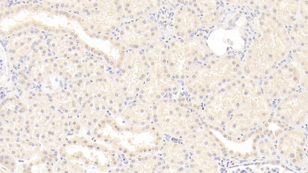
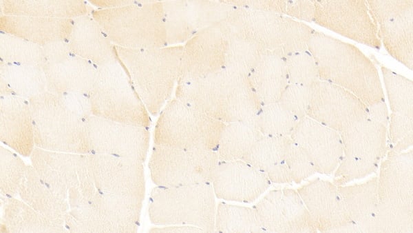
(DAB staining on IHCP;Sample: Human Skeletal muscle Tissue; Primary Ab: 20ug/ml Mouse AntiHuman ISLR AntibodySecond Ab: 2ug/mL HRPLinked Caprine AntiMouse IgG Polyclonal Antibody(Catalog: SAA544Mu19))
IHC (Immunohiostchemistry)
(DAB staining on IHCP;Sample: Human Skeletal muscle Tissue; Primary Ab: 20ug/ml Mouse AntiHuman ISLR AntibodySecond Ab: 2ug/mL HRPLinked Caprine AntiMouse IgG Polyclonal Antibody(Catalog: SAA544Mu19))
Immunoglobulin Superfamily Containing Leucine Rich Repeat Protein (ISLR), Monoclonal Antibody (Cat# AAA151883)
Full Name
Monoclonal Antibody to Immunoglobulin Superfamily Containing Leucine Rich Repeat Protein (ISLR)
Gene Names
ISLR; HsT17563
Reactivity
Human, Pig
Applications
Immunoprecipitation, Immunocytochemistry, Immunohistochemistry, Western Blot
Purity
Protein A + Protein G affinity chromatography
Pricing
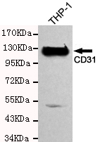 IP (Immunoprecipitation)
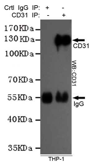
(Immunoprecipitation analysis of THP-1 cell lysates using CD31 mouse mAb.)
IP (Immunoprecipitation)
(Immunoprecipitation analysis of THP-1 cell lysates using CD31 mouse mAb.)
CD31, Monoclonal Antibody (Cat# AAA320368)
Full Name
CD31 Mouse mAb
Gene Names
PECAM1; CD31; PECA1; GPIIA'; PECAM-1; endoCAM; CD31/EndoCAM
Reactivity
Human
Applications
Immunoprecipitation, Western Blot
Purity
The antibody was affinity-purified from mouse ascites by affinity-chromatography using epitope-specific immunogen.
Pricing
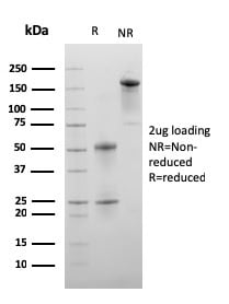
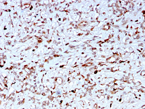
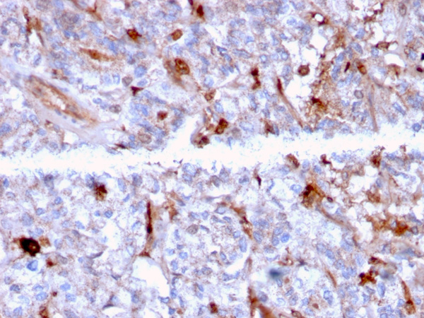
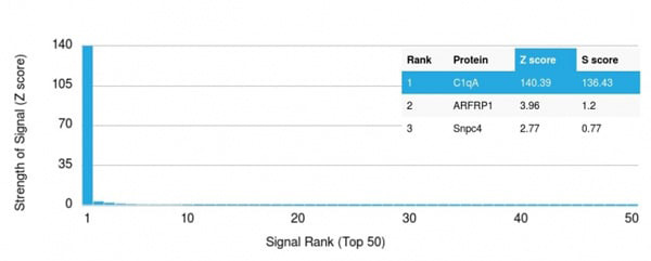 Application Data
(Analysis of Protein Array containing more than 19,000 full-length human proteins using C1QA Mouse Monoclonal Antibody (C1QA/2956). Z- and S- Score: The Z-score represents the strength of a signal that a monoclonal antibody (MAb) (in combination with a fluorescently-tagged anti-IgG secondary antibody) produces when binding to a particular protein on the HuProtTM array. Z-scores are described in units of standard deviations (SD's) above the mean value of all signals generated on that array. If targets on HuProtTM are arranged in descending order of the Z-score, the S-score is the difference (also in units of SD's) between the Z-score. S-score therefore represents the relative target specificity of a MAb to its intended target. A MAb is considered to specific to its intended target, if the MAb has an S-score of at least 2.5. For example, if a MAb binds to protein X with a Z-score of 43 and to protein Y with a Z-score of 14, then the S-score for the binding of that MAb to protein X is equal to 29.)
Application Data
(Analysis of Protein Array containing more than 19,000 full-length human proteins using C1QA Mouse Monoclonal Antibody (C1QA/2956). Z- and S- Score: The Z-score represents the strength of a signal that a monoclonal antibody (MAb) (in combination with a fluorescently-tagged anti-IgG secondary antibody) produces when binding to a particular protein on the HuProtTM array. Z-scores are described in units of standard deviations (SD's) above the mean value of all signals generated on that array. If targets on HuProtTM are arranged in descending order of the Z-score, the S-score is the difference (also in units of SD's) between the Z-score. S-score therefore represents the relative target specificity of a MAb to its intended target. A MAb is considered to specific to its intended target, if the MAb has an S-score of at least 2.5. For example, if a MAb binds to protein X with a Z-score of 43 and to protein Y with a Z-score of 14, then the S-score for the binding of that MAb to protein X is equal to 29.)
C1QA/Complement C1q A-Chain, Monoclonal Antibody (Cat# AAA215224)
Full Name
C1QA/Complement C1q A-Chain
Reactivity
Human
Applications
Immunohistochemistry
Purity
Purified Ab with BSA and Azide at 200ug/ml OR Purified Ab WITHOUT BSA and Azide at 1.0mg/ml
Pricing
 IHC (Immunohistochemisry)
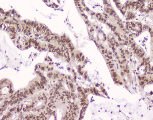
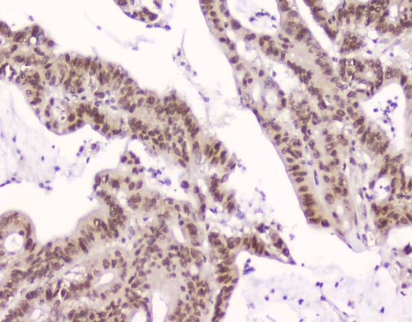
(Figure 3. IHC analysis of CRM1 using anti-CRM1 antibody (M01180).CRM1 was detected in paraffin-embedded section of human intestinal cancer tissue. Heat mediated antigen retrieval was performed in citrate buffer (pH6, epitope retrieval solution) for 20 mins. The tissue section was blocked with 10% goat serum. The tissue section was then incubated with 2ug/ml mouse anti-CRM1 Antibody (M01180) overnight at 4 degree C. Biotinylated goat anti-mouse IgG was used as secondary antibody and incubated for 30 minutes at 37 degree C. The tissue section was developed using Strepavidin-Biotin-Complex (SABC) with DAB as the chromogen.)
IHC (Immunohistochemisry)
(Figure 3. IHC analysis of CRM1 using anti-CRM1 antibody (M01180).CRM1 was detected in paraffin-embedded section of human intestinal cancer tissue. Heat mediated antigen retrieval was performed in citrate buffer (pH6, epitope retrieval solution) for 20 mins. The tissue section was blocked with 10% goat serum. The tissue section was then incubated with 2ug/ml mouse anti-CRM1 Antibody (M01180) overnight at 4 degree C. Biotinylated goat anti-mouse IgG was used as secondary antibody and incubated for 30 minutes at 37 degree C. The tissue section was developed using Strepavidin-Biotin-Complex (SABC) with DAB as the chromogen.)
CRM1, Monoclonal Antibody (Cat# AAA125138)
Full Name
Anti-CRM1 Antibody (monoclonal, 5G3)
Gene Names
XPO1; emb; CRM1; exp1
Reactivity
Human
Applications
Flow Cytometry, Immunocytochemistry, Immunohistochemistry, Western Blot
Purity
Immunogen Affinity Purified
Pricing
 Bioactivity
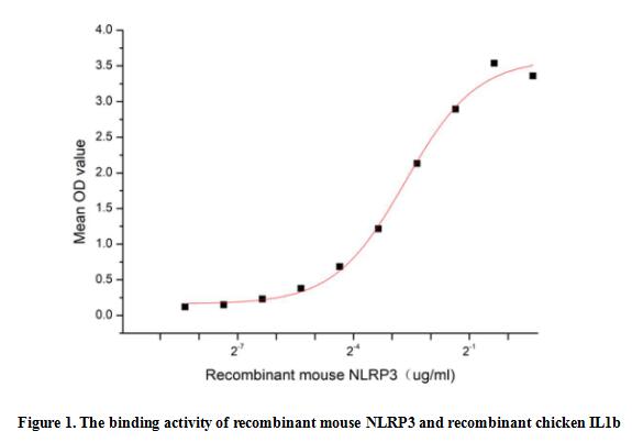
(NLR Family, Pyrin Domain Containing Protein 3 (NLRP3), also known as NALP3, CIAS1, PYPAF or Cryopyrin, is a cytosolic multiprotein complex composed of the innate immune receptor protein NLRP3, adapter protein ASC, and inflammatory protease caspase-1 that responds to microbial infection, endogenous danger signals, and environmental stimuli. Aberrant activation of the NLRP3 inflammasome is associated with the pathogenesis of various inflammatory diseases, such as diabetes, cancer, and Alzheimer's disease. Interleukin 1 Beta (IL1b) can interact with NLRP3, thus a functional binding ELISA assay was conducted to detect the interaction of recombinant mouse NLRP3 and recombinant chicken IL1b. Briefly, NLRP3 was diluted serially in PBS with 0.01% BSA (pH 7.4). Duplicate samples of 100 ul were then transferred to IL1b-coated microtiter wells and incubated for 1h at 37 degree C. Wells were washed with PBST and incubated for 1h with anti-NLRP3 pAb, then aspirated and washed 3 times. After incubation with HRP labelled secondary antibody for 1h at 37 degree C, wells were aspirated and washed 5 times. With the addition of substrate solution, wells were incubated 15-25 minutes at 37 degree C. Finally, add 50 uL stop solution to the wells and read at 450/630 nm immediately. The binding activity of recombinant mouse NLRP3 and recombinant chicken IL1b was shown in Figure 1, the EC50 for this effect is 0.16 ug/mL.)
Bioactivity
(NLR Family, Pyrin Domain Containing Protein 3 (NLRP3), also known as NALP3, CIAS1, PYPAF or Cryopyrin, is a cytosolic multiprotein complex composed of the innate immune receptor protein NLRP3, adapter protein ASC, and inflammatory protease caspase-1 that responds to microbial infection, endogenous danger signals, and environmental stimuli. Aberrant activation of the NLRP3 inflammasome is associated with the pathogenesis of various inflammatory diseases, such as diabetes, cancer, and Alzheimer's disease. Interleukin 1 Beta (IL1b) can interact with NLRP3, thus a functional binding ELISA assay was conducted to detect the interaction of recombinant mouse NLRP3 and recombinant chicken IL1b. Briefly, NLRP3 was diluted serially in PBS with 0.01% BSA (pH 7.4). Duplicate samples of 100 ul were then transferred to IL1b-coated microtiter wells and incubated for 1h at 37 degree C. Wells were washed with PBST and incubated for 1h with anti-NLRP3 pAb, then aspirated and washed 3 times. After incubation with HRP labelled secondary antibody for 1h at 37 degree C, wells were aspirated and washed 5 times. With the addition of substrate solution, wells were incubated 15-25 minutes at 37 degree C. Finally, add 50 uL stop solution to the wells and read at 450/630 nm immediately. The binding activity of recombinant mouse NLRP3 and recombinant chicken IL1b was shown in Figure 1, the EC50 for this effect is 0.16 ug/mL.)
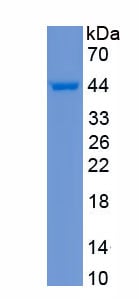
NLR Family, Pyrin Domain Containing Protein 3 (NLRP3), Active Protein (Cat# AAA161932)
Full Name
Active NLR Family, Pyrin Domain Containing Protein 3 (NLRP3)
Gene Names
Nlrp3; FCU; MWS; FCAS; Cias1; Mmig1; NALP3; Pypaf1; AII/AVP; AGTAVPRL
Applications
Activity Assay, Cell Culture
Purity
Greater than 95% by SDS-PAGE
Pricing
 Application Data
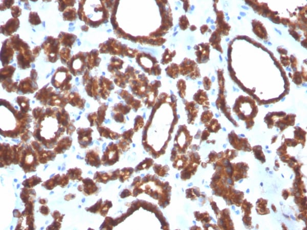
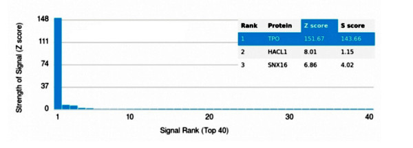
(Analysis of Protein Array containing more than 19,000 full-length human proteins using Thyroid Peroxidase Mouse Monoclonal Antibody (TPO/3697). Z- and S- Score: The Z-score represents the strength of a signal that a monoclonal antibody (MAb) (in combination with a fluorescently-tagged anti-IgG secondary antibody) produces when binding to a particular protein on the HuProtTM array. Z-scores are described in units of standard deviations (SD's) above the mean value of all signals generated on that array. If targets on HuProtTM are arranged in descending order of the Z-score, the S-score is the difference (also in units of SD's) between the Z-score. S-score therefore represents the relative target specificity of a MAb to its intended target. A MAb is considered to specific to its intended target, if the MAb has an S-score of at least 2.5. For example, if a MAb binds to protein X with a Z-score of 43 and to protein Y with a Z-score of 14, then the S-score for the binding of that MAb to protein X is equal to 29.)
Application Data
(Analysis of Protein Array containing more than 19,000 full-length human proteins using Thyroid Peroxidase Mouse Monoclonal Antibody (TPO/3697). Z- and S- Score: The Z-score represents the strength of a signal that a monoclonal antibody (MAb) (in combination with a fluorescently-tagged anti-IgG secondary antibody) produces when binding to a particular protein on the HuProtTM array. Z-scores are described in units of standard deviations (SD's) above the mean value of all signals generated on that array. If targets on HuProtTM are arranged in descending order of the Z-score, the S-score is the difference (also in units of SD's) between the Z-score. S-score therefore represents the relative target specificity of a MAb to its intended target. A MAb is considered to specific to its intended target, if the MAb has an S-score of at least 2.5. For example, if a MAb binds to protein X with a Z-score of 43 and to protein Y with a Z-score of 14, then the S-score for the binding of that MAb to protein X is equal to 29.)
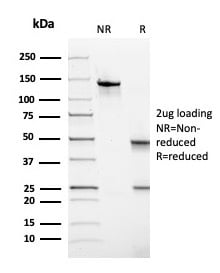
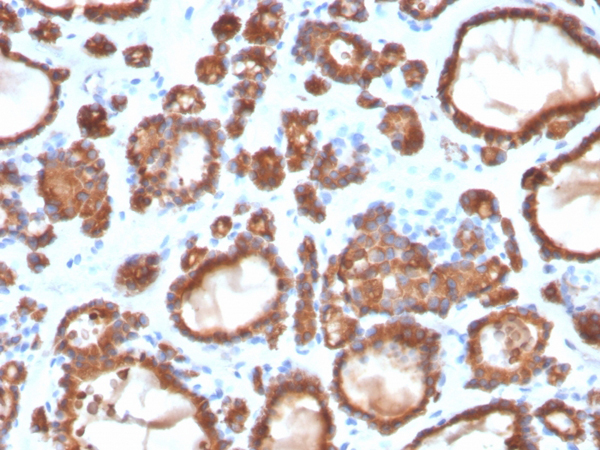
TPO (Thyroid Peroxidase), Monoclonal Antibody (Cat# AAA215468)
Full Name
TPO (Thyroid Peroxidase) (Thyroid Marker)
Gene Names
TPO; MSA; TPX; TDH2A
Reactivity
Human
Applications
Immunohistochemistry
Purity
Purified Ab WITHOUT BSA and Azide at 1.0mg/ml
Pricing
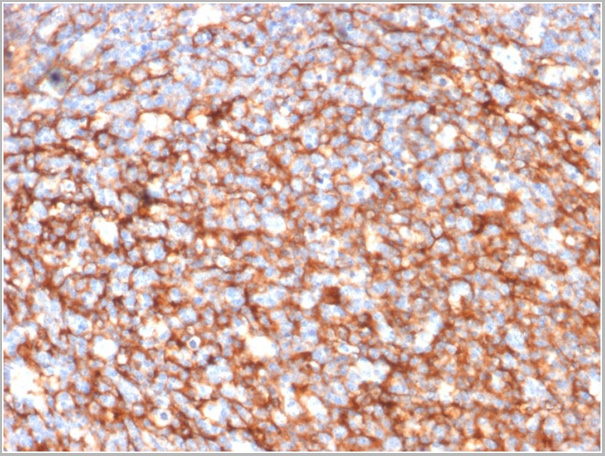
 Application Data
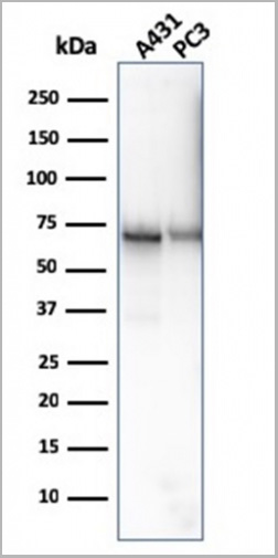
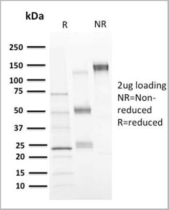
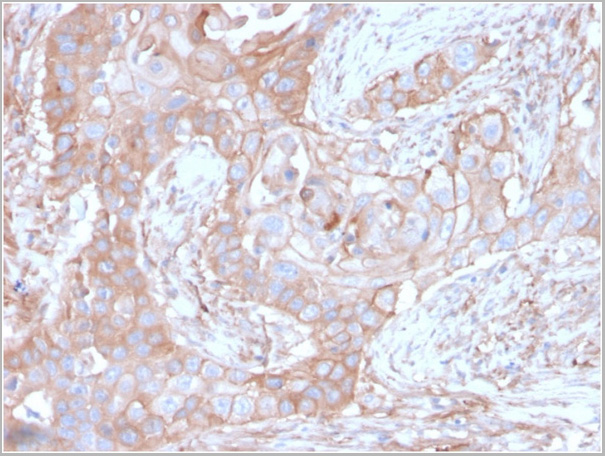
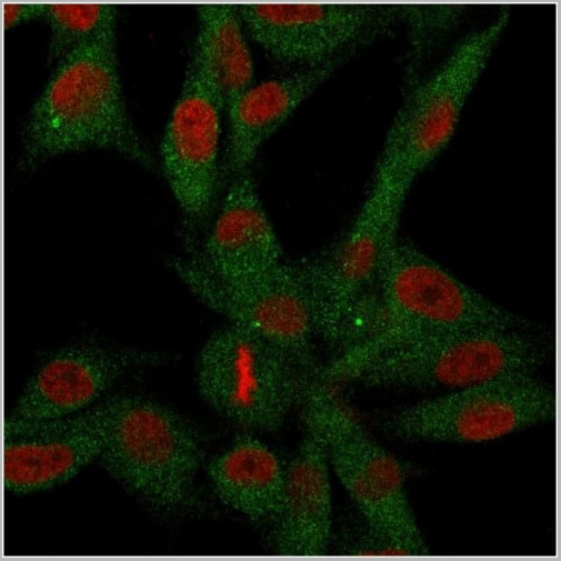
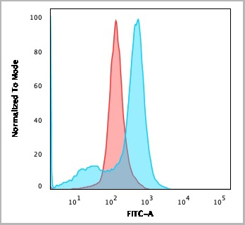
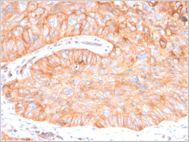
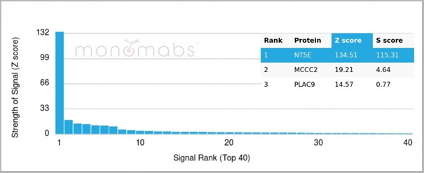
(Analysis of Protein Array containing more than 19,000 full-length human proteins using CD73 Mouse Recombinant Monoclonal Antibody (CD73/2646) Z- and S- Score: The Z-score represents the strength of a signal that a monoclonal antibody (MAb) (in combination with a fluorescently-tagged anti-IgG secondary antibody) produces when binding to a particular protein on the HuProtTM array. Z-scores are described in units of standard deviations (SD’s) above the mean value of all signals generated on that array. If targets on HuProtTM are arranged in descending order of the Z-score, the S-score is the difference (also in units of SD’s) between the Z-score. S-score therefore represents the relative target specificity of a MAb to its intended target. A MAb is considered to specific to its intended target, if the MAb has an S-score of at least 2.5. For example, if a MAb binds to protein X with a Z-score of 43 and to protein Y with a Z-score of 14, then the S-score for the binding of that MAb to protein X is equal to 29.)
Application Data
(Analysis of Protein Array containing more than 19,000 full-length human proteins using CD73 Mouse Recombinant Monoclonal Antibody (CD73/2646) Z- and S- Score: The Z-score represents the strength of a signal that a monoclonal antibody (MAb) (in combination with a fluorescently-tagged anti-IgG secondary antibody) produces when binding to a particular protein on the HuProtTM array. Z-scores are described in units of standard deviations (SD’s) above the mean value of all signals generated on that array. If targets on HuProtTM are arranged in descending order of the Z-score, the S-score is the difference (also in units of SD’s) between the Z-score. S-score therefore represents the relative target specificity of a MAb to its intended target. A MAb is considered to specific to its intended target, if the MAb has an S-score of at least 2.5. For example, if a MAb binds to protein X with a Z-score of 43 and to protein Y with a Z-score of 14, then the S-score for the binding of that MAb to protein X is equal to 29.)
CD73, Monoclonal Antibody (Cat# AAA23929)
Full Name
CD73 (Immuno-Oncology Target)
Gene Names
NT5E; NT; eN; NT5; NTE; eNT; CD73; E5NT; CALJA
Reactivity
Human
Applications
Flow Cytometry, Immunofluorescence, Western Blot, Immunohistochemistry
Purity
Purified Ab with BSA and Azide at 200ug/ml OR Purified Ab WITHOUT BSA and Azide at 1.0mg/ml
Pricing
 Application Data
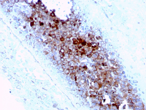
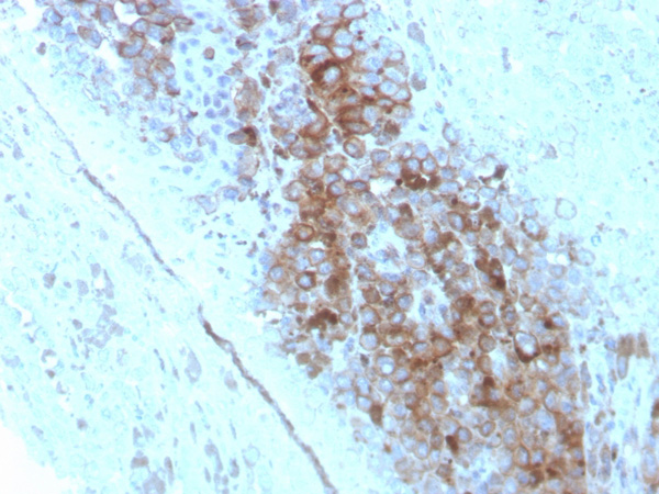
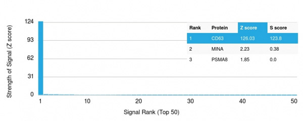
(Analysis of Protein Array containing more than 19,000 full-length human proteins using CD63 Mouse Monoclonal Antibody (LAMP3/2789). Z- and S- Score: The Z-score represents the strength of a signal that a monoclonal antibody (MAb) (in combination with a fluorescently-tagged anti-IgG secondary antibody) produces when binding to a particular protein on the HuProtTM array. Z-scores are described in units of standard deviations (SD's) above the mean value of all signals generated on that array. If targets on HuProtTM are arranged in descending order of the Z-score, the S-score is the difference (also in units of SD's) between the Z-score. S-score therefore represents the relative target specificity of a MAb to its intended target. A MAb is considered to specific to its intended target, if the MAb has an S-score of at least 2.5. For example, if a MAb binds to protein X with a Z-score of 43 and to protein Y with a Z-score of 14, then the S-score for the binding of that MAb to protein X is equal to 29.)
Application Data
(Analysis of Protein Array containing more than 19,000 full-length human proteins using CD63 Mouse Monoclonal Antibody (LAMP3/2789). Z- and S- Score: The Z-score represents the strength of a signal that a monoclonal antibody (MAb) (in combination with a fluorescently-tagged anti-IgG secondary antibody) produces when binding to a particular protein on the HuProtTM array. Z-scores are described in units of standard deviations (SD's) above the mean value of all signals generated on that array. If targets on HuProtTM are arranged in descending order of the Z-score, the S-score is the difference (also in units of SD's) between the Z-score. S-score therefore represents the relative target specificity of a MAb to its intended target. A MAb is considered to specific to its intended target, if the MAb has an S-score of at least 2.5. For example, if a MAb binds to protein X with a Z-score of 43 and to protein Y with a Z-score of 14, then the S-score for the binding of that MAb to protein X is equal to 29.)
CD63, Monoclonal Antibody (Cat# AAA215313)
Full Name
CD63 (Late Endosomes Marker)
Gene Names
CD63; MLA1; ME491; LAMP-3; OMA81H; TSPAN30
Reactivity
Human, Mouse
Applications
Immunohistochemistry, Western Blot
Purity
Purified Ab with BSA and Azide at 200ug/ml OR Purified Ab WITHOUT BSA and Azide at 1.0mg/ml
Pricing
 Bioactivity
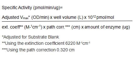
(Glucose-6-phosphate dehydrogenase (G6PD) converts D-glucose 6-phosphate (G6P) into 6-phosphoglucono-delta-lactone and generate co-enzyme nicotinamide adenine dinucleotide phosphate (NADPH). G6PD is the rate-limiting enzyme of the pentose phosphate pathway that supplies reducing energy to cells by maintaining the level of NADPH, which in turn maintains the level of glutathione in these cells that helps protect the red blood cells against oxidative damage from compounds like hydrogen peroxide. More importantly, NADPH is used for biosynthesis of fatty acids or isoprenoids. G6PD is generally found as a dimer of two identical monomers. Depending on conditions, such as pH, these dimers can themselves dimerize to form tetramers. Each monomer in the complex has a substrate binding site that binds to G6P, and a catalytic coenzyme binding site that binds to NADP /NADPH using the Rossman fold. Its activity is stimulated by the substrate G6P and NADP. The activity of recombinant human G6PD was measured by its ability to dehydrogenate glucose-6-phosphate. The reaction was performed in 25 mM Tris, 50 mM NaCl, 10 mM MgCl2, pH 8.0 (Assay Buffer), initiated by addition 50 uL of various concentrations of G6PD (diluted by Assay Buffer) to 50 ul substrate mixture consists of 1 mM NADP and 1 mM G6P. The final well serves as a negative control with no G6PD, replaced with 50 uL assay buffer. Then read at a wavelength of 340 nm in kinetic mode for 5 minutes. The specific activity of recombinant human G6PD is >280 pmol/min/ug.)
Bioactivity
(Glucose-6-phosphate dehydrogenase (G6PD) converts D-glucose 6-phosphate (G6P) into 6-phosphoglucono-delta-lactone and generate co-enzyme nicotinamide adenine dinucleotide phosphate (NADPH). G6PD is the rate-limiting enzyme of the pentose phosphate pathway that supplies reducing energy to cells by maintaining the level of NADPH, which in turn maintains the level of glutathione in these cells that helps protect the red blood cells against oxidative damage from compounds like hydrogen peroxide. More importantly, NADPH is used for biosynthesis of fatty acids or isoprenoids. G6PD is generally found as a dimer of two identical monomers. Depending on conditions, such as pH, these dimers can themselves dimerize to form tetramers. Each monomer in the complex has a substrate binding site that binds to G6P, and a catalytic coenzyme binding site that binds to NADP /NADPH using the Rossman fold. Its activity is stimulated by the substrate G6P and NADP. The activity of recombinant human G6PD was measured by its ability to dehydrogenate glucose-6-phosphate. The reaction was performed in 25 mM Tris, 50 mM NaCl, 10 mM MgCl2, pH 8.0 (Assay Buffer), initiated by addition 50 uL of various concentrations of G6PD (diluted by Assay Buffer) to 50 ul substrate mixture consists of 1 mM NADP and 1 mM G6P. The final well serves as a negative control with no G6PD, replaced with 50 uL assay buffer. Then read at a wavelength of 340 nm in kinetic mode for 5 minutes. The specific activity of recombinant human G6PD is >280 pmol/min/ug.)
Glucose-6-phosphate Dehydrogenase (G6PD), Active Protein (Cat# AAA161771)
Full Name
Active Glucose-6-phosphate Dehydrogenase (G6PD)
Gene Names
G6PD; G6PD1
Applications
Activity Assay, Cell Culture
Purity
Greater than 90% by SDS-PAGE
Pricing
 Bioactivity
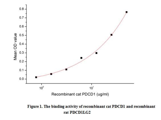
(PD-1 (Programmed Death-1 receptor), also known as CD279, is a receptor expressed on T cells responsible for modulating T cell activation. Like CTLA4, PD-1 is classified as an immune checkpoint inhibitory receptor. When PD-1 protein binds to PD-L1, it initiates a negative signaling cascade inhibiting activation of T cells. The cytoplasmic tail contains two tyrosine residues that form the immunoreceptor tyrosine-based inhibitory motif (ITIM) and immunoreceptor tyrosine-based switch motif (ITSM) that are important for mediating PD-1 signaling. Normally, PD-1 helps keep T cells from attacking other cells in the body. However, many cancers take advantage of this by expressing high amounts of PD-L1 allowing cancer cells to evade the body's own immune response. Blocking the PD-1:PD-L1 interaction has proven successful in treating many different cancer types. A functional binding ELISA assay was conducted to detect the interaction of recombinant cat PDCD1 and recombinant rat PDCD1LG2. Briefly, biotin-linked PDCD1 were diluted serially in PBS, with 0.01% BSA (pH 7.4). Duplicate samples of 100 ul were then transferred to PDCD1LG2-coated microtiter wells and incubated for 1h at 37 degree C. Wells were washed with PBST 3 times and incubation with Streptavidin-HRP for 30min, then wells were aspirated and washed 5 times. With the addition of substrate solution, wells were incubated 15-25 minutes at 37 degree C. Finally, add 50 ul stop solution to the wells and read at 450 nm immediately. The binding activity of PDCD1 and PDCD1LG2 was shown in Figure 1, and this effect was in a dose dependent manner.)
Bioactivity
(PD-1 (Programmed Death-1 receptor), also known as CD279, is a receptor expressed on T cells responsible for modulating T cell activation. Like CTLA4, PD-1 is classified as an immune checkpoint inhibitory receptor. When PD-1 protein binds to PD-L1, it initiates a negative signaling cascade inhibiting activation of T cells. The cytoplasmic tail contains two tyrosine residues that form the immunoreceptor tyrosine-based inhibitory motif (ITIM) and immunoreceptor tyrosine-based switch motif (ITSM) that are important for mediating PD-1 signaling. Normally, PD-1 helps keep T cells from attacking other cells in the body. However, many cancers take advantage of this by expressing high amounts of PD-L1 allowing cancer cells to evade the body's own immune response. Blocking the PD-1:PD-L1 interaction has proven successful in treating many different cancer types. A functional binding ELISA assay was conducted to detect the interaction of recombinant cat PDCD1 and recombinant rat PDCD1LG2. Briefly, biotin-linked PDCD1 were diluted serially in PBS, with 0.01% BSA (pH 7.4). Duplicate samples of 100 ul were then transferred to PDCD1LG2-coated microtiter wells and incubated for 1h at 37 degree C. Wells were washed with PBST 3 times and incubation with Streptavidin-HRP for 30min, then wells were aspirated and washed 5 times. With the addition of substrate solution, wells were incubated 15-25 minutes at 37 degree C. Finally, add 50 ul stop solution to the wells and read at 450 nm immediately. The binding activity of PDCD1 and PDCD1LG2 was shown in Figure 1, and this effect was in a dose dependent manner.)
Programmed Cell Death Protein 1 (PD1), Active Protein (Cat# AAA161773)
Full Name
Active Programmed Cell Death Protein 1 (PD1)
Applications
Activity Assay, Cell Culture
Purity
Greater than 90% by SDS-PAGE
Pricing
 FCM/FACS (Flow Cytometry)
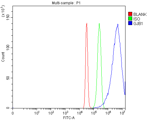
(Figure 2. Flow Cytometry analysis of HepG2 cells using anti-Connexin-32/GJB1 antibody .Overlay histogram showing HepG2 cells stained with A01050 (Blue line). The cells were fixed with 4% paraformaldehyde and blocked with 10% normal goat serum. And then incubated with rabbit anti-Connexin-32/GJB1 Antibody for 30 min at 20 degree C. DyLight488 conjugated goat anti-rabbit IgG was used as secondary antibody for 30 minutes at 20 degree C. Isotype control antibody (Green line) was rabbit IgG (1ug/1x106) used under the same conditions. Unlabelled sample (Red line) was also used as a control.)
FCM/FACS (Flow Cytometry)
(Figure 2. Flow Cytometry analysis of HepG2 cells using anti-Connexin-32/GJB1 antibody .Overlay histogram showing HepG2 cells stained with A01050 (Blue line). The cells were fixed with 4% paraformaldehyde and blocked with 10% normal goat serum. And then incubated with rabbit anti-Connexin-32/GJB1 Antibody for 30 min at 20 degree C. DyLight488 conjugated goat anti-rabbit IgG was used as secondary antibody for 30 minutes at 20 degree C. Isotype control antibody (Green line) was rabbit IgG (1ug/1x106) used under the same conditions. Unlabelled sample (Red line) was also used as a control.)
Connexin-32/GJB1, Polyclonal Antibody (Cat# AAA127031)
Full Name
Anti-Connexin-32/GJB1 Antibody Picoband
Gene Names
GJB1; CMTX; CX32; CMTX1
Reactivity
Human
Applications
Flow Cytometry, Western Blot
Purity
Immunogen affinity purified.
Pricing
 Bioactivity
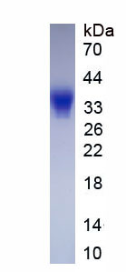
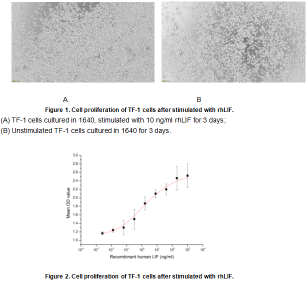
(Leukemia inhibitory factor (LIF), is an interleukin 6 class cytokine that affects cell growth by inhibiting differentiation. LIF as a cytokine also has another fuction including: the growth promotion and cell differentiation of different types of target cells, influence on bone metabolism, cachexia, neural development, embryogenesis and inflammation. The activity of LIF is usually measured by a cell proliferation assay using TF-1 cells. TF-1 cells were seeded into triplicate wells of 96-well plates at a density of 20000 cells/well with 10% serum standard 1640 which contains various concentrations of recombinant human LIF. After incubated for 3 days, cells were observed by inverted microscope and cell proliferation was measured by Cell Counting Kit-8 (CCK-8). Briefly, 10 ul of CCK-8 solution was added to each well of the plate, then the absorbance at 450 nm was measured using a microplate reader after incubating the plate for 2-4 hours at 37 degree C. Proliferation of TF-1 cells after incubation with LIF for 3 days observed by inverted microscope was shown in Figure 1. Cell viability was assessed by CCK-8 (Cell Counting Kit-8) assay after incubation with rhLIF for 3 days. The result was shown in Figure 2. It was obvious that rhLIF significantly increased cell viability of TF-1 cells. The ED50 is 1.86 ng/ml.)
Bioactivity
(Leukemia inhibitory factor (LIF), is an interleukin 6 class cytokine that affects cell growth by inhibiting differentiation. LIF as a cytokine also has another fuction including: the growth promotion and cell differentiation of different types of target cells, influence on bone metabolism, cachexia, neural development, embryogenesis and inflammation. The activity of LIF is usually measured by a cell proliferation assay using TF-1 cells. TF-1 cells were seeded into triplicate wells of 96-well plates at a density of 20000 cells/well with 10% serum standard 1640 which contains various concentrations of recombinant human LIF. After incubated for 3 days, cells were observed by inverted microscope and cell proliferation was measured by Cell Counting Kit-8 (CCK-8). Briefly, 10 ul of CCK-8 solution was added to each well of the plate, then the absorbance at 450 nm was measured using a microplate reader after incubating the plate for 2-4 hours at 37 degree C. Proliferation of TF-1 cells after incubation with LIF for 3 days observed by inverted microscope was shown in Figure 1. Cell viability was assessed by CCK-8 (Cell Counting Kit-8) assay after incubation with rhLIF for 3 days. The result was shown in Figure 2. It was obvious that rhLIF significantly increased cell viability of TF-1 cells. The ED50 is 1.86 ng/ml.)
Leukemia Inhibitory Factor (LIF), Active Protein (Cat# AAA161682)
Full Name
Active Leukemia Inhibitory Factor (LIF)
Gene Names
LIF; CDF; DIA; HILDA; MLPLI
Applications
Activity Assay, Cell Culture
Purity
Greater than 90% by SDS-PAGE
Pricing
 FCM/FACS (Flow Cytometry)
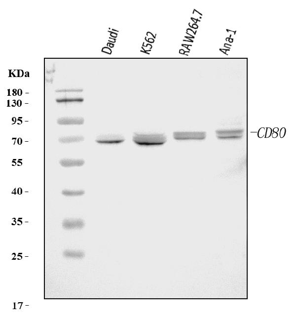
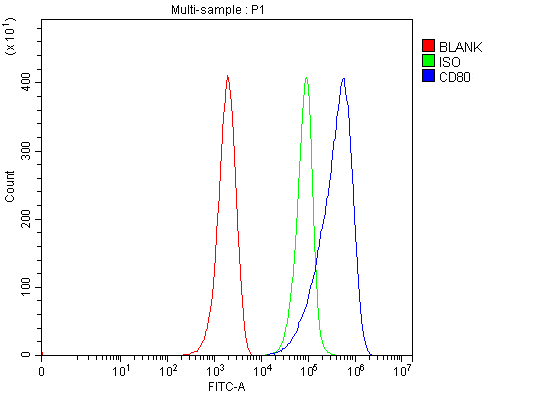
(Figure 2. Flow Cytometry analysis of ANA-1 cells using anti-Cd80 antibody (AAA125979).Overlay histogram showing ANA-1 cells stained with AAA125979 (Blue line). The cells were blocked with 10% normal goat serum. And then incubated with rabbit anti-Cd80 Antibody (AAA125979, 1 ug/1x10^6 cells) for 30 min at 20 degree C. DyLight488 conjugated goat anti-rabbit IgG was used as secondary antibody for 30 minutes at 20 degree C. Isotype control antibody (Green line) was rabbit IgG (1 ug/1x10^6) used under the same conditions. Unlabelled sample (Red line) was also used as a control.)
FCM/FACS (Flow Cytometry)
(Figure 2. Flow Cytometry analysis of ANA-1 cells using anti-Cd80 antibody (AAA125979).Overlay histogram showing ANA-1 cells stained with AAA125979 (Blue line). The cells were blocked with 10% normal goat serum. And then incubated with rabbit anti-Cd80 Antibody (AAA125979, 1 ug/1x10^6 cells) for 30 min at 20 degree C. DyLight488 conjugated goat anti-rabbit IgG was used as secondary antibody for 30 minutes at 20 degree C. Isotype control antibody (Green line) was rabbit IgG (1 ug/1x10^6) used under the same conditions. Unlabelled sample (Red line) was also used as a control.)
Cd80, Polyclonal Antibody (Cat# AAA125979)
Full Name
Anti-Cd80 Antibody Picoband
Gene Names
Cd80; B71; Ly53; TSA1; Cd28l; Ly-53; MIC17
Reactivity
Human, Mouse
Applications
Western Blot, Flow Cytometry
Purity
Immunogen affinity purified.
Pricing
 Standard Curve (Sample)
PLGF, CLIA Kit (Cat# AAA318557)
Full Name
PLGF (Human) LumiAb ELISA Kit
Gene Names
PGF; PGFL; PIGF; PLGF; PlGF-2; D12S1900; SHGC-10760
Reactivity
Human
Pricing
 FCM/FACS (Flow Cytometry)
IRS1, Monoclonal Antibody (Cat# AAA125498)
Full Name
Anti-Human IRS1 DyLight 488 Conjugated Antibody (Monoclonal, 10I3)
Gene Names
Irs1; G972R; IRS-1
Reactivity
Human
Applications
Flow Cytometry
Purity
Immunogen affinity purified.
Pricing
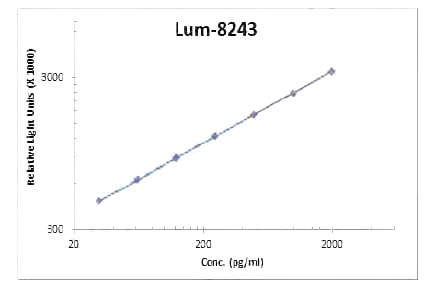 Standard Curve (Sample)
IL-23, CLIA Kit (Cat# AAA318595)
Full Name
IL-23 (Human) LumiAb ELISA Kit
Gene Names
IL23A; P19; SGRF; IL-23; IL-23A; IL23P19
Reactivity
Human
Pricing
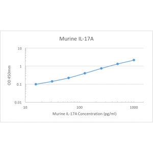 Standard Curve (Sample)
IL-17A, ELISA Kit (Cat# AAA79424)
Full Name
Murine IL-17A Pre-Coated ELISA Kit
Gene Names
Il17a; Il17; Ctla8; IL-17; Ctla-8; IL-17A
Reactivity
Murine
Pricing
 Application Data
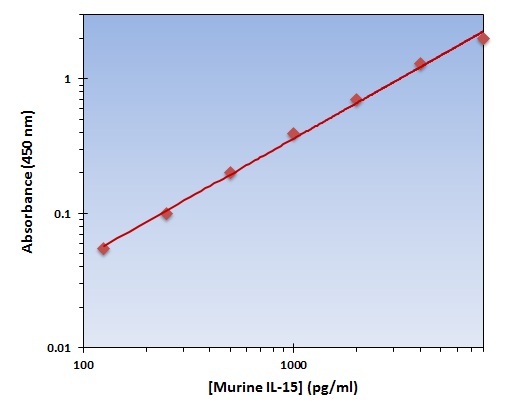
(The OmniKine Murine IL-15 ELISA Kit allows for the detection and quantification of endogenous levels of natural and/or recombinant Murine IL-15 proteins within the range of 125-8000 pg/ml.)
Application Data
(The OmniKine Murine IL-15 ELISA Kit allows for the detection and quantification of endogenous levels of natural and/or recombinant Murine IL-15 proteins within the range of 125-8000 pg/ml.)
IL-15, ELISA Kit (Cat# AAA315766)
Full Name
IL-15 (Murine) OmniKine ELISA Kit
Gene Names
Il15; IL-15; AI503618
Reactivity
Mouse
Pricing
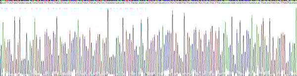
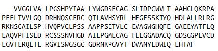 Amino Acid Info
(Amino Acid info)
Amino Acid Info
(Amino Acid info)
Coagulation Factor XII (F12), Recombinant Protein (Cat# AAA153377)
Full Name
Recombinant Coagulation Factor XII (F12)
Gene Names
F12; HAF
Applications
Western Blot
Purity
Greater than 90% by SDS-PAGE
Pricing
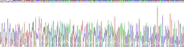
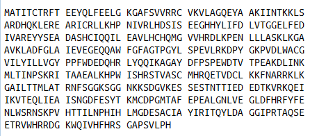
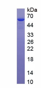
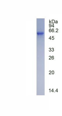 SDS-PAGE
(SDS-PAGE)
SDS-PAGE
(SDS-PAGE)
Calcium/Calmodulin Dependent Protein Kinase II Alpha (CAMK2a), Recombinant Protein (Cat# AAA152298)
Full Name
Recombinant Calcium/Calmodulin Dependent Protein Kinase II Alpha (CAMK2a)
Applications
Western Blot
Purity
Greater than 90% by SDS-PAGE
Pricing
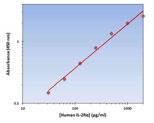 Application Data
(The OmniKine Human IL-2Ralpha ELISA Kit allows for the detection and quantification of endogenous levels of natural and/or recombinant Human IL-2Ralpha proteins within the range of 32-2000 pg/ml.)
Application Data
(The OmniKine Human IL-2Ralpha ELISA Kit allows for the detection and quantification of endogenous levels of natural and/or recombinant Human IL-2Ralpha proteins within the range of 32-2000 pg/ml.)
IL-2Ralpha, ELISA Kit (Cat# AAA315730)
Full Name
IL-2Ralpha (Human) OmniKine ELISA Kit
Gene Names
IL2RA; p55; CD25; IL2R; IMD41; TCGFR; IDDM10
Reactivity
Human
Pricing
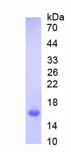
 Gene Sequence
Solute Carrier Family 30 Member 8 (SLC30A8), Recombinant Protein (Cat# AAA153614)
Full Name
Recombinant Solute Carrier Family 30 Member 8 (SLC30A8)
Gene Names
SLC30A8; ZNT8; ZnT-8
Applications
Western Blot
Purity
> 80%
Pricing
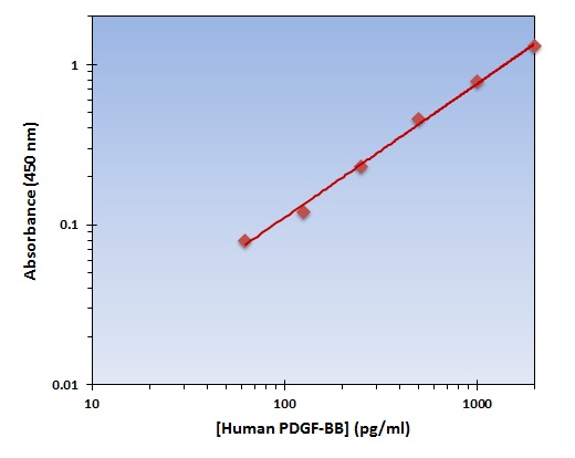 Application Data
(The OmniKine Human PDGF-BB ELISA Kit allows for the detection and quantification of endogenous levels of natural and/or recombinant Human PDGF-BB proteins within the range of 62-2000 pg/ml.)
Application Data
(The OmniKine Human PDGF-BB ELISA Kit allows for the detection and quantification of endogenous levels of natural and/or recombinant Human PDGF-BB proteins within the range of 62-2000 pg/ml.)
PDGF-BB, ELISA Kit (Cat# AAA315647)
Full Name
PDGF-BB (Human) OmniKine ELISA Kit
Gene Names
PDGFB; SIS; SSV; IBGC5; PDGF2; c-sis; PDGF-2
Reactivity
Human
Pricing
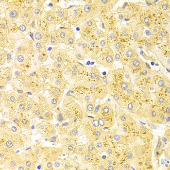
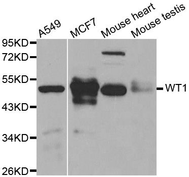
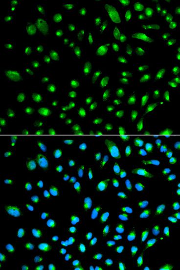 IF (Immunofluorescence)
(Immunofluorescence analysis of HeLa cells using WT1 antibody. Blue: DAPI for nuclear staining.)
IF (Immunofluorescence)
(Immunofluorescence analysis of HeLa cells using WT1 antibody. Blue: DAPI for nuclear staining.)
WT1, Polyclonal Antibody (Cat# AAA36915)
Full Name
WT1 Polyclonal Antibody
Gene Names
WT1; GUD; AWT1; WAGR; WT33; NPHS4; WIT-2; EWS-WT1
Reactivity
Human, Mouse
Applications
Immunofluorescence, Immunohistochemistry, Western Blot
Purity
Affinity Purification
Pricing
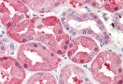
 WB (Western Blot)
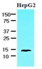
(Cell lysates of HepG2 (30 ug) were resolved by SDS-PAGE, transferred to NC membrane and probed with anti-human FABP1 (1:1000). Proteins were visualized using a goat anti-mouse secondary antibody conjugated to HRP and an ECL detection system.)
WB (Western Blot)
(Cell lysates of HepG2 (30 ug) were resolved by SDS-PAGE, transferred to NC membrane and probed with anti-human FABP1 (1:1000). Proteins were visualized using a goat anti-mouse secondary antibody conjugated to HRP and an ECL detection system.)
FABP1 / L-FABP, Monoclonal Antibody (Cat# AAA51816)
Full Name
Mouse Monoclonal [clone 2G4] (IgG1,k) to Human FABP1 / L-FABP
Gene Names
FABP1; FABPL; L-FABP
Reactivity
Human
Applications
Western Blot, Immunohistochemistry
Purity
Protein G Purified
Pricing
 IF (Immunofluorescence)
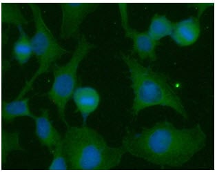
(ICC/IF analysis of RAS in HeLa cells. The cell was stained with AAA48181 (1:100). The secondary antibody (green) was used Alexa Fluor 488. DAPI was stained the cell nucleus (blue).)
IF (Immunofluorescence)
(ICC/IF analysis of RAS in HeLa cells. The cell was stained with AAA48181 (1:100). The secondary antibody (green) was used Alexa Fluor 488. DAPI was stained the cell nucleus (blue).)
RAS, Monoclonal Antibody (Cat# AAA48181)
Full Name
RAS antibody
Gene Names
KRAS; NS; NS3; CFC2; RALD; K-Ras; KRAS1; KRAS2; RASK2; KI-RAS; C-K-RAS; K-RAS2A; K-RAS2B; K-RAS4A; K-RAS4B; c-Ki-ras2
Reactivity
Human
Applications
Flow Cytometry, Immunofluorescence, Immunocytochemistry, Western Blot
Purity
By protein-A affinity chromatography
Pricing
 IHC (Immunohistochemisry)
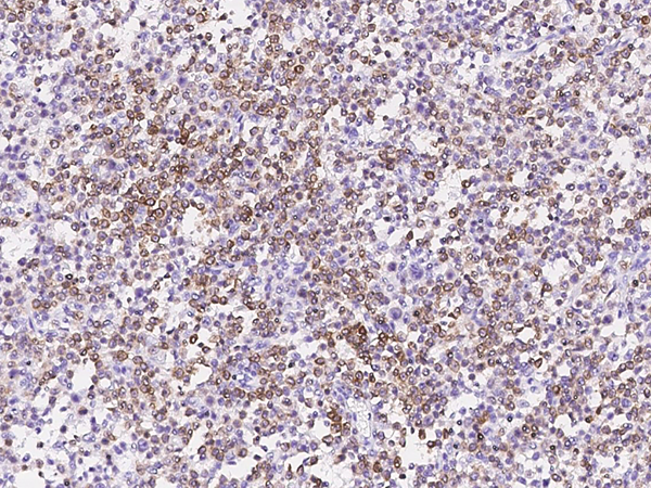
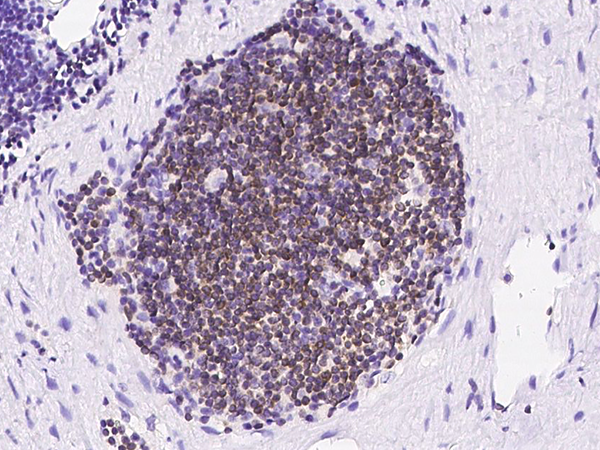
(Immunochemical staining of human BCL2 in human lymph node with mouse monoclonal antibody at 1:60 dilution, formalin-fixed paraffin embedded sections.)
IHC (Immunohistochemisry)
(Immunochemical staining of human BCL2 in human lymph node with mouse monoclonal antibody at 1:60 dilution, formalin-fixed paraffin embedded sections.)
Bcl-2, Monoclonal Antibody (Cat# AAA254662)
Full Name
Anti-Bcl-2 Antibody, Mouse Monoclonal
Gene Names
BCL2; Bcl-2; PPP1R50
Reactivity
Human
Applications
Immunohistochemistry, Western Blot
Purity
Protein A
Pricing
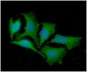 IF (Immunofluorescence)
(ICC/IF analysis of LRPAP in HeLa cells line, stained with DAPI (Blue) for nucleus staining and monoclonal anti-human LRPAP1 antibody (1:100) with goat anti-mouse IgG-Alexa fluor 488 conjugate (Green).)
IF (Immunofluorescence)
(ICC/IF analysis of LRPAP in HeLa cells line, stained with DAPI (Blue) for nucleus staining and monoclonal anti-human LRPAP1 antibody (1:100) with goat anti-mouse IgG-Alexa fluor 488 conjugate (Green).)
LRPAP1, Monoclonal Antibody (Cat# AAA48150)
Full Name
LRPAP1 antibody
Gene Names
LRPAP1; RAP; MRAP; A2RAP; HBP44; MYP23; A2MRAP; alpha-2-MRAP
Reactivity
Human
Applications
Immunofluorescence, Immunocytochemistry, Flow Cytometry, Western Blot
Purity
By protein-A affinity chromatography
Pricing

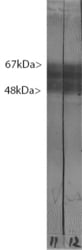 WB (Western Blot)
(Blots of crude rat brain extract stained with MAPT/TAU antibody, revealing, as expected, multiple bands in the range 48-67 kDa.)
WB (Western Blot)
(Blots of crude rat brain extract stained with MAPT/TAU antibody, revealing, as expected, multiple bands in the range 48-67 kDa.)
MAPT/Tau, Monoclonal Antibody (Cat# AAA162349)
Full Name
Anti-MAPT/Tau Antibody (clone MCA-2E9)
Reactivity
Human
Predicted: Mouse, Rat
Predicted: Mouse, Rat
Applications
Western Blot, Immunohistochemistry, Immunofluorescence, Immunocytochemistry
Purity
Affinity Purified
Pricing
 IF (Immunofluorescence)
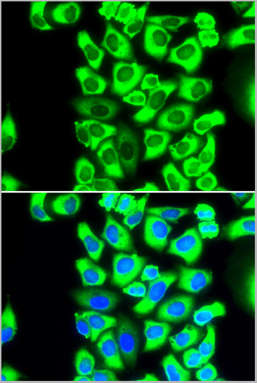
(Immunofluorescence analysis of MCF-7 cells using RPS3A antibody (AAA28136). Blue: DAPI for nuclear staining.)
IF (Immunofluorescence)
(Immunofluorescence analysis of MCF-7 cells using RPS3A antibody (AAA28136). Blue: DAPI for nuclear staining.)
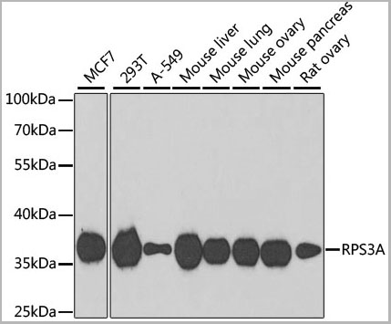
RPS3A, Antibody (Cat# AAA28136)
Full Name
RPS3A Polyclonal Antibody
Gene Names
RPS3A; S3A; FTE1; MFTL
Applications
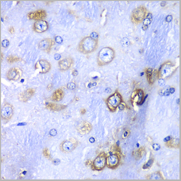
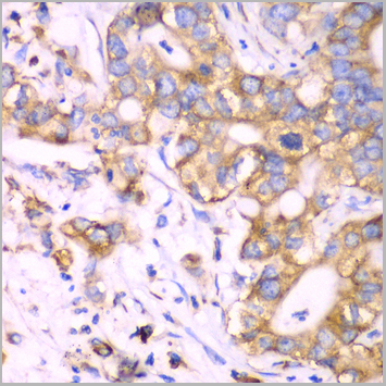
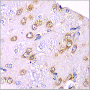
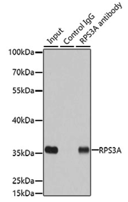
Western Blot, Immunohistochemistry, Immunofluorescence, Immunoprecipitation
Purity
Affinity Purification
Pricing
 IF (Immunofluorescence)
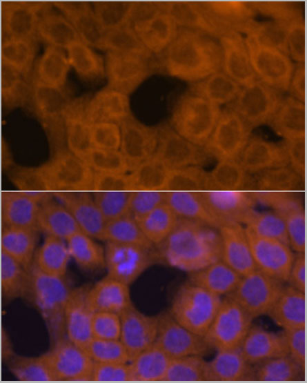
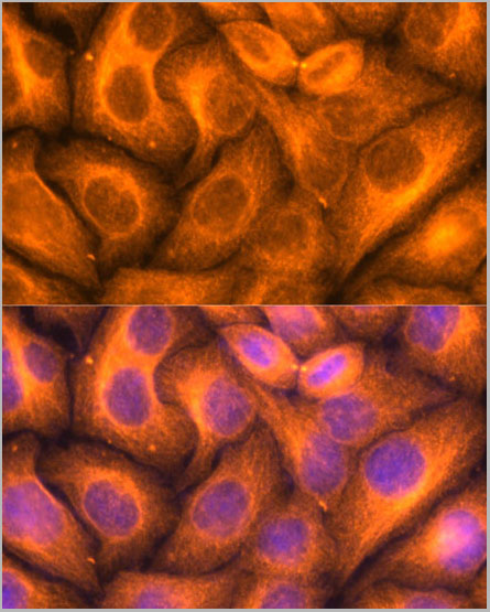
(Immunofluorescence analysis of HeLa cells using β-Tubulin antibody at dilution of 1:100. Blue: DAPI for nuclear staining.)
IF (Immunofluorescence)
(Immunofluorescence analysis of HeLa cells using β-Tubulin antibody at dilution of 1:100. Blue: DAPI for nuclear staining.)
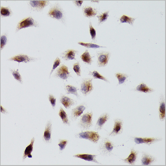
beta-Tubulin, Polyclonal Antibody (Cat# AAA28394)
Full Name
beta-Tubulin Rabbit pAb
Gene Names
TUBB; M40; TUBB1; TUBB5; OK/SW-cl.56
Reactivity
Human, Mouse, Rat
Applications
Western Blot, Immunohistochemistry, Immunofluorescence
Purity
Affinity purification
Pricing
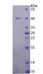 SDS-PAGE
Mdm2 p53 Binding Protein Homolog, Recombinant Protein (Cat# AAA144146)
Full Name
Recombinant Mdm2 p53 Binding Protein Homolog (MDM2)
Applications
Western Blot
Purity
> 95%
Pricing

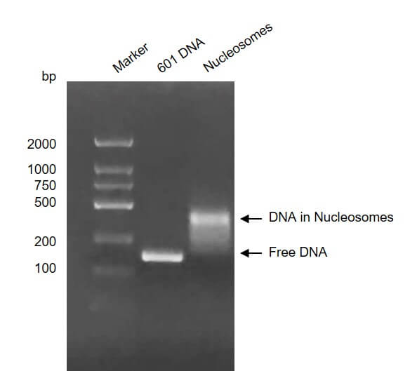
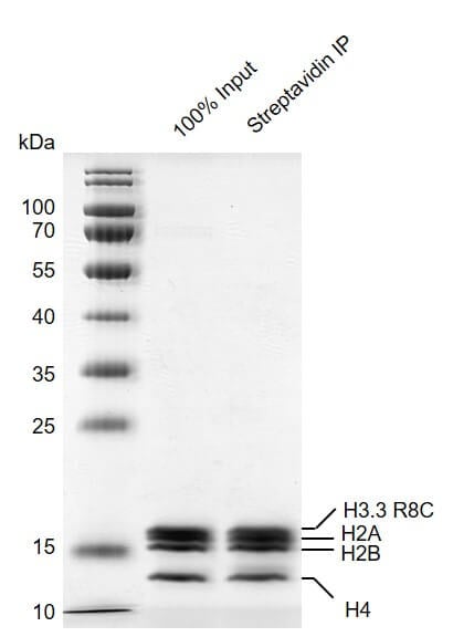 SDS-PAGE
(Streptavidin pull down assay for Recombinant Mononucleosomes H3.3 (R8C) - biotin 24 ug biotinylated mononucleosomes were incubated with 10 ul streptavidin beads for 1 hr at 4 degree C. Streptavidin beads were washed 3 times with 1 ml binding buffer. Then the beads were added 60 ul 2×SDS loading buffer and boiled for 10 min at 95 degree C. 2.4 ul samples were loaded and run on a 12.5% SDS-PAGE gel and stained by Commassie blue. The SDS-PAGE gel result showed that almost all of biotinylated mononucleosomes were pulled down by streptavidin beads.)
SDS-PAGE
(Streptavidin pull down assay for Recombinant Mononucleosomes H3.3 (R8C) - biotin 24 ug biotinylated mononucleosomes were incubated with 10 ul streptavidin beads for 1 hr at 4 degree C. Streptavidin beads were washed 3 times with 1 ml binding buffer. Then the beads were added 60 ul 2×SDS loading buffer and boiled for 10 min at 95 degree C. 2.4 ul samples were loaded and run on a 12.5% SDS-PAGE gel and stained by Commassie blue. The SDS-PAGE gel result showed that almost all of biotinylated mononucleosomes were pulled down by streptavidin beads.)
Mononucleosomes H3.3, Recombinant Protein (Cat# AAA60207)
Full Name
Recombinant Mononucleosomes H3.3 (R8C)-biotin
Gene Names
H3F3A; H3F3; H3.3A
Pricing
 SDS-PAGE
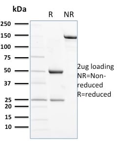
(SDS-PAGE Analysis Purified CD56 Mouse Monoclonal Antibody (ERIC-1). Confirmation of Purity and Integrity of Antibody.)
SDS-PAGE
(SDS-PAGE Analysis Purified CD56 Mouse Monoclonal Antibody (ERIC-1). Confirmation of Purity and Integrity of Antibody.)
CD56/NCAM1/NKH1, Monoclonal Antibody (Cat# AAA215099)
Full Name
CD56/NCAM1/NKH1 (Neuronal Cell Marker)
Gene Names
NCAM1; CD56; NCAM; MSK39
Reactivity
Human, Rat
Applications
Immunohistochemistry
Purity
1.0mg/mL of Purified Ab WITHOUT BSA and Azide.
Pricing
 ELISA
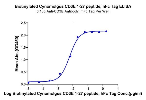
(Biotinylated Cynomolgus CD3E/CD3 epsilon 1-27 peptide Protein ELISA DataImmobilized Anti-CD3 Antibody, mFc Tag at 1?g/ml (100?l/well) on the plate. Dose response curve for Biotinylated Cynomolgus CD3E 1-27 peptide, hFc Tag with the EC50 of 6ng/ml determined by ELISA.)
ELISA
(Biotinylated Cynomolgus CD3E/CD3 epsilon 1-27 peptide Protein ELISA DataImmobilized Anti-CD3 Antibody, mFc Tag at 1?g/ml (100?l/well) on the plate. Dose response curve for Biotinylated Cynomolgus CD3E 1-27 peptide, hFc Tag with the EC50 of 6ng/ml determined by ELISA.)
CD3E/CD3 epsilon 1-27 peptide, Recombinant Protein (Cat# AAA76287)
Full Name
Biotinylated Cynomolgus CD3E/CD3 epsilon 1-27 peptide Protein Recombinant
Purity
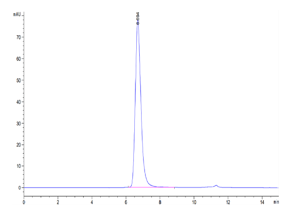
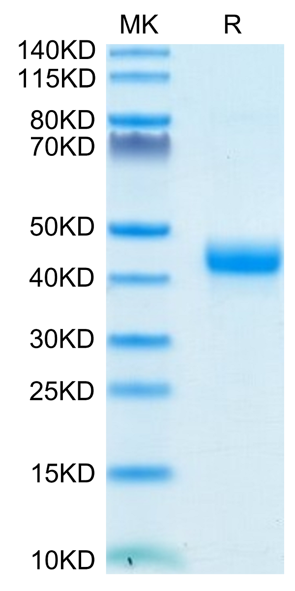
>95%, as determined by SDS-PAGE and HPLC
Pricing

 ELISA
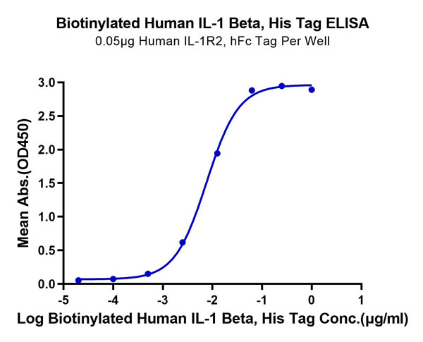
(Biotinylated Human IL-1 Beta/IL-1F2 Protein ELISA DataImmobilized Human IL-1R2, hFc Tag at 0.5?g/ml (100?l/well) on the plate. Dose response curve for Biotinylated Human IL-1 Beta, His Tag with the EC50 of 7.6ng/ml determined by ELISA.)
ELISA
(Biotinylated Human IL-1 Beta/IL-1F2 Protein ELISA DataImmobilized Human IL-1R2, hFc Tag at 0.5?g/ml (100?l/well) on the plate. Dose response curve for Biotinylated Human IL-1 Beta, His Tag with the EC50 of 7.6ng/ml determined by ELISA.)
IL-1 Beta/IL-1F2, Recombinant Protein (Cat# AAA76301)
Full Name
Biotinylated Human IL-1 Beta/IL-1F2 Protein Recombinant
Gene Names
IL1B; IL-1; IL1F2; IL1-BETA
Purity
>95%, as determined by SDS-PAGE and HPLC
Pricing
 ELISA
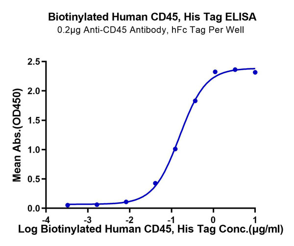
(Biotinylated Human CD45/PTPRC Protein ELISA DataImmobilized Anti-CD45 Antibody at 2?g/ml (100?l/well) on the plate. Dose response curve for Biotinylated Human CD45, His Tag with the EC50 of 0.16?g/ml determined by ELISA.)
ELISA
(Biotinylated Human CD45/PTPRC Protein ELISA DataImmobilized Anti-CD45 Antibody at 2?g/ml (100?l/well) on the plate. Dose response curve for Biotinylated Human CD45, His Tag with the EC50 of 0.16?g/ml determined by ELISA.)
CD45/PTPRC, Recombinant Protein (Cat# AAA76313)
Full Name
Biotinylated Human CD45/PTPRC Protein Recombinant
Gene Names
PTPRC; LCA; LY5; B220; CD45; L-CA; T200; CD45R; GP180
Purity
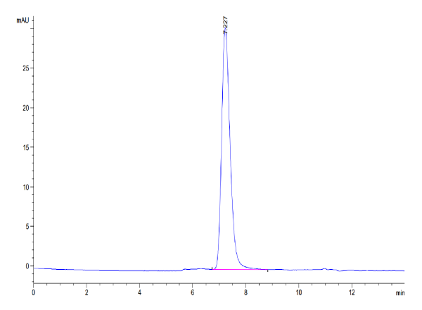
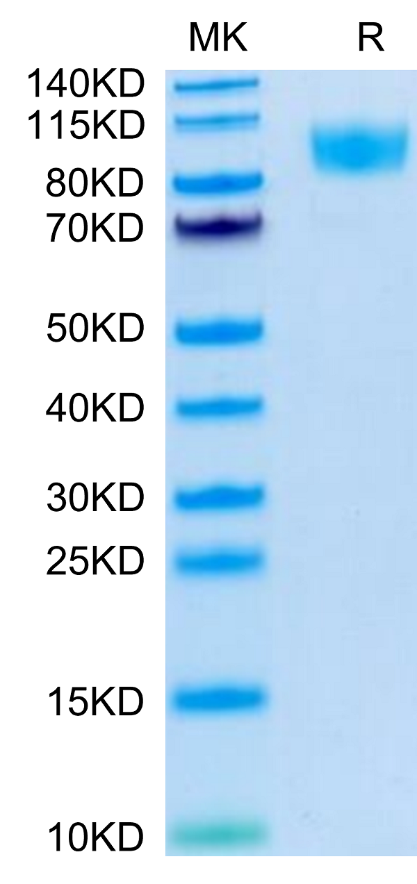
>95%, as determined by SDS-PAGE and HPLC
Pricing