Filters
▼Clonality
▼Type
▼Reactivity
▼Gene Name
▼Isotype
▼Host
▼Application
▼Clone
▼Polyclonal Antibodies
At AAA Biotech also known as AAA Bio or AAABio, we provide a broad range of purified polyclonal antibodies (pAbs) that are able to all be browsed online through our website. Due to their high specificity and strong binding affinity, these antibodies are ideal for wide swathes of research and experimental applications.
Our polyclonal antibodies can easily support your work, whether you use them for Western Blotting, Immunocytochemistry (with or without Immunofluorescence used in conjunction), Immunohistochemistry, Immunoprecipitation, and ELISA tests. We highly encourage you to browse our range of pAbs and choose the one that best suits your experimental model.
Viewing 1000-1050 of 118597 product results
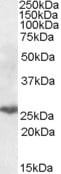
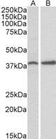
 WB (Western Blot)
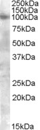
(FOXG1 antibody (pAb) tested by Western blot. Detection of Goosecoid by Western blot analysis. Lane 1: Whole-cell extract of Hek 293 cells (40 ug). Lane 2: Whole-cell extract of Jurkat cells (40 ug). Both were probed with FOXG1 antibody (pAb) at a 1:500 dilution.)
WB (Western Blot)
(FOXG1 antibody (pAb) tested by Western blot. Detection of Goosecoid by Western blot analysis. Lane 1: Whole-cell extract of Hek 293 cells (40 ug). Lane 2: Whole-cell extract of Jurkat cells (40 ug). Both were probed with FOXG1 antibody (pAb) at a 1:500 dilution.)
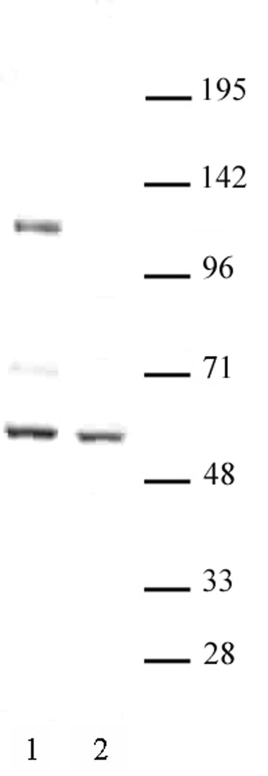
FOXG1, Polyclonal Antibody (Cat# AAA59996)
 WB (Western Blot)
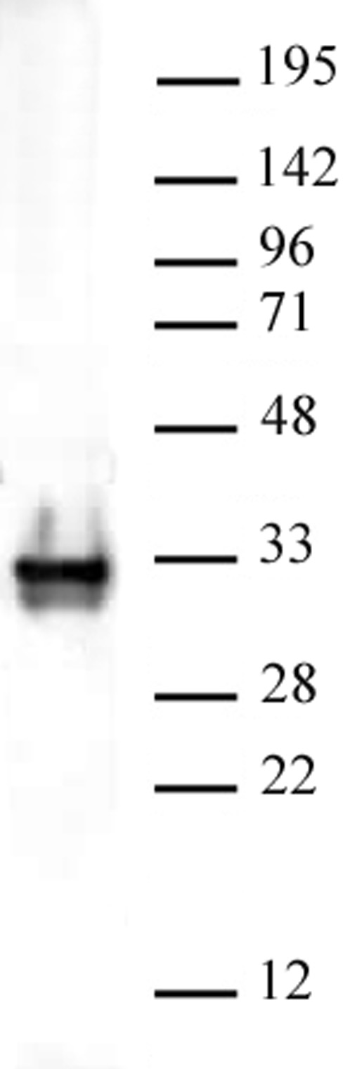
(CENP-B antibody (pAb) tested by Western blot. CENP-B detection by Western blot. The analysis was performed using 30 ug HeLa nuclear cell extract and CENP-B at a 1:500 dilution.)
WB (Western Blot)
(CENP-B antibody (pAb) tested by Western blot. CENP-B detection by Western blot. The analysis was performed using 30 ug HeLa nuclear cell extract and CENP-B at a 1:500 dilution.)
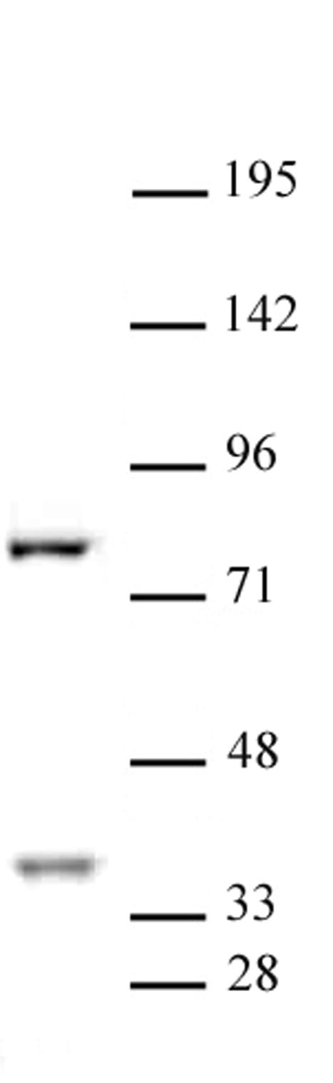
CENP-B, Polyclonal Antibody (Cat# AAA60003)

 WB (Western Blot)
(YAP1 antibody (pAb) tested by Western blot. YAP1 detection by Western blot. The analysis was performed using 40 ug HeLa whole-cell extract and YAP1 at a 1:500 dilution.)
WB (Western Blot)
(YAP1 antibody (pAb) tested by Western blot. YAP1 detection by Western blot. The analysis was performed using 40 ug HeLa whole-cell extract and YAP1 at a 1:500 dilution.)
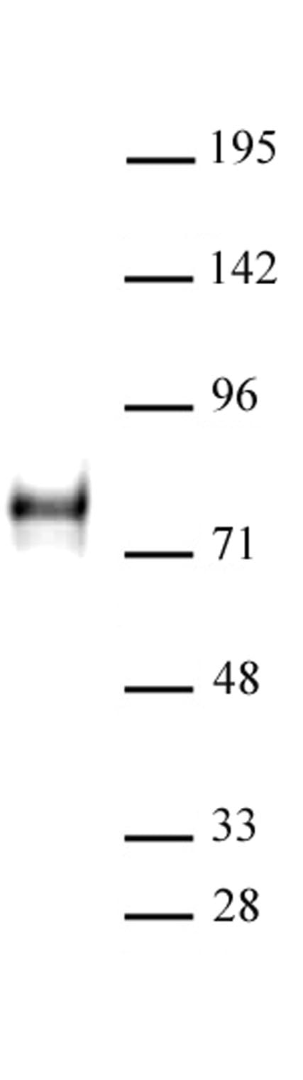
YAP1, Polyclonal Antibody (Cat# AAA60004)
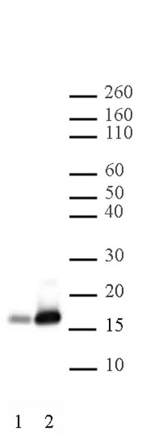
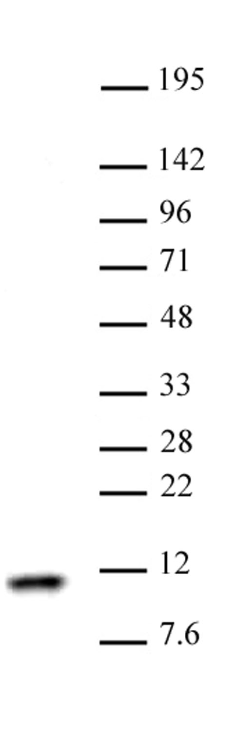 WB (Western Blot)
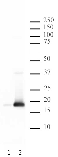
(Histone H4 antibody (pAb) tested by Western blot Nuclear extract of HeLa cells (30 ug) probed with Histone H4 antibody (1:1,000).)
WB (Western Blot)
(Histone H4 antibody (pAb) tested by Western blot Nuclear extract of HeLa cells (30 ug) probed with Histone H4 antibody (1:1,000).)
Histone H4, Polyclonal Antibody (Cat# AAA60006)

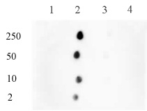
 DB (Dot Blot)
(Histone H3 dimethyl Lys4 pAb tested by dot blot analysis. Dot blot analysis was used to confirm the specificity of Histone H3 dimethyl Lys4 pAb for dimethyl-lysine 4 of histone H3. Peptides corresponding to regions around major sites of histone H3 methylation (lysine 4, lysine 9, lysine 27) were spotted onto PVDF and probed with the antibody at a dilution of 1:5,000. The amount of peptide (in picomoles) spotted is indicated next to each row. Lane 1: Unmodified Lys4 peptide. Lane 2: Monomethyl Lys4 peptide. Lane 3: Dimethyl Lys4 peptide. Lane 4: Trimethyl Lys4 peptide. Lane 5: Monomethyl Lys9 peptide. Lane 6: Unmodified Lys9 peptide. Lane 7: Dimethyl Lys9 peptide. Lane 8: Trimethyl Lys9 peptide. Lane 9: Unmodified Lys27 peptide. Lane 10: Monomethyl Lys27 peptide. Lane 11: Dimethyl Lys27 peptide. Lane 12: Trimethyl Lys27 peptide.)
DB (Dot Blot)
(Histone H3 dimethyl Lys4 pAb tested by dot blot analysis. Dot blot analysis was used to confirm the specificity of Histone H3 dimethyl Lys4 pAb for dimethyl-lysine 4 of histone H3. Peptides corresponding to regions around major sites of histone H3 methylation (lysine 4, lysine 9, lysine 27) were spotted onto PVDF and probed with the antibody at a dilution of 1:5,000. The amount of peptide (in picomoles) spotted is indicated next to each row. Lane 1: Unmodified Lys4 peptide. Lane 2: Monomethyl Lys4 peptide. Lane 3: Dimethyl Lys4 peptide. Lane 4: Trimethyl Lys4 peptide. Lane 5: Monomethyl Lys9 peptide. Lane 6: Unmodified Lys9 peptide. Lane 7: Dimethyl Lys9 peptide. Lane 8: Trimethyl Lys9 peptide. Lane 9: Unmodified Lys27 peptide. Lane 10: Monomethyl Lys27 peptide. Lane 11: Dimethyl Lys27 peptide. Lane 12: Trimethyl Lys27 peptide.)
Histone H3K4me2, Polyclonal Antibody (Cat# AAA59802)
 WB (Western Blot)
(ASH2L pAb tested by Western blot. ASH2L detection by Western blot. The analysis of ASH2L was performed using HeLa nuclear extract and ASH2L pAb at a 1:2,000 dilution.)
WB (Western Blot)
(ASH2L pAb tested by Western blot. ASH2L detection by Western blot. The analysis of ASH2L was performed using HeLa nuclear extract and ASH2L pAb at a 1:2,000 dilution.)
ASH2L, Polyclonal Antibody (Cat# AAA59826)
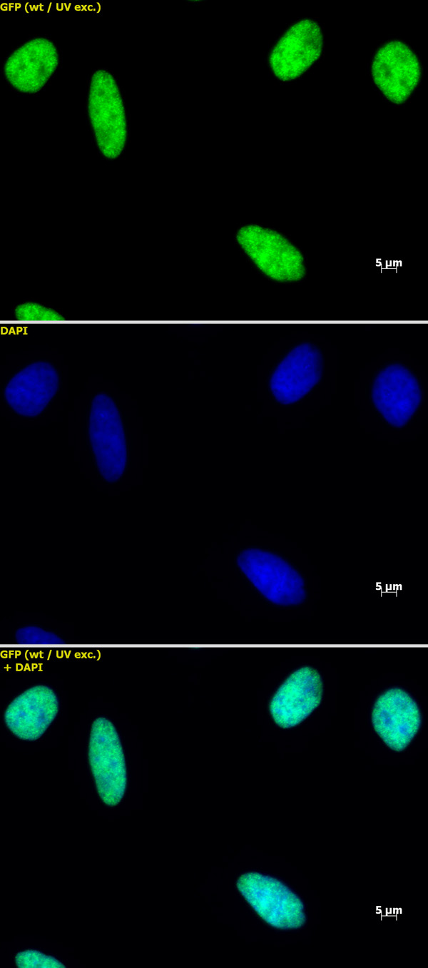
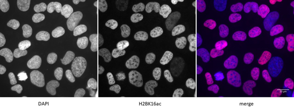 IF (Immunofluorescence)
(Detection of H2BK16ac by immunofluorescence U2OS cells were stained with H2BK16ac antibody at a dilution of 1:500. Left panel: DAPI. Middle panel: H2BK16ac antibody staining. Right panel: merge.)
IF (Immunofluorescence)
(Detection of H2BK16ac by immunofluorescence U2OS cells were stained with H2BK16ac antibody at a dilution of 1:500. Left panel: DAPI. Middle panel: H2BK16ac antibody staining. Right panel: merge.)
Histone H2BK16ac, Polyclonal Antibody (Cat# AAA59833)
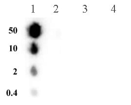
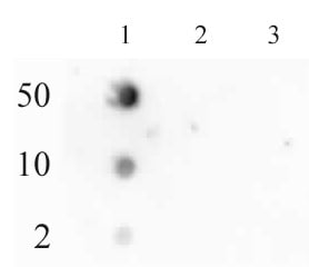
 DB (Dot Blot)
(Histone H3 phospho Thr3 pAb tested by dot blot analysis. Dot blot analysis was used to confirm the specificity of Histone H3 phospho Thr3 pAb for phospho-Thr3 of histone H3. Decreasing amounts of peptides corresponding to regions around major sites of histone H3 threonine phosphorylation were spotted onto PVDF and probed with the antibody at a dilution of 1:2,000. Lane 1: Peptide phosphorylated at threonine 3. Lane 2: Unmodified threonine 3 peptide. Lane 3: Peptide phosphorylated at threonine 11. Lane 4: Unmodified threonine 11 peptide. Lane 5: Peptide phosphorylated at threonine 45. Lane 6: Unmodified threonine 45 peptide.)
DB (Dot Blot)
(Histone H3 phospho Thr3 pAb tested by dot blot analysis. Dot blot analysis was used to confirm the specificity of Histone H3 phospho Thr3 pAb for phospho-Thr3 of histone H3. Decreasing amounts of peptides corresponding to regions around major sites of histone H3 threonine phosphorylation were spotted onto PVDF and probed with the antibody at a dilution of 1:2,000. Lane 1: Peptide phosphorylated at threonine 3. Lane 2: Unmodified threonine 3 peptide. Lane 3: Peptide phosphorylated at threonine 11. Lane 4: Unmodified threonine 11 peptide. Lane 5: Peptide phosphorylated at threonine 45. Lane 6: Unmodified threonine 45 peptide.)
Histone H3T3ph, Polyclonal Antibody (Cat# AAA59842)
 WB (Western Blot)
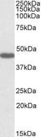
(PP2A pAb tested by Western blot. Nuclear extract of HeLa cells was probed with PP2A pAb (1:1,000 dilution).)
WB (Western Blot)
(PP2A pAb tested by Western blot. Nuclear extract of HeLa cells was probed with PP2A pAb (1:1,000 dilution).)
PP2A, Polyclonal Antibody (Cat# AAA59846)
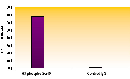
 DB (Dot Blot)
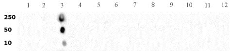
(Histone H3 phospho Ser10 antibody tested by dot blot analysis. Dot blot analysis was used to confirm the specificity of Histone H3 phospho Ser10 antibody for histone H3 phospho Ser10. Phosphorylated peptides corresponding to the immunogen and a related sequence derived from histone H3 were spotted onto PVDF and probed with the antibody at 1:50,000. The amount of peptide (picomoles) spotted is indicated next to each row. Lane 1: phospho-Ser10 peptide. Lane 2: unmodified Ser10 peptide. Lane 3: phospho-Ser28 peptide 4 peptide. Lane 4: unmodified Ser28 peptide.)
DB (Dot Blot)
(Histone H3 phospho Ser10 antibody tested by dot blot analysis. Dot blot analysis was used to confirm the specificity of Histone H3 phospho Ser10 antibody for histone H3 phospho Ser10. Phosphorylated peptides corresponding to the immunogen and a related sequence derived from histone H3 were spotted onto PVDF and probed with the antibody at 1:50,000. The amount of peptide (picomoles) spotted is indicated next to each row. Lane 1: phospho-Ser10 peptide. Lane 2: unmodified Ser10 peptide. Lane 3: phospho-Ser28 peptide 4 peptide. Lane 4: unmodified Ser28 peptide.)
Histone H3S10ph, Polyclonal Antibody (Cat# AAA59852)
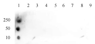
 DB (Dot Blot)
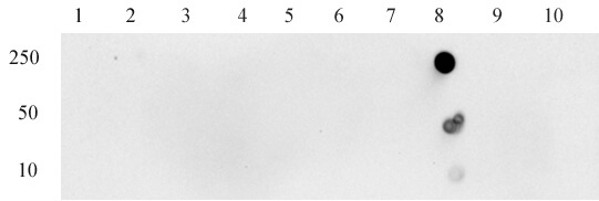
(Specificity Analysis of Histone H3K14bz antibody (pAb): Dot blot analysis was used to confirm the specificity of the antibody (pAb) for benzoylated (bz) lysine14 of histone H3. Peptides corresponding to regions around major sites of histone H3 methylation were spotted onto nitrocellulose and probed with the antibody at a dilution of 1:1,000. The amount of peptide (picomoles) spotted is indicated next to each row. Columns spotted as follows: 1: H3K23 unmod. 2: H3K23bz. 3: H4K8 unmod 4: H3K8bz. 5: H3K12 unmod. 6: H3K12bz. 7: H3K14 unmod. 8: H3K14bz. 9: H2AK9 unmod. 10: H2AK9bz.)
DB (Dot Blot)
(Specificity Analysis of Histone H3K14bz antibody (pAb): Dot blot analysis was used to confirm the specificity of the antibody (pAb) for benzoylated (bz) lysine14 of histone H3. Peptides corresponding to regions around major sites of histone H3 methylation were spotted onto nitrocellulose and probed with the antibody at a dilution of 1:1,000. The amount of peptide (picomoles) spotted is indicated next to each row. Columns spotted as follows: 1: H3K23 unmod. 2: H3K23bz. 3: H4K8 unmod 4: H3K8bz. 5: H3K12 unmod. 6: H3K12bz. 7: H3K14 unmod. 8: H3K14bz. 9: H2AK9 unmod. 10: H2AK9bz.)
Histone H3K14bz, Polyclonal Antibody (Cat# AAA59859)
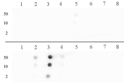
 DB (Dot Blot)
(Histone H3 dimethyl Lys14 antibody tested by dot blot analysis. Dot blot analysis was used to confirm the specificity of Histone H3 dimethyl Lys14 antibody for histone H3 dimethylated at Lys14. Methylated peptides corresponding to the immunogen and related peptides were spotted onto PVDF and probed with the antibody at a dilution of 1:5,000. The amount of peptide (picomoles) spotted is indicated in the left lane next to each row. Top panel: Lane 1: Unmodified lysine 4. Lane 2: Monomethyl lysine 4. Lane 3: Dimethyl lysine 4. Lane 4: Trimethyl lysine 4. Lane 5: Monomethyl lysine 4 #2. Lane 6: Unmodified corresponding to amino acids 6-22 of human histone H3. Lane 7: Monomethyl lysine 9. Lane 8: Dimethyl lysine 9. Lane 9: Trimethyl lysine 9. Bottom panel: Lane 1: Unmodified lysine 27. Lane 2: Monomethyl lysine 27. Lane 3: Dimethyl lysine 27. Lane 4: Trimethyl lysine 27. Lane 5: Monomethyl lysine 14 #2. Lane 6: Dimethyl lysine 14. No detection of peptides (mono-, di-, or trimethylated) corresponding to lysine 56 of histone H3 was observed (data not shown).)
DB (Dot Blot)
(Histone H3 dimethyl Lys14 antibody tested by dot blot analysis. Dot blot analysis was used to confirm the specificity of Histone H3 dimethyl Lys14 antibody for histone H3 dimethylated at Lys14. Methylated peptides corresponding to the immunogen and related peptides were spotted onto PVDF and probed with the antibody at a dilution of 1:5,000. The amount of peptide (picomoles) spotted is indicated in the left lane next to each row. Top panel: Lane 1: Unmodified lysine 4. Lane 2: Monomethyl lysine 4. Lane 3: Dimethyl lysine 4. Lane 4: Trimethyl lysine 4. Lane 5: Monomethyl lysine 4 #2. Lane 6: Unmodified corresponding to amino acids 6-22 of human histone H3. Lane 7: Monomethyl lysine 9. Lane 8: Dimethyl lysine 9. Lane 9: Trimethyl lysine 9. Bottom panel: Lane 1: Unmodified lysine 27. Lane 2: Monomethyl lysine 27. Lane 3: Dimethyl lysine 27. Lane 4: Trimethyl lysine 27. Lane 5: Monomethyl lysine 14 #2. Lane 6: Dimethyl lysine 14. No detection of peptides (mono-, di-, or trimethylated) corresponding to lysine 56 of histone H3 was observed (data not shown).)
Histone H3K14me2, Polyclonal Antibody (Cat# AAA59860)
 DB (Dot Blot)
(Histone H3 monomethyl Lys122 pAb tested by dot blot analysis. Dot blot analysis was used to confirm the specificity of 39367 for monomethyl Lys122 histone H3. Methylated peptides corresponding to the immunogen were spotted onto PVDF and probed with 39367 at 1:5,000. The amount of peptide (picomoles) spotted is indicated next to each row. Lane 1: unmodified Lys122 H3 peptide. Lane 2: monomethyl-Lys122 H3 peptide. Lane 3: dimethyl-Lys122 H3 peptide. Lane 4: trimethyl-Lys122 H3 peptide.)
DB (Dot Blot)
(Histone H3 monomethyl Lys122 pAb tested by dot blot analysis. Dot blot analysis was used to confirm the specificity of 39367 for monomethyl Lys122 histone H3. Methylated peptides corresponding to the immunogen were spotted onto PVDF and probed with 39367 at 1:5,000. The amount of peptide (picomoles) spotted is indicated next to each row. Lane 1: unmodified Lys122 H3 peptide. Lane 2: monomethyl-Lys122 H3 peptide. Lane 3: dimethyl-Lys122 H3 peptide. Lane 4: trimethyl-Lys122 H3 peptide.)
Histone H3K122me1, Polyclonal Antibody (Cat# AAA59862)

 DB (Dot Blot)
(Histone H3K4ac antibody (pAb) tested by dot blot analysis. Dot blot analysis was used to confirm the specificity of Histone H3K4ac antibody. Acetylated peptides corresponding to the immunogen and related peptides were spotted onto PVDF and probed with the antibody at 1:1,000. The amount of peptide (picomoles) spotted is indicated next to each row. Column 1: H3K4ac peptide. Column 2: unmodified H3K9 peptide. Column 3: H3K9ac peptide. Column 4: unmodified H3K14 peptide. Column 5: H3K14ac peptide. Column 6: H3K18ac peptide. Column 7: H3K23ac peptide. Column 8: H3K27ac peptide.)
DB (Dot Blot)
(Histone H3K4ac antibody (pAb) tested by dot blot analysis. Dot blot analysis was used to confirm the specificity of Histone H3K4ac antibody. Acetylated peptides corresponding to the immunogen and related peptides were spotted onto PVDF and probed with the antibody at 1:1,000. The amount of peptide (picomoles) spotted is indicated next to each row. Column 1: H3K4ac peptide. Column 2: unmodified H3K9 peptide. Column 3: H3K9ac peptide. Column 4: unmodified H3K14 peptide. Column 5: H3K14ac peptide. Column 6: H3K18ac peptide. Column 7: H3K23ac peptide. Column 8: H3K27ac peptide.)
Histone H3K4ac, Polyclonal Antibody (Cat# AAA59867)

 DB (Dot Blot)
(RNA Pol II CTD phospho Ser2 antibody (pAb) tested by dot blot analysis. Dot blot analysis was used to confirm the specificity of RNA Pol II CTD phospho Ser2 pAb. Peptides corresponding to the immunogen and related peptides were spotted onto PVDF and probed with the antibody at a dilution of 1:2,000. The amount of peptide (picomoles) spotted is indicated next to each row. Lane 1: Phospho Ser2 of RNA Pol II CTD peptide. Lane 2: Unmodified RNA Pol II CTD peptide. Lane 3: Phospho Ser5 of RNA Pol II CTD peptide.)
DB (Dot Blot)
(RNA Pol II CTD phospho Ser2 antibody (pAb) tested by dot blot analysis. Dot blot analysis was used to confirm the specificity of RNA Pol II CTD phospho Ser2 pAb. Peptides corresponding to the immunogen and related peptides were spotted onto PVDF and probed with the antibody at a dilution of 1:2,000. The amount of peptide (picomoles) spotted is indicated next to each row. Lane 1: Phospho Ser2 of RNA Pol II CTD peptide. Lane 2: Unmodified RNA Pol II CTD peptide. Lane 3: Phospho Ser5 of RNA Pol II CTD peptide.)
RNA pol II CTD phospho Ser2, Polyclonal Antibody (Cat# AAA59880)

 DB (Dot Blot)
(Histone H4 acetyl Lys5 antibody tested by dot blot analysis. Dot blot analysis was used to confirm the specificity of Histone H4 acetyl Lys5 antibody for acetyl Lys5 histone H4. Acetylated peptides corresponding to the immunogen and related peptides were spotted onto PVDF and probed with the antibody at a dilution of 1:5,000. The amount of peptide (picomoles) spotted is indicated next to each row. Lane 1: acetyl-Lys5 peptide. Histone H4 acetyl Lys5 antibody tested by dot blot analysis. Dot blot analysis was used to confirm the specificity of Histone H4 acetyl Lys5 antibody for acetyl Lys5 histone H4. Acetylated peptides corresponding to the immunogen and related peptides were spotted onto PVDF and probed with the antibody at a dilution of 1:5,000. The amount of peptide (picomoles) spotted is indicated next to each row. Lane 1: acetyl-Lys5 peptide. Lane 2: unmodified Lys5 peptide. Lane 3: acetyl-Lys8 peptide. Lane 4: unmodified Lys8 peptide. Lane 5: acetyl-Lys12 peptide. Lane 6: unmodified Lys12 peptide. Lane 7: acetyl-Lys16 peptide. Lane 8: unmodified Lys16 peptide.Lane 2: unmodified Lys5 peptide. Lane 3: acetyl-Lys8 peptide. Lane 4: unmodified Lys8 peptide. Lane 5: acetyl-Lys12 peptide. Lane 6: unmodified Lys12 peptide. Lane 7: acetyl-Lys16 peptide. Lane 8: unmodified Lys16 peptide.)
DB (Dot Blot)
(Histone H4 acetyl Lys5 antibody tested by dot blot analysis. Dot blot analysis was used to confirm the specificity of Histone H4 acetyl Lys5 antibody for acetyl Lys5 histone H4. Acetylated peptides corresponding to the immunogen and related peptides were spotted onto PVDF and probed with the antibody at a dilution of 1:5,000. The amount of peptide (picomoles) spotted is indicated next to each row. Lane 1: acetyl-Lys5 peptide. Histone H4 acetyl Lys5 antibody tested by dot blot analysis. Dot blot analysis was used to confirm the specificity of Histone H4 acetyl Lys5 antibody for acetyl Lys5 histone H4. Acetylated peptides corresponding to the immunogen and related peptides were spotted onto PVDF and probed with the antibody at a dilution of 1:5,000. The amount of peptide (picomoles) spotted is indicated next to each row. Lane 1: acetyl-Lys5 peptide. Lane 2: unmodified Lys5 peptide. Lane 3: acetyl-Lys8 peptide. Lane 4: unmodified Lys8 peptide. Lane 5: acetyl-Lys12 peptide. Lane 6: unmodified Lys12 peptide. Lane 7: acetyl-Lys16 peptide. Lane 8: unmodified Lys16 peptide.Lane 2: unmodified Lys5 peptide. Lane 3: acetyl-Lys8 peptide. Lane 4: unmodified Lys8 peptide. Lane 5: acetyl-Lys12 peptide. Lane 6: unmodified Lys12 peptide. Lane 7: acetyl-Lys16 peptide. Lane 8: unmodified Lys16 peptide.)
Histone H4K5ac, Polyclonal Antibody (Cat# AAA59886)
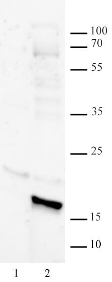
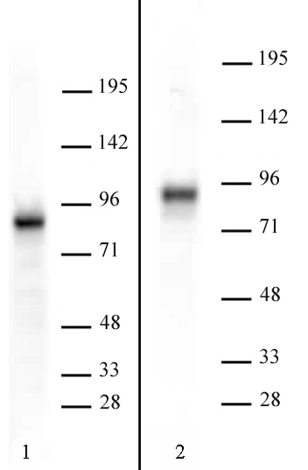
 WB (Western Blot)
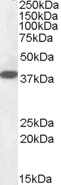
(Histone macroH2A1 pAb tested by Western blot. HeLa nuclear extract (20 ug per lane) was probed with Histone macroH2A1 pAb at a dilution of 1:2,000.)
WB (Western Blot)
(Histone macroH2A1 pAb tested by Western blot. HeLa nuclear extract (20 ug per lane) was probed with Histone macroH2A1 pAb at a dilution of 1:2,000.)
Histone macroH2A1, Polyclonal Antibody (Cat# AAA59890)
 WB (Western Blot)
(Histone H1 pAb tested by Western blot. HeLa nuclear extract (20 ug) probed with Histone H1 pAb (1:2,000 dilution).)
WB (Western Blot)
(Histone H1 pAb tested by Western blot. HeLa nuclear extract (20 ug) probed with Histone H1 pAb (1:2,000 dilution).)
Histone H1, Polyclonal Antibody (Cat# AAA59920)
 DB (Dot Blot)
(Histone H3 dimethyl Arg17 asymmetric pAb tested by dot blot analysis. Dot blot analysis was used to confirm the specificity of Histone H3 dimethyl Arg17 asymmetric pAb for dimethyl Arg17 of histone H3. Peptides corresponding to the immunogen and related peptides were spotted onto PVDF and probed with Histone H3 dimethyl Arg17 asymmetric pAb at a dilution of 1:30,000. The amount of peptide (picomoles) spotted is indicated next to each row. Top panel Lane 1: unmodified Arg2. Lane 2: monomethyl-Arg2 H3. Lane 3: dimethyl-Arg2 H3 (sym). Lane 4: dimethyl-Arg2 H3 (asym). Lane 5: unmodified Arg8. Lane 6: monomethyl-Arg8 H3. Lane 7: dimethyl-Arg8 H3 (sym). Lane 8: dimethyl-Arg8 H3 (asym). Bottom panel Lane 1: unmodified Arg17. Lane 2: monomethyl-Arg17 H3. Lane 3: dimethyl-Arg17 H3 (asym). Lane 4: dimethyl-Arg17 H3 (sym). Lane 5: unmodified Arg26. Lane 6: monomethyl-Arg26 H3. Lane 7: dimethyl-Arg26 H3 (asym). Lane 8: dimethyl-Arg26 H3 (sym).)
DB (Dot Blot)
(Histone H3 dimethyl Arg17 asymmetric pAb tested by dot blot analysis. Dot blot analysis was used to confirm the specificity of Histone H3 dimethyl Arg17 asymmetric pAb for dimethyl Arg17 of histone H3. Peptides corresponding to the immunogen and related peptides were spotted onto PVDF and probed with Histone H3 dimethyl Arg17 asymmetric pAb at a dilution of 1:30,000. The amount of peptide (picomoles) spotted is indicated next to each row. Top panel Lane 1: unmodified Arg2. Lane 2: monomethyl-Arg2 H3. Lane 3: dimethyl-Arg2 H3 (sym). Lane 4: dimethyl-Arg2 H3 (asym). Lane 5: unmodified Arg8. Lane 6: monomethyl-Arg8 H3. Lane 7: dimethyl-Arg8 H3 (sym). Lane 8: dimethyl-Arg8 H3 (asym). Bottom panel Lane 1: unmodified Arg17. Lane 2: monomethyl-Arg17 H3. Lane 3: dimethyl-Arg17 H3 (asym). Lane 4: dimethyl-Arg17 H3 (sym). Lane 5: unmodified Arg26. Lane 6: monomethyl-Arg26 H3. Lane 7: dimethyl-Arg26 H3 (asym). Lane 8: dimethyl-Arg26 H3 (sym).)
Histone H3R17me2a, Polyclonal Antibody (Cat# AAA59921)

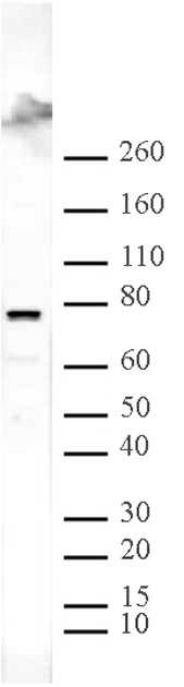
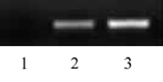
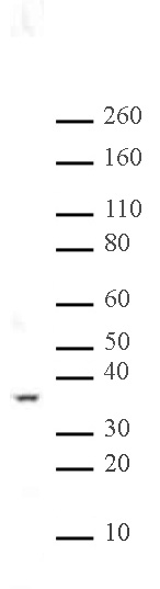
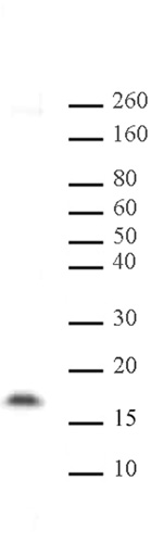
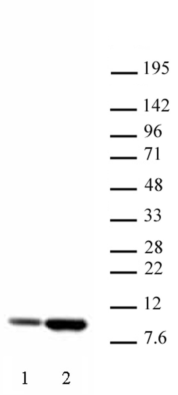
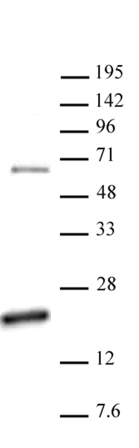
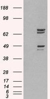
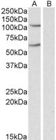
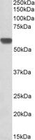
 WB (Western Blot)
(TORC2 antibody (pAb) tested by Western blot. Detection of TORC2 by Western blot analysis. Whole cell extract (20 ug) of NIH/3T3 (lane 1) and HEK293 (lane 2) blotted with TORC2 antibody at a dilution of 1:2,000.)
WB (Western Blot)
(TORC2 antibody (pAb) tested by Western blot. Detection of TORC2 by Western blot analysis. Whole cell extract (20 ug) of NIH/3T3 (lane 1) and HEK293 (lane 2) blotted with TORC2 antibody at a dilution of 1:2,000.)
TORC2, Polyclonal Antibody (Cat# AAA59928)
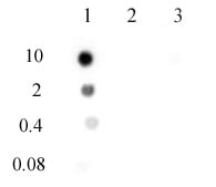
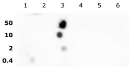
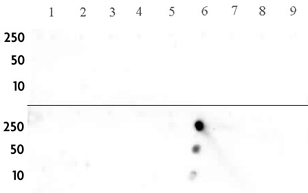
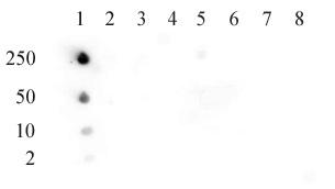
 DB (Dot Blot)
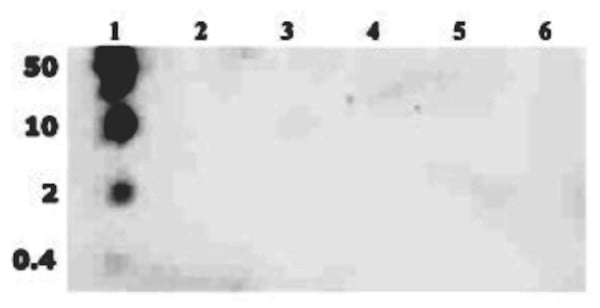
(N6-Methyladenosine (m6A) antibody (pAb) tested by DNA dot blot analysis. Single-stranded DNA oligonucleotides (amount of oligo in pmoles listed on the left side of the blot) were spotted on to a positively charged nylon membrane and blotted with N-6-methyladenosine antibody (1 ug/ml dilution). Lane 1: DNA containing a single 6-methyladenosine. Lane 2: DNA containing unmethylated adenosine. Lane 3: DNA containing a single 1-methyladenosine.)
DB (Dot Blot)
(N6-Methyladenosine (m6A) antibody (pAb) tested by DNA dot blot analysis. Single-stranded DNA oligonucleotides (amount of oligo in pmoles listed on the left side of the blot) were spotted on to a positively charged nylon membrane and blotted with N-6-methyladenosine antibody (1 ug/ml dilution). Lane 1: DNA containing a single 6-methyladenosine. Lane 2: DNA containing unmethylated adenosine. Lane 3: DNA containing a single 1-methyladenosine.)
N6-Methyladenosine, Polyclonal Antibody (Cat# AAA60036)

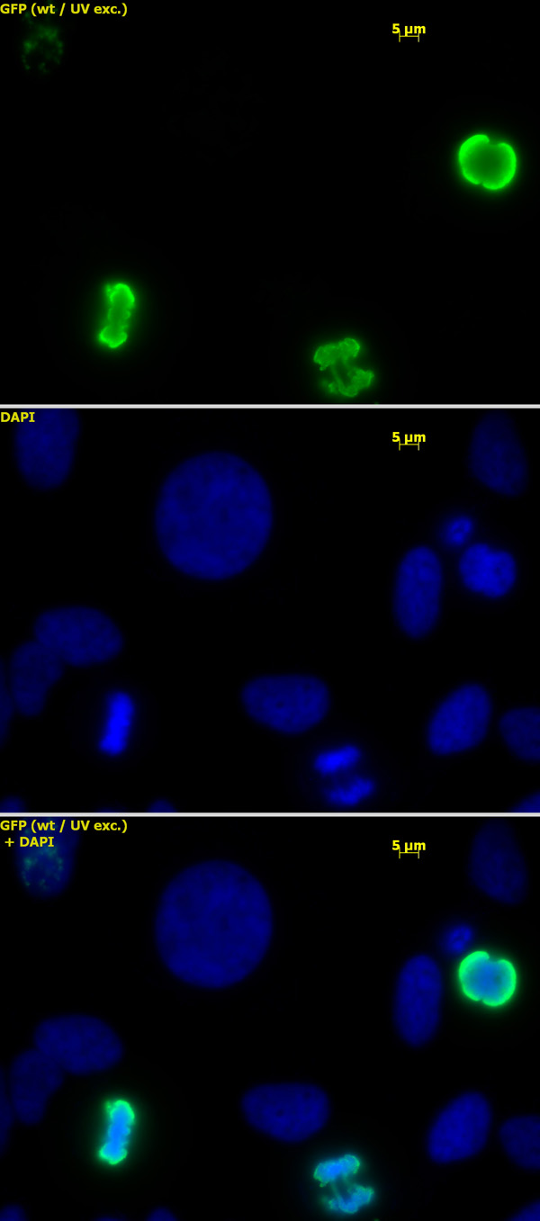
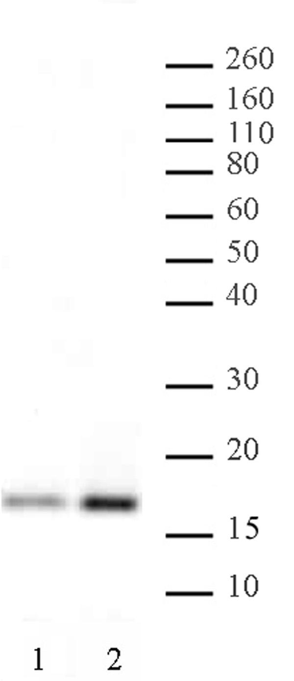
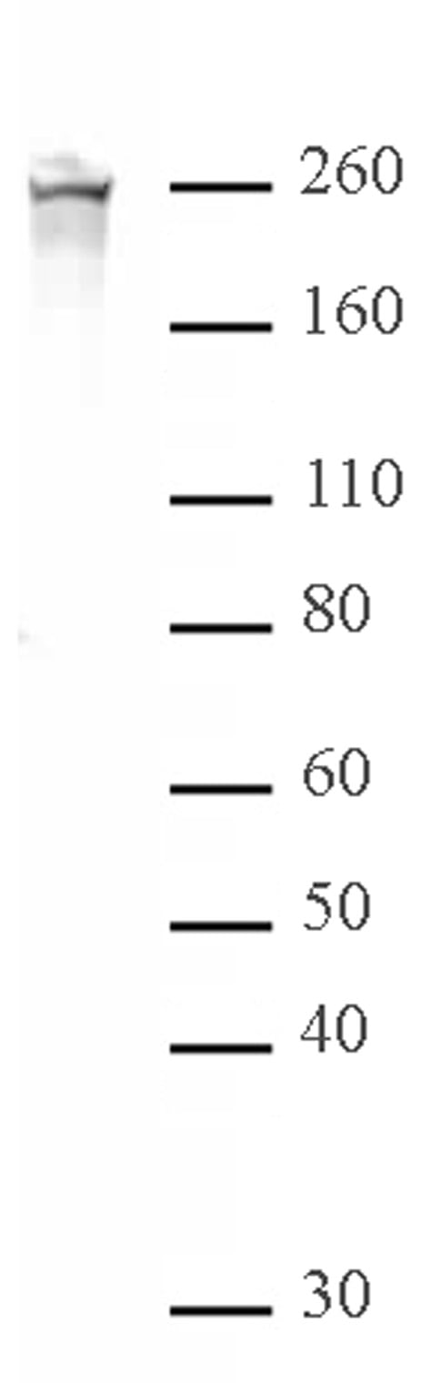
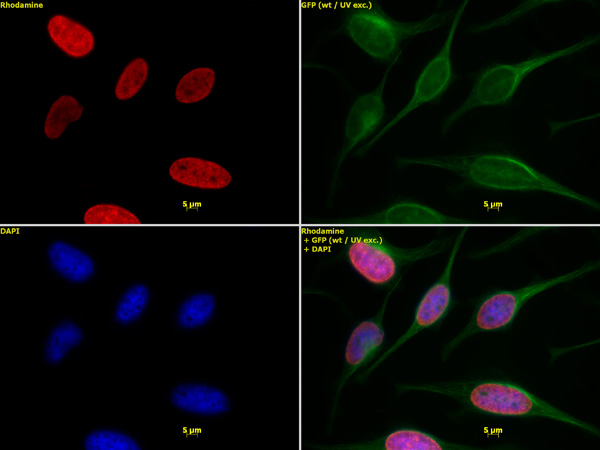
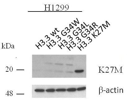 WB (Western Blot)
(Western blot of Histone H3.3K27M pAb. H1299 cell extracts transfected with human Histone H3.3 wild-type (wt) or various Histone H3.3 single amino acid substitutions were probed with Histone H3.3K27M antibody (pAb) at 2 ug/ml. Beta-actin was used as a loading control.)
WB (Western Blot)
(Western blot of Histone H3.3K27M pAb. H1299 cell extracts transfected with human Histone H3.3 wild-type (wt) or various Histone H3.3 single amino acid substitutions were probed with Histone H3.3K27M antibody (pAb) at 2 ug/ml. Beta-actin was used as a loading control.)
Histone H3.3K27M, Polyclonal Antibody (Cat# AAA60091)
Calcitonin (Salmon), Polyclonal Antibody (Cat# AAA58827)
IGF-2 (Human), Polyclonal Antibody (Cat# AAA58831)
PTH [39-84] IgG fraction, Polyclonal Antibody (Cat# AAA58834)
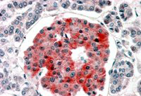
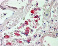
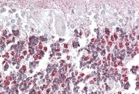
 IHC (Immunohistochemisry)
((3.8ug/ml) staining of paraffin embedded Human Cerebellum Steamed antigen retrieval with citrate buffer pH 6, AP-staining.)
IHC (Immunohistochemisry)
((3.8ug/ml) staining of paraffin embedded Human Cerebellum Steamed antigen retrieval with citrate buffer pH 6, AP-staining.)
SHP2/PTPN11, Polyclonal Antibody (Cat# AAA61286)
 WB (Western Blot)
((0.1ug/ml) staining of Human Ovary lysate (35ug protein in RIPA buffer). Primary incubation was 1 hour. Detected by chemiluminescence.)
WB (Western Blot)
((0.1ug/ml) staining of Human Ovary lysate (35ug protein in RIPA buffer). Primary incubation was 1 hour. Detected by chemiluminescence.)
Fibulin 5/FBLN5, Polyclonal Antibody (Cat# AAA61289)
 Application Data
((3.75 ug/mL) staining of paraffin embedded Human Cortex. Steamed antigen retrieval with citrate buffer pH, AP-staining.)
Application Data
((3.75 ug/mL) staining of paraffin embedded Human Cortex. Steamed antigen retrieval with citrate buffer pH, AP-staining.)
NCAM2/OCAM, Polyclonal Antibody (Cat# AAA61292)
Expected from sequence similarity: Human, Mouse, Rat, Dog, Cow
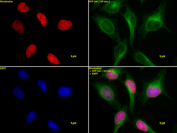
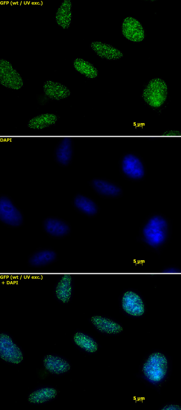
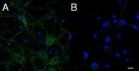 IF (Immunofluorescence)
(A) (2.5mg/ml) staining of primary rat cortical neurons showed localization of IFT74 to vesicles in the cell body and along the neuronal processes. B) Control. (Data were kindly provided by Dr. Bryan Traynor. ))
IF (Immunofluorescence)
(A) (2.5mg/ml) staining of primary rat cortical neurons showed localization of IFT74 to vesicles in the cell body and along the neuronal processes. B) Control. (Data were kindly provided by Dr. Bryan Traynor. ))
CMG1/CCDC2/IFT74, Polyclonal Antibody (Cat# AAA61299)
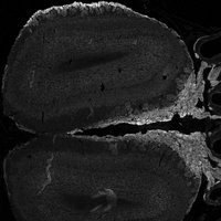
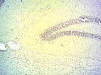 IHC (Immunohistochemistry)
((2ug/ml) staining of PFA-fixed cryo-sectioned Mouse Hippocampus. Microwaved antigen retrieval with citrate buffer pH 4.5, HRP-staining.)
IHC (Immunohistochemistry)
((2ug/ml) staining of PFA-fixed cryo-sectioned Mouse Hippocampus. Microwaved antigen retrieval with citrate buffer pH 4.5, HRP-staining.)
DIO2, Polyclonal Antibody (Cat# AAA61301)
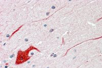
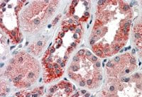
 IHC (Immunohiostchemistry)
((3.8ug/ml) staining of paraffin embedded Human Pancreas. Steamed antigen retrieval with citrate buffer pH 6, AP-staining.)
IHC (Immunohiostchemistry)
((3.8ug/ml) staining of paraffin embedded Human Pancreas. Steamed antigen retrieval with citrate buffer pH 6, AP-staining.)
KCNJ11/KATP, Polyclonal Antibody (Cat# AAA61304)
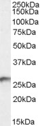
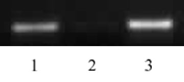
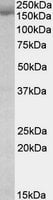
 WB (Western Blot)
(HEK293 overexpressing BDH2 (RC210586) and probed (mock transfection in first lane), tested by Origene.)
WB (Western Blot)
(HEK293 overexpressing BDH2 (RC210586) and probed (mock transfection in first lane), tested by Origene.)
BDH2/DHRS6, Polyclonal Antibody (Cat# AAA61322)
 IHC (Immunohiostchemistry)
((2.5ug/ml) staining of paraffin embedded Human Lung. Steamed antigen retrieval with citrate buffer pH 6, AP-staining.)
IHC (Immunohiostchemistry)
((2.5ug/ml) staining of paraffin embedded Human Lung. Steamed antigen retrieval with citrate buffer pH 6, AP-staining.)
CES1, Polyclonal Antibody (Cat# AAA61329)
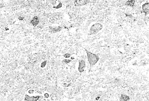
 IHC (Immunohiostchemistry)
((5ug/ml) staining of paraffin embedded Human Liver. Microwaved antigen retrieval with Tris/EDTA buffer pH9, HRP-staining.)
IHC (Immunohiostchemistry)
((5ug/ml) staining of paraffin embedded Human Liver. Microwaved antigen retrieval with Tris/EDTA buffer pH9, HRP-staining.)
Apolipoprotein L6, Polyclonal Antibody (Cat# AAA61337)
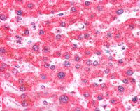
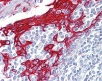
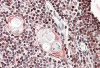 IHC (Immunohiostchemistry)
((3.8ug/ml) staining of paraffin embedded Human Thymus. Steamed antigen retrieval with citrate buffer pH 6, AP-staining.)
IHC (Immunohiostchemistry)
((3.8ug/ml) staining of paraffin embedded Human Thymus. Steamed antigen retrieval with citrate buffer pH 6, AP-staining.)
SATB1, Polyclonal Antibody (Cat# AAA61338)
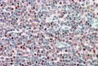 IHC (Immunohiostchemistry)
((5ug/ml) staining of paraffin embedded Human Tonsil. Steamed antigen retrieval with citrate buffer pH 6, AP-staining.)
IHC (Immunohiostchemistry)
((5ug/ml) staining of paraffin embedded Human Tonsil. Steamed antigen retrieval with citrate buffer pH 6, AP-staining.)
MECL1, Polyclonal Antibody (Cat# AAA61349)

 IHC (Immunohiostchemistry)
((2.5ug/ml) staining of paraffin embedded Human Tonsil. Steamed antigen retrieval with citrate buffer pH 6, AP-staining.)
IHC (Immunohiostchemistry)
((2.5ug/ml) staining of paraffin embedded Human Tonsil. Steamed antigen retrieval with citrate buffer pH 6, AP-staining.)
KRT13, Polyclonal Antibody (Cat# AAA61355)
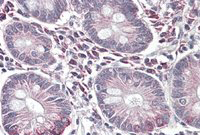 IHC (Immunohistochemistry)
((3.8ug/ml) staining of paraffin embedded Human Small Intestine. Steamed antigen retrieval with citrate buffer pH 6, AP-staining.)
IHC (Immunohistochemistry)
((3.8ug/ml) staining of paraffin embedded Human Small Intestine. Steamed antigen retrieval with citrate buffer pH 6, AP-staining.)
SLC10A2/ASBT, Polyclonal Antibody (Cat# AAA61357)
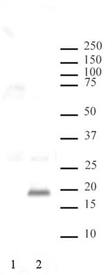
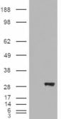
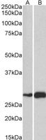
 WB (Western Blot)
((0.3ug/ml) staining of A549 lysate (35ug protein in RIPA buffer). Primary incubation was 1 hour. Detected by chemiluminescence.)
WB (Western Blot)
((0.3ug/ml) staining of A549 lysate (35ug protein in RIPA buffer). Primary incubation was 1 hour. Detected by chemiluminescence.)
FGFR2, Polyclonal Antibody (Cat# AAA61361)
SRCAP, Polyclonal Antibody (Cat# AAA61363)
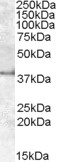
 WB (Western Blot)
((1ug/ml) staining of Rat Stomach lysate (35ug protein in RIPA buffer). Primary incubation was 1 hour. Detected by chemiluminescence.)
WB (Western Blot)
((1ug/ml) staining of Rat Stomach lysate (35ug protein in RIPA buffer). Primary incubation was 1 hour. Detected by chemiluminescence.)
PROKR2, Polyclonal Antibody (Cat# AAA61377)
TAS1R3/T1R3, Polyclonal Antibody (Cat# AAA61382)
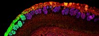
 IHC (Immunohiostchemistry)
((3.8ug/ml) staining of paraffin embedded Human Cerebellum. Steamed antigen retrieval with citrate buffer pH 6, AP-staining.)
IHC (Immunohiostchemistry)
((3.8ug/ml) staining of paraffin embedded Human Cerebellum. Steamed antigen retrieval with citrate buffer pH 6, AP-staining.)
Laforin, Polyclonal Antibody (Cat# AAA61383)
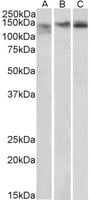 WB (Western Blot)
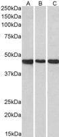
((0.3ug/ml) staining of Mouse Spleen (A), Mouse Thymus (B) and Rat Spleen (C) lysates (35ug protein in RIPA buffer). Primary incubation was 1 hour. Detected by chemiluminescence.)
WB (Western Blot)
((0.3ug/ml) staining of Mouse Spleen (A), Mouse Thymus (B) and Rat Spleen (C) lysates (35ug protein in RIPA buffer). Primary incubation was 1 hour. Detected by chemiluminescence.)
Stromal antigen 2/STAG2, Polyclonal Antibody (Cat# AAA61386)
 WB (Western Blot)
((0.3ug/ml) staining of Mouse Ovary (A) and Rat Uterus (B) lysate (35ug protein in RIPA buffer). Primary incubation was 1 hour. Detected by chemiluminescence.)
WB (Western Blot)
((0.3ug/ml) staining of Mouse Ovary (A) and Rat Uterus (B) lysate (35ug protein in RIPA buffer). Primary incubation was 1 hour. Detected by chemiluminescence.)
PRKCDBP, Polyclonal Antibody (Cat# AAA61392)
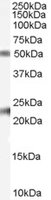
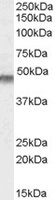
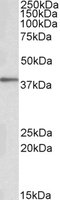
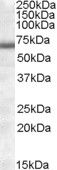
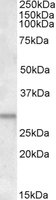
 WB (Western Blot)
((0.1ug/ml) staining of Mouse Brain lysate (35ug protein in RIPA buffer). Primary incubation was 1 hour. Detected by chemiluminescence.)
WB (Western Blot)
((0.1ug/ml) staining of Mouse Brain lysate (35ug protein in RIPA buffer). Primary incubation was 1 hour. Detected by chemiluminescence.)
ATG16L1, Polyclonal Antibody (Cat# AAA61397)
 IHC (Immunohiostchemistry)
((3.8ug/ml) staining of paraffin embedded Human Kidney. Steamed antigen retrieval with citrate buffer pH 6, AP-staining.)
IHC (Immunohiostchemistry)
((3.8ug/ml) staining of paraffin embedded Human Kidney. Steamed antigen retrieval with citrate buffer pH 6, AP-staining.)
IREB2/IRP2, Polyclonal Antibody (Cat# AAA61399)
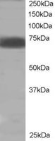
 WB (Western Blot)
(AAA61420 (1ug/ml) staining of Human Heart lysate (35ug protein in RIPA buffer). Primary incubation was 1 hour. Detected by chemiluminescence.)
WB (Western Blot)
(AAA61420 (1ug/ml) staining of Human Heart lysate (35ug protein in RIPA buffer). Primary incubation was 1 hour. Detected by chemiluminescence.)
COL4A3BP, Polyclonal Antibody (Cat# AAA61420)
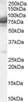
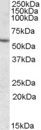
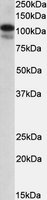
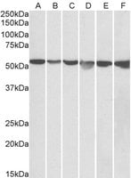 WB (Western Blot)
((0. 05ug/ml) staining of HeLa (A), A431 (B), A549 (C), MCF7 (D), Jurkat (E) and K562 (F) lysates (35ug protein in RIPA buffer). Primary incubation was 1 hour. Detected by chemiluminescence.)
WB (Western Blot)
((0. 05ug/ml) staining of HeLa (A), A431 (B), A549 (C), MCF7 (D), Jurkat (E) and K562 (F) lysates (35ug protein in RIPA buffer). Primary incubation was 1 hour. Detected by chemiluminescence.)
GPI/Neuroleukin, Polyclonal Antibody (Cat# AAA61429)
 WB (Western Blot)
((1ug/ml) staining of Mouse (A) and Rat (B) Skeletal Muscle lysate (35ug protein in RIPA buffer). Primary incubation was 1 hour. Detected by chemiluminescence.)
WB (Western Blot)
((1ug/ml) staining of Mouse (A) and Rat (B) Skeletal Muscle lysate (35ug protein in RIPA buffer). Primary incubation was 1 hour. Detected by chemiluminescence.)
ATF5, Polyclonal Antibody (Cat# AAA61443)
What are Polyclonal Antibodies?
Polyclonal antibodies are antibodies that come from multiple B cell clones of a host animal. The typical hosts used for the majority of polyclonal antibody production are rabbits, goats, sheep, and donkeys. These polyclonal antibodies, once having identified their target, will bind to different epitopes located at different regions or sequences on the same protein/antigen. This ability to bind multiple epitopes is what makes polyclonal antibodies highly sensitive, as explained in our detailed guide on polyclonal antibodies and why they matter.
As a result, they are ideal at locating and binding to the target, even if the target is in very low concentrations (due to many different antibodies being able to bind to the same target molecule, which allows for significant amplification of a downstream signal).
Polyclonal antibodies are typically produced by injecting an antigen into a host animal, which causes the animal’s immune system to attack the foreign antigen by mass generating antibodies against it. After a period of time, serum is collected from the animal and purified using physicochemical fractionation, class-specific affinity purification, and/or antigen-affinity purification.
Key Uses of Polyclonal Antibodies
- Western Blotting: This method is used to find specific proteins in biological samples after separating them by size.
- Immunohistochemistry: IHC helps visualize the location of proteins in tissue sections using various staining techniques.
- ELISA: (Enzyme-Linked Immunosorbent Assay) is typically used to identify specific protein quantities in a sample. ELISAs can be either “Quantitative” or “Qualitative”.
- Flow Cytometry: technique that identifies and measures the specific protein on the surface or inside the cells in a fluid suspension.
- Immunoprecipitation: IP isolates and studies a specific protein from a complex mixture using antibodies.
Why Buy Polyclonal Antibodies from AAA Biotech?
1. Ideal for Various Applications
Our antibodies are generally going to be validated for use in multiple types of assays, including ELISA, Western Blotting, Immunohistochemistry, Immunoprecipitation, amongst others. They are ideal for a wide range of research applications.
2. Rigorous Quality Control
All of the antibodies in our catalog undergo strict quality testing to ensure specificity, sensitivity, and consistent performance. We are confident in the ability of our antibodies to provide you with accurate results.
3. Wide Assortment of Antibodies
Antibodies in our catalog can be found for both common and exotic species, and these antibodies are also available in both conjugated and recombinant forms to suit many diverse experimental needs.
4. Highly Purified
Our antibodies are available in purified forms with over 85% purity, as confirmed by SDS-PAGE. They are also available with tags such as His, Flag, GST, or MBP. We cater to customers worldwide.
FAQ
1. How are polyclonal antibodies produced?
Traditionally, polyclonal antibodies are produced by injecting an antigen into a host animal (such as a rabbit or goat), which then triggers an immune response from the host animal. The animal’s B cells produce antibodies that will recognize different parts of the injected antigen. These antibodies are then collected from the animal’s blood and purified for use.
2. How do polyclonal antibodies differ from monoclonal antibodies?
Polyclonal antibodies are a mix of antibodies that bind to different locations (epitopes) of the same antigen, while monoclonal antibodies are identical and bind to just one specific epitope. This makes polyclonal antibodies more versatile and better at detecting proteins that may be present in low quantities or in altered/modified forms.
3. How should I store polyclonal antibodies?
Polyclonal antibodies should be stored at 4°C for short-term use (up to a few weeks) and at -20°C or -80°C for long-term storage. Avoid repeated freeze-thaw cycles by dividing them into small aliquots. Always check the datasheet for specific storage instructions.