Filters
Clonality
Type
Reactivity
Gene Name
Isotype
Host
Application
Clone
7106 results for " Mouse" - showing 6950-7000


 FCM (Flow Cytometry)
(Figure 6. Flow Cytometry analysis of CACO-2 cells using anti-TJP2/ZO2 antibody (AAA19265).Overlay histogram showing CACO-2 cells stained with AAA19265 (Blue line). The cells were blocked with 10% normal goat serum. And then incubated with rabbit anti-TJP2/ZO2 Antibody (AAA19265, 1μg/1x106 cells) for 30 min at 20 degree C. DyLight®488 conjugated goat anti-rabbit IgG (5-10μg/1x106 cells) was used as secondary antibody for 30 minutes at 20 degree C. Isotype control antibody (Green line) was rabbit IgG (1μg/1x106) used under the same conditions. Unlabelled sample (Red line) was also used as a control.)
FCM (Flow Cytometry)
(Figure 6. Flow Cytometry analysis of CACO-2 cells using anti-TJP2/ZO2 antibody (AAA19265).Overlay histogram showing CACO-2 cells stained with AAA19265 (Blue line). The cells were blocked with 10% normal goat serum. And then incubated with rabbit anti-TJP2/ZO2 Antibody (AAA19265, 1μg/1x106 cells) for 30 min at 20 degree C. DyLight®488 conjugated goat anti-rabbit IgG (5-10μg/1x106 cells) was used as secondary antibody for 30 minutes at 20 degree C. Isotype control antibody (Green line) was rabbit IgG (1μg/1x106) used under the same conditions. Unlabelled sample (Red line) was also used as a control.)
TJP2/ZO2, Polyclonal Antibody (Cat# AAA19265)
Full Name
Anti-TJP2/ZO2 Antibody
Gene Names
TJP2; ZO2; X104; PFIC4; DFNA51; DUP9q21.11; C9DUPq21.11
Reactivity
Human, Monkey, Mouse, Rat
Applications
WB, IHC-P, ICC, IF, FC/FACS/FCM
Purity
Immunogen affinity purified.
Pricing


 FCM (Flow Cytometry)
(Figure 8. Flow Cytometry analysis of K562 cells using anti-HP1 alpha/CBX5 antibody (AAA19266).Overlay histogram showing K562 cells stained with AAA19266 (Blue line). The cells were blocked with 10% normal goat serum. And then incubated with rabbit anti-HP1 alpha/CBX5 Antibody (AAA19266,1μg/1x106 cells) for 30 min at 20 degree C. DyLight®488 conjugated goat anti-rabbit IgG (5-10μg/1x106 cells) was used as secondary antibody for 30 minutes at 20 degree C. Isotype control antibody (Green line) was rabbit IgG (1μg/1x106) used under the same conditions. Unlabelled sample (Red line) was also used as a control.)
FCM (Flow Cytometry)
(Figure 8. Flow Cytometry analysis of K562 cells using anti-HP1 alpha/CBX5 antibody (AAA19266).Overlay histogram showing K562 cells stained with AAA19266 (Blue line). The cells were blocked with 10% normal goat serum. And then incubated with rabbit anti-HP1 alpha/CBX5 Antibody (AAA19266,1μg/1x106 cells) for 30 min at 20 degree C. DyLight®488 conjugated goat anti-rabbit IgG (5-10μg/1x106 cells) was used as secondary antibody for 30 minutes at 20 degree C. Isotype control antibody (Green line) was rabbit IgG (1μg/1x106) used under the same conditions. Unlabelled sample (Red line) was also used as a control.)
HP1 alpha/CBX5, Polyclonal Antibody (Cat# AAA19266)
Full Name
Anti-HP1 alpha/CBX5 Antibody
Gene Names
CBX5; HP1; HP1A
Reactivity
Human, Mouse, Rat
Applications
WB, IHC-P, ICC, IF, FC/FACS/FCM, EIA
Purity
Immunogen affinity purified.
Pricing


 FCM (Flow Cytometry)
(Figure 8. Flow Cytometry analysis of C6 cells using anti-Clathrin heavy chain/CLTC antibody (AAA19270).Overlay histogram showing C6 cells stained with AAA19270 (Blue line). The cells were blocked with 10% normal goat serum. And then incubated with rabbit anti-Clathrin heavy chain/CLTC Antibody (AAA19270,1μg/1x106 cells) for 30 min at 20 degree C. DyLight®488 conjugated goat anti-rabbit IgG (5-10μg/1x106 cells) was used as secondary antibody for 30 minutes at 20 degree C. Isotype control antibody (Green line) was rabbit IgG (1μg/1x106) used under the same conditions. Unlabelled sample (Red line) was also used as a control.)
FCM (Flow Cytometry)
(Figure 8. Flow Cytometry analysis of C6 cells using anti-Clathrin heavy chain/CLTC antibody (AAA19270).Overlay histogram showing C6 cells stained with AAA19270 (Blue line). The cells were blocked with 10% normal goat serum. And then incubated with rabbit anti-Clathrin heavy chain/CLTC Antibody (AAA19270,1μg/1x106 cells) for 30 min at 20 degree C. DyLight®488 conjugated goat anti-rabbit IgG (5-10μg/1x106 cells) was used as secondary antibody for 30 minutes at 20 degree C. Isotype control antibody (Green line) was rabbit IgG (1μg/1x106) used under the same conditions. Unlabelled sample (Red line) was also used as a control.)
Clathrin heavy chain/CLTC, Polyclonal Antibody (Cat# AAA19270)
Full Name
Anti-Clathrin heavy chain/CLTC Antibody
Gene Names
CLTC; Hc; CHC; CHC17; CLH-17; CLTCL2
Reactivity
Human, Mouse, Rat
Applications
WB, IHC-P, ICC, IF, FC/FACS/FCM, EIA
Purity
Immunogen affinity purified.
Pricing


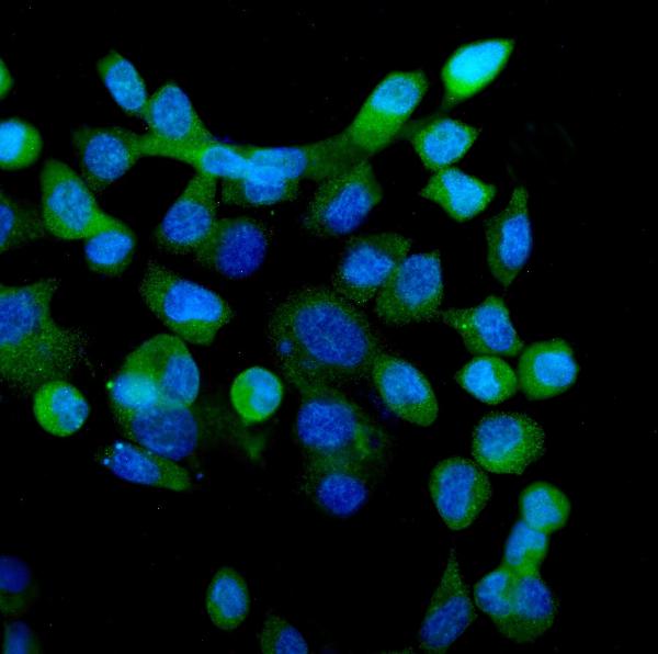
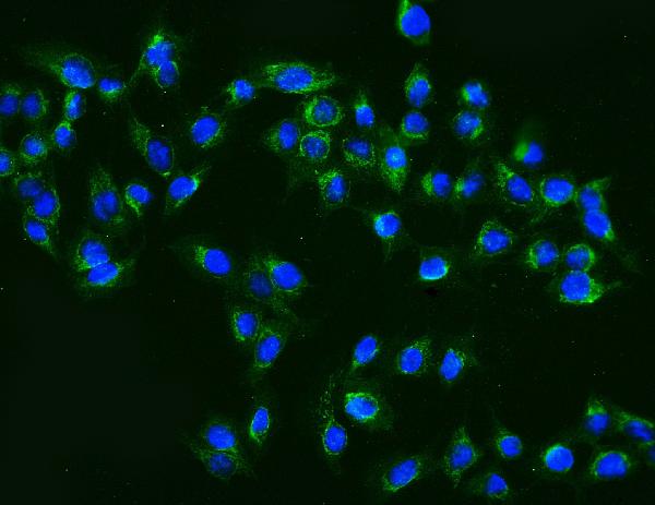
 IF (Immunofluorescence)
(Figure 9. IF analysis of ApoER2/LRP8 using anti- ApoER2/LRP8 antibody (AAA19275).ApoER2/LRP8 was detected in immunocytochemical section of HepG2 cells. Enzyme antigen retrieval was performed using IHC enzyme antigen retrieval reagent for 15 mins. The cells were blocked with 10% goat serum. And then incubated with 4μg/mL rabbit anti- ApoER2/LRP8 Antibody (AAA19275) overnight at 4 degree C. DyLight®488 Conjugated Goat Anti-Rabbit IgG was used as secondary antibody at 1:100 dilution and incubated for 30 minutes at 37 degree C. The section was counterstained with DAPI. Visualize using a fluorescence microscope and filter sets appropriate for the label used.)
IF (Immunofluorescence)
(Figure 9. IF analysis of ApoER2/LRP8 using anti- ApoER2/LRP8 antibody (AAA19275).ApoER2/LRP8 was detected in immunocytochemical section of HepG2 cells. Enzyme antigen retrieval was performed using IHC enzyme antigen retrieval reagent for 15 mins. The cells were blocked with 10% goat serum. And then incubated with 4μg/mL rabbit anti- ApoER2/LRP8 Antibody (AAA19275) overnight at 4 degree C. DyLight®488 Conjugated Goat Anti-Rabbit IgG was used as secondary antibody at 1:100 dilution and incubated for 30 minutes at 37 degree C. The section was counterstained with DAPI. Visualize using a fluorescence microscope and filter sets appropriate for the label used.)
ApoER2/LRP8, Polyclonal Antibody (Cat# AAA19275)
Full Name
Anti-ApoER2/LRP8 Antibody
Gene Names
LRP8; MCI1; LRP-8; APOER2; HSZ75190
Reactivity
Human, Mouse, Rat
Applications
WB, IHC-P, ICC, IF, FC/FACS/FCM, EIA
Purity
Immunogen affinity purified.
Pricing


 FCM (Flow Cytometry)
(Figure 12. Flow Cytometry analysis of U937 cells using anti-DYNLL1/PIN antibody (AAA19276).Overlay histogram showing U937 cells stained with AAA19276 (Blue line). The cells were blocked with 10% normal goat serum. And then incubated with rabbit anti-DYNLL1/PIN Antibody (AAA19276, 1μg/1x106 cells) for 30 min at 20 degree C. DyLight®488 conjugated goat anti-rabbit IgG (5-10μg/1x106 cells) was used as secondary antibody for 30 minutes at 20 degree C. Isotype control antibody (Green line) was rabbit IgG (1μg/1x106) used under the same conditions. Unlabelled sample (Red line) was also used as a control.)
FCM (Flow Cytometry)
(Figure 12. Flow Cytometry analysis of U937 cells using anti-DYNLL1/PIN antibody (AAA19276).Overlay histogram showing U937 cells stained with AAA19276 (Blue line). The cells were blocked with 10% normal goat serum. And then incubated with rabbit anti-DYNLL1/PIN Antibody (AAA19276, 1μg/1x106 cells) for 30 min at 20 degree C. DyLight®488 conjugated goat anti-rabbit IgG (5-10μg/1x106 cells) was used as secondary antibody for 30 minutes at 20 degree C. Isotype control antibody (Green line) was rabbit IgG (1μg/1x106) used under the same conditions. Unlabelled sample (Red line) was also used as a control.)
DYNLL1/PIN, Polyclonal Antibody (Cat# AAA19276)
Full Name
Anti-DYNLL1/PIN Antibody
Gene Names
DYNLL1; LC8; PIN; DLC1; DLC8; LC8a; DNCL1; hdlc1; DNCLC1
Reactivity
Human, Mouse, Rat
Applications
WB, IHC-P, ICC, IF, FC/FACS/FCM, EIA
Purity
Immunogen affinity purified.
Pricing
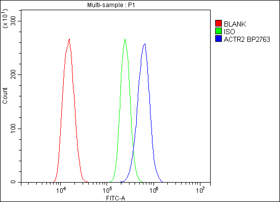


 FCM (Flow Cytometry)
(Figure 6. Flow Cytometry analysis of C6 cells using anti-Arp2/ACTR2 antibody (AAA19284).Overlay histogram showing C6 cells stained with AAA19284 (Blue line). The cells were blocked with 10% normal goat serum. And then incubated with rabbit anti-Arp2/ACTR2 Antibody (AAA19284,1μg/1x106 cells) for 30 min at 20 degree C. DyLight®488 conjugated goat anti-rabbit IgG (5-10μg/1x106 cells) was used as secondary antibody for 30 minutes at 20 degree C. Isotype control antibody (Green line) was rabbit IgG (1μg/1x106) used under the same conditions. Unlabelled sample (Red line) was also used as a control.)
FCM (Flow Cytometry)
(Figure 6. Flow Cytometry analysis of C6 cells using anti-Arp2/ACTR2 antibody (AAA19284).Overlay histogram showing C6 cells stained with AAA19284 (Blue line). The cells were blocked with 10% normal goat serum. And then incubated with rabbit anti-Arp2/ACTR2 Antibody (AAA19284,1μg/1x106 cells) for 30 min at 20 degree C. DyLight®488 conjugated goat anti-rabbit IgG (5-10μg/1x106 cells) was used as secondary antibody for 30 minutes at 20 degree C. Isotype control antibody (Green line) was rabbit IgG (1μg/1x106) used under the same conditions. Unlabelled sample (Red line) was also used as a control.)
Arp2/ACTR2, Polyclonal Antibody (Cat# AAA19284)
Full Name
Anti-Arp2/ACTR2 Antibody
Gene Names
ACTR2; ARP2
Reactivity
Human, Mouse, Monkey, Rat
Applications
WB, IHC-P, ICC, IF, FC/FACS/FCM, EIA
Purity
Immunogen affinity purified.
Pricing


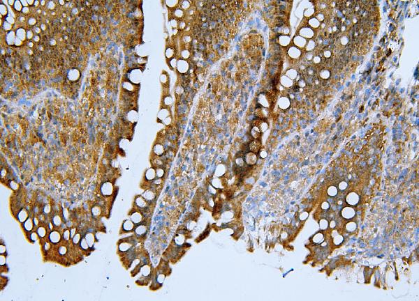
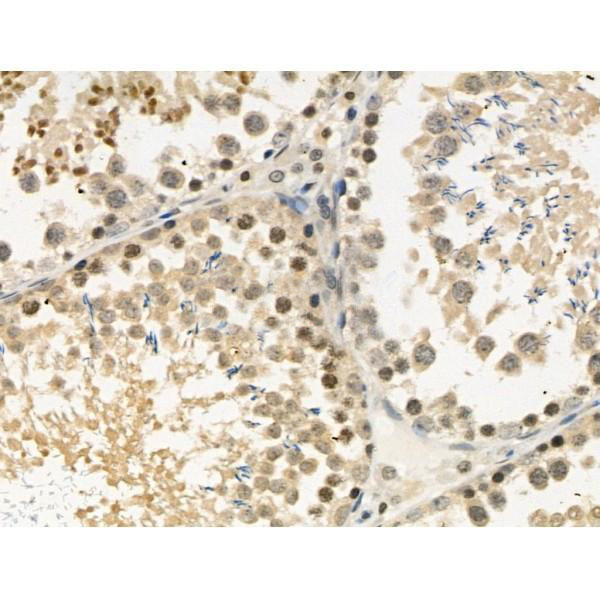
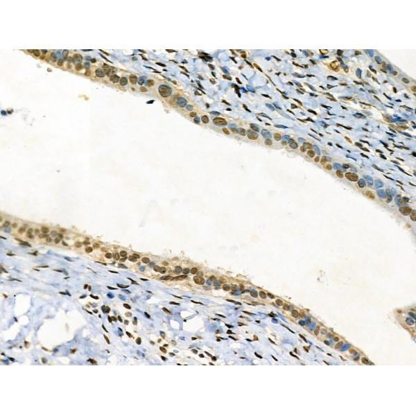
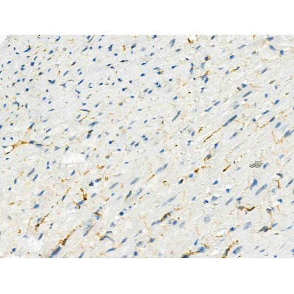
 IHC (Immunohistochemistry)
(Figure 8. IHC analysis of TAGLN/Transgelin using anti-TAGLN/Transgelin antibody (AAA19286).TAGLN/Transgelin was detected in paraffin-embedded section of rat testis tissue. Heat mediated antigen retrieval was performed in EDTA buffer (pH8. 0, epitope retrieval solution). The tissue section was blocked with 10% goat serum. The tissue section was then incubated with 2μg/ml rabbit anti-TAGLN/Transgelin Antibody (AAA19286) overnight at 4 degree C. Biotinylated goat anti-rabbit IgG was used as secondary antibody and incubated for 30 minutes at 37 degree C. The tissue section was developed using Strepavidin-Biotin-Complex (SABC) (Catalog # with DAB as the chromogen.)
IHC (Immunohistochemistry)
(Figure 8. IHC analysis of TAGLN/Transgelin using anti-TAGLN/Transgelin antibody (AAA19286).TAGLN/Transgelin was detected in paraffin-embedded section of rat testis tissue. Heat mediated antigen retrieval was performed in EDTA buffer (pH8. 0, epitope retrieval solution). The tissue section was blocked with 10% goat serum. The tissue section was then incubated with 2μg/ml rabbit anti-TAGLN/Transgelin Antibody (AAA19286) overnight at 4 degree C. Biotinylated goat anti-rabbit IgG was used as secondary antibody and incubated for 30 minutes at 37 degree C. The tissue section was developed using Strepavidin-Biotin-Complex (SABC) (Catalog # with DAB as the chromogen.)
TAGLN/Transgelin, Polyclonal Antibody (Cat# AAA19286)
Full Name
Anti-TAGLN/Transgelin Antibody
Gene Names
TAGLN; SM22; SMCC; TAGLN1; WS3-10
Reactivity
Human, Mouse, Rat
Applications
WB, IHC-P, ICC, IF, FC/FACS/FCM, EIA
Purity
Immunogen affinity purified.
Pricing


 FCM (Flow Cytometry)
(Figure 10. Flow Cytometry analysis of K562 cells using anti-HNRNPH3 antibody (AAA19338).Overlay histogram showing K562 cells stained with AAA19338 (Blue line). The cells were blocked with 10% normal goat serum. And then incubated with rabbit anti-HNRNPH3 Antibody (AAA19338, 1μg/1x106 cells) for 30 min at 20 degree C. DyLight®488 conjugated goat anti-rabbit IgG (5-10μg/1x106 cells) was used as secondary antibody for 30 minutes at 20 degree C. Isotype control antibody (Green line) was rabbit IgG (1μg/1x106) used under the same conditions. Unlabelled sample (Red line) was also used as a control.)
FCM (Flow Cytometry)
(Figure 10. Flow Cytometry analysis of K562 cells using anti-HNRNPH3 antibody (AAA19338).Overlay histogram showing K562 cells stained with AAA19338 (Blue line). The cells were blocked with 10% normal goat serum. And then incubated with rabbit anti-HNRNPH3 Antibody (AAA19338, 1μg/1x106 cells) for 30 min at 20 degree C. DyLight®488 conjugated goat anti-rabbit IgG (5-10μg/1x106 cells) was used as secondary antibody for 30 minutes at 20 degree C. Isotype control antibody (Green line) was rabbit IgG (1μg/1x106) used under the same conditions. Unlabelled sample (Red line) was also used as a control.)
HNRNPH3, Polyclonal Antibody (Cat# AAA19338)
Full Name
Anti-HNRNPH3 Antibody
Gene Names
HNRNPH3; 2H9; HNRPH3
Reactivity
Human, Mouse, Rat
Applications
WB, IHC-P, ICC, IF, FC/FACS/FCM, EIA
Purity
Immunogen affinity purified.
Pricing


 FCM (Flow Cytometry)
(Figure 8. Flow Cytometry analysis of U937 cells using anti-SEC14L3/TAP2 antibody (AAA19342).Overlay histogram showing U937 cells stained with AAA19342 (Blue line). The cells were blocked with 10% normal goat serum. And then incubated with rabbit anti-SEC14L3/TAP2 Antibody (AAA19342, 1μg/1x106 cells) for 30 min at 20 degree C. DyLight®488 conjugated goat anti-rabbit IgG (5-10μg/1x106 cells) was used as secondary antibody for 30 minutes at 20 degree C. Isotype control antibody (Green line) was rabbit IgG (1μg/1x106) used under the same conditions. Unlabelled sample (Red line) was also used as a control.)
FCM (Flow Cytometry)
(Figure 8. Flow Cytometry analysis of U937 cells using anti-SEC14L3/TAP2 antibody (AAA19342).Overlay histogram showing U937 cells stained with AAA19342 (Blue line). The cells were blocked with 10% normal goat serum. And then incubated with rabbit anti-SEC14L3/TAP2 Antibody (AAA19342, 1μg/1x106 cells) for 30 min at 20 degree C. DyLight®488 conjugated goat anti-rabbit IgG (5-10μg/1x106 cells) was used as secondary antibody for 30 minutes at 20 degree C. Isotype control antibody (Green line) was rabbit IgG (1μg/1x106) used under the same conditions. Unlabelled sample (Red line) was also used as a control.)
SEC14L3/TAP2, Polyclonal Antibody (Cat# AAA19342)
Full Name
Anti-SEC14L3/TAP2 Antibody
Gene Names
SEC14L3; TAP2
Reactivity
Human, Mouse, Rat
Applications
WB, IHC-P, ICC, IF, FC/FACS/FCM, EIA
Purity
Immunogen affinity purified.
Pricing


 FCM (Flow Cytometry)
(Figure 7. Flow Cytometry analysis of U87 cells using anti-MPI antibody (AAA19349).Overlay histogram showing U87 cells stained with AAA19349 (Blue line). The cells were blocked with 10% normal goat serum. And then incubated with mouse anti- MPI Antibody (AAA19349, 1μg/1x106 cells) for 30 min at 20 degree C. DyLight®488 conjugated goat anti-mouse IgG (BA1126, 5-10μg/1x106 cells) was used as secondary antibody for 30 minutes at 20 degree C. Isotype control antibody (Green line) was mouse IgG (1μg/1x106) used under the same conditions. Unlabelled sample (Red line) was also used as a control.)
FCM (Flow Cytometry)
(Figure 7. Flow Cytometry analysis of U87 cells using anti-MPI antibody (AAA19349).Overlay histogram showing U87 cells stained with AAA19349 (Blue line). The cells were blocked with 10% normal goat serum. And then incubated with mouse anti- MPI Antibody (AAA19349, 1μg/1x106 cells) for 30 min at 20 degree C. DyLight®488 conjugated goat anti-mouse IgG (BA1126, 5-10μg/1x106 cells) was used as secondary antibody for 30 minutes at 20 degree C. Isotype control antibody (Green line) was mouse IgG (1μg/1x106) used under the same conditions. Unlabelled sample (Red line) was also used as a control.)
MPI, Monoclonal Antibody (Cat# AAA19349)
Full Name
Anti-MPI Antibody (monoclonal, 5G5)
Gene Names
MPI; PMI; PMI1; CDG1B
Reactivity
Human, Rat
Applications
WB, IHC-P, ICC, IF, FC/FACS/FCM
Purity
Immunogen affinity purified.
Pricing


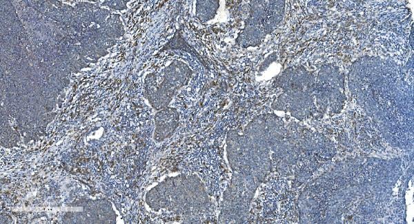


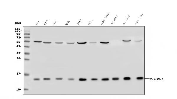
 FCM (Flow Cytometry)
(Figure 10. Flow Cytometry analysis of A431 cells using anti-TIMM8A/DDP antibody (AAA19317).Overlay histogram showing A431 cells stained with AAA19317 (Blue line). The cells were blocked with 10% normal goat serum. And then incubated with rabbit anti-TIMM8A/DDP Antibody (AAA19317, 1μg/1x106 cells) for 30 min at 20 degree C. DyLight®488 conjugated goat anti-rabbit IgG (5-10μg/1x106 cells) was used as secondary antibody for 30 minutes at 20 degree C. Isotype control antibody (Green line) was rabbit IgG (1μg/1x106) used under the same conditions. Unlabelled sample (Red line) was also used as a control.)
FCM (Flow Cytometry)
(Figure 10. Flow Cytometry analysis of A431 cells using anti-TIMM8A/DDP antibody (AAA19317).Overlay histogram showing A431 cells stained with AAA19317 (Blue line). The cells were blocked with 10% normal goat serum. And then incubated with rabbit anti-TIMM8A/DDP Antibody (AAA19317, 1μg/1x106 cells) for 30 min at 20 degree C. DyLight®488 conjugated goat anti-rabbit IgG (5-10μg/1x106 cells) was used as secondary antibody for 30 minutes at 20 degree C. Isotype control antibody (Green line) was rabbit IgG (1μg/1x106) used under the same conditions. Unlabelled sample (Red line) was also used as a control.)
TIMM8A/DDP, Polyclonal Antibody (Cat# AAA19317)
Full Name
Anti-TIMM8A/DDP Antibody
Gene Names
TIMM8A; DDP; MTS; DDP1; DFN1; TIM8
Reactivity
Human, Mouse, Rat, Monkey
Applications
WB, IHC-P, ICC, IF, EIA
Purity
Immunogen affinity purified.
Pricing


 FCM (Flow Cytometry)
(Figure 6. Flow Cytometry analysis of A549 cells using anti-MCM5 antibody (AAA19377).Overlay histogram showing A549 cells stained with AAA19377 (Blue line). The cells were blocked with 10% normal goat serum. And then incubated with mouse anti- MCM5 Antibody (AAA19377, 1μg/1x106 cells) for 30 min at 20 degree C. DyLight®488 conjugated goat anti-mouse IgG (BA1126, 5-10μg/1x106 cells) was used as secondary antibody for 30 minutes at 20 degree C. Isotype control antibody (Green line) was mouse IgG (1μg/1x106) used under the same conditions. Unlabelled sample (Red line) was also used as a control.)
FCM (Flow Cytometry)
(Figure 6. Flow Cytometry analysis of A549 cells using anti-MCM5 antibody (AAA19377).Overlay histogram showing A549 cells stained with AAA19377 (Blue line). The cells were blocked with 10% normal goat serum. And then incubated with mouse anti- MCM5 Antibody (AAA19377, 1μg/1x106 cells) for 30 min at 20 degree C. DyLight®488 conjugated goat anti-mouse IgG (BA1126, 5-10μg/1x106 cells) was used as secondary antibody for 30 minutes at 20 degree C. Isotype control antibody (Green line) was mouse IgG (1μg/1x106) used under the same conditions. Unlabelled sample (Red line) was also used as a control.)
MCM5, Monoclonal Antibody (Cat# AAA19377)
Full Name
Anti-MCM5 Antibody (monoclonal, 4G10)
Gene Names
MCM5; CDC46; P1-CDC46
Reactivity
Human, Mouse, Rat
Applications
WB, IHC-P, ICC, IF, FC/FACS/FCM
Purity
Immunogen affinity purified.
Pricing


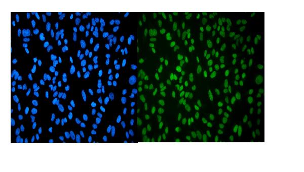
 IF (Immunofluorescence)
(Figure 8. IF analysis of eRF1/ETF1 using anti- eRF1/ETF1 antibody (AAA19383).eRF1/ETF1 was detected in immunocytochemical section of A431 cells. Enzyme antigen retrieval was performed using IHC enzyme antigen retrieval reagent for 15 mins. The cells were blocked with 10% goat serum. And then incubated with 5μg/mL mouse anti- eRF1/ETF1 Antibody (AAA19383) overnight at 4 degree C. DyLight®488 Conjugated Goat Anti-Mouse IgG (BA1126) was used as secondary antibody at 1:100 dilution and incubated for 30 minutes at 37 degree C. The section was counterstained with DAPI. Visualize using a fluorescence microscope and filter sets appropriate for the label used.)
IF (Immunofluorescence)
(Figure 8. IF analysis of eRF1/ETF1 using anti- eRF1/ETF1 antibody (AAA19383).eRF1/ETF1 was detected in immunocytochemical section of A431 cells. Enzyme antigen retrieval was performed using IHC enzyme antigen retrieval reagent for 15 mins. The cells were blocked with 10% goat serum. And then incubated with 5μg/mL mouse anti- eRF1/ETF1 Antibody (AAA19383) overnight at 4 degree C. DyLight®488 Conjugated Goat Anti-Mouse IgG (BA1126) was used as secondary antibody at 1:100 dilution and incubated for 30 minutes at 37 degree C. The section was counterstained with DAPI. Visualize using a fluorescence microscope and filter sets appropriate for the label used.)
eRF1/ETF1, Monoclonal Antibody (Cat# AAA19383)
Full Name
Anti-eRF1/ETF1 Antibody (monoclonal, 3E5)
Gene Names
ETF1; ERF; RF1; ERF1; TB3-1; D5S1995; SUP45L1
Reactivity
Human, Mouse, Rat
Applications
WB, IHC-P, ICC, IF, FC/FACS/FCM
Purity
Immunogen affinity purified.
Pricing


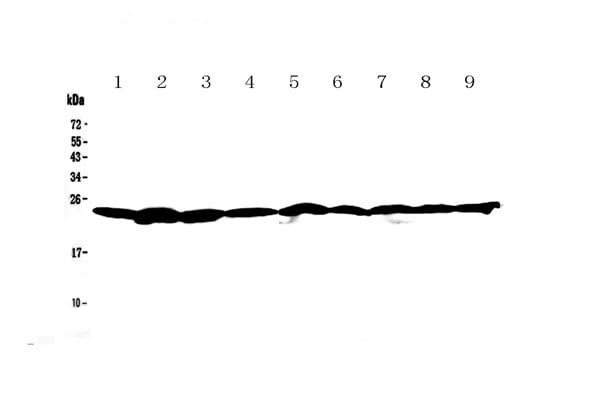 WB (Western Blot)
(Figure 1. Western blot analysis of Ran using anti-Ran antibody (AAA19131). Electrophoresis was performed on a 5-20% SDS-PAGE gel at 70V (Stacking gel) / 90V (Resolving gel) for 2-3 hours. The sample well of each lane was loaded with 50ug of sample under reducing conditions. Lane 1: rat brain tissue lysate,Lane 2: rat testis tissue lysate,Lane 3: rat thymus tissue lysate,Lane 4: mouse brain tissue lysate,Lane 5: mouse testis tissue lysate,Lane 6: mouse thymus tissue lysate,Lane 7: human A549 whole cell lysate,Lane 8: human 22RV1 whole cell lysate,Lane 9: human Hela whole cell lysate. After Electrophoresis, proteins were transferred to a Nitrocellulose membrane at 150mA for 50-90 minutes. Blocked the membrane with 5% Non-fat Milk/ TBS for 1.5 hour at RT. The membrane was incubated with rabbit anti-Ran antigen affinity purified polyclonal antibody at 0.5ug/mL overnight at 4 degree C, then washed with TBS-0.1%Tween 3 times with 5 minutes each and probed with a goat anti-rabbit IgG-HRP secondary antibody at a dilution of 1:10000 for 1.5 hour at RT. The signal is developed using an Enhanced Chemiluminescent detection (ECL) kit with Tanon 5200 system. A specific band was detected for Ran at approximately 24KD. The expected band size for Ran is at 24KD.)
WB (Western Blot)
(Figure 1. Western blot analysis of Ran using anti-Ran antibody (AAA19131). Electrophoresis was performed on a 5-20% SDS-PAGE gel at 70V (Stacking gel) / 90V (Resolving gel) for 2-3 hours. The sample well of each lane was loaded with 50ug of sample under reducing conditions. Lane 1: rat brain tissue lysate,Lane 2: rat testis tissue lysate,Lane 3: rat thymus tissue lysate,Lane 4: mouse brain tissue lysate,Lane 5: mouse testis tissue lysate,Lane 6: mouse thymus tissue lysate,Lane 7: human A549 whole cell lysate,Lane 8: human 22RV1 whole cell lysate,Lane 9: human Hela whole cell lysate. After Electrophoresis, proteins were transferred to a Nitrocellulose membrane at 150mA for 50-90 minutes. Blocked the membrane with 5% Non-fat Milk/ TBS for 1.5 hour at RT. The membrane was incubated with rabbit anti-Ran antigen affinity purified polyclonal antibody at 0.5ug/mL overnight at 4 degree C, then washed with TBS-0.1%Tween 3 times with 5 minutes each and probed with a goat anti-rabbit IgG-HRP secondary antibody at a dilution of 1:10000 for 1.5 hour at RT. The signal is developed using an Enhanced Chemiluminescent detection (ECL) kit with Tanon 5200 system. A specific band was detected for Ran at approximately 24KD. The expected band size for Ran is at 24KD.)
Ran, Polyclonal Antibody (Cat# AAA19131)
Full Name
Anti-Ran Picoband Antibody
Gene Names
RAN; TC4; Gsp1; ARA24
Reactivity
Human, Mouse, Rat
No cross reactivity with other proteins.
No cross reactivity with other proteins.
Applications
IHC, WB
Purity
Immunogen affinity purified
Pricing


 Application Data
(At 25 degree C. The primary antibody was diluted at 1/200 and incubated with the sample for 1 hour at 37 degree C. An Alexa Fluor 594 conjugated goat anti-rabbit IgG (H+L) Ab, diluted at 1/600, was used as the secondary antibody.)
Application Data
(At 25 degree C. The primary antibody was diluted at 1/200 and incubated with the sample for 1 hour at 37 degree C. An Alexa Fluor 594 conjugated goat anti-rabbit IgG (H+L) Ab, diluted at 1/600, was used as the secondary antibody.)
FOXO4, Polyclonal Antibody (Cat# AAA31400)
Full Name
Phospho-FOXO4 (Thr455) Antibody
Gene Names
FOXO4; AFX; AFX1; MLLT7
Reactivity
Human, Mouse, Rat
Predicted Reactivity: Bovine (91%), Horse (83%), Sheep (91%), Dog (91%)
Predicted Reactivity: Bovine (91%), Horse (83%), Sheep (91%), Dog (91%)
Applications
WB, IHC, IF, ICC, EIA
Purity
The antibody is from purified rabbit serum by affinity purification via sequential chromatography on phospho-peptide and non-phospho-peptide affinity columns.
Pricing


 Application Data
(At 25 degree C. The primary antibody was diluted at 1/200 and incubated with the sample for 1 hour at 37 degree C. An Alexa Fluor 594 conjugated goat anti-rabbit IgG (H+L) antibody(Red), diluted at 1/600, was used as secondary antibody.)
Application Data
(At 25 degree C. The primary antibody was diluted at 1/200 and incubated with the sample for 1 hour at 37 degree C. An Alexa Fluor 594 conjugated goat anti-rabbit IgG (H+L) antibody(Red), diluted at 1/600, was used as secondary antibody.)
RAD17, Polyclonal Antibody (Cat# AAA31431)
Full Name
Phospho-RAD17 (Ser646) Antibody
Gene Names
RAD17; CCYC; R24L; RAD24; HRAD17; RAD17SP
Reactivity
Human, Mouse, Rat
Predicted Reactivity: Pig (100%), Bovine (100%), Horse (100%), Sheep (100%)
Predicted Reactivity: Pig (100%), Bovine (100%), Horse (100%), Sheep (100%)
Applications
WB, IHC, IF, ICC, EIA
Purity
The antibody is from purified rabbit serum by affinity purification via sequential chromatography on phospho-peptide and non-phospho-peptide affinity columns.
Pricing


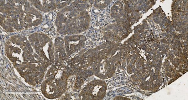
 IHC (Immunohistochemistry)
(At 1/100 staining Human gastric cancer and adjacent normal tissues by IHC-P. The sample was formaldehyde fixed and a heat mediated antigen retrieval step in citrate buffer was performed. The sample was then blocked and incubated with the primary antibody at 4 degree C overnight. An HRP conjugated anti-Rabbit antibody was used as the secondary antibody.)
IHC (Immunohistochemistry)
(At 1/100 staining Human gastric cancer and adjacent normal tissues by IHC-P. The sample was formaldehyde fixed and a heat mediated antigen retrieval step in citrate buffer was performed. The sample was then blocked and incubated with the primary antibody at 4 degree C overnight. An HRP conjugated anti-Rabbit antibody was used as the secondary antibody.)
AIRE, Polyclonal Antibody (Cat# AAA31324)
Full Name
Phospho-AIRE (Ser156) Antibody
Gene Names
AIRE; APS1; APSI; PGA1; AIRE1; APECED
Reactivity
Human, Mouse, Rat
Applications
IHC, EIA
Purity
The antibody is from purified rabbit serum by affinity purification via sequential chromatography on phospho-peptide and non-phospho-peptide affinity columns.
Pricing


 IHC (Immunohistochemistry)
(At 1/100 staining Mouse kidney tissue by IHC-P. The sample was formaldehyde fixed and a heat mediated antigen retrieval step in citrate buffer was performed. The sample was then blocked and incubated with the primary antibody at 4 degree C overnight. An HRP conjugated anti-Rabbit antibody was used as the secondary antibody.)
IHC (Immunohistochemistry)
(At 1/100 staining Mouse kidney tissue by IHC-P. The sample was formaldehyde fixed and a heat mediated antigen retrieval step in citrate buffer was performed. The sample was then blocked and incubated with the primary antibody at 4 degree C overnight. An HRP conjugated anti-Rabbit antibody was used as the secondary antibody.)
YB1, Polyclonal Antibody (Cat# AAA31294)
Full Name
Phospho-YB1 (Ser102) Antibody
Gene Names
YBX1; YB1; BP-8; CSDB; DBPB; YB-1; CSDA2; NSEP1; NSEP-1; MDR-NF1
Reactivity
Human, Mouse, Rat
Applications
IHC, EIA
Purity
The antibody is from purified rabbit serum by affinity purification via sequential chromatography on phospho-peptide and non-phospho-peptide affinity columns.
Pricing


 Application Data
(At 25 degree C. Samples were then incubated with primary Ab(At 37 degree C. An AlexaFluor594 conjugated goat anti-rabbit IgG(H+L) Ab(Red) and an AlexaFluor488 conjugated goat anti-mouse IgG(H+L) Ab(Green) were used as the secondary antibody.The nuclear counter stain is DAPI (blue).)
Application Data
(At 25 degree C. Samples were then incubated with primary Ab(At 37 degree C. An AlexaFluor594 conjugated goat anti-rabbit IgG(H+L) Ab(Red) and an AlexaFluor488 conjugated goat anti-mouse IgG(H+L) Ab(Green) were used as the secondary antibody.The nuclear counter stain is DAPI (blue).)
GSK3 beta, Polyclonal Antibody (Cat# AAA31297)
Full Name
Phospho-GSK3 beta (Thr390) Antibody
Reactivity
Human, Mouse, Rat
Applications
WB, IHC, IF, ICC, EIA
Purity
The antibody is from purified rabbit serum by affinity purification via sequential chromatography on phospho-peptide and non-phospho-peptide affinity columns.
Pricing


 Application Data
(At 25 degree C. Samples were then incubated with primary Ab(At 37 degree C. An AlexaFluor594 conjugated goat anti-rabbit IgG(H+L) Ab(Red) and an AlexaFluor488 conjugated goat anti-mouse IgG(H+L) Ab(Green) were used as the secondary antibody.The nuclear counter stain is DAPI (blue).)
Application Data
(At 25 degree C. Samples were then incubated with primary Ab(At 37 degree C. An AlexaFluor594 conjugated goat anti-rabbit IgG(H+L) Ab(Red) and an AlexaFluor488 conjugated goat anti-mouse IgG(H+L) Ab(Green) were used as the secondary antibody.The nuclear counter stain is DAPI (blue).)
Bub1, Polyclonal Antibody (Cat# AAA31312)
Full Name
Phospho-Bub1 (Ser459) Antibody
Gene Names
BUB1; BUB1A; BUB1L; hBUB1
Reactivity
Human, Rat
Applications
IHC, IF, ICC, EIA
Purity
The antibody is from purified rabbit serum by affinity purification via sequential chromatography on phospho-peptide and non-phospho-peptide affinity columns.
Pricing


 IHC (Immunohistchemistry)
(At 1/100 staining Mouse kidney tissue by IHC-P. The sample was formaldehyde fixed and a heat mediated antigen retrieval step in citrate buffer was performed. The sample was then blocked and incubated with the primary antibody at 4 degree C overnight. An HRP conjugated anti-Rabbit antibody was used as the secondary antibody.)
IHC (Immunohistchemistry)
(At 1/100 staining Mouse kidney tissue by IHC-P. The sample was formaldehyde fixed and a heat mediated antigen retrieval step in citrate buffer was performed. The sample was then blocked and incubated with the primary antibody at 4 degree C overnight. An HRP conjugated anti-Rabbit antibody was used as the secondary antibody.)
ARRB1, Polyclonal Antibody (Cat# AAA31314)
Full Name
Phospho-ARRB1 (Ser412) Antibody
Gene Names
ARRB1; ARB1; ARR1
Reactivity
Human, Mouse, Rat
Applications
WB, IHC, EIA
Purity
The antibody is from purified rabbit serum by affinity purification via sequential chromatography on phospho-peptide and non-phospho-peptide affinity columns.
Pricing


 IP (Immunoprecipitation)
(HSPA1A was immunoprecipitated using:Lane A:0.5 mg Hela Whole Cell Lysate2 uL anti-HSPA1A rabbit monoclonal antibody and 15 ul of 50 % Protein G agarose.Primary antibody:Anti-HSPA1A rabbit monoclonal antibody,at 1:200 dilution Secondary antibody:Dylight 800-labeled antibody to rabbit IgG (H+L), at 1:5000 dilution Developed using the odssey technique.Performed under reducing conditions.Predicted band size: 70 kDaObserved band size: 70 kDa)
IP (Immunoprecipitation)
(HSPA1A was immunoprecipitated using:Lane A:0.5 mg Hela Whole Cell Lysate2 uL anti-HSPA1A rabbit monoclonal antibody and 15 ul of 50 % Protein G agarose.Primary antibody:Anti-HSPA1A rabbit monoclonal antibody,at 1:200 dilution Secondary antibody:Dylight 800-labeled antibody to rabbit IgG (H+L), at 1:5000 dilution Developed using the odssey technique.Performed under reducing conditions.Predicted band size: 70 kDaObserved band size: 70 kDa)
HSP70, Monoclonal Antibody (Cat# AAA27747)
Full Name
Recombinant Anti-HSP70 Antibody, Rabbit Monoclonal
Gene Names
HSPA1B; HSP72; HSPA1; HSX70; HSP70-1; HSP70-2; HSP70.1; HSP70.2; HSP70-1B
Reactivity
Human
Applications
WB, EIA, IHC-P, FC/FACS/FCM, ICC, IF, IP
Purity
Protein A
Pricing


 Application Data
(Published Customer Image:Mouse CD31 antibody, clone ER-MP12 used for the demonstration of vasculature in mouse brain by immunofluorescence.Image caption:Inhibition of 2-AG hydrolysis reduces LPS-induced BBB permeability. a, b Fibrinogen levels in b plasma and the a ratio of brain to plasma fibrinogen were assessed by ELISA. n?=?5/7 mice per group. c, d Fluorescent immunostaining in the striatum for fibrinogen (red) and vascular marker (CD31; green) demonstrated leakage of fibrinogen into the brain with vehicle treatment, whereas vascular integrity was preserved when (e, f) MAGL was inhibited. g Extravascular fibrinogen was semi-quantitated in fluorescently labeled sections of the striatum. Bar graphs were plotted with mean?+/-SEM and data analyzed using one-way analysis of variance (ANOVA) with Tukey post-hoc comparisons. n = 5/7 mice per group. Significance is shown as *p?)
Application Data
(Published Customer Image:Mouse CD31 antibody, clone ER-MP12 used for the demonstration of vasculature in mouse brain by immunofluorescence.Image caption:Inhibition of 2-AG hydrolysis reduces LPS-induced BBB permeability. a, b Fibrinogen levels in b plasma and the a ratio of brain to plasma fibrinogen were assessed by ELISA. n?=?5/7 mice per group. c, d Fluorescent immunostaining in the striatum for fibrinogen (red) and vascular marker (CD31; green) demonstrated leakage of fibrinogen into the brain with vehicle treatment, whereas vascular integrity was preserved when (e, f) MAGL was inhibited. g Extravascular fibrinogen was semi-quantitated in fluorescently labeled sections of the striatum. Bar graphs were plotted with mean?+/-SEM and data analyzed using one-way analysis of variance (ANOVA) with Tukey post-hoc comparisons. n = 5/7 mice per group. Significance is shown as *p?)
CD31, Monoclonal Antibody (Cat# AAA12258)
Full Name
Rat Anti Mouse CD31: FITC
Gene Names
Pecam1; Cd31; Pecam; C85791; PECAM-1
Reactivity
Mouse
Applications
FC/FACS
Purity
Purified IgG prepared by affinity chromatography on Protein G from tissue culture supernatant
Pricing


 Application Data
(Published Customer Image:Rat anti Mouse Gr-1 antibody, clone RB6-8C5 used for the identification of neutrophils by immunofluorescence.Image caption:S. typhimurium-Infected Macrophages Containing Phagocytosed Neutrophils and T Cells Confocal fluorescence microscopy of 50-mum-thick liver sections from 1-wk-infected Slc11a1 wild-type mice. (A-C) S.Typhimurium (O-antigen, arrows) are red, macrophages (F4-80 and MOMA-2) are blue, DNA (DAPI) is gray, phalloidin is green, and neutrophils (Gr-1/Ly-6G/RB6-8C5) are pink (arrowheads). (A) Collapsed image from a 40-mum Z-stack. Scale bar is 20 mum. (B and C) Sections from (A) that are 4 mum apart. The video from which (A-C) were derived (Video S2) is available online. (D-G) T cells within multinucleate macrophages.Macrophages (F4-80 and MOMA-2) are blue (D, G, and H), T cells (CD3zeta) are red (D, G, arrowheads), DAPI is gray (E, G), actin-bound phalloidin is green (F, G). (G) Is a composite of (D, E, and F). Scale bars are 16 mum. (H) An image from a different mouse stained and labeled as described for (D-G). Scale bar is 8 mum. A video showing a T cell inside of a macrophage is available online (Video S3).From: Nix RN, Altschuler SE, Henson PM, Detweiler CS (2007) Hemophagocytic Macrophages Harbor Salmonella enterica during Persistent Infection.PLoS Pathog 3(12): e193.)
Application Data
(Published Customer Image:Rat anti Mouse Gr-1 antibody, clone RB6-8C5 used for the identification of neutrophils by immunofluorescence.Image caption:S. typhimurium-Infected Macrophages Containing Phagocytosed Neutrophils and T Cells Confocal fluorescence microscopy of 50-mum-thick liver sections from 1-wk-infected Slc11a1 wild-type mice. (A-C) S.Typhimurium (O-antigen, arrows) are red, macrophages (F4-80 and MOMA-2) are blue, DNA (DAPI) is gray, phalloidin is green, and neutrophils (Gr-1/Ly-6G/RB6-8C5) are pink (arrowheads). (A) Collapsed image from a 40-mum Z-stack. Scale bar is 20 mum. (B and C) Sections from (A) that are 4 mum apart. The video from which (A-C) were derived (Video S2) is available online. (D-G) T cells within multinucleate macrophages.Macrophages (F4-80 and MOMA-2) are blue (D, G, and H), T cells (CD3zeta) are red (D, G, arrowheads), DAPI is gray (E, G), actin-bound phalloidin is green (F, G). (G) Is a composite of (D, E, and F). Scale bars are 16 mum. (H) An image from a different mouse stained and labeled as described for (D-G). Scale bar is 8 mum. A video showing a T cell inside of a macrophage is available online (Video S3).From: Nix RN, Altschuler SE, Henson PM, Detweiler CS (2007) Hemophagocytic Macrophages Harbor Salmonella enterica during Persistent Infection.PLoS Pathog 3(12): e193.)
Gr-1, Monoclonal Antibody (Cat# AAA12257)
Full Name
Rat Anti Mouse Gr-1: FITC
Reactivity
Mouse
Applications
FC/FACS
Purity
Purified IgG prepared by affinity chromatography on Protein G from tissue culture supernatant
Pricing


 FCM (Flow Cytometry)
(Figure 8. Flow Cytometry analysis of U87 cells using anti-Transketolase/TKT antibody (AAA19254).Overlay histogram showing U87 cells stained with AAA19254 (Blue line). The cells were blocked with 10% normal goat serum. And then incubated with rabbit anti-Transketolase/TKT Antibody (AAA19254,1μg/1x106 cells) for 30 min at 20 degree C. DyLight®488 conjugated goat anti-rabbit IgG (5-10μg/1x106 cells) was used as secondary antibody for 30 minutes at 20 degree C. Isotype control antibody (Green line) was rabbit IgG (1μg/1x106) used under the same conditions. Unlabelled sample (Red line) was also used as a control.)
FCM (Flow Cytometry)
(Figure 8. Flow Cytometry analysis of U87 cells using anti-Transketolase/TKT antibody (AAA19254).Overlay histogram showing U87 cells stained with AAA19254 (Blue line). The cells were blocked with 10% normal goat serum. And then incubated with rabbit anti-Transketolase/TKT Antibody (AAA19254,1μg/1x106 cells) for 30 min at 20 degree C. DyLight®488 conjugated goat anti-rabbit IgG (5-10μg/1x106 cells) was used as secondary antibody for 30 minutes at 20 degree C. Isotype control antibody (Green line) was rabbit IgG (1μg/1x106) used under the same conditions. Unlabelled sample (Red line) was also used as a control.)
Transketolase/TKT, Polyclonal Antibody (Cat# AAA19254)
Full Name
Anti-Transketolase/TKT Antibody
Gene Names
TKT; TK; TKT1; HEL107
Reactivity
Human, Mouse, Rat
Applications
WB, IHC-P, ICC, IF, FC/FACS/FCM, EIA
Purity
Immunogen affinity purified.
Pricing


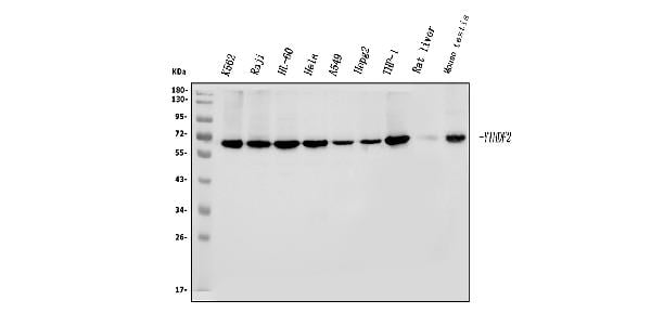
 FCM (Flow Cytometry)
(Figure 8. Flow Cytometry analysis of THP-1 cells using anti-YTHDF2 antibody (AAA19260).Overlay histogram showing THP-1 cells stained with AAA19260 (Blue line). The cells were blocked with 10% normal goat serum. And then incubated with rabbit anti-YTHDF2 Antibody (AAA19260, 1μg/1x106 cells) for 30 min at 20 degree C. DyLight®488 conjugated goat anti-rabbit IgG (5-10μg/1x106 cells) was used as secondary antibody for 30 minutes at 20 degree C. Isotype control antibody (Green line) was rabbit IgG (1μg/1x106) used under the same conditions. Unlabelled sample (Red line) was also used as a control.)
FCM (Flow Cytometry)
(Figure 8. Flow Cytometry analysis of THP-1 cells using anti-YTHDF2 antibody (AAA19260).Overlay histogram showing THP-1 cells stained with AAA19260 (Blue line). The cells were blocked with 10% normal goat serum. And then incubated with rabbit anti-YTHDF2 Antibody (AAA19260, 1μg/1x106 cells) for 30 min at 20 degree C. DyLight®488 conjugated goat anti-rabbit IgG (5-10μg/1x106 cells) was used as secondary antibody for 30 minutes at 20 degree C. Isotype control antibody (Green line) was rabbit IgG (1μg/1x106) used under the same conditions. Unlabelled sample (Red line) was also used as a control.)
YTHDF2, Polyclonal Antibody (Cat# AAA19260)
Full Name
Anti-YTHDF2 Antibody
Gene Names
YTHDF2; HGRG8; NY-REN-2
Reactivity
Human, Mouse, Rat
Applications
WB, IHC-P, ICC, IF, FC/FACS/FCM
Purity
Immunogen affinity purified.
Pricing


 FCM (Flow Cytometry)
(Figure 10. Flow Cytometry analysis of K562 cells using anti-RCC1 antibody (AAA19263).Overlay histogram showing K562 cells stained with AAA19263 (Blue line). The cells were blocked with 10% normal goat serum. And then incubated with rabbit anti-RCC1 Antibody (AAA19263,1μg/1x106 cells) for 30 min at 20 degree C. DyLight®488 conjugated goat anti-rabbit IgG (5-10μg/1x106 cells) was used as secondary antibody for 30 minutes at 20 degree C. Isotype control antibody (Green line) was rabbit IgG (1μg/1x106) used under the same conditions. Unlabelled sample (Red line) was also used as a control.)
FCM (Flow Cytometry)
(Figure 10. Flow Cytometry analysis of K562 cells using anti-RCC1 antibody (AAA19263).Overlay histogram showing K562 cells stained with AAA19263 (Blue line). The cells were blocked with 10% normal goat serum. And then incubated with rabbit anti-RCC1 Antibody (AAA19263,1μg/1x106 cells) for 30 min at 20 degree C. DyLight®488 conjugated goat anti-rabbit IgG (5-10μg/1x106 cells) was used as secondary antibody for 30 minutes at 20 degree C. Isotype control antibody (Green line) was rabbit IgG (1μg/1x106) used under the same conditions. Unlabelled sample (Red line) was also used as a control.)
RCC1, Polyclonal Antibody (Cat# AAA19263)
Full Name
Anti-RCC1 Antibody
Gene Names
RCC1; CHC1; RCC1-I; SNHG3-RCC1
Reactivity
Human, Mouse, Rat
Applications
WB, IHC-P, ICC, IF, FC/FACS/FCM, EIA
Purity
Immunogen affinity purified.
Pricing


 FCM (Flow Cytometry)
(Figure 7. Flow Cytometry analysis of 293T cells using anti-OTULIN antibody (AAA19319).Overlay histogram showing 293T cells stained with AAA19319 (Blue line). The cells were blocked with 10% normal goat serum. And then incubated with rabbit anti-OTULIN Antibody (AAA19319, 1μg/1x106 cells) for 30 min at 20 degree C. DyLight®488 conjugated goat anti-rabbit IgG (5-10μg/1x106 cells) was used as secondary antibody for 30 minutes at 20 degree C. Isotype control antibody (Green line) was rabbit IgG (1μg/1x106) used under the same conditions. Unlabelled sample (Red line) was also used as a control.)
FCM (Flow Cytometry)
(Figure 7. Flow Cytometry analysis of 293T cells using anti-OTULIN antibody (AAA19319).Overlay histogram showing 293T cells stained with AAA19319 (Blue line). The cells were blocked with 10% normal goat serum. And then incubated with rabbit anti-OTULIN Antibody (AAA19319, 1μg/1x106 cells) for 30 min at 20 degree C. DyLight®488 conjugated goat anti-rabbit IgG (5-10μg/1x106 cells) was used as secondary antibody for 30 minutes at 20 degree C. Isotype control antibody (Green line) was rabbit IgG (1μg/1x106) used under the same conditions. Unlabelled sample (Red line) was also used as a control.)
OTULIN, Polyclonal Antibody (Cat# AAA19319)
Full Name
Anti-OTULIN Antibody
Gene Names
OTULIN; GUM; FAM105B
Reactivity
Human, Mouse, Rat
Applications
WB, IHC-P, ICC, IF, FC/FACS/FCM, EIA
Purity
Immunogen affinity purified.
Pricing


 FCM (Flow Cytometry)
(Figure 7. Flow Cytometry analysis of HELA cells using anti-DNAJC10 antibody (AAA19320).Overlay histogram showing HELA cells stained with AAA19320 (Blue line). The cells were blocked with 10% normal goat serum. And then incubated with rabbit anti-DNAJC10 Antibody (AAA19320, 1μg/1x106 cells) for 30 min at 20 degree C. DyLight®488 conjugated goat anti-rabbit IgG (5-10μg/1x106 cells) was used as secondary antibody for 30 minutes at 20 degree C. Isotype control antibody (Green line) was rabbit IgG (1μg/1x106) used under the same conditions. Unlabelled sample (Red line) was also used as a control.)
FCM (Flow Cytometry)
(Figure 7. Flow Cytometry analysis of HELA cells using anti-DNAJC10 antibody (AAA19320).Overlay histogram showing HELA cells stained with AAA19320 (Blue line). The cells were blocked with 10% normal goat serum. And then incubated with rabbit anti-DNAJC10 Antibody (AAA19320, 1μg/1x106 cells) for 30 min at 20 degree C. DyLight®488 conjugated goat anti-rabbit IgG (5-10μg/1x106 cells) was used as secondary antibody for 30 minutes at 20 degree C. Isotype control antibody (Green line) was rabbit IgG (1μg/1x106) used under the same conditions. Unlabelled sample (Red line) was also used as a control.)
DNAJC10, Polyclonal Antibody (Cat# AAA19320)
Full Name
Anti-DNAJC10 Antibody
Gene Names
DNAJC10; JPDI; MTHr; ERdj5; PDIA19
Reactivity
Human, Mouse, Rat
Applications
WB, IHC-P, ICC, IF, FC/FACS/FCM, EIA
Purity
Immunogen affinity purified.
Pricing


 FCM (Flow Cytometry)
(Figure 10. Flow Cytometry analysis of U20S cells using anti-PDIA5 antibody (AAA19331).Overlay histogram showing U20S cells stained with AAA19331 (Blue line). The cells were blocked with 10% normal goat serum. And then incubated with rabbit anti-PDIA5 Antibody (AAA19331, 1μg/1x106 cells) for 30 min at 20 degree C. DyLight®488 conjugated goat anti-rabbit IgG (5-10μg/1x106 cells) was used as secondary antibody for 30 minutes at 20 degree C. Isotype control antibody (Green line) was rabbit IgG (1μg/1x106) used under the same conditions. Unlabelled sample (Red line) was also used as a control.)
FCM (Flow Cytometry)
(Figure 10. Flow Cytometry analysis of U20S cells using anti-PDIA5 antibody (AAA19331).Overlay histogram showing U20S cells stained with AAA19331 (Blue line). The cells were blocked with 10% normal goat serum. And then incubated with rabbit anti-PDIA5 Antibody (AAA19331, 1μg/1x106 cells) for 30 min at 20 degree C. DyLight®488 conjugated goat anti-rabbit IgG (5-10μg/1x106 cells) was used as secondary antibody for 30 minutes at 20 degree C. Isotype control antibody (Green line) was rabbit IgG (1μg/1x106) used under the same conditions. Unlabelled sample (Red line) was also used as a control.)
PDIA5, Polyclonal Antibody (Cat# AAA19331)
Full Name
Anti-PDIA5 Antibody
Gene Names
PDIA5; PDIR
Reactivity
Human, Mouse, Rat
Applications
WB, IHC-P, ICC, IF, FC/FACS/FCM, EIA
Purity
Immunogen affinity purified.
Pricing


 FCM (Flow Cytometry)
(Figure 9. Flow Cytometry analysis of K562 cells using anti-MCM6 antibody (AAA19372).Overlay histogram showing K562 cells stained with AAA19372 (Blue line). The cells were blocked with 10% normal goat serum. And then incubated with mouse anti- MCM6 Antibody (AAA19372, 1μg/1x106 cells) for 30 min at 20 degree C. DyLight®488 conjugated goat anti-mouse IgG (BA1126, 5-10μg/1x106 cells) was used as secondary antibody for 30 minutes at 20 degree C. Isotype control antibody (Green line) was mouse IgG (1μg/1x106) used under the same conditions. Unlabelled sample (Red line) was also used as a control.)
FCM (Flow Cytometry)
(Figure 9. Flow Cytometry analysis of K562 cells using anti-MCM6 antibody (AAA19372).Overlay histogram showing K562 cells stained with AAA19372 (Blue line). The cells were blocked with 10% normal goat serum. And then incubated with mouse anti- MCM6 Antibody (AAA19372, 1μg/1x106 cells) for 30 min at 20 degree C. DyLight®488 conjugated goat anti-mouse IgG (BA1126, 5-10μg/1x106 cells) was used as secondary antibody for 30 minutes at 20 degree C. Isotype control antibody (Green line) was mouse IgG (1μg/1x106) used under the same conditions. Unlabelled sample (Red line) was also used as a control.)
MCM6, Monoclonal Antibody (Cat# AAA19372)
Full Name
Anti-MCM6 Antibody (monoclonal, 10I9)
Gene Names
MCM6; Mis5; P105MCM; MCG40308
Reactivity
Human
Applications
WB, IHC-P, ICC, IF, FC/FACS/FCM
Purity
Immunogen affinity purified.
Pricing


 FCM (Flow Cytometry)
(Figure 10. Flow Cytometry analysis of A549 cells using anti- U2AF65/U2AF2 antibody (AAA19375).Overlay histogram showing A549 cells stained with AAA19375 (Blue line). The cells were blocked with 10% normal goat serum. And then incubated with mouse anti-U2AF65/U2AF2 Antibody (AAA19375, 1μg/1x106 cells) for 30 min at 20 degree C. DyLight®488 conjugated goat anti-mouse IgG (BA1126, 5-10μg/1x106 cells) was used as secondary antibody for 30 minutes at 20 degree C. Isotype control antibody (Green line) was mouse IgG (1μg/1x106) used under the same conditions. Unlabelled sample (Red line) was also used as a control.)
FCM (Flow Cytometry)
(Figure 10. Flow Cytometry analysis of A549 cells using anti- U2AF65/U2AF2 antibody (AAA19375).Overlay histogram showing A549 cells stained with AAA19375 (Blue line). The cells were blocked with 10% normal goat serum. And then incubated with mouse anti-U2AF65/U2AF2 Antibody (AAA19375, 1μg/1x106 cells) for 30 min at 20 degree C. DyLight®488 conjugated goat anti-mouse IgG (BA1126, 5-10μg/1x106 cells) was used as secondary antibody for 30 minutes at 20 degree C. Isotype control antibody (Green line) was mouse IgG (1μg/1x106) used under the same conditions. Unlabelled sample (Red line) was also used as a control.)
U2AF65/U2AF2, Monoclonal Antibody (Cat# AAA19375)
Full Name
Anti-U2AF65/U2AF2 Antibody (monoclonal, 10F4)
Gene Names
U2AF2; U2AF65
Reactivity
Human, Mouse, Rat
Applications
WB, IHC-P, ICC, IF, FC/FACS/FCM
Purity
Immunogen affinity purified.
Pricing


 FCM (Flow Cytometry)
(Figure 9. Flow Cytometry analysis of THP-1 cells using anti-CHCHD10 antibody (AAA19310).Overlay histogram showing THP-1 cells stained with AAA19310 (Blue line). The cells were blocked with 10% normal goat serum. And then incubated with rabbit anti-CHCHD10 Antibody (AAA19310, 1μg/1x106 cells) for 30 min at 20 degree C. DyLight®488 conjugated goat anti-rabbit IgG (5-10μg/1x106 cells) was used as secondary antibody for 30 minutes at 20 degree C. Isotype control antibody (Green line) was rabbit IgG (1μg/1x106) used under the same conditions. Unlabelled sample (Red line) was also used as a control.)
FCM (Flow Cytometry)
(Figure 9. Flow Cytometry analysis of THP-1 cells using anti-CHCHD10 antibody (AAA19310).Overlay histogram showing THP-1 cells stained with AAA19310 (Blue line). The cells were blocked with 10% normal goat serum. And then incubated with rabbit anti-CHCHD10 Antibody (AAA19310, 1μg/1x106 cells) for 30 min at 20 degree C. DyLight®488 conjugated goat anti-rabbit IgG (5-10μg/1x106 cells) was used as secondary antibody for 30 minutes at 20 degree C. Isotype control antibody (Green line) was rabbit IgG (1μg/1x106) used under the same conditions. Unlabelled sample (Red line) was also used as a control.)
CHCHD10, Polyclonal Antibody (Cat# AAA19310)
Full Name
Anti-CHCHD10 Antibody
Gene Names
CHCHD10; FTDALS2; N27C7-4; C22orf16
Reactivity
Human, Mouse, Rat, Monkey
Applications
WB, IHC-P, ICC, IF, FC/FACS/FCM, EIA
Purity
Immunogen affinity purified.
Pricing


 FCM (Flow Cytometry)
(Figure 9. Flow Cytometry analysis of U251 cells using anti-VPRBP/DCAF1 antibody (AAA19343).Overlay histogram showing U251 cells stained with AAA19343 (Blue line). The cells were blocked with 10% normal goat serum. And then incubated with rabbit anti-VPRBP/DCAF1 Antibody (AAA19343, 1μg/1x106 cells) for 30 min at 20 degree C. DyLight®488 conjugated goat anti-rabbit IgG (5-10μg/1x106 cells) was used as secondary antibody for 30 minutes at 20 degree C. Isotype control antibody (Green line) was rabbit IgG (1μg/1x106) used under the same conditions. Unlabelled sample (Red line) was also used as a control.)
FCM (Flow Cytometry)
(Figure 9. Flow Cytometry analysis of U251 cells using anti-VPRBP/DCAF1 antibody (AAA19343).Overlay histogram showing U251 cells stained with AAA19343 (Blue line). The cells were blocked with 10% normal goat serum. And then incubated with rabbit anti-VPRBP/DCAF1 Antibody (AAA19343, 1μg/1x106 cells) for 30 min at 20 degree C. DyLight®488 conjugated goat anti-rabbit IgG (5-10μg/1x106 cells) was used as secondary antibody for 30 minutes at 20 degree C. Isotype control antibody (Green line) was rabbit IgG (1μg/1x106) used under the same conditions. Unlabelled sample (Red line) was also used as a control.)
VPRBP/DCAF1, Polyclonal Antibody (Cat# AAA19343)
Full Name
Anti-VPRBP/DCAF1 Antibody
Gene Names
DCAF1; RIP; VPRBP
Reactivity
Human, Mouse, Rat
Applications
WB, IHC-P, ICC, IF, FC/FACS/FCM, EIA
Purity
Immunogen affinity purified.
Pricing


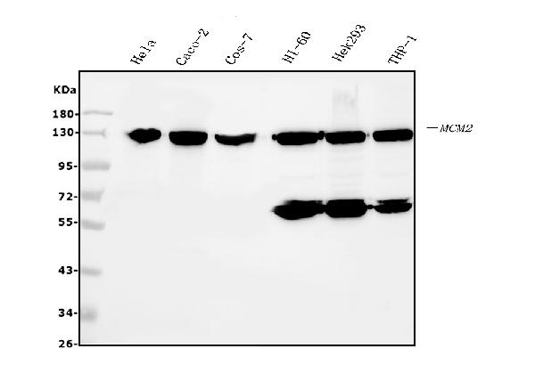
 FCM (Flow Cytometry)
(Figure 6. Flow Cytometry analysis of HL-60 cells using anti- MCM2 antibody (AAA19351).Overlay histogram showing HL-60 cells stained with AAA19351 (Blue line). The cells were blocked with 10% normal goat serum. And then incubated with mouse anti-MCM2 Antibody (AAA19351, 1μg/1x106 cells) for 30 min at 20 degree C. DyLight®488 conjugated goat anti-mouse IgG (BA1126, 5-10μg/1x106 cells) was used as secondary antibody for 30 minutes at 20 degree C. Isotype control antibody (Green line) was mouse IgG (1μg/1x106) used under the same conditions. Unlabelled sample (Red line) was also used as a control.)
FCM (Flow Cytometry)
(Figure 6. Flow Cytometry analysis of HL-60 cells using anti- MCM2 antibody (AAA19351).Overlay histogram showing HL-60 cells stained with AAA19351 (Blue line). The cells were blocked with 10% normal goat serum. And then incubated with mouse anti-MCM2 Antibody (AAA19351, 1μg/1x106 cells) for 30 min at 20 degree C. DyLight®488 conjugated goat anti-mouse IgG (BA1126, 5-10μg/1x106 cells) was used as secondary antibody for 30 minutes at 20 degree C. Isotype control antibody (Green line) was mouse IgG (1μg/1x106) used under the same conditions. Unlabelled sample (Red line) was also used as a control.)
MCM2, Monoclonal Antibody (Cat# AAA19351)
Full Name
Anti-MCM2 Antibody (monoclonal, 11C4)
Gene Names
MCM2; BM28; CCNL1; CDCL1; cdc19; D3S3194; MITOTIN
Reactivity
Human, Monkey
Applications
WB, IHC-P, ICC, IF, FC/FACS/FCM
Purity
Immunogen affinity purified.
Pricing


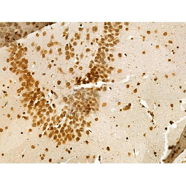
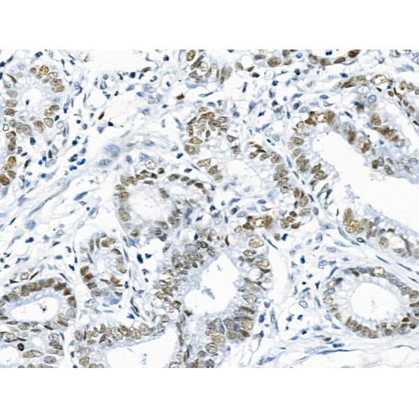
 IHC (Immunohistochemistry)
(Figure 13. IHC analysis of EEF2/Elongation factor 2 using anti-EEF2/Elongation factor 2 antibody (AAA19229).EEF2/Elongation factor 2 was detected in paraffin-embedded section of rat brain tissue. Heat mediated antigen retrieval was performed in EDTA buffer (pH8. 0, epitope retrieval solution). The tissue section was blocked with 10% goat serum. The tissue section was then incubated with 2μg/ml rabbit anti-EEF2/Elongation factor 2 Antibody (AAA19229) overnight at 4 degree C. Biotinylated goat anti-rabbit IgG was used as secondary antibody and incubated for 30 minutes at 37 degree C. The tissue section was developed using Strepavidin-Biotin-Complex (SABC) (Catalog # with DAB as the chromogen.)
IHC (Immunohistochemistry)
(Figure 13. IHC analysis of EEF2/Elongation factor 2 using anti-EEF2/Elongation factor 2 antibody (AAA19229).EEF2/Elongation factor 2 was detected in paraffin-embedded section of rat brain tissue. Heat mediated antigen retrieval was performed in EDTA buffer (pH8. 0, epitope retrieval solution). The tissue section was blocked with 10% goat serum. The tissue section was then incubated with 2μg/ml rabbit anti-EEF2/Elongation factor 2 Antibody (AAA19229) overnight at 4 degree C. Biotinylated goat anti-rabbit IgG was used as secondary antibody and incubated for 30 minutes at 37 degree C. The tissue section was developed using Strepavidin-Biotin-Complex (SABC) (Catalog # with DAB as the chromogen.)
EEF2/Elongation factor 2, Polyclonal Antibody (Cat# AAA19229)
Full Name
Anti-EEF2/Elongation factor 2 Antibody
Gene Names
EEF2; EF2; EF-2; EEF-2
Reactivity
Human, Mouse, Rat, Monkey
Applications
WB, IHC-P, ICC, IF, FC/FACS/FCM
Purity
Immunogen affinity purified.
Pricing


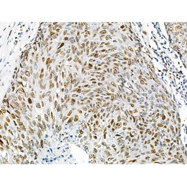
 IHC (Immunohistochemistry)
(Figure 8. IHC analysis of MEK2/MAP2K2 using anti-MEK2/MAP2K2 antibody (AAA19230).MEK2/MAP2K2 was detected in paraffin-embedded section of mouse brain tissue. Heat mediated antigen retrieval was performed in EDTA buffer (pH8. 0, epitope retrieval solution). The tissue section was blocked with 10% goat serum. The tissue section was then incubated with 2μg/ml rabbit anti-MEK2/MAP2K2 Antibody (AAA19230) overnight at 4 degree C. Biotinylated goat anti-rabbit IgG was used as secondary antibody and incubated for 30 minutes at 37 degree C. The tissue section was developed using Strepavidin-Biotin-Complex (SABC) (Catalog # with DAB as the chromogen.)
IHC (Immunohistochemistry)
(Figure 8. IHC analysis of MEK2/MAP2K2 using anti-MEK2/MAP2K2 antibody (AAA19230).MEK2/MAP2K2 was detected in paraffin-embedded section of mouse brain tissue. Heat mediated antigen retrieval was performed in EDTA buffer (pH8. 0, epitope retrieval solution). The tissue section was blocked with 10% goat serum. The tissue section was then incubated with 2μg/ml rabbit anti-MEK2/MAP2K2 Antibody (AAA19230) overnight at 4 degree C. Biotinylated goat anti-rabbit IgG was used as secondary antibody and incubated for 30 minutes at 37 degree C. The tissue section was developed using Strepavidin-Biotin-Complex (SABC) (Catalog # with DAB as the chromogen.)
MEK2/MAP2K2, Polyclonal Antibody (Cat# AAA19230)
Full Name
Anti-MEK2/MAP2K2 Antibody
Gene Names
MAP2K2; CFC4; MEK2; MKK2; MAPKK2; PRKMK2
Reactivity
Human, Mouse, Rat
Applications
WB, IHC-P, ICC, IF, FC/FACS/FCM, EIA
Purity
Immunogen affinity purified.
Pricing


 FCM (Flow Cytometry)
(Figure 8. Flow Cytometry analysis of HL-60 cells using anti-REA/PHB2 antibody (AAA19273).Overlay histogram showing HL-60 cells stained with AAA19273 (Blue line). The cells were blocked with 10% normal goat serum. And then incubated with rabbit anti-REA/PHB2 Antibody (AAA19273,1μg/1x106 cells) for 30 min at 20 degree C. DyLight®488 conjugated goat anti-rabbit IgG (5-10μg/1x106 cells) was used as secondary antibody for 30 minutes at 20 degree C. Isotype control antibody (Green line) was rabbit IgG (1μg/1x106) used under the same conditions. Unlabelled sample (Red line) was also used as a control.)
FCM (Flow Cytometry)
(Figure 8. Flow Cytometry analysis of HL-60 cells using anti-REA/PHB2 antibody (AAA19273).Overlay histogram showing HL-60 cells stained with AAA19273 (Blue line). The cells were blocked with 10% normal goat serum. And then incubated with rabbit anti-REA/PHB2 Antibody (AAA19273,1μg/1x106 cells) for 30 min at 20 degree C. DyLight®488 conjugated goat anti-rabbit IgG (5-10μg/1x106 cells) was used as secondary antibody for 30 minutes at 20 degree C. Isotype control antibody (Green line) was rabbit IgG (1μg/1x106) used under the same conditions. Unlabelled sample (Red line) was also used as a control.)
REA/PHB2, Polyclonal Antibody (Cat# AAA19273)
Full Name
Anti-REA/PHB2 Antibody
Gene Names
PHB2; BAP; REA; p22; Bap37; BCAP37; PNAS-141
Reactivity
Human, Mouse, Rat
Applications
WB, IHC-P, ICC, IF, FC/FACS/FCM, EIA
Purity
Immunogen affinity purified.
Pricing


 FCM (Flow Cytometry)
(Figure 8. Flow Cytometry analysis of U87 cells using anti-PSMB5/MB1 antibody (AAA19274).Overlay histogram showing U87 cells stained with AAA19274 (Blue line). The cells were blocked with 10% normal goat serum. And then incubated with rabbit anti-PSMB5/MB1 Antibody (AAA19274, 1μg/1x106 cells) for 30 min at 20 degree C. DyLight®488 conjugated goat anti-rabbit IgG (5-10μg/1x106 cells) was used as secondary antibody for 30 minutes at 20 degree C. Isotype control antibody (Green line) was rabbit IgG (1μg/1x106) used under the same conditions. Unlabelled sample (Red line) was also used as a control.)
FCM (Flow Cytometry)
(Figure 8. Flow Cytometry analysis of U87 cells using anti-PSMB5/MB1 antibody (AAA19274).Overlay histogram showing U87 cells stained with AAA19274 (Blue line). The cells were blocked with 10% normal goat serum. And then incubated with rabbit anti-PSMB5/MB1 Antibody (AAA19274, 1μg/1x106 cells) for 30 min at 20 degree C. DyLight®488 conjugated goat anti-rabbit IgG (5-10μg/1x106 cells) was used as secondary antibody for 30 minutes at 20 degree C. Isotype control antibody (Green line) was rabbit IgG (1μg/1x106) used under the same conditions. Unlabelled sample (Red line) was also used as a control.)
PSMB5/MB1, Polyclonal Antibody (Cat# AAA19274)
Full Name
Anti-PSMB5/MB1 Antibody
Gene Names
PSMB5; X; MB1; LMPX
Reactivity
Human, Mouse, Rat
Applications
WB, IHC-P, ICC, IF, FC/FACS/FCM, EIA
Purity
Immunogen affinity purified.
Pricing


 FCM (Flow Cytometry)
(Figure 8. Flow Cytometry analysis of K562 cells using anti-PDIA6 antibody (AAA19282).Overlay histogram showing K562 cells stained with AAA19282 (Blue line). The cells were blocked with 10% normal goat serum. And then incubated with rabbit anti-PDIA6 Antibody (AAA19282, 1μg/1x106 cells) for 30 min at 20 degree C. DyLight®488 conjugated goat anti-rabbit IgG (5-10μg/1x106 cells) was used as secondary antibody for 30 minutes at 20 degree C. Isotype control antibody (Green line) was rabbit IgG (1μg/1x106) used under the same conditions. Unlabelled sample (Red line) was also used as a control.)
FCM (Flow Cytometry)
(Figure 8. Flow Cytometry analysis of K562 cells using anti-PDIA6 antibody (AAA19282).Overlay histogram showing K562 cells stained with AAA19282 (Blue line). The cells were blocked with 10% normal goat serum. And then incubated with rabbit anti-PDIA6 Antibody (AAA19282, 1μg/1x106 cells) for 30 min at 20 degree C. DyLight®488 conjugated goat anti-rabbit IgG (5-10μg/1x106 cells) was used as secondary antibody for 30 minutes at 20 degree C. Isotype control antibody (Green line) was rabbit IgG (1μg/1x106) used under the same conditions. Unlabelled sample (Red line) was also used as a control.)
PDIA6, Polyclonal Antibody (Cat# AAA19282)
Full Name
Anti-PDIA6 Antibody
Gene Names
PDIA6; P5; ERP5; TXNDC7
Reactivity
Human, Mouse, Rat
Applications
WB, IHC-P, ICC, IF, FC/FACS/FCM, EIA
Purity
Immunogen affinity purified.
Pricing


 Application Data
(At 25 degree C. Samples were then incubated with primary Ab(At 37 degree C. An AlexaFluor594 conjugated goat anti-rabbit IgG(H+L) Ab(Red) and an AlexaFluor488 conjugated goat anti-mouse IgG(H+L) Ab(Green) were used as the secondary antibody.The nuclear counter stain is DAPI (blue).)
Application Data
(At 25 degree C. Samples were then incubated with primary Ab(At 37 degree C. An AlexaFluor594 conjugated goat anti-rabbit IgG(H+L) Ab(Red) and an AlexaFluor488 conjugated goat anti-mouse IgG(H+L) Ab(Green) were used as the secondary antibody.The nuclear counter stain is DAPI (blue).)
LRRK2, Polyclonal Antibody (Cat# AAA31287)
Full Name
Phospho-LRRK2 (Ser935) Antibody
Gene Names
LRRK2; PARK8; RIPK7; ROCO2; AURA17; DARDARIN
Reactivity
Human, Mouse, Rat
Applications
WB, IHC, IF, ICC, EIA
Purity
The antibody is from purified rabbit serum by affinity purification via sequential chromatography on phospho-peptide and non-phospho-peptide affinity columns.
Pricing


 Application Data
(At 25 degree C. The primary antibody was diluted at 1/200 and incubated with the sample for 1 hour at 37 degree C. An Alexa Fluor 594 conjugated goat anti-rabbit IgG (H+L) antibody(Red), diluted at 1/600, was used as secondary antibody.)
Application Data
(At 25 degree C. The primary antibody was diluted at 1/200 and incubated with the sample for 1 hour at 37 degree C. An Alexa Fluor 594 conjugated goat anti-rabbit IgG (H+L) antibody(Red), diluted at 1/600, was used as secondary antibody.)
Ret, Polyclonal Antibody (Cat# AAA31389)
Full Name
Phospho-Ret (Tyr1096) Antibody
Gene Names
RET; PTC; MTC1; HSCR1; MEN2A; MEN2B; RET51; CDHF12; CDHR16; RET-ELE1
Reactivity
Human, Mouse, Rat
Predicted Reactivity: Pig (100%), Bovine (100%), Sheep (100%), Rabbit (100%), Dog (100%)
Predicted Reactivity: Pig (100%), Bovine (100%), Sheep (100%), Rabbit (100%), Dog (100%)
Applications
WB, IHC, IF, ICC, EIA
Purity
The antibody is from purified rabbit serum by affinity purification via sequential chromatography on phospho-peptide and non-phospho-peptide affinity columns.
Pricing


 Application Data
(At 25 degree C. Samples were then incubated with primary Ab(At 37 degree C. An AlexaFluor594 conjugated goat anti-rabbit IgG(H+L) Ab(Red) and an AlexaFluor488 conjugated goat anti-mouse IgG(H+L) Ab(Green) were used as the secondary antibody.The nuclear counter stain is DAPI(blue).)
Application Data
(At 25 degree C. Samples were then incubated with primary Ab(At 37 degree C. An AlexaFluor594 conjugated goat anti-rabbit IgG(H+L) Ab(Red) and an AlexaFluor488 conjugated goat anti-mouse IgG(H+L) Ab(Green) were used as the secondary antibody.The nuclear counter stain is DAPI(blue).)
INCENP, Polyclonal Antibody (Cat# AAA31457)
Full Name
Phospho-INCENP (Thr59) Antibody
Reactivity
Human, Mouse, Rat
Predicted Reactivity: Pig (100%), Zebrafish (100%), Horse (100%), Sheep (89%), Rabbit (100%), Dog (100%), Chicken (100%)
Predicted Reactivity: Pig (100%), Zebrafish (100%), Horse (100%), Sheep (89%), Rabbit (100%), Dog (100%), Chicken (100%)
Applications
WB, IHC, IF, ICC, EIA
Purity
The antibody is from purified rabbit serum by affinity purification via sequential chromatography on phospho-peptide and non-phospho-peptide affinity columns.
Pricing


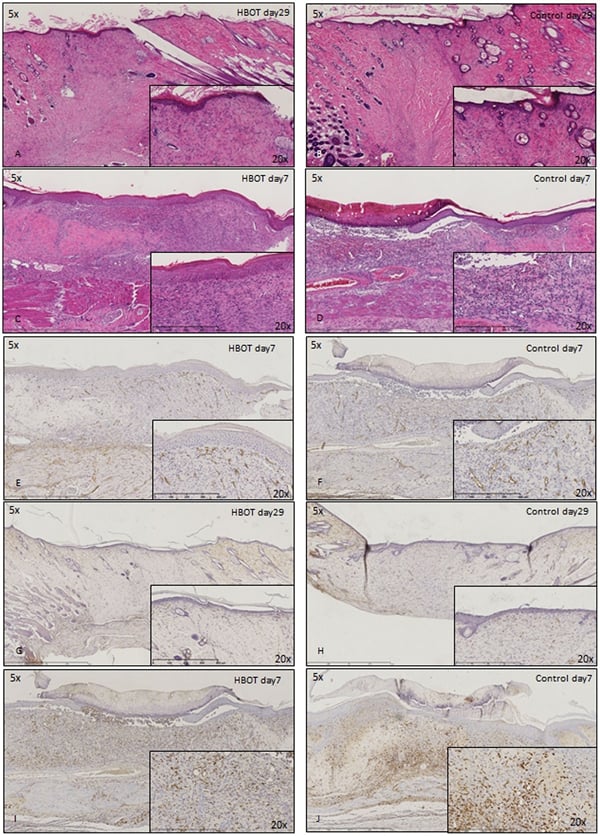 Application Data
(Published customer image: Histological staining of control and HBOT wounds at post-wounding days 7 and 29. A -D) H&E staining. E -H) CD34 immunohistochemistry. I+J) CD68 immunohistochemistry.From: uk B, Tong M, Fijneman EMG, van Neck JW (2014) Hyperbaric Oxygen Therapy to Treat Diabetes Impaired Wound Healing in Rats. PLoS ONE 9(10): e108533.)
Application Data
(Published customer image: Histological staining of control and HBOT wounds at post-wounding days 7 and 29. A -D) H&E staining. E -H) CD34 immunohistochemistry. I+J) CD68 immunohistochemistry.From: uk B, Tong M, Fijneman EMG, van Neck JW (2014) Hyperbaric Oxygen Therapy to Treat Diabetes Impaired Wound Healing in Rats. PLoS ONE 9(10): e108533.)
CD68, Monoclonal Antibody (Cat# AAA12150)
Full Name
MOUSE ANTI RAT CD68:RPE
Applications
FC/FACS
Pricing


 Application Data
(Published customer image: Increased accumulation of repair-associated macrophages surrounding collaterals in ischemic hind limbs is PAR2-dependent. (A) Stainings of CD206-positive macrophages (green) and SMA-positive vessels (red) in non-ischemic (control) and ischemic (ligated) hind limbs of WT, PAR1-/- and PAR2-/- mice are shown. Nuclei were visualized with DAPI (blue). Arrows indicate single macrophages in the non-ischemic adductor. Quantification of the average number of repair-associated macrophages per vessel is indicated on the right. (B) Correlation between the number of CD206-positive macrophages in the ischemic tissues and the expression of CD11b and (C) CD115 on monocytes. ** p)
Application Data
(Published customer image: Increased accumulation of repair-associated macrophages surrounding collaterals in ischemic hind limbs is PAR2-dependent. (A) Stainings of CD206-positive macrophages (green) and SMA-positive vessels (red) in non-ischemic (control) and ischemic (ligated) hind limbs of WT, PAR1-/- and PAR2-/- mice are shown. Nuclei were visualized with DAPI (blue). Arrows indicate single macrophages in the non-ischemic adductor. Quantification of the average number of repair-associated macrophages per vessel is indicated on the right. (B) Correlation between the number of CD206-positive macrophages in the ischemic tissues and the expression of CD11b and (C) CD115 on monocytes. ** p)
CD206, Monoclonal Antibody (Cat# AAA12120)
Full Name
RAT ANTI MOUSE CD206:FITC
Gene Names
Mrc1; MR; CD206; AW259686
Applications
FC/FACS
Pricing


 Application Data
(Published customer image: Increased accumulation of repair-associated macrophages surrounding collaterals in ischemic hind limbs is PAR2-dependent. (A) Stainings of CD206-positive macrophages (green) and SMA-positive vessels (red) in non-ischemic (control) and ischemic (ligated) hind limbs of WT, PAR1-/- and PAR2-/- mice are shown. Nuclei were visualized with DAPI (blue). Arrows indicate single macrophages in the non-ischemic adductor. Quantification of the average number of repair-associated macrophages per vessel is indicated on the right. (B) Correlation between the number of CD206-positive macrophages in the ischemic tissues and the expression of CD11b and (C) CD115 on monocytes. ** p)
Application Data
(Published customer image: Increased accumulation of repair-associated macrophages surrounding collaterals in ischemic hind limbs is PAR2-dependent. (A) Stainings of CD206-positive macrophages (green) and SMA-positive vessels (red) in non-ischemic (control) and ischemic (ligated) hind limbs of WT, PAR1-/- and PAR2-/- mice are shown. Nuclei were visualized with DAPI (blue). Arrows indicate single macrophages in the non-ischemic adductor. Quantification of the average number of repair-associated macrophages per vessel is indicated on the right. (B) Correlation between the number of CD206-positive macrophages in the ischemic tissues and the expression of CD11b and (C) CD115 on monocytes. ** p)
CD206, Monoclonal Antibody (Cat# AAA12117)
Full Name
RAT ANTI MOUSE CD206:Biotin
Gene Names
Mrc1; MR; CD206; AW259686
Applications
FC/FACS
Pricing


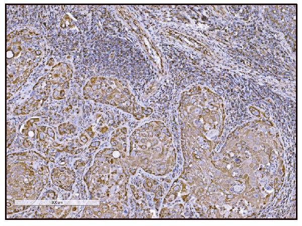


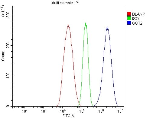


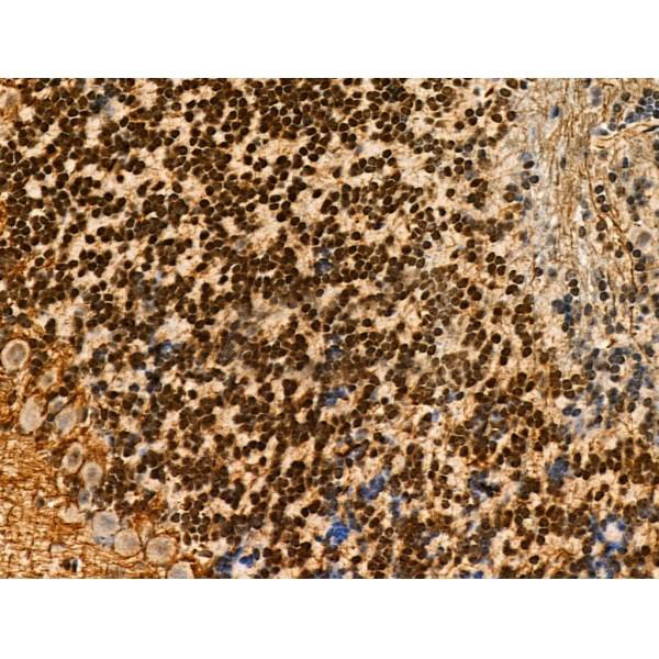
 IHC (Immunohistochemistry)
(Figure 14. IHC analysis of FABP-1/GOT2 using anti-FABP-1/GOT2 antibody (AAA19309).FABP-1/GOT2 was detected in paraffin-embedded section of rat lymph node tissue. Heat mediated antigen retrieval was performed in EDTA buffer (pH8. 0, epitope retrieval solution). The tissue section was blocked with 10% goat serum. The tissue section was then incubated with 2μg/ml rabbit anti-FABP-1/GOT2 Antibody (AAA19309) overnight at 4 degree C. Biotinylated goat anti-rabbit IgG was used as secondary antibody and incubated for 30 minutes at 37 degree C. The tissue section was developed using Strepavidin-Biotin-Complex (SABC) (Catalog # with DAB as the chromogen.)
IHC (Immunohistochemistry)
(Figure 14. IHC analysis of FABP-1/GOT2 using anti-FABP-1/GOT2 antibody (AAA19309).FABP-1/GOT2 was detected in paraffin-embedded section of rat lymph node tissue. Heat mediated antigen retrieval was performed in EDTA buffer (pH8. 0, epitope retrieval solution). The tissue section was blocked with 10% goat serum. The tissue section was then incubated with 2μg/ml rabbit anti-FABP-1/GOT2 Antibody (AAA19309) overnight at 4 degree C. Biotinylated goat anti-rabbit IgG was used as secondary antibody and incubated for 30 minutes at 37 degree C. The tissue section was developed using Strepavidin-Biotin-Complex (SABC) (Catalog # with DAB as the chromogen.)
FABP-1/GOT2, Polyclonal Antibody (Cat# AAA19309)
Full Name
Anti-FABP-1/GOT2 Antibody
Gene Names
GOT2; KAT4; KATIV; mitAAT
Reactivity
Human, Mouse, Rat
Applications
WB, IHC-P, ICC, IF, FC/FACS/FCM, EIA
Purity
Immunogen affinity purified.
Pricing


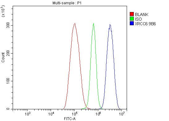 FCM (Flow Cytometry)
(Figure 7. Flow Cytometry analysis of THP-1 cells using anti-Ku70 antibody (AAA19361).Overlay histogram showing THP-1 cells stained with AAA19361 (Blue line). The cells were blocked with 10% normal goat serum. And then incubated with mouse anti- Ku70 Antibody (AAA19361, 1μg/1x106 cells) for 30 min at 20 degree C. DyLight®488 conjugated goat anti-mouse IgG (BA1126, 5-10μg/1x106 cells) was used as secondary antibody for 30 minutes at 20 degree C. Isotype control antibody (Green line) was mouse IgG (1μg/1x106) used under the same conditions. Unlabelled sample (Red line) was also used as a control.)
FCM (Flow Cytometry)
(Figure 7. Flow Cytometry analysis of THP-1 cells using anti-Ku70 antibody (AAA19361).Overlay histogram showing THP-1 cells stained with AAA19361 (Blue line). The cells were blocked with 10% normal goat serum. And then incubated with mouse anti- Ku70 Antibody (AAA19361, 1μg/1x106 cells) for 30 min at 20 degree C. DyLight®488 conjugated goat anti-mouse IgG (BA1126, 5-10μg/1x106 cells) was used as secondary antibody for 30 minutes at 20 degree C. Isotype control antibody (Green line) was mouse IgG (1μg/1x106) used under the same conditions. Unlabelled sample (Red line) was also used as a control.)
Ku70, Monoclonal Antibody (Cat# AAA19361)
Full Name
Anti-Ku70 Antibody (monoclonal, 9B6)
Gene Names
XRCC6; ML8; KU70; TLAA; CTC75; CTCBF; G22P1
Reactivity
Human
Applications
WB, IHC-P, ICC, IF, FC/FACS/FCM
Purity
Immunogen affinity purified.
Pricing


 FCM (Flow Cytometry)
(Figure 11. Flow Cytometry analysis of THP-1 cells using anti-EPRS1/PARS antibody (AAA19268).Overlay histogram showing THP-1 cells stained with AAA19268 (Blue line). The cells were blocked with 10% normal goat serum. And then incubated with rabbit anti-EPRS1/PARS Antibody (AAA19268, 1μg/1x106 cells) for 30 min at 20 degree C. DyLight®488 conjugated goat anti-rabbit IgG (5-10μg/1x106 cells) was used as secondary antibody for 30 minutes at 20 degree C. Isotype control antibody (Green line) was rabbit IgG (1μg/1x106) used under the same conditions. Unlabelled sample (Red line) was also used as a control.)
FCM (Flow Cytometry)
(Figure 11. Flow Cytometry analysis of THP-1 cells using anti-EPRS1/PARS antibody (AAA19268).Overlay histogram showing THP-1 cells stained with AAA19268 (Blue line). The cells were blocked with 10% normal goat serum. And then incubated with rabbit anti-EPRS1/PARS Antibody (AAA19268, 1μg/1x106 cells) for 30 min at 20 degree C. DyLight®488 conjugated goat anti-rabbit IgG (5-10μg/1x106 cells) was used as secondary antibody for 30 minutes at 20 degree C. Isotype control antibody (Green line) was rabbit IgG (1μg/1x106) used under the same conditions. Unlabelled sample (Red line) was also used as a control.)
EPRS1/PARS, Polyclonal Antibody (Cat# AAA19268)
Full Name
Anti-EPRS1/PARS Antibody
Gene Names
EPRS; EARS; PARS; QARS; QPRS; PIG32; GLUPRORS
Reactivity
Human, Mouse, Rat
Applications
WB, IHC-P, ICC, IF, FC/FACS/FCM, EIA
Purity
Immunogen affinity purified.
Pricing


 IF (Immunofluorescence)
(Confocal immunofluorescent analysis of GAPDH Antibody (C-term R248) with Hela cell followed by Alexa Fluor 488-conjugated goat anti-rabbit lgG (green). Actin filaments have been labeled with Alexa Fluor 555 phalloidin (red).DAPI was used to stain the cell nuclear (blue).)
IF (Immunofluorescence)
(Confocal immunofluorescent analysis of GAPDH Antibody (C-term R248) with Hela cell followed by Alexa Fluor 488-conjugated goat anti-rabbit lgG (green). Actin filaments have been labeled with Alexa Fluor 555 phalloidin (red).DAPI was used to stain the cell nuclear (blue).)
GAPDH, Polyclonal Antibody (Cat# AAA28693)
Full Name
GAPDH Antibody (C-term R248)
Gene Names
GAPDH; G3PD; GAPD; HEL-S-162eP
Reactivity
Human (Predicted Reactivity: Chicken, Mouse, Pig, Rat)
Applications
WB, EIA, IHC, FC/FACS, IF
Purity
Purified Rabbit Polyclonal Antibody (Pab)
Pricing