
Filters
Clonality
Type
Reactivity
Gene Name
Isotype
Host
Application
Clone
10000 results for "Recombinant Proteins" - showing 2550-2600
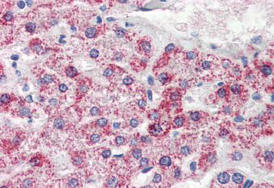
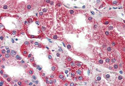
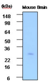 WB (Western Blot)
(Cell lysates of Mouse brain (30 ug) were resolved by SDS-PAGE, transferred to PVDF membrane and probed with anti-human PSP (1:1000). Proteins were visualized using a goat anti-mouse secondary antibody conjugated to HRP and an ECL detection system.)
WB (Western Blot)
(Cell lysates of Mouse brain (30 ug) were resolved by SDS-PAGE, transferred to PVDF membrane and probed with anti-human PSP (1:1000). Proteins were visualized using a goat anti-mouse secondary antibody conjugated to HRP and an ECL detection system.)
PSPH / PSP, Monoclonal Antibody (Cat# AAA51840)
Full Name
Mouse Monoclonal [clone 3G12] (IgG1,k) to Human PSPH / PSP
Gene Names
PSPH; PSP; PSPHD
Reactivity
Human, Mouse
Applications
Western Blot, Immunohistochemistry
Purity
Protein G Purified
Pricing
 IF (Immunofluorescence)
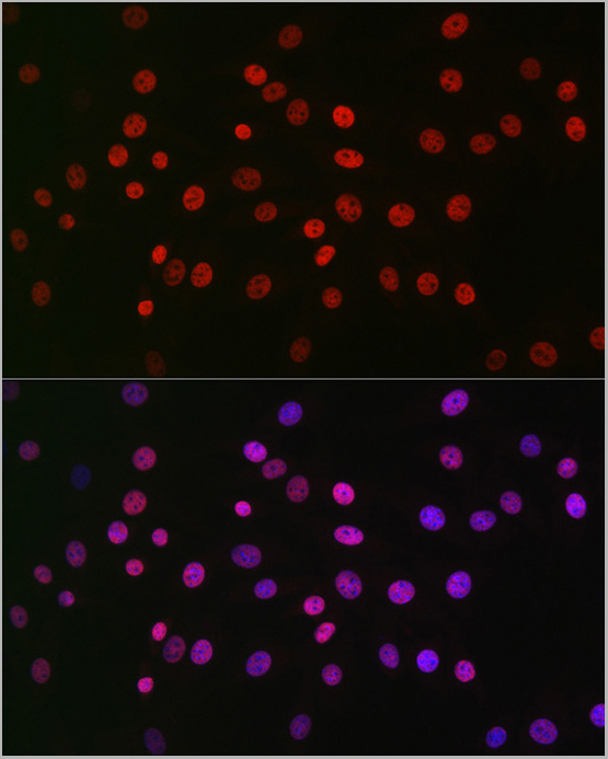
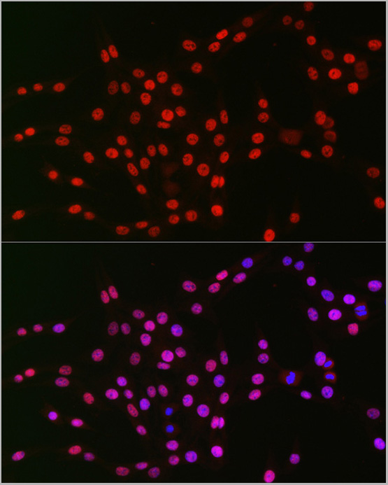
(Immunofluorescence analysis of PC-12 cells using Rev-Erbα/NR1D1 Rabbit pAb at dilution of 1:50 (40x lens). Blue: DAPI for nuclear staining.)
IF (Immunofluorescence)
(Immunofluorescence analysis of PC-12 cells using Rev-Erbα/NR1D1 Rabbit pAb at dilution of 1:50 (40x lens). Blue: DAPI for nuclear staining.)
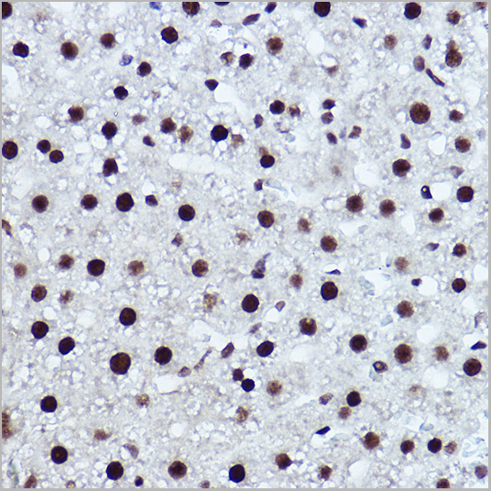
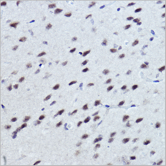
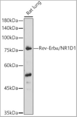
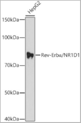
Rev-Erbalpha/NR1D1, Polyclonal Antibody (Cat# AAA28372)
Full Name
Rev-Erbalpha/NR1D1 Rabbit pAb
Gene Names
NR1D1; EAR1; hRev; THRA1; THRAL; ear-1
Applications
Western Blot, Immunohistochemistry, Immunofluorescence, Immunocytochemistry
Purity
Affinity purification
Pricing
 IP (Immunoprecipitation)
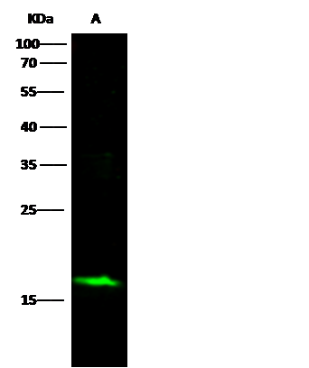
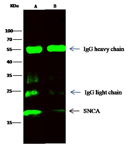
(SNCA was immunoprecipitated using:Lane A:0.5 mg Hela Whole Cell LysateLane B:0.5 mg 293T Whole Cell Lysate2 uL anti-SNCA rabbit monoclonal antibody and 15 ul of 50 % Protein G agarose.Primary antibody:Anti-SNCA rabbit monoclonal antibody,at 1:200 dilutionSecondary antibody:Dylight 800-labeled antibody to rabbit IgG (H+L), at 1:5000 dilutionDeveloped using the odssey technique.Performed under reducing conditions.Predicted band size: 16 kDaObserved band size: 18 kDa)
IP (Immunoprecipitation)
(SNCA was immunoprecipitated using:Lane A:0.5 mg Hela Whole Cell LysateLane B:0.5 mg 293T Whole Cell Lysate2 uL anti-SNCA rabbit monoclonal antibody and 15 ul of 50 % Protein G agarose.Primary antibody:Anti-SNCA rabbit monoclonal antibody,at 1:200 dilutionSecondary antibody:Dylight 800-labeled antibody to rabbit IgG (H+L), at 1:5000 dilutionDeveloped using the odssey technique.Performed under reducing conditions.Predicted band size: 16 kDaObserved band size: 18 kDa)
alpha-Synuclein, Monoclonal Antibody (Cat# AAA255515)
Full Name
Recombinant Anti-alpha-Synuclein Antibody, Rabbit Monoclonal
Gene Names
SNCA; PD1; NACP; PARK1; PARK4
Reactivity
Human
Applications
Immunoprecipitation, Western Blot
Purity
Protein A
Pricing

 ELISA
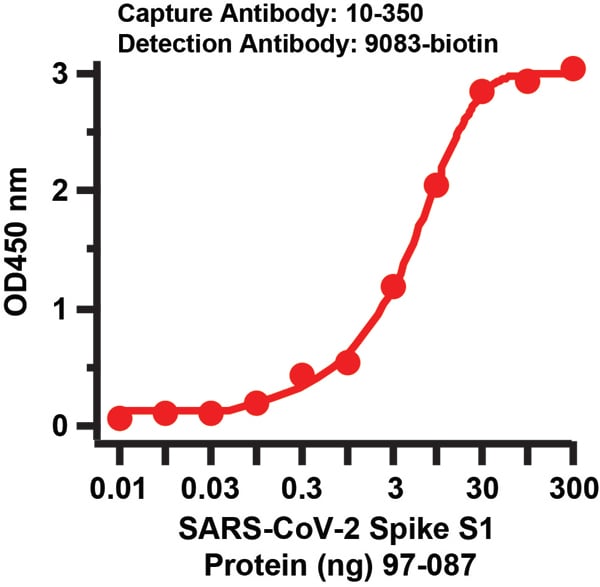
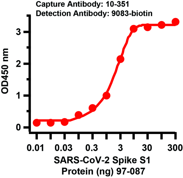
(Figure 3 Sandwich ELISA for SARS-CoV-2 (COVID-19) Matched Pair Spike S1 AntibodiesAntibodies: SARS-CoV-2 (COVID-19) Spike Antibodies, 10-351 and 9083-biotin. A sandwich ELISA was performed using SARS-CoV-2 Spike S1 antibody (10-351, 2ug/ml) as capture antibody, the Spike S1 recombinant protein as the binding protein (97-087), and the anti-SARS-CoV-2 Spike S1 antibody (9083-biotin, 1ug/ml) as the detection antibody. Secondary: Streptavidin-HRP at 1:10000 dilution. Detection range is from 0.03 ng to 300 ng. EC50 = 1.96 ng)
ELISA
(Figure 3 Sandwich ELISA for SARS-CoV-2 (COVID-19) Matched Pair Spike S1 AntibodiesAntibodies: SARS-CoV-2 (COVID-19) Spike Antibodies, 10-351 and 9083-biotin. A sandwich ELISA was performed using SARS-CoV-2 Spike S1 antibody (10-351, 2ug/ml) as capture antibody, the Spike S1 recombinant protein as the binding protein (97-087), and the anti-SARS-CoV-2 Spike S1 antibody (9083-biotin, 1ug/ml) as the detection antibody. Secondary: Streptavidin-HRP at 1:10000 dilution. Detection range is from 0.03 ng to 300 ng. EC50 = 1.96 ng)
COVID 19 Spike S1 Coronavirus, Polyclonal Antibody (Cat# AAA40916)
Full Name
SARS-CoV-2 (COVID-19) Spike S1 Antibody (biotin)
Reactivity
Virus
Applications
ELISA
Purity
SARS-CoV-2 (COVID-19) Spike S1 Antibody (biotin) is affinity chromatography purified via peptide column
Pricing
 Application Data
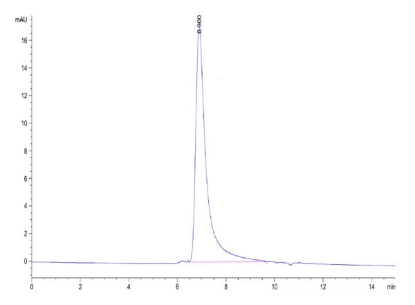
(Biotinylated Human OX40/TNFRSF4/CD134 Protein SEC-HPLCThe purity of Biotinylated Human OX40 is greater than 95% as determined by SEC HPLC.)
Application Data
(Biotinylated Human OX40/TNFRSF4/CD134 Protein SEC-HPLCThe purity of Biotinylated Human OX40 is greater than 95% as determined by SEC HPLC.)
OX40/TNFRSF4/CD134, Recombinant Protein (Cat# AAA76243)
Full Name
Biotinylated Human OX40/TNFRSF4/CD134 Protein Recombinant
Gene Names
TNFRSF4; OX40; ACT35; CD134; IMD16; TXGP1L
Purity
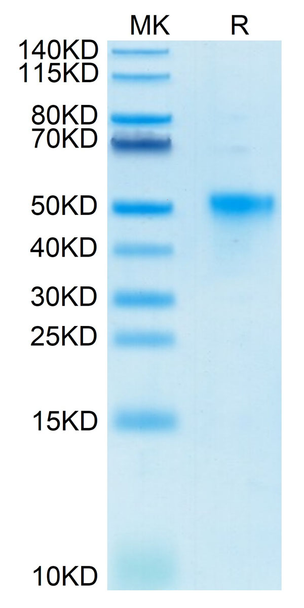
>95%, as determined by SDS-PAGE and HPLC
Pricing
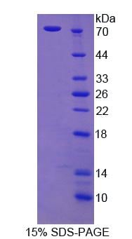 SDS-PAGE
Cortactin, Recombinant Protein (Cat# AAA144144)
Full Name
Recombinant Cortactin (CTTN)
Reactivity
Rattus norvegicus (Rat)
Applications
Western Blot
Purity
> 90%
Pricing
 Application Data
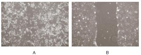
(Interleukin 11 (IL-11) is a multifunctional cytokine. It is a key regulator of multiple events in hematopoiesis, most notably the stimulation of megakaryocyte maturation. IL-11 has been demonstrated to improve platelet recovery after chemotherapy-induced thrombocytopenia, induce acute phase proteins, modulate antigen-antibody responses, participate in the regulation of bone cell proliferation and differentiation IL-11 causes bone-resorption. Besides, IL-11 have been proved can promote migration of A549 cells, 5×10⁴ cells were seeded into 6 well plates. After cell confluent, replace with DMEM without serum overnight. Using a (yellow) pipette tip make a straight scratch, simulating a wound, then washing the wells three times with PBS. Adding 1% serum standard DMEM containing various concentrations of recombinan human IL-11 to each well, incubating the plate for 48 hours at 37, 5% CO2. The results observed by inverted microscope was shown in Figure.(A) A549 cells cultured in DMEM with 100ng/mL IL-11 for 48h; (B) A549 cells cultured in DMEM before addition IL-11.Figure. Wound healing assay of A549 cells after stimulated with IL-11.)
Application Data
(Interleukin 11 (IL-11) is a multifunctional cytokine. It is a key regulator of multiple events in hematopoiesis, most notably the stimulation of megakaryocyte maturation. IL-11 has been demonstrated to improve platelet recovery after chemotherapy-induced thrombocytopenia, induce acute phase proteins, modulate antigen-antibody responses, participate in the regulation of bone cell proliferation and differentiation IL-11 causes bone-resorption. Besides, IL-11 have been proved can promote migration of A549 cells, 5×10⁴ cells were seeded into 6 well plates. After cell confluent, replace with DMEM without serum overnight. Using a (yellow) pipette tip make a straight scratch, simulating a wound, then washing the wells three times with PBS. Adding 1% serum standard DMEM containing various concentrations of recombinan human IL-11 to each well, incubating the plate for 48 hours at 37, 5% CO2. The results observed by inverted microscope was shown in Figure.(A) A549 cells cultured in DMEM with 100ng/mL IL-11 for 48h; (B) A549 cells cultured in DMEM before addition IL-11.Figure. Wound healing assay of A549 cells after stimulated with IL-11.)
Interleukin 11, Active Protein (Cat# AAA150057)
Full Name
Active Interleukin 11 (IL11)
Gene Names
IL11; AGIF; IL-11
Applications
Activity Assay, Cell Culture
Purity
>97%
Pricing
 Bioactivity
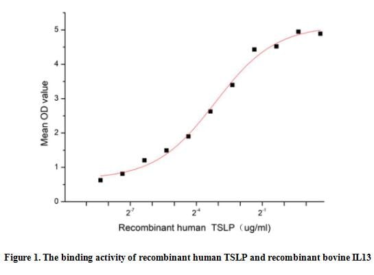
(Thymic stromal lymphopoietin (TSLP) is a member of the IL-2 cytokine family and a distant paralog of IL-7. TSLP is a pleiotropic cytokine that acts on multiple cell lineages, including dendritic cells, T cells, B cells, neutrophils, mast cells, eosinophils and innate lymphoid cells, affecting their maturation, survival and recruitment. It is best known for its role in promoting type 2 immune responses such as in allergic diseases. Interleukin 13 (IL13) is a critical downstream element for TSLP-driven allergic inflammation. Thus a functional binding ELISA assay was conducted to detect the interaction of recombinant human TSLP and recombinant bovine IL13. Briefly, TSLP was diluted serially in PBS with 0.01% BSA (pH 7.4). Duplicate samples of 100 ul were then transferred to IL13-coated microtiter wells and incubated for 1h at 37 degree C. Wells were washed with PBST and incubated for 1h with anti-TSLP pAb, then aspirated and washed 3 times. After incubation with HRP labelled secondary antibody for 1h at 37 degree C, wells were aspirated and washed 5 times. With the addition of substrate solution, wells were incubated 15-25 minutes at 37 degree C. Finally, add 50 uL stop solution to the wells and read at 450/630 nm immediately. The binding activity of recombinant human TSLP and recombinant bovine IL13 was shown in Figure 1, the EC50 for this effect is 0.11 ug/mL.)
Bioactivity
(Thymic stromal lymphopoietin (TSLP) is a member of the IL-2 cytokine family and a distant paralog of IL-7. TSLP is a pleiotropic cytokine that acts on multiple cell lineages, including dendritic cells, T cells, B cells, neutrophils, mast cells, eosinophils and innate lymphoid cells, affecting their maturation, survival and recruitment. It is best known for its role in promoting type 2 immune responses such as in allergic diseases. Interleukin 13 (IL13) is a critical downstream element for TSLP-driven allergic inflammation. Thus a functional binding ELISA assay was conducted to detect the interaction of recombinant human TSLP and recombinant bovine IL13. Briefly, TSLP was diluted serially in PBS with 0.01% BSA (pH 7.4). Duplicate samples of 100 ul were then transferred to IL13-coated microtiter wells and incubated for 1h at 37 degree C. Wells were washed with PBST and incubated for 1h with anti-TSLP pAb, then aspirated and washed 3 times. After incubation with HRP labelled secondary antibody for 1h at 37 degree C, wells were aspirated and washed 5 times. With the addition of substrate solution, wells were incubated 15-25 minutes at 37 degree C. Finally, add 50 uL stop solution to the wells and read at 450/630 nm immediately. The binding activity of recombinant human TSLP and recombinant bovine IL13 was shown in Figure 1, the EC50 for this effect is 0.11 ug/mL.)
Thymic Stromal Lymphopoietin (TSLP), Active Protein (Cat# AAA161842)
Full Name
Active Thymic Stromal Lymphopoietin (TSLP)
Applications
Activity Assay, Cell Culture
Purity
Greater than 95% by SDS-PAGE
Pricing

 Bioactivity
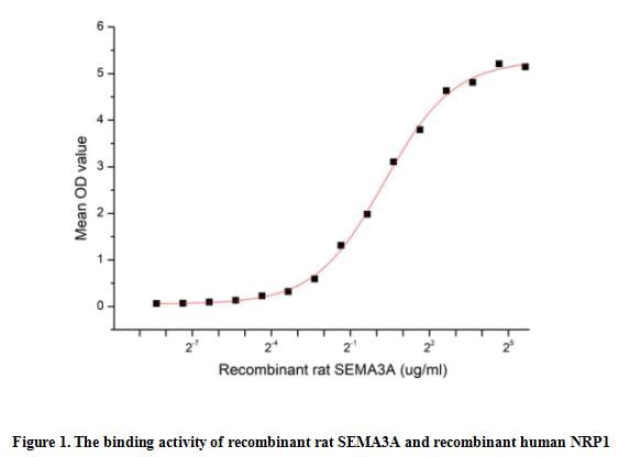
(The Semaphorin 3A(SEMA3A) which belongs to the semaphorin family can function as either a chemorepulsive agent, inhibiting axonal outgrowth, or as a chemoattractive agent, stimulating the growth of apical dendrites. In both cases, the protein is vital for normal neuronal pattern development. Semaphorin 3A is secreted protein containing a Sema domain, an immunoglobulin C2-like domain and a basic domain near the carboxyl tail. It can be secreted by neurons and surrounding tissue to guide migrating cells and axons in the developing nervous system. Besides, Neuropilin 1 (NRP1) has been identified as an interactor of SEMA3A, thus a functional binding ELISA assay was conducted to detect the interaction of recombinant rat SEMA3A and recombinant human NRP1. Briefly, SEMA3A were diluted serially in PBS, with 0.01% BSA (pH 7.4). Duplicate samples of 100 ul were then transferred to NRP1-coated microtiter wells and incubated for 2h at 37 degree C. Wells were washed with PBST and incubated for 1h with anti-SEMA3A pAb, then aspirated and washed 3 times. After incubation with HRP labelled secondary antibody, wells were aspirated and washed 3 times. With the addition of substrate solution, wells were incubated 15-25 minutes at 37 degree C. Finally, add 50 ul stop solution to the wells and read at 450 nm immediately. The binding activity of recombinant rat SEMA3A and recombinant human NRP1 was shown in Figure 1, the EC50 for this effect is 1.22 ug/mL.)
Bioactivity
(The Semaphorin 3A(SEMA3A) which belongs to the semaphorin family can function as either a chemorepulsive agent, inhibiting axonal outgrowth, or as a chemoattractive agent, stimulating the growth of apical dendrites. In both cases, the protein is vital for normal neuronal pattern development. Semaphorin 3A is secreted protein containing a Sema domain, an immunoglobulin C2-like domain and a basic domain near the carboxyl tail. It can be secreted by neurons and surrounding tissue to guide migrating cells and axons in the developing nervous system. Besides, Neuropilin 1 (NRP1) has been identified as an interactor of SEMA3A, thus a functional binding ELISA assay was conducted to detect the interaction of recombinant rat SEMA3A and recombinant human NRP1. Briefly, SEMA3A were diluted serially in PBS, with 0.01% BSA (pH 7.4). Duplicate samples of 100 ul were then transferred to NRP1-coated microtiter wells and incubated for 2h at 37 degree C. Wells were washed with PBST and incubated for 1h with anti-SEMA3A pAb, then aspirated and washed 3 times. After incubation with HRP labelled secondary antibody, wells were aspirated and washed 3 times. With the addition of substrate solution, wells were incubated 15-25 minutes at 37 degree C. Finally, add 50 ul stop solution to the wells and read at 450 nm immediately. The binding activity of recombinant rat SEMA3A and recombinant human NRP1 was shown in Figure 1, the EC50 for this effect is 1.22 ug/mL.)
Semaphorin 3A (SEMA3A), Active Protein (Cat# AAA161937)
Full Name
Active Semaphorin 3A (SEMA3A)
Applications
Activity Assay, Cell Culture
Purity
Greater than 90% by SDS-PAGE
Pricing
 IHC (Immunohiostchemistry)
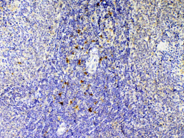
(Figure 2. IHC analysis of IL12B using anti-IL12B antibody (AAA124625).IL12B was detected in paraffin-embedded section of rat spleen tissue. Heat mediated antigen retrieval was performed in citrate buffer (pH6, epitope retrieval solution) for 20 mins. The tissue section was blocked with 10% goat serum. The tissue section was then incubated with 1ug/ml rabbit anti-IL12B Antibody (AAA124625) overnight at 4 degree C. Biotinylated goat anti-rabbit IgG was used as secondary antibody and incubated for 30 minutes at 37 degree C. The tissue section was developed using Strepavidin-Biotin-Complex (SABC) with DAB as the chromogen.)
IHC (Immunohiostchemistry)
(Figure 2. IHC analysis of IL12B using anti-IL12B antibody (AAA124625).IL12B was detected in paraffin-embedded section of rat spleen tissue. Heat mediated antigen retrieval was performed in citrate buffer (pH6, epitope retrieval solution) for 20 mins. The tissue section was blocked with 10% goat serum. The tissue section was then incubated with 1ug/ml rabbit anti-IL12B Antibody (AAA124625) overnight at 4 degree C. Biotinylated goat anti-rabbit IgG was used as secondary antibody and incubated for 30 minutes at 37 degree C. The tissue section was developed using Strepavidin-Biotin-Complex (SABC) with DAB as the chromogen.)
IL12B/Il 12, Polyclonal Antibody (Cat# AAA124625)
Full Name
Anti-IL12B/Il 12 Picoband Antibody
Gene Names
Il12b; Il12
Reactivity
Mouse, Rat
No cross reactivity with other proteins.
No cross reactivity with other proteins.
Applications
Western Blot, Immunohistochemistry
Purity
Immunogen affinity purified
Pricing
 WB (Western Blot)
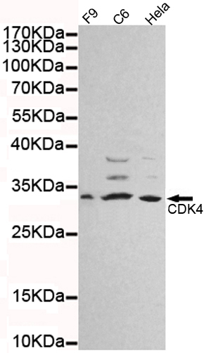
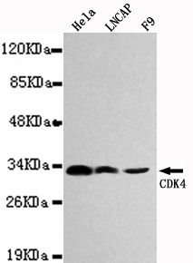
(Western blot detection of CDK4 in Hela.Lncap and F9 cell lysates using CDK4 mouse mAb (1:1000 diluted).Predicted band size:34KDa.Observed band size:33KDa.)
WB (Western Blot)
(Western blot detection of CDK4 in Hela.Lncap and F9 cell lysates using CDK4 mouse mAb (1:1000 diluted).Predicted band size:34KDa.Observed band size:33KDa.)
CDK4, Monoclonal Antibody (Cat# AAA320333)
Full Name
CDK4 Mouse mAb
Gene Names
CDK4; CMM3; PSK-J3
Reactivity
Human, Mouse, Rat
Applications
Western Blot
Purity
The antibody was affinity-purified from mouse ascites by affinity-chromatography using epitope-specific immunogen.
Pricing
 WB (Western Blot)
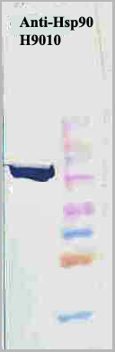
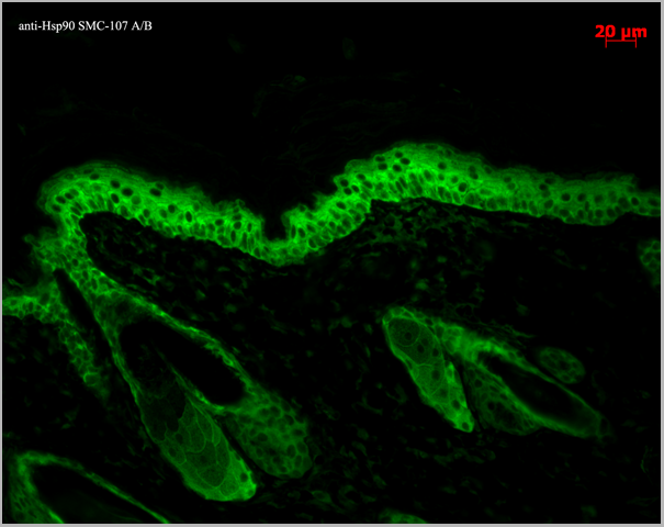
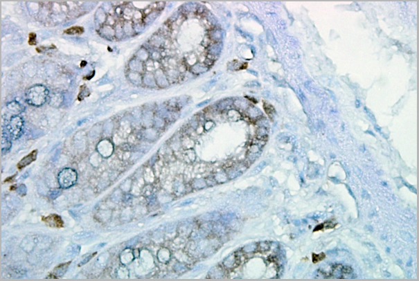
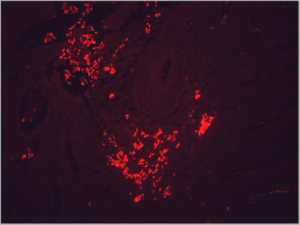
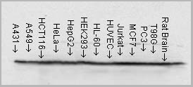
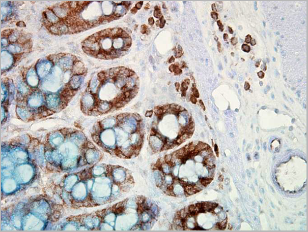
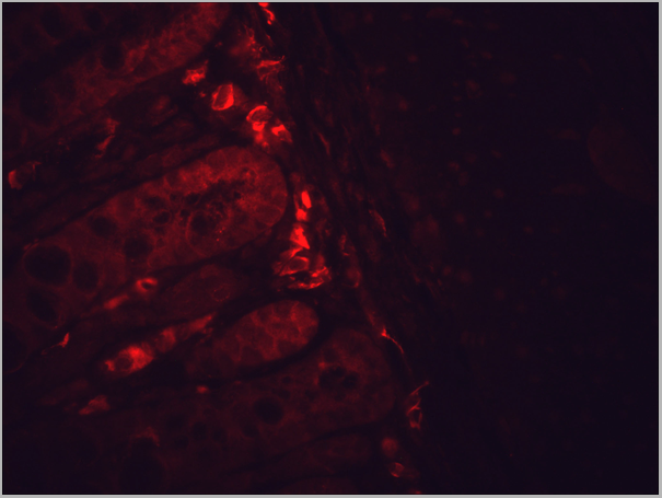
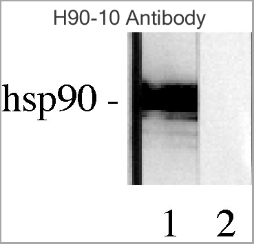
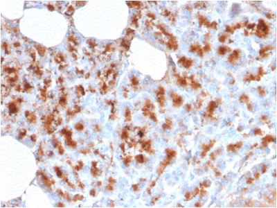
(Western blot analysis of Human Lysates showing detection of Hsp90 protein using Mouse Anti-Hsp90 Monoclonal Antibody, Clone H9010. Primary Antibody: Mouse Anti-Hsp90 Monoclonal Antibody at 1:1000. Comparison of clone H9010 behavior with Hsp90 human beta (1) and Hsp90 human alpha (2). Courtesy of: David Toft, Mayo Clinic.)
WB (Western Blot)
(Western blot analysis of Human Lysates showing detection of Hsp90 protein using Mouse Anti-Hsp90 Monoclonal Antibody, Clone H9010. Primary Antibody: Mouse Anti-Hsp90 Monoclonal Antibody at 1:1000. Comparison of clone H9010 behavior with Hsp90 human beta (1) and Hsp90 human alpha (2). Courtesy of: David Toft, Mayo Clinic.)
Hsp90, Monoclonal Antibody (Cat# AAA17785)
Full Name
Hsp90 Antibody: Biotin
Gene Names
HSP90AB1; HSP84; HSPC2; HSPCB; D6S182; HSP90B
Reactivity
Human, Mouse, Rat, Rabbit, Chicken, Dog, Fish, Shark, Hamster
Applications
Western Blot, Immunohistochemistry, Immunocytochemistry, Immunofluorescence, Antibody Microarray
Pricing
 Application Data
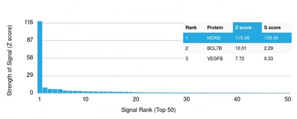
(Analysis of Protein Array containing more than 19,000 full-length human proteins using MDM2 Mouse Monoclonal Antibody (MDM2/2414).Z- and S- Score: The Z-score represents the strength of a signal that a monoclonal antibody (MAb) (in combination with a fluorescently-tagged anti-IgG secondary antibody) produces when binding to a particular protein on the HuProtTM array. Z-scores are described in units of standard deviations (SD's) above the mean value of all signals generated on that array. If targets on HuProtTM are arranged in descending order of the Z-score, the S-score is the difference (also in units of SD's) between the Z-score. S-score therefore represents the relative target specificity of a MAb to its intended target. A MAb is considered to specific to its intended target, if the MAb has an S-score of at least 2.5. For example, if a MAb binds to protein X with a Z-score of 43 and to protein Y with a Z-score of 14, then the S-score for the binding of that MAb to protein X is equal to 29.)
Application Data
(Analysis of Protein Array containing more than 19,000 full-length human proteins using MDM2 Mouse Monoclonal Antibody (MDM2/2414).Z- and S- Score: The Z-score represents the strength of a signal that a monoclonal antibody (MAb) (in combination with a fluorescently-tagged anti-IgG secondary antibody) produces when binding to a particular protein on the HuProtTM array. Z-scores are described in units of standard deviations (SD's) above the mean value of all signals generated on that array. If targets on HuProtTM are arranged in descending order of the Z-score, the S-score is the difference (also in units of SD's) between the Z-score. S-score therefore represents the relative target specificity of a MAb to its intended target. A MAb is considered to specific to its intended target, if the MAb has an S-score of at least 2.5. For example, if a MAb binds to protein X with a Z-score of 43 and to protein Y with a Z-score of 14, then the S-score for the binding of that MAb to protein X is equal to 29.)
MDM2, Monoclonal Antibody (Cat# AAA214776)
Full Name
MDM2
Gene Names
MDM2; HDMX; hdm2; ACTFS
Reactivity
Human. Others not known.
Applications
Immunohistochemistry, Western Blot
Purity
Purified Ab with BSA and Azide at 200ug/ml OR Purified Ab WITHOUT BSA and Azide at 1.0mg/ml
Pricing
 WB (Western Blot)
(Western Blot; Sample: Lane1: Human Leukocyte lysate; Lane2: Human Lymphocyte lysate; Lane3: Human PBMC lysate Primary Ab: 0.1ug/ml Mouse AntiHuman S100A9 Antibody Second Ab: 0.2ug/mL HRPLinked Caprine AntiMouse IgG Polyclonal Antibody (Catalog: SAA544Mu19))
WB (Western Blot)
(Western Blot; Sample: Lane1: Human Leukocyte lysate; Lane2: Human Lymphocyte lysate; Lane3: Human PBMC lysate Primary Ab: 0.1ug/ml Mouse AntiHuman S100A9 Antibody Second Ab: 0.2ug/mL HRPLinked Caprine AntiMouse IgG Polyclonal Antibody (Catalog: SAA544Mu19))
S100 Calcium Binding Protein A9 (S100A9), Monoclonal Antibody (Cat# AAA151749)
Full Name
Monoclonal Antibody to S100 Calcium Binding Protein A9 (S100A9)
Reactivity
Human, Rat
Applications
Immunoprecipitation, Immunocytochemistry, Immunohistochemistry, Western Blot
Purity
Protein A + Protein G affinity chromatography
Pricing


 WB (Western Blot)
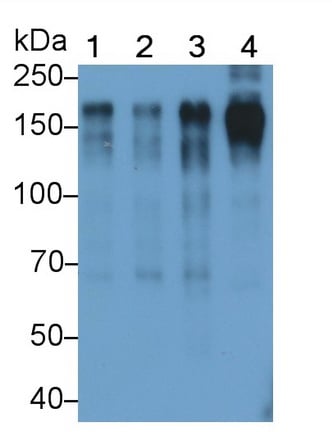
(Western Blot; Sample: Lane1: Hela cell lysate; Lane2: MCF7 cell lysate; Lane3: A431 cell lysate; Lane4: Human Placenta lysatePrimary Ab: 0.01ug/ml Mouse AntiHuman EGFR AntibodySecond Ab: 0.2ug/mL HRPLinked Caprine AntiMouse IgG Polyclonal Antibody(Catalog: SAA544Mu19))
WB (Western Blot)
(Western Blot; Sample: Lane1: Hela cell lysate; Lane2: MCF7 cell lysate; Lane3: A431 cell lysate; Lane4: Human Placenta lysatePrimary Ab: 0.01ug/ml Mouse AntiHuman EGFR AntibodySecond Ab: 0.2ug/mL HRPLinked Caprine AntiMouse IgG Polyclonal Antibody(Catalog: SAA544Mu19))
Epidermal Growth Factor Receptor (EGFR), Monoclonal Antibody (Cat# AAA151619)
Full Name
Monoclonal Antibody to Epidermal Growth Factor Receptor (EGFR)
Gene Names
EGFR; ERBB; HER1; mENA; ERBB1; PIG61
Reactivity
Human, Pig
Applications
Immunoprecipitation, Immunocytochemistry, Immunohistochemistry, Western Blot
Purity
Protein A + Protein G affinity chromatography
Pricing
 Bioactivity
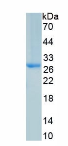
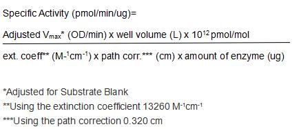
(Complement Factor D (CFD) is a serine protease that catalyzes the initial proteolytic step in the alternative pathway of complement. Expressed in adipose tissue at high levels, factor D is also known as adipsin. It is an exceptionally specific protease and the only known protein substrate is factor B in complex with C3. Factor D protease activity is regulated by reversible conformational changes, which differs from the majority of serine proteases whose regulation involves either activation by processing of the zymogens or inactivation by binding of the inhibitors. Compared to its physiologically important proteolytic activity, factor D has much lower activity toward synthetic peptide substrates. However, thioester substrates have been routinely used for assessing factor D activity. The full-length (amino acid residues 1-263) of rat CFD was expressed which activity was measured by its ability to cleaves a thioester substrate Z-Lys-SBzl•HCl. The reaction was performed in 50 mM Tris, 1 M NaCl, pH 7.5 (Assay Buffer), initiated by addition 50 uL of various concentrations of CFD (diluted by Assay Buffer) to 50 ul substrate mixture of 0.2mM Z-Lys-SBzl•HCl and 0.2 mM DTNB. The final well serves as a negative control with no CFD, replaced with 50 ul assay buffer. Then read in kinetic mode for 5 minutes at an absorbance of 405 nm. The specific activity of recombinant rat CFD is > 10000 pmol/min/ug.)
Bioactivity
(Complement Factor D (CFD) is a serine protease that catalyzes the initial proteolytic step in the alternative pathway of complement. Expressed in adipose tissue at high levels, factor D is also known as adipsin. It is an exceptionally specific protease and the only known protein substrate is factor B in complex with C3. Factor D protease activity is regulated by reversible conformational changes, which differs from the majority of serine proteases whose regulation involves either activation by processing of the zymogens or inactivation by binding of the inhibitors. Compared to its physiologically important proteolytic activity, factor D has much lower activity toward synthetic peptide substrates. However, thioester substrates have been routinely used for assessing factor D activity. The full-length (amino acid residues 1-263) of rat CFD was expressed which activity was measured by its ability to cleaves a thioester substrate Z-Lys-SBzl•HCl. The reaction was performed in 50 mM Tris, 1 M NaCl, pH 7.5 (Assay Buffer), initiated by addition 50 uL of various concentrations of CFD (diluted by Assay Buffer) to 50 ul substrate mixture of 0.2mM Z-Lys-SBzl•HCl and 0.2 mM DTNB. The final well serves as a negative control with no CFD, replaced with 50 ul assay buffer. Then read in kinetic mode for 5 minutes at an absorbance of 405 nm. The specific activity of recombinant rat CFD is > 10000 pmol/min/ug.)
Complement Factor D (CFD), Active Protein (Cat# AAA161865)
Full Name
Active Complement Factor D (CFD)
Gene Names
Cfd; Df; Adn; EVE
Applications
Activity Assay, Cell Culture
Purity
Greater than 95% by SDS-PAGE
Pricing
 IF (Immunofluorescence)
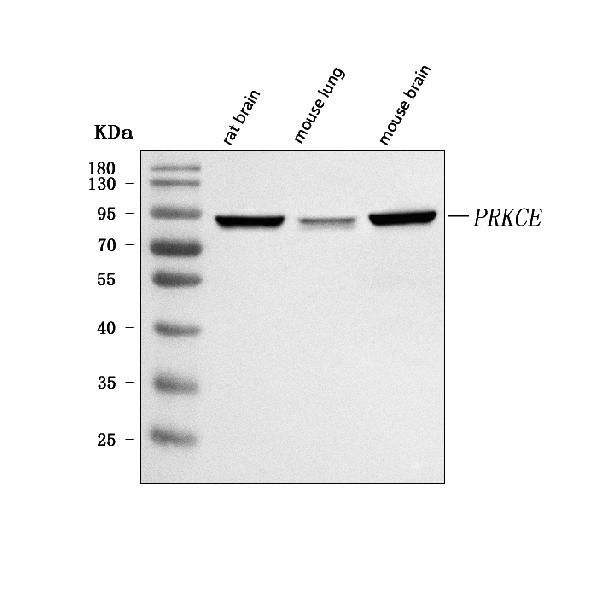
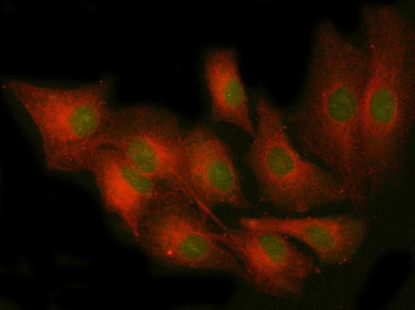
(Figure 2. IF analysis of PRKCE4 using anti-PRKCE4 antibody (AAA127039) and anti-Tubulin Alpha antibody (M03989-3).PRKCE4 was detected in immunocytochemical section of A549 cell. Enzyme antigen retrieval was performed using IHC enzyme antigen retrieval reagent for 15 mins. The cells were blocked with 10% goat serum. And then incubated with 5ug/mL rabbit anti-PRKCE4 Antibody (AAA127039) and mouse anti-Tubulin Alpha antibody (M03989-3) overnight at 4 degree C. DyLight488 Conjugated Goat Anti-Rabbit IgG and Cy3 Conjugated Goat Anti-Mouse IgG (BA1031) were used as secondary antibody at 1:500 dilution and incubated for 30 minutes at 37 degree C. Visualize using a fluorescence microscope and filter sets appropriate for the label used.)
IF (Immunofluorescence)
(Figure 2. IF analysis of PRKCE4 using anti-PRKCE4 antibody (AAA127039) and anti-Tubulin Alpha antibody (M03989-3).PRKCE4 was detected in immunocytochemical section of A549 cell. Enzyme antigen retrieval was performed using IHC enzyme antigen retrieval reagent for 15 mins. The cells were blocked with 10% goat serum. And then incubated with 5ug/mL rabbit anti-PRKCE4 Antibody (AAA127039) and mouse anti-Tubulin Alpha antibody (M03989-3) overnight at 4 degree C. DyLight488 Conjugated Goat Anti-Rabbit IgG and Cy3 Conjugated Goat Anti-Mouse IgG (BA1031) were used as secondary antibody at 1:500 dilution and incubated for 30 minutes at 37 degree C. Visualize using a fluorescence microscope and filter sets appropriate for the label used.)
PRKCE, Polyclonal Antibody (Cat# AAA127039)
Full Name
Anti-PRKCE Antibody Picoband
Gene Names
PRKCE; PKCE; nPKC-epsilon
Reactivity
Human, Mouse, Rat
Applications
Western Blot, Immunofluorescence, Immunocytochemistry
Purity
Immunogen affinity purified.
Pricing
 FCM/FACS (Flow Cytometry)
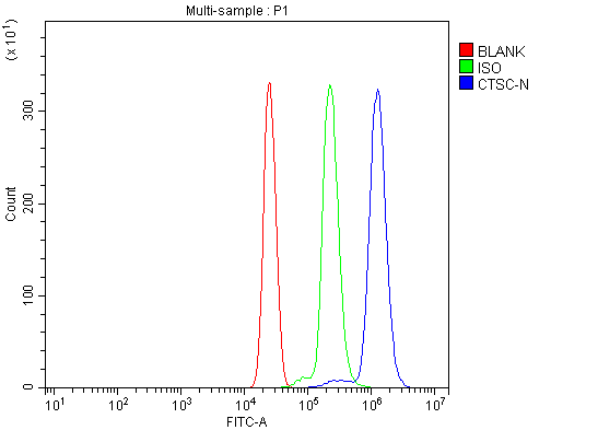
(Figure 2. Flow Cytometry analysis of U937 cells using anti-CTSC antibody (AAA126202).Overlay histogram showing U937 cells stained with AAA126202 (Blue line). The cells were blocked with 10% normal goat serum. And then incubated with rabbit anti-CTSC Antibody (AAA126202, 1 ug/1x10^6 cells) for 30 min at 20 degree C. DyLight488 conjugated goat anti-rabbit IgG was used as secondary antibody for 30 minutes at 20 degree C. Isotype control antibody (Green line) was rabbit IgG (1 ug/1x10^6) used under the same conditions. Unlabelled sample (Red line) was also used as a control.)
FCM/FACS (Flow Cytometry)
(Figure 2. Flow Cytometry analysis of U937 cells using anti-CTSC antibody (AAA126202).Overlay histogram showing U937 cells stained with AAA126202 (Blue line). The cells were blocked with 10% normal goat serum. And then incubated with rabbit anti-CTSC Antibody (AAA126202, 1 ug/1x10^6 cells) for 30 min at 20 degree C. DyLight488 conjugated goat anti-rabbit IgG was used as secondary antibody for 30 minutes at 20 degree C. Isotype control antibody (Green line) was rabbit IgG (1 ug/1x10^6) used under the same conditions. Unlabelled sample (Red line) was also used as a control.)
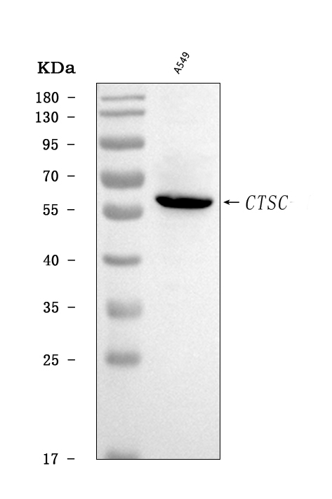
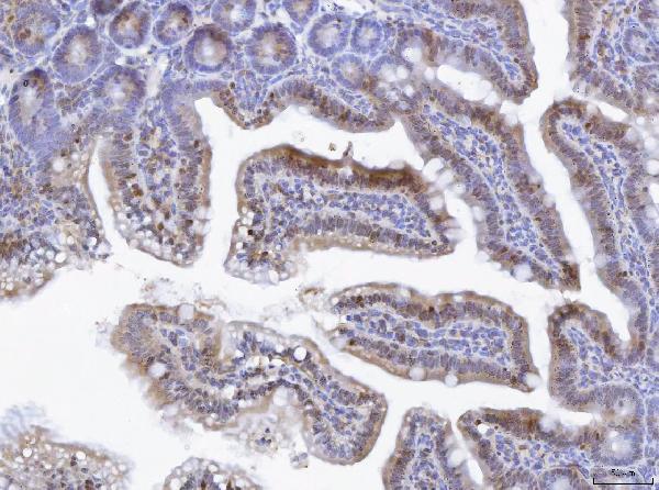
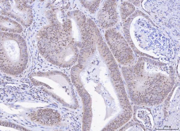
CTSC, Polyclonal Antibody (Cat# AAA126202)
Full Name
Anti-CTSC Antibody Picoband
Gene Names
CTSC; JP; HMS; JPD; PLS; CPPI; DPP1; DPPI; PALS; DPP-I; PDON1
Reactivity
Human
Applications
Flow Cytometry, Western Blot
Purity
Immunogen affinity purified.
Pricing
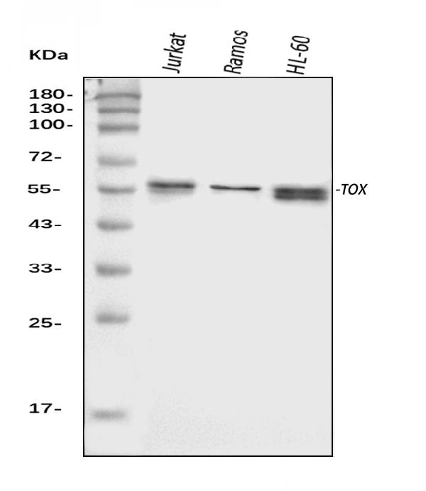
 FCM/FACS (Flow Cytometry)
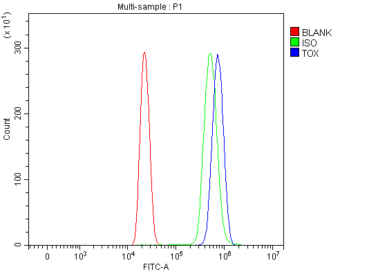
(Figure 4. Flow Cytometry analysis of HL-60 cells using anti-TOX antibody (AAA126522).Overlay histogram showing HL-60 cells stained with AAA126522 (Blue line). The cells were blocked with 10% normal goat serum. And then incubated with rabbit anti-TOX Antibody (AAA126522, 1 ug/1x10^6 cells) for 30 min at 20 degree C. DyLight488 conjugated goat anti-rabbit IgG was used as secondary antibody for 30 minutes at 20 degree C. Isotype control antibody (Green line) was rabbit IgG (1 ug/1x10^6) used under the same conditions. Unlabelled sample (Red line) was also used as a control.)
FCM/FACS (Flow Cytometry)
(Figure 4. Flow Cytometry analysis of HL-60 cells using anti-TOX antibody (AAA126522).Overlay histogram showing HL-60 cells stained with AAA126522 (Blue line). The cells were blocked with 10% normal goat serum. And then incubated with rabbit anti-TOX Antibody (AAA126522, 1 ug/1x10^6 cells) for 30 min at 20 degree C. DyLight488 conjugated goat anti-rabbit IgG was used as secondary antibody for 30 minutes at 20 degree C. Isotype control antibody (Green line) was rabbit IgG (1 ug/1x10^6) used under the same conditions. Unlabelled sample (Red line) was also used as a control.)
TOX, Polyclonal Antibody (Cat# AAA126522)
Full Name
Anti-TOX Antibody Picoband
Gene Names
TOX; TOX1
Reactivity
Human, Mouse
Applications
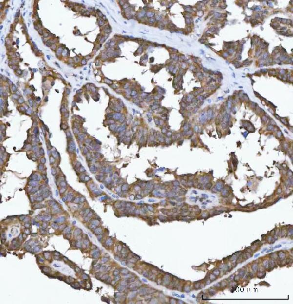
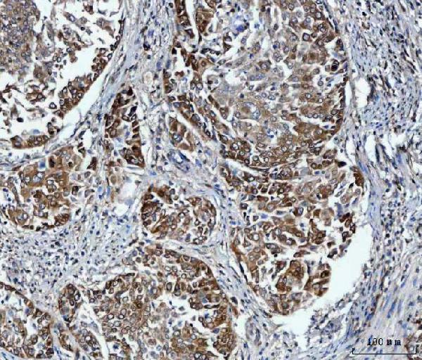
Flow Cytometry, Immunohistochemistry, Western Blot
Purity
Immunogen affinity purified.
Pricing
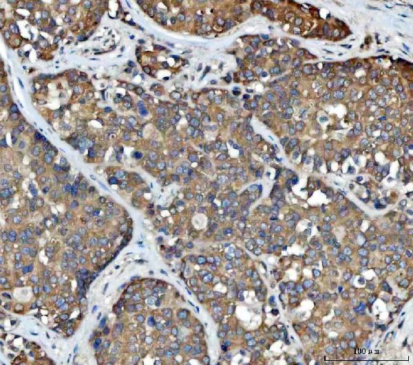
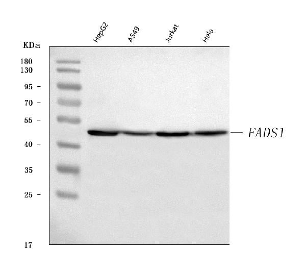
 FCM/FACS (Flow Cytometry)
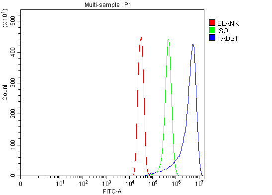
(Figure 5. Flow Cytometry analysis of SiHa cells using anti-FADS1 antibody (AAA126233).Overlay histogram showing SiHa cells stained with AAA126233 (Blue line). The cells were blocked with 10% normal goat serum. And then incubated with rabbit anti-FADS1 Antibody (AAA126233, 1 ug/1x10^6 cells) for 30 min at 20 degree C. DyLight488 conjugated goat anti-rabbit IgG was used as secondary antibody for 30 minutes at 20 degree C. Isotype control antibody (Green line) was rabbit IgG (1 ug/1x10^6) used under the same conditions. Unlabelled sample (Red line) was also used as a control.)
FCM/FACS (Flow Cytometry)
(Figure 5. Flow Cytometry analysis of SiHa cells using anti-FADS1 antibody (AAA126233).Overlay histogram showing SiHa cells stained with AAA126233 (Blue line). The cells were blocked with 10% normal goat serum. And then incubated with rabbit anti-FADS1 Antibody (AAA126233, 1 ug/1x10^6 cells) for 30 min at 20 degree C. DyLight488 conjugated goat anti-rabbit IgG was used as secondary antibody for 30 minutes at 20 degree C. Isotype control antibody (Green line) was rabbit IgG (1 ug/1x10^6) used under the same conditions. Unlabelled sample (Red line) was also used as a control.)
FADS1, Polyclonal Antibody (Cat# AAA126233)
Full Name
Anti-FADS1 Antibody Picoband
Gene Names
FADS1; D5D; TU12; FADS6; FADSD5; LLCDL1
Reactivity
Human
Applications
Flow Cytometry, Immunohistochemistry, Western Blot
Purity
Immunogen affinity purified.
Pricing
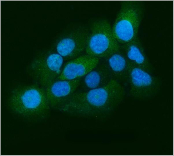
 FCM (Flow Cytometry)
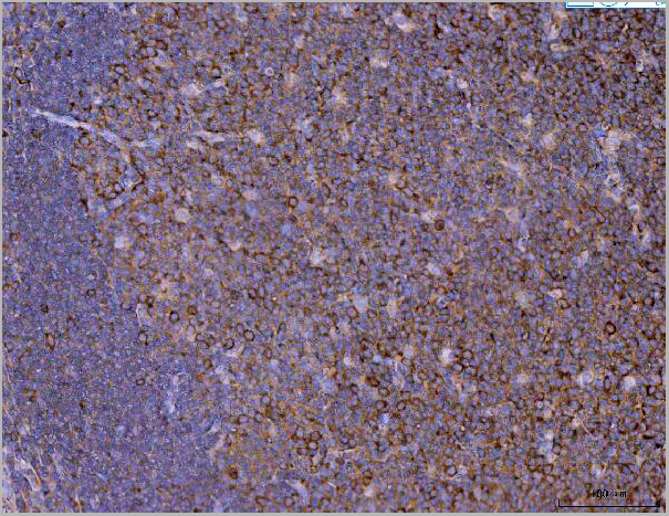
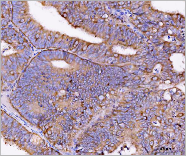
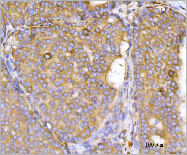
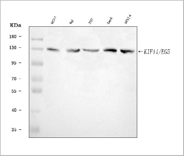
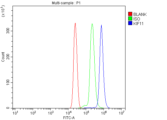
(Figure 6. Flow Cytometry analysis of U937 cells using anti-Eg5/KIF11 antibody (AAA19431).Overlay histogram showing U937 cells stained with AAA19431 (Blue line). The cells were blocked with 10% normal goat serum. And then incubated with rabbit anti-Eg5/KIF11 Antibody (AAA19431, 1 ug/1x10^6 cells) for 30 min at 20 degree C. DyLight488 conjugated goat anti-rabbit IgG was used as secondary antibody for 30 minutes at 20 degree C. Isotype control antibody (Green line) was rabbit IgG (1 ug/1x10^6) used under the same conditions. Unlabelled sample (Red line) was also used as a control.)
FCM (Flow Cytometry)
(Figure 6. Flow Cytometry analysis of U937 cells using anti-Eg5/KIF11 antibody (AAA19431).Overlay histogram showing U937 cells stained with AAA19431 (Blue line). The cells were blocked with 10% normal goat serum. And then incubated with rabbit anti-Eg5/KIF11 Antibody (AAA19431, 1 ug/1x10^6 cells) for 30 min at 20 degree C. DyLight488 conjugated goat anti-rabbit IgG was used as secondary antibody for 30 minutes at 20 degree C. Isotype control antibody (Green line) was rabbit IgG (1 ug/1x10^6) used under the same conditions. Unlabelled sample (Red line) was also used as a control.)
Eg5/KIF11, Polyclonal Antibody (Cat# AAA19431)
Full Name
Anti-Eg5/KIF11 Antibody Picoband
Gene Names
KIF11; EG5; HKSP; KNSL1; MCLMR; TRIP5
Reactivity
Human
Applications
Western Blot, Immunohistochemistry, Immunocytochemistry, Immunofluorescence, Flow Cytometry
Purity
Immunogen affinity purified.
Pricing
 Bioactivity
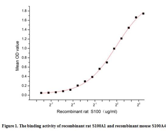
(Small calcium binding protein that S100 Calcium Binding Protein (S100), also known as S100 Alpha (S100A1), is a member of the S100 family of calcium-binding proteins. As with most S100 proteins, S100A1 proteins are localized in the cytoplasm and/or nucleus of a wide range of cells, and it possess a variety of intracellular and extracellular functions. They interact with multiple receptors and signal transducers to regulate pathways that govern inflammation, cell differentiation, proliferation, energy metabolism, apoptosis, calcium homeostasis, cell cytoskeleton and microbial resistance. S100 Calcium Binding Protein A4 (S100A4) is one of targets of S100A1. Thus a functional binding ELISA assay was conducted to detect the interaction of recombinant rat S100A1 and recombinant mouse S100A4. Briefly, S100A1 was diluted serially in PBS with 0.01% BSA (pH 7.4). Duplicate samples of 100 ul were then transferred to S100A4-coated microtiter wells and incubated for 1h at 37 degree C. Wells were washed with PBST and incubated for 1h with anti-S100A1 pAb, then aspirated and washed 3 times. After incubation with HRP labelled secondary antibody for 1h at 37 degree C, wells were aspirated and washed 5 times. With the addition of substrate solution, wells were incubated 15-25 minutes at 37 degree C. Finally, add 50 uL stop solution to the wells and read at 450/630 nm immediately. The binding activity of recombinant rat S100A1 and recombinant mouse S100A4 was shown in Figure 1, the EC50 for this effect is 3.92 ug/mL.)
Bioactivity
(Small calcium binding protein that S100 Calcium Binding Protein (S100), also known as S100 Alpha (S100A1), is a member of the S100 family of calcium-binding proteins. As with most S100 proteins, S100A1 proteins are localized in the cytoplasm and/or nucleus of a wide range of cells, and it possess a variety of intracellular and extracellular functions. They interact with multiple receptors and signal transducers to regulate pathways that govern inflammation, cell differentiation, proliferation, energy metabolism, apoptosis, calcium homeostasis, cell cytoskeleton and microbial resistance. S100 Calcium Binding Protein A4 (S100A4) is one of targets of S100A1. Thus a functional binding ELISA assay was conducted to detect the interaction of recombinant rat S100A1 and recombinant mouse S100A4. Briefly, S100A1 was diluted serially in PBS with 0.01% BSA (pH 7.4). Duplicate samples of 100 ul were then transferred to S100A4-coated microtiter wells and incubated for 1h at 37 degree C. Wells were washed with PBST and incubated for 1h with anti-S100A1 pAb, then aspirated and washed 3 times. After incubation with HRP labelled secondary antibody for 1h at 37 degree C, wells were aspirated and washed 5 times. With the addition of substrate solution, wells were incubated 15-25 minutes at 37 degree C. Finally, add 50 uL stop solution to the wells and read at 450/630 nm immediately. The binding activity of recombinant rat S100A1 and recombinant mouse S100A4 was shown in Figure 1, the EC50 for this effect is 3.92 ug/mL.)
S100 Calcium Binding Protein (S100), Active Protein (Cat# AAA161651)
Full Name
Active S100 Calcium Binding Protein (S100)
Applications
Activity Assay, Cell Culture
Purity
Greater than 90% by SDS-PAGE
Pricing
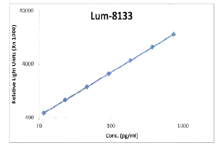 Standard Curve (Sample)
IL-21, CLIA Kit (Cat# AAA318541)
Full Name
IL-21 (Human) LumiAb ELISA Kit
Gene Names
IL21; Za11; IL-21; CVID11
Reactivity
Human
Pricing
 WB (Western Blot)
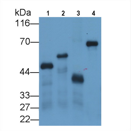
(Western Blot;Sample: Four differentRecombinant proteins with GST Tag (Lane 1-4).)
WB (Western Blot)
(Western Blot;Sample: Four differentRecombinant proteins with GST Tag (Lane 1-4).)
Glutathione S Transferase (GST), Monoclonal Antibody (Cat# AAA150822)
Full Name
Monoclonal Antibody to Glutathione S Transferase (GST)
Reactivity
General
Applications
Immunoprecipitation, Immunocytochemistry, Immunohistochemistry, Western Blot
Purity
Protein A + Protein G affinity chromatography
Pricing
 Application Data
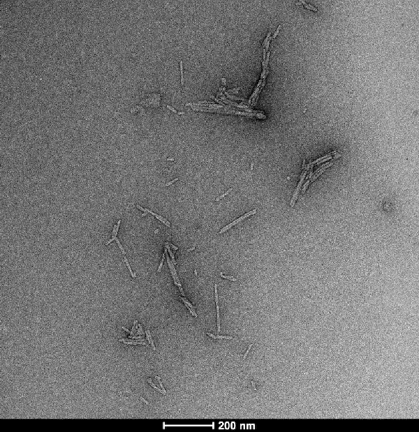
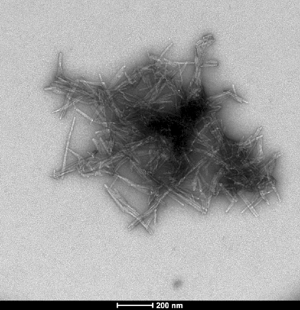
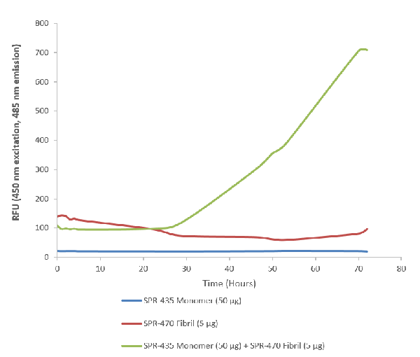
(ThioflavinTassayofSOD1pre-formedfibrils(PFFs)(AAA254095)combinedwithSOD1monomers .Pre-formedfibrilsseedtheaggregationofmonomers,leadingtoanincreaseinThioflavinTfluorescence)
Application Data
(ThioflavinTassayofSOD1pre-formedfibrils(PFFs)(AAA254095)combinedwithSOD1monomers .Pre-formedfibrilsseedtheaggregationofmonomers,leadingtoanincreaseinThioflavinTfluorescence)
Superoxide dismutase, Recombinant Protein (Cat# AAA254095)
Full Name
Human Recombinant Superoxide dismutase Protein Pre-formed Fibrils
Gene Names
SOD1; ALS; SOD; ALS1; IPOA; hSod1; HEL-S-44; homodimer
Applications
Western Blot
Purity
>95%; Ion-exchange Purified
Pricing
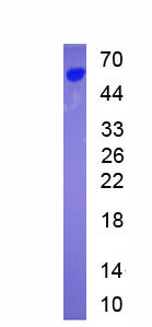
 Amino Acid Info
Insulin Receptor (INSR), Recombinant Protein (Cat# AAA153404)
Full Name
Recombinant Insulin Receptor (INSR)
Gene Names
INSR; HHF5; CD220
Applications
Western Blot
Purity
Greater than 90% by SDS-PAGE
Pricing
 Bioactivity
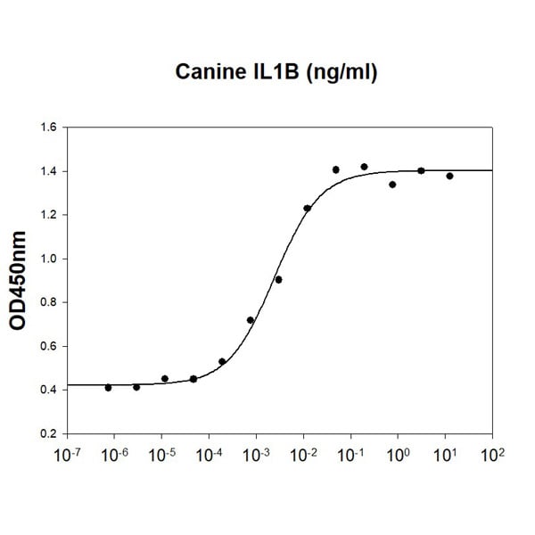
(Canine IL-1B stimulates cell proliferation of the D10.G4.1 mouse helper T cells. The ED50 range range )
Bioactivity
(Canine IL-1B stimulates cell proliferation of the D10.G4.1 mouse helper T cells. The ED50 range range )
IL-1 beta/IL-1F2, Active Protein (Cat# AAA48366)
Full Name
Recombinant canine IL-1 beta/IL-1F2 protein
Gene Names
IL1B; IL-1
Applications
Bioactivity, SDS-PAGE
Purity
>95% by SDS-PAGE
Pricing
 Bioactivity
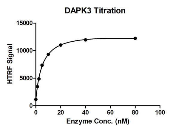
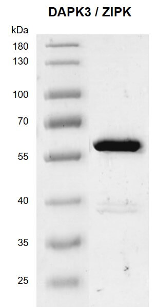
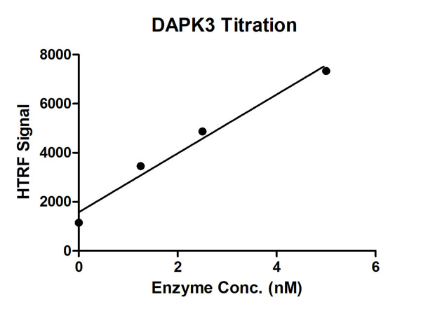
(HTRF assay for DAPK3/ZIPK; activity 1 uM STK S1 substrate was incubated with different concentrations of DAPK3/ZIPK; protein in a 10 ul reaction system containing 1×Enzymatic Buffer, 10 mM MgCl2, 1 mM DTT and 100 uM ATP for 1 hour. The detection reagents were added and incubated with the reactions for 30 min. All the operations and reactions were performed at room temperature.)
Bioactivity
(HTRF assay for DAPK3/ZIPK; activity 1 uM STK S1 substrate was incubated with different concentrations of DAPK3/ZIPK; protein in a 10 ul reaction system containing 1×Enzymatic Buffer, 10 mM MgCl2, 1 mM DTT and 100 uM ATP for 1 hour. The detection reagents were added and incubated with the reactions for 30 min. All the operations and reactions were performed at room temperature.)
DAPK3/ZIPK, Active Protein (Cat# AAA60341)
Full Name
Recombinant DAPK3/ZIPK protein
Purity
>=85%
Pricing

 SDS-PAGE
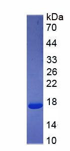
(Figure. SDS-PAGE)
SDS-PAGE
(Figure. SDS-PAGE)
Serum Amyloid A (SAA), Recombinant Protein (Cat# AAA150678)
Full Name
Recombinant Serum Amyloid A (SAA)
Applications
Western Blot
Purity
>90%
Pricing
Artemin, Active Protein (Cat# AAA75568)
Full Name
Artemin protein
Gene Names
ARTN; EVN; NBN; ENOVIN
Purity
> 98% pure
Pricing
West Nile Virus E, Active Protein (Cat# AAA24010)
Full Name
West Nile Virus E protein (His tag)
Applications
Western Blot
Purity
> 95% pure
Pricing
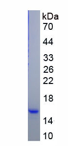
 Amino Acid Info
Eukaryotic Phospholipase A2, Pancreas (pPLA2), Protein (Cat# AAA153221)
Full Name
Eukaryotic Phospholipase A2, Pancreas (pPLA2)
Gene Names
PLA2G1B; PLA2; sPLA(2)-IB
Applications
Western Blot
Purity
Greater than 90% by SDS-PAGE
Pricing
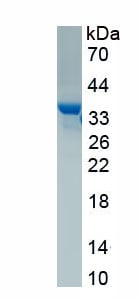 SDS-PAGE
(Figure. SDS-PAGE)
SDS-PAGE
(Figure. SDS-PAGE)
Aprotinin (AP), Protein (Cat# AAA161948)
Full Name
Eukaryotic Aprotinin (AP)
Applications
Western Blot
Purity
Greater than 95% by SDS-PAGE
Pricing
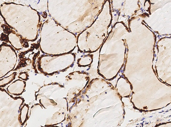
 IF (Immunofluorescence)
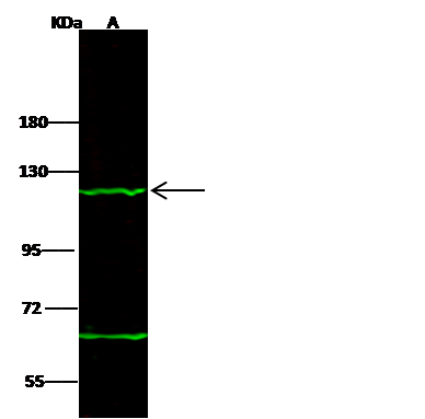
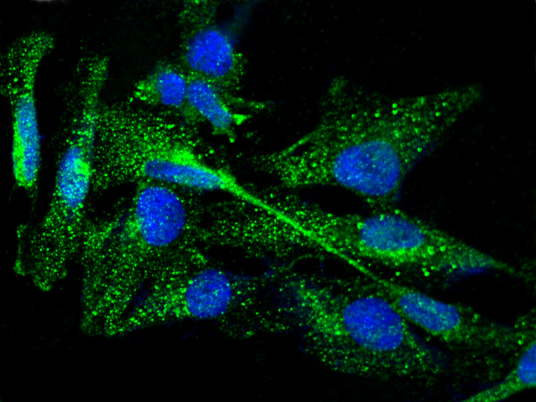
(Immunofluorescence staining of TPO in U251MG cells. Cells were fixed with 4% PFA, permeabilzed with 0.3% Triton X-100 in PBS,blocked with 10% serum, and incubated with rabbit anti-Human TPO polyclonal antibody (dilution ratio 1:1000) at 4 degree C overnight. Then cells were stained with the Alexa Fluor$488-conjugated Goat Anti-rabbit IgG secondary antibody (green) and counterstained with DAPI (blue).Positive staining was localized to cytoplasm.)
IF (Immunofluorescence)
(Immunofluorescence staining of TPO in U251MG cells. Cells were fixed with 4% PFA, permeabilzed with 0.3% Triton X-100 in PBS,blocked with 10% serum, and incubated with rabbit anti-Human TPO polyclonal antibody (dilution ratio 1:1000) at 4 degree C overnight. Then cells were stained with the Alexa Fluor$488-conjugated Goat Anti-rabbit IgG secondary antibody (green) and counterstained with DAPI (blue).Positive staining was localized to cytoplasm.)
Thyroid peroxidase, Polyclonal Antibody (Cat# AAA255653)
Full Name
Anti-Thyroid peroxidase Antibody, Rabbit Polyclonal
Gene Names
TPO; MSA; TPX; TDH2A
Reactivity
Human
Applications
Immunofluorescence, Immunocytochemistry, Immunohistochemistry, Western Blot
Pricing
 WB (Western Blot)
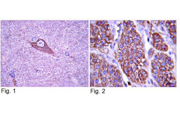
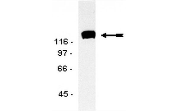
(Western blot analysis of EGF-stimulated A431 cel lysate probed with anti-TrkA (2 ug/ml))
WB (Western Blot)
(Western blot analysis of EGF-stimulated A431 cel lysate probed with anti-TrkA (2 ug/ml))
TrkA, Polyclonal Antibody (Cat# AAA75070)
Full Name
TrkA antibody
Reactivity
Reacts with Rat, Mouse and Human. Reactivity to other species is unknown. Recognizes TrKa, MW 140kDa. Does not crossreact with TrkB or TrkC.
Applications
Western Blot, Immunoprecipitation, Immunocytochemistry
Purity
TrkA antibody was purified by Protein A chromatography.
Pricing
 Bioactivity
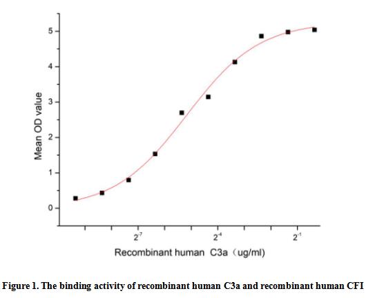
(Complement Component 3a (C3a) is a component of the complement system, this protein is also known as the C3a anaphylatoxin, modulates inflammation and possesses antimicrobial activity. It's been identified that C3A can interact with Complement Factor I (CFI), thus a functional binding ELISA assay was conducted to detect the interaction of recombinant human C3a and recombinant human CFI. Briefly, C3a was diluted serially in PBS with 0.01% BSA (pH 7.4). Duplicate samples of 100 ul were then transferred to CFI-coated microtiter wells and incubated for 1h at 37 degree C. Wells were washed with PBST and incubated for 1h with anti-C3a pAb, then aspirated and washed 3 times. After incubation with HRP labelled secondary antibody for 1h at 37 degree C, wells were aspirated and washed 5 times. With the addition of substrate solution, wells were incubated 15-25 minutes at 37 degree C. Finally, add 50 uL stop solution to the wells and read at 450/630 nm immediately. The binding activity of recombinant human C3a and recombinant human CFI was shown in Figure 1, the EC50 for this effect is 0.03 ug/mL.)
Bioactivity
(Complement Component 3a (C3a) is a component of the complement system, this protein is also known as the C3a anaphylatoxin, modulates inflammation and possesses antimicrobial activity. It's been identified that C3A can interact with Complement Factor I (CFI), thus a functional binding ELISA assay was conducted to detect the interaction of recombinant human C3a and recombinant human CFI. Briefly, C3a was diluted serially in PBS with 0.01% BSA (pH 7.4). Duplicate samples of 100 ul were then transferred to CFI-coated microtiter wells and incubated for 1h at 37 degree C. Wells were washed with PBST and incubated for 1h with anti-C3a pAb, then aspirated and washed 3 times. After incubation with HRP labelled secondary antibody for 1h at 37 degree C, wells were aspirated and washed 5 times. With the addition of substrate solution, wells were incubated 15-25 minutes at 37 degree C. Finally, add 50 uL stop solution to the wells and read at 450/630 nm immediately. The binding activity of recombinant human C3a and recombinant human CFI was shown in Figure 1, the EC50 for this effect is 0.03 ug/mL.)
Complement Component 3a (C3a), Active Protein (Cat# AAA161726)
Full Name
Active Complement Component 3a (C3a)
Gene Names
C3; ASP; C3a; C3b; AHUS5; ARMD9; CPAMD1
Applications
Activity Assay, Cell Culture
Purity
Greater than 95% by SDS-PAGE
Pricing
 Bioactivity
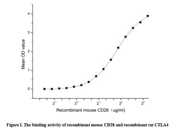
(The cluster of differentiation (CD) antigen CD28 is a central co-stimulatory molecule for TCR-mediated activation such as cytokine production and T-cell proliferation upon ligand binding and TCR stimulation. It is a member of the immunoglobulin subfamily, other members of the subfamily include ICOS, CTLA4, PD1, PD1H, TIGIT and BTLA. Cytotoxic T-Lymphocyte Associated Antigen 4 (CTLA4) and CD28 are highly homologous and compete for the same ligands, CTLA4 can also bind with CD28 to form a dimer. Thus a functional binding ELISA assay was conducted to detect the interaction of recombinant mouse CD28 and recombinant rat CTLA4. Briefly, rmCD28 was diluted serially in PBS with 0.01% BSA (pH 7.4). Duplicate samples of 100 ul were then transferred to CTLA4-coated microtiter wells and incubated for 1h at 37 degree C. Wells were washed with PBST and incubated for 1h with anti-CD28 pAb, then aspirated and washed 3 times. After incubation with HRP labelled secondary antibody for 1h at 37 degree C, wells were aspirated and washed 5 times. With the addition of substrate solution, wells were incubated 15-25 minutes at 37 degree C. Finally, add 50 uL stop solution to the wells and read at 450/630 nm immediately. The binding activity of recombinant mouse CD28 and recombinant rat CTLA4 was shown in Figure 1, the EC50 for this effect is 2.82 ug/mL.)
Bioactivity
(The cluster of differentiation (CD) antigen CD28 is a central co-stimulatory molecule for TCR-mediated activation such as cytokine production and T-cell proliferation upon ligand binding and TCR stimulation. It is a member of the immunoglobulin subfamily, other members of the subfamily include ICOS, CTLA4, PD1, PD1H, TIGIT and BTLA. Cytotoxic T-Lymphocyte Associated Antigen 4 (CTLA4) and CD28 are highly homologous and compete for the same ligands, CTLA4 can also bind with CD28 to form a dimer. Thus a functional binding ELISA assay was conducted to detect the interaction of recombinant mouse CD28 and recombinant rat CTLA4. Briefly, rmCD28 was diluted serially in PBS with 0.01% BSA (pH 7.4). Duplicate samples of 100 ul were then transferred to CTLA4-coated microtiter wells and incubated for 1h at 37 degree C. Wells were washed with PBST and incubated for 1h with anti-CD28 pAb, then aspirated and washed 3 times. After incubation with HRP labelled secondary antibody for 1h at 37 degree C, wells were aspirated and washed 5 times. With the addition of substrate solution, wells were incubated 15-25 minutes at 37 degree C. Finally, add 50 uL stop solution to the wells and read at 450/630 nm immediately. The binding activity of recombinant mouse CD28 and recombinant rat CTLA4 was shown in Figure 1, the EC50 for this effect is 2.82 ug/mL.)
Cluster Of Differentiation 28 (CD28), Active Protein (Cat# AAA161757)
Full Name
Active Cluster Of Differentiation 28 (CD28)
Applications
Activity Assay, Cell Culture
Purity
Greater than 90% by SDS-PAGE
Pricing
 SDS-PAGE
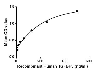
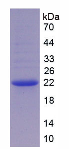
Insulin Like Growth Factor Binding Protein 3 (IGFBP3), Active Protein (Cat# AAA149226)
Full Name
Active Insulin Like Growth Factor Binding Protein 3 (IGFBP3)
Gene Names
IGFBP3; IBP3; BP-53
Applications
Activity Assay, Cell Culture
Purity
> 90%
Pricing
 ELISA
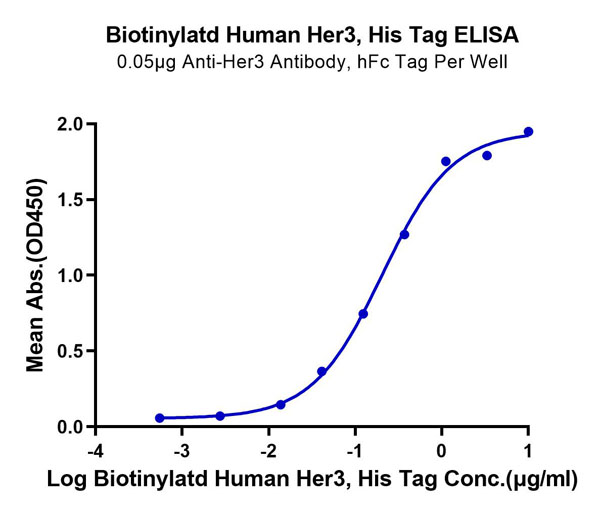
(Biotinylated Human Her3/ErbB3 Protein ELISA DataImmobilized Anti-Her3 Antibody, hFc Tag at 0.5?g/ml (100?l/well) on the plate. Dose response curve for Biotinylatd Human Her3, His Tag with the EC50 of 0.20?g/ml determined by ELISA.)
ELISA
(Biotinylated Human Her3/ErbB3 Protein ELISA DataImmobilized Anti-Her3 Antibody, hFc Tag at 0.5?g/ml (100?l/well) on the plate. Dose response curve for Biotinylatd Human Her3, His Tag with the EC50 of 0.20?g/ml determined by ELISA.)
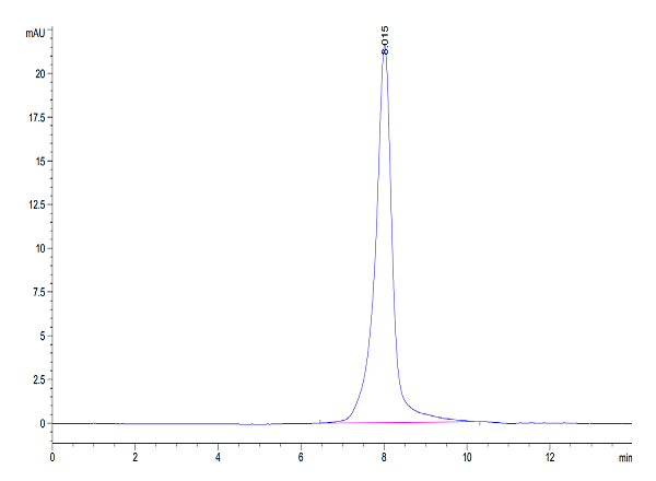
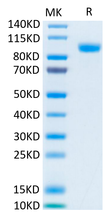
Her2/ErbB2 Domain 4, Recombinant Protein (Cat# AAA76335)
Full Name
Biotinylated Human Her2/ErbB2 Domain 4 Protein Recombinant
Gene Names
ERBB2; NEU; NGL; HER2; TKR1; CD340; HER-2; MLN 19; HER-2/neu
Purity
>95%, as determined by SDS-PAGE and HPLC
Pricing
 IHC (Immunohiostchemistry)
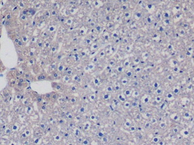
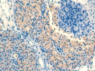
(DAB staining on IHCP;Sample: Rat Ovary Tissue; Primary Ab: 10ug/ml Mouse AntiRat TGFb1 AntibodySecond Ab: 2ug/mL HRPLinked Caprine AntiMouse IgG Polyclonal Antibody(Catalog: SAA544Mu19))
IHC (Immunohiostchemistry)
(DAB staining on IHCP;Sample: Rat Ovary Tissue; Primary Ab: 10ug/ml Mouse AntiRat TGFb1 AntibodySecond Ab: 2ug/mL HRPLinked Caprine AntiMouse IgG Polyclonal Antibody(Catalog: SAA544Mu19))
Transforming Growth Factor Beta 1 (TGFb1), Monoclonal Antibody (Cat# AAA151488)
Full Name
Monoclonal Antibody to Transforming Growth Factor Beta 1 (TGFb1)
Gene Names
Tgfb1; Tgfb
Reactivity
Rat
Applications
Immunoprecipitation, Immunocytochemistry, Immunohistochemistry, Western Blot
Purity
Protein A + Protein G affinity chromatography
Pricing
 Bioactivity
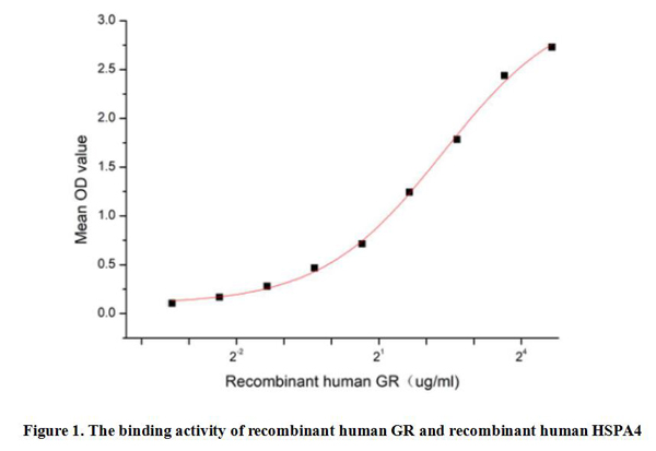
(Glucocorticoid Receptor (GR) is a member of the steroid receptor superfamily, which includes receptors for other steroid hormones such as estrogens, progestogens, androgens and mineralocorticoids. GR is widely distributed in various types of human cells, especially in liver, muscle, adipose tissue, lung, brain and other organs. GR plays a role in signaling within cells. When glucocorticoids enter cells and bind to GR, GR undergoes conformational changes that activate its transcriptional activity. It has been identified that the binding of Heat Shock 70kDa Protein 4 (HSPA4) to GR plays a key role in glucocorticoid signaling, which not only participates in the stabilization and activation of receptors, but also may affect the effect of drugs. Thus a functional binding ELISA assay was conducted to detect the interaction of recombinant human GR and recombinant human HSPA4. Briefly, GR was diluted serially in PBS with 0.01% BSA (pH 7.4). Duplicate samples of 100 ul were then transferred to HSPA4-coated microtiter wells and incubated for 1h at 37 degree C. Wells were washed with PBST and incubated for 1h with anti-GR pAb, then aspirated and washed 3 times. After incubation with HRP labelled secondary antibody for 1h at 37 degree C, wells were aspirated and washed 5 times. With the addition of substrate solution, wells were incubated 15-25 minutes at 37 degree C. Finally, add 50 uL stop solution to the wells and read at 450/630 nm immediately. The binding activity of recombinant human GR and recombinant human HSPA4 was shown in Figure 1, the EC50 for this effect is 5.1 ug/mL.)
Bioactivity
(Glucocorticoid Receptor (GR) is a member of the steroid receptor superfamily, which includes receptors for other steroid hormones such as estrogens, progestogens, androgens and mineralocorticoids. GR is widely distributed in various types of human cells, especially in liver, muscle, adipose tissue, lung, brain and other organs. GR plays a role in signaling within cells. When glucocorticoids enter cells and bind to GR, GR undergoes conformational changes that activate its transcriptional activity. It has been identified that the binding of Heat Shock 70kDa Protein 4 (HSPA4) to GR plays a key role in glucocorticoid signaling, which not only participates in the stabilization and activation of receptors, but also may affect the effect of drugs. Thus a functional binding ELISA assay was conducted to detect the interaction of recombinant human GR and recombinant human HSPA4. Briefly, GR was diluted serially in PBS with 0.01% BSA (pH 7.4). Duplicate samples of 100 ul were then transferred to HSPA4-coated microtiter wells and incubated for 1h at 37 degree C. Wells were washed with PBST and incubated for 1h with anti-GR pAb, then aspirated and washed 3 times. After incubation with HRP labelled secondary antibody for 1h at 37 degree C, wells were aspirated and washed 5 times. With the addition of substrate solution, wells were incubated 15-25 minutes at 37 degree C. Finally, add 50 uL stop solution to the wells and read at 450/630 nm immediately. The binding activity of recombinant human GR and recombinant human HSPA4 was shown in Figure 1, the EC50 for this effect is 5.1 ug/mL.)
Glucocorticoid Receptor (GR), Active Protein (Cat# AAA161853)
Full Name
Active Glucocorticoid Receptor (GR)
Gene Names
NR3C1; GR; GCR; GRL; GCCR
Applications
Activity Assay, Cell Culture
Purity
Greater than 95% by SDS-PAGE
Pricing
 WB (Western Blot)
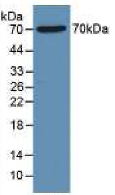
(Sample: Recombinant HDAC1, Human;Antibody: Rabbit Anti-Human HDAC1 Ab)
WB (Western Blot)
(Sample: Recombinant HDAC1, Human;Antibody: Rabbit Anti-Human HDAC1 Ab)
Histone Deacetylase 1 (HDAC1), Active Protein (Cat# AAA149259)
Full Name
Active Histone Deacetylase 1 (HDAC1)
Gene Names
HDAC1; HD1; RPD3; KDAC1; GON-10; RPD3L1
Reactivity
Homo sapiens (Human)
Applications
Activity Assay, Cell Culture
Purity
>95%
Pricing
 SDS-PAGE
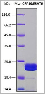
(SDS-PAGE analysis (15%) of 2 ul of recombinant CFP10: ESAT6. Purity is approx. 95% as determined by gel electrophoresis. The band which appears just below, corresponds to a in vivo N-terminus or C-terminus degradation product of this same protein, as it is showed in a western blot performed with a his-tag monoclonal antibody.)
SDS-PAGE
(SDS-PAGE analysis (15%) of 2 ul of recombinant CFP10: ESAT6. Purity is approx. 95% as determined by gel electrophoresis. The band which appears just below, corresponds to a in vivo N-terminus or C-terminus degradation product of this same protein, as it is showed in a western blot performed with a his-tag monoclonal antibody.)
CFP10:ESAT6, Recombinant Protein (Cat# AAA14240)
Full Name
Recombinant chimeric antigen CFP10:ESAT6 for Mycobacterium tuberculosis (Koch's bacillus)
Pricing
 FCM (Flow Cytometry)
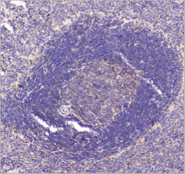
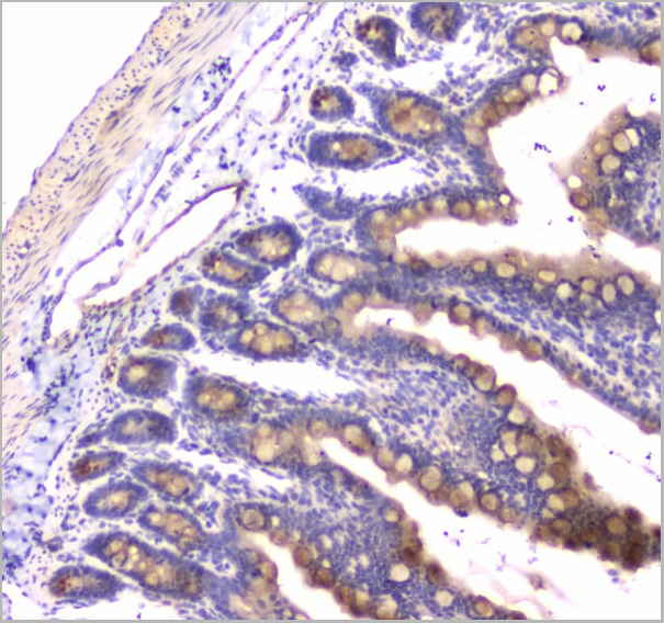
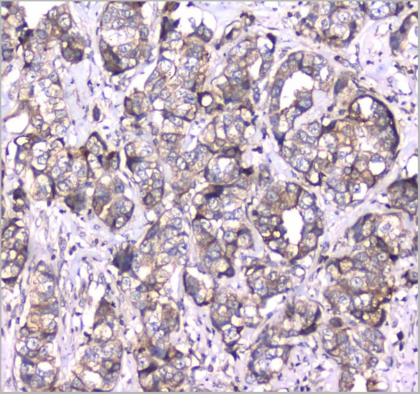
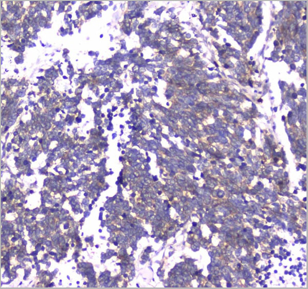
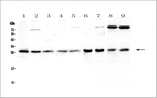
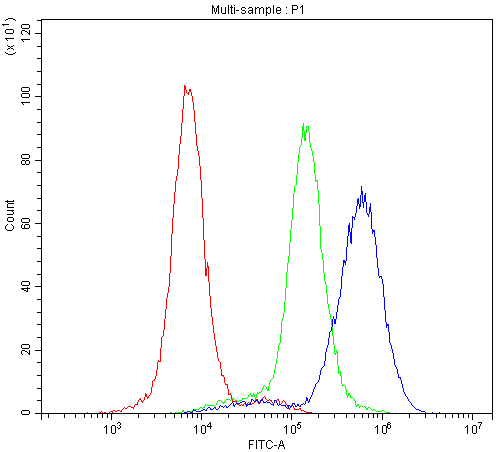
(Figure 7. Flow Cytometry analysis of K562 cells using anti-RAB27A antibody (AAA19152).Overlay histogram showing K562 cells stained with AAA19152 (Blue line).The cells were blocked with 10% normal goat serum. And then incubated with rabbit anti-RAB27A Antibody (AAA19152,1ug/1x10^6 cells) for 30 min at 20 degree C. DyLight ®488 conjugated goat anti-rabbit IgG (5-10ug/1x10^6 cells) was used as secondary antibody for 30 minutes at 20 degree C. Isotype control antibody (Green line) was rabbit IgG (1ug/1x106) used under the same conditions. Unlabelled sample (Red line) was also used as a control.)
FCM (Flow Cytometry)
(Figure 7. Flow Cytometry analysis of K562 cells using anti-RAB27A antibody (AAA19152).Overlay histogram showing K562 cells stained with AAA19152 (Blue line).The cells were blocked with 10% normal goat serum. And then incubated with rabbit anti-RAB27A Antibody (AAA19152,1ug/1x10^6 cells) for 30 min at 20 degree C. DyLight ®488 conjugated goat anti-rabbit IgG (5-10ug/1x10^6 cells) was used as secondary antibody for 30 minutes at 20 degree C. Isotype control antibody (Green line) was rabbit IgG (1ug/1x106) used under the same conditions. Unlabelled sample (Red line) was also used as a control.)
RAB27A, Polyclonal Antibody (Cat# AAA19152)
Full Name
Anti-RAB27A Picoband antibody
Gene Names
RAB27A; GS2; RAM; RAB27; HsT18676
Reactivity
Human, Mouse, Rat
No cross reactivity with other proteins.
No cross reactivity with other proteins.
Applications
Flow Cytometry, Immunohistochemistry, Immunocytochemistry, Western Blot
Pricing
 Bioactivity
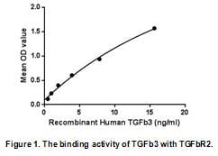
(Transforming growth factor beta-3 (TGFb3) is a type of protein, known as a cytokine, which is involved in cell differentiation, embryogenesis and development. It belongs to a large family of cytokines called the Transforming growth factor beta superfamily. TGFb3 is believed to regulate molecules involved in cellular adhesion and extracellular matrix (ECM) formation during the process of palate development. It also plays an essential role in controlling the development of lungs in mammals, by also regulating cell adhesion and ECM formation in this tissue, and controls wound healing by regulating the movements of epidermal and dermal cells in injured skin. Besides, Transforming Growth Factor Beta Receptor II (TGFbR2) has been identified as an interactor of TGFb3, thus a binding ELISA assay was conducted to detect the interaction of recombinant human TGFb3 and recombinant human TGFbR2. Briefly, TGFb3 were diluted serially in PBS, with 0.01% BSA (pH 7.4). Duplicate samples of 100uL were then transferred to TGFbR2-coated microtiter wells and incubated for 2h at 37 degree C. Wells were washed with PBST and incubated for 1h with anti-TGFb3 pAb, then aspirated and washed 3 times. After incubation with HRP labelled secondary antibody, wells were aspirated and washed 3 times. With the addition of substrate solution, wells were incubated 15-25 minutes at 37 degree C. Finally, add 50uL stop solution to the wells and read at 450nm immediately. The binding activity of TGFb3 and TGFbR2 was shown in Figure 1, and this effect was in a dose dependent manner.)
Bioactivity
(Transforming growth factor beta-3 (TGFb3) is a type of protein, known as a cytokine, which is involved in cell differentiation, embryogenesis and development. It belongs to a large family of cytokines called the Transforming growth factor beta superfamily. TGFb3 is believed to regulate molecules involved in cellular adhesion and extracellular matrix (ECM) formation during the process of palate development. It also plays an essential role in controlling the development of lungs in mammals, by also regulating cell adhesion and ECM formation in this tissue, and controls wound healing by regulating the movements of epidermal and dermal cells in injured skin. Besides, Transforming Growth Factor Beta Receptor II (TGFbR2) has been identified as an interactor of TGFb3, thus a binding ELISA assay was conducted to detect the interaction of recombinant human TGFb3 and recombinant human TGFbR2. Briefly, TGFb3 were diluted serially in PBS, with 0.01% BSA (pH 7.4). Duplicate samples of 100uL were then transferred to TGFbR2-coated microtiter wells and incubated for 2h at 37 degree C. Wells were washed with PBST and incubated for 1h with anti-TGFb3 pAb, then aspirated and washed 3 times. After incubation with HRP labelled secondary antibody, wells were aspirated and washed 3 times. With the addition of substrate solution, wells were incubated 15-25 minutes at 37 degree C. Finally, add 50uL stop solution to the wells and read at 450nm immediately. The binding activity of TGFb3 and TGFbR2 was shown in Figure 1, and this effect was in a dose dependent manner.)
Transforming Growth Factor Beta 3 (TGFb3), Active Protein (Cat# AAA152205)
Full Name
Active Transforming Growth Factor Beta 3 (TGFb3)
Gene Names
TGFB3; ARVD; TGF-beta3
Applications
Activity Assay, Cell Culture
Purity
Greater than 95% by SDS-PAGE
Pricing
 FCM (Flow Cytometry)
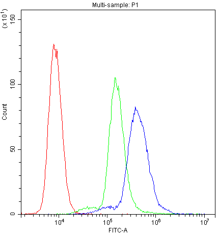
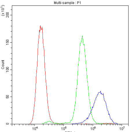
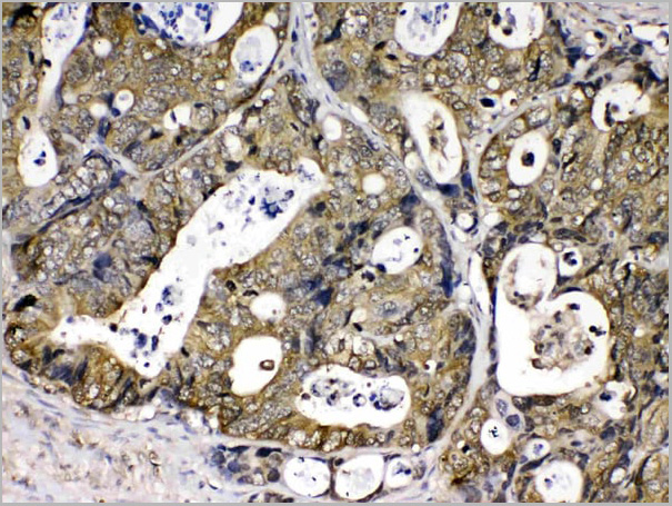
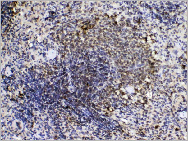
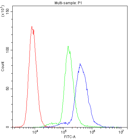
(Figure 7. Flow Cytometry analysis of K562 cells using anti-PPIA antibody (AAA19148).Overlay histogram showing K562 cells stained with AAA19148 (Blue line).The cells were blocked with 10% normal goat serum. And then incubated with rabbit anti-PPIA Antibody (AAA19148,1ug/1x10^6 cells) for 30 min at 20 degree C. DyLight ®488 conjugated goat anti-rabbit IgG (5-10ug/1x10^6 cells) was used as secondary antibody for 30 minutes at 20 degree C. Isotype control antibody (Green line) was rabbit IgG (1ug/1x106) used under the same conditions. Unlabelled sample (Red line) was also used as a control.)
FCM (Flow Cytometry)
(Figure 7. Flow Cytometry analysis of K562 cells using anti-PPIA antibody (AAA19148).Overlay histogram showing K562 cells stained with AAA19148 (Blue line).The cells were blocked with 10% normal goat serum. And then incubated with rabbit anti-PPIA Antibody (AAA19148,1ug/1x10^6 cells) for 30 min at 20 degree C. DyLight ®488 conjugated goat anti-rabbit IgG (5-10ug/1x10^6 cells) was used as secondary antibody for 30 minutes at 20 degree C. Isotype control antibody (Green line) was rabbit IgG (1ug/1x106) used under the same conditions. Unlabelled sample (Red line) was also used as a control.)
Cyclophilin A, Polyclonal Antibody (Cat# AAA19148)
Full Name
Anti-Cyclophilin A Picoband Antibody
Gene Names
PPIA; CYPA; CYPH; HEL-S-69p
Reactivity
Human, Mouse, Rat
No cross reactivity with other proteins
No cross reactivity with other proteins
Applications
Flow Cytometry, Immunohistochemistry, Immunocytochemistry, Western Blot
Purity
Immunogen affinity purified
Pricing
 Bioactivity
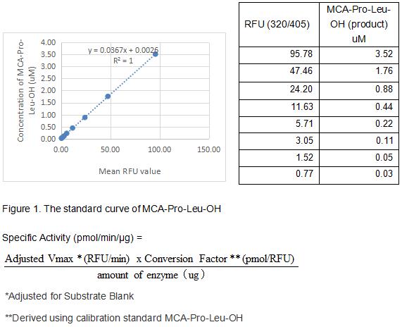
(Matrix metalloproteinases are a family of zinc and calcium dependent endopeptidases with the combined ability to degrade all the components of the extracellular matrix. MMP-3 (stromelysin-1), can degrade a broad range of substrates including collagen alpha chains, aggrecan, laminin, fibronectin, and elastin. MMP-3 does not cleave the triple helical region of interstitial collagens, a characteristic which distinguishes the stromelysins from the collagenases. MMP-3 is expressed by fibroblasts, chrondrocytes, osteoblasts, endothelial cells, smooth muscle cells and macrophages. Structurally, MMP-3 may be divided into several distinct domains; a pro-domain which is cleaved upon activation; a catalytic domain containing the zinc binding site; a short hinge region and a carboxyl terminal (hemopexin-like) domain. The activity of recombinant human MMP-3 is measured by its ability to cleave a fluorogenic peptide substrate Mca-Arg-Pro-Lys-Pro-Val-Glu-Nval-Trp-Arg-Lys(Dnp)-NH2 in the assay buffer 50 mM Tris, 10 mM CaCl2, 150 mM NaCl, 0.05% (w/v) Brij-35, pH 7.5. The rhMMP3 is diluted to 200 ug/ml in assay buffer, then activated with 5 ug/ml chymotrypsin at 37 degree C for 30min followed by adding 2 mM PMSF to stop activation. The activated rhMMP3 is diluted to 6.25 ug/mL in assay buffer. Loading into a black well plate 50 uL of 6.25 ug/mL rhMMP3 and start the reaction by adding 50 uL of 20 uM substrate, with a substrate blank containing 50 uL assay buffer, 50 uL substrate, and no rhMMP3. Then read at excitiation and emission wavelengths of 320 nm and 405 nm, respectively, in kinetic mode for 5 minutes. The specific activity of recombinant human MMP3 is > 90 pmol/min/ug.)
Bioactivity
(Matrix metalloproteinases are a family of zinc and calcium dependent endopeptidases with the combined ability to degrade all the components of the extracellular matrix. MMP-3 (stromelysin-1), can degrade a broad range of substrates including collagen alpha chains, aggrecan, laminin, fibronectin, and elastin. MMP-3 does not cleave the triple helical region of interstitial collagens, a characteristic which distinguishes the stromelysins from the collagenases. MMP-3 is expressed by fibroblasts, chrondrocytes, osteoblasts, endothelial cells, smooth muscle cells and macrophages. Structurally, MMP-3 may be divided into several distinct domains; a pro-domain which is cleaved upon activation; a catalytic domain containing the zinc binding site; a short hinge region and a carboxyl terminal (hemopexin-like) domain. The activity of recombinant human MMP-3 is measured by its ability to cleave a fluorogenic peptide substrate Mca-Arg-Pro-Lys-Pro-Val-Glu-Nval-Trp-Arg-Lys(Dnp)-NH2 in the assay buffer 50 mM Tris, 10 mM CaCl2, 150 mM NaCl, 0.05% (w/v) Brij-35, pH 7.5. The rhMMP3 is diluted to 200 ug/ml in assay buffer, then activated with 5 ug/ml chymotrypsin at 37 degree C for 30min followed by adding 2 mM PMSF to stop activation. The activated rhMMP3 is diluted to 6.25 ug/mL in assay buffer. Loading into a black well plate 50 uL of 6.25 ug/mL rhMMP3 and start the reaction by adding 50 uL of 20 uM substrate, with a substrate blank containing 50 uL assay buffer, 50 uL substrate, and no rhMMP3. Then read at excitiation and emission wavelengths of 320 nm and 405 nm, respectively, in kinetic mode for 5 minutes. The specific activity of recombinant human MMP3 is > 90 pmol/min/ug.)
Matrix Metalloproteinase 3 (MMP3), Active Protein (Cat# AAA161687)
Full Name
Active Matrix Metalloproteinase 3 (MMP3)
Gene Names
MMP3; SL-1; STMY; STR1; CHDS6; MMP-3; STMY1
Applications
Activity Assay, Cell Culture
Purity
Greater than 95% by SDS-PAGE
Pricing
 Bioactivity
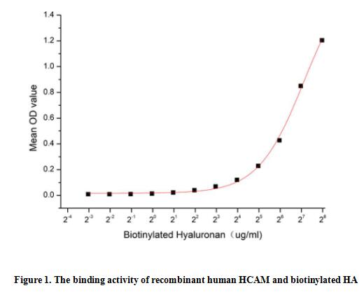
(Homing Associated Cell Adhesion Molecule (HCAM), also known as CD44, is a ubiquitous multistructural and multifunctional cells surface adhesion molecule involved in cell-cell and cell-matrix interactions. CD44 is broadly expressed, including in the membranes of B cells, granulocytes, monocytes, and erythrocytes as well as on many thymocytes and mature T cells, besides it is highly expressed in many cancers and regulates metastasis via recruitment of CD44 to the cell surface. This protein is a receptor for hyaluronic acid (HA) and can also interact with other ligands, such as osteopontin, collagens, and matrix metalloproteinases (MMPs). Thus a functional binding ELISA assay was conducted to detect the interaction of recombinant human HCAM and biotinylated hyaluronan (HA). Briefly, biotin-linked HA was diluted serially in PBS, with 0.01% BSA (pH 7.4). Duplicate samples of 100 ul were then transferred to HCAM-coated microtiter wells and incubated for 2h at 37 degree C. Wells were washed with PBST 3 times and incubation with Streptavidin-HRP for 1 hour, then wells were aspirated and washed 5 times. With the addition of substrate solution, wells were incubated 15-25 minutes at 37 degree C. Finally, add 50 ul stop solution to the wells and read at 450/630 nm immediately. The binding activity of recombinant human HCAM and biotinylated HA was shown in Figure 1, and this effect was in a dose dependent manner.)
Bioactivity
(Homing Associated Cell Adhesion Molecule (HCAM), also known as CD44, is a ubiquitous multistructural and multifunctional cells surface adhesion molecule involved in cell-cell and cell-matrix interactions. CD44 is broadly expressed, including in the membranes of B cells, granulocytes, monocytes, and erythrocytes as well as on many thymocytes and mature T cells, besides it is highly expressed in many cancers and regulates metastasis via recruitment of CD44 to the cell surface. This protein is a receptor for hyaluronic acid (HA) and can also interact with other ligands, such as osteopontin, collagens, and matrix metalloproteinases (MMPs). Thus a functional binding ELISA assay was conducted to detect the interaction of recombinant human HCAM and biotinylated hyaluronan (HA). Briefly, biotin-linked HA was diluted serially in PBS, with 0.01% BSA (pH 7.4). Duplicate samples of 100 ul were then transferred to HCAM-coated microtiter wells and incubated for 2h at 37 degree C. Wells were washed with PBST 3 times and incubation with Streptavidin-HRP for 1 hour, then wells were aspirated and washed 5 times. With the addition of substrate solution, wells were incubated 15-25 minutes at 37 degree C. Finally, add 50 ul stop solution to the wells and read at 450/630 nm immediately. The binding activity of recombinant human HCAM and biotinylated HA was shown in Figure 1, and this effect was in a dose dependent manner.)
Homing Associated Cell Adhesion Molecule (HCAM), Active Protein (Cat# AAA161762)
Full Name
Active Homing Associated Cell Adhesion Molecule (HCAM)
Gene Names
CD44; IN; LHR; MC56; MDU2; MDU3; MIC4; Pgp1; CDW44; CSPG8; HCELL; HUTCH-I; ECMR-III
Applications
Activity Assay, Cell Culture
Purity
Greater than 90% by SDS-PAGE
Pricing
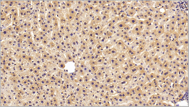
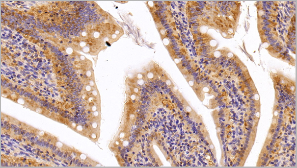
 IHC (Immunohistchemistry)
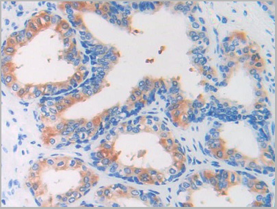
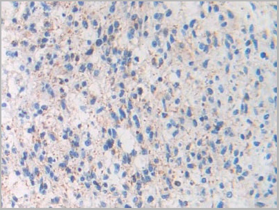
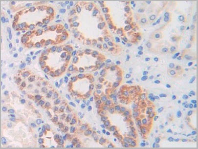
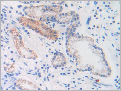
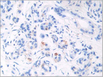
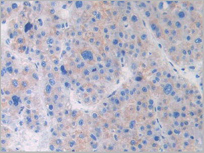
(DAB staining on IHCP;Sample: Human Liver cancer Tissue; Primary Ab: 30ug/ml Mouse AntiHuman BNP AntibodySecond Ab: 2ug/mL HRPLinked Caprine AntiMouse IgG Polyclonal Antibody(Catalog: SAA544Mu19))
IHC (Immunohistchemistry)
(DAB staining on IHCP;Sample: Human Liver cancer Tissue; Primary Ab: 30ug/ml Mouse AntiHuman BNP AntibodySecond Ab: 2ug/mL HRPLinked Caprine AntiMouse IgG Polyclonal Antibody(Catalog: SAA544Mu19))
Brain Natriuretic Peptide (BNP), Monoclonal Antibody (Cat# AAA21183)
Full Name
Monoclonal Antibody to Brain Natriuretic Peptide (BNP)
Gene Names
NPPB; BNP
Reactivity
Human
Applications
Western Blot, Immunohistochemistry, Immunocytochemistry, Immunoprecipitation
Purity
Protein A + Protein G affinity chromatography
Pricing
 IF (Immunofluorescence)
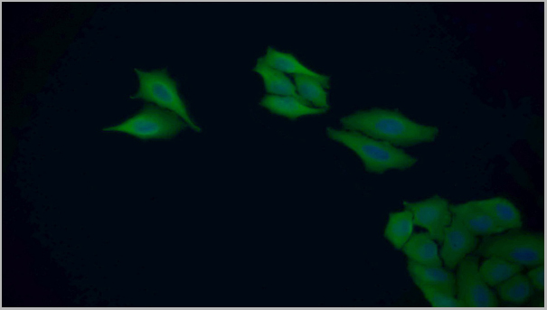
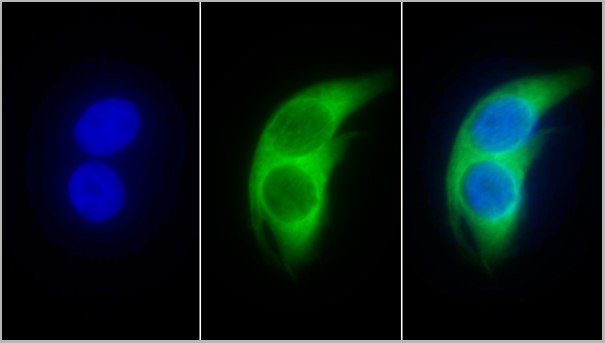
(FITC staining on IF;Sample: Human HepG2 cell; Primary Ab: 20ug/ml Rabbit AntiHuman GSTp AntibodySecond Ab: 2ug/ml FITCLinked Caprine AntiRabbit IgG Polyclonal Antibody(Catalog: SAA544Rb18))
IF (Immunofluorescence)
(FITC staining on IF;Sample: Human HepG2 cell; Primary Ab: 20ug/ml Rabbit AntiHuman GSTp AntibodySecond Ab: 2ug/ml FITCLinked Caprine AntiRabbit IgG Polyclonal Antibody(Catalog: SAA544Rb18))
Glutathione S Transferase Pi (GSTp), Polyclonal Antibody (Cat# AAA21213)
Full Name
Polyclonal Antibody to Glutathione S Transferase Pi (GSTp)
Gene Names
GSTP1; PI; DFN7; GST3; GSTP; FAEES3
Reactivity
Human, Rat
Applications
Western Blot, Immunohistochemistry, Immunocytochemistry, Immunoprecipitation
Purity
Antigenspecific affinity chromatography followed by Protein A affinity chromatography
Pricing