Filters
▼Clonality
▼Type
▼Reactivity
▼Gene Name
▼Isotype
▼Host
▼Application
▼Clone
▼Polyclonal Antibodies
At AAA Biotech also known as AAA Bio or AAABio, we provide a broad range of purified polyclonal antibodies (pAbs) that are able to all be browsed online through our website. Due to their high specificity and strong binding affinity, these antibodies are ideal for wide swathes of research and experimental applications.
Our polyclonal antibodies can easily support your work, whether you use them for Western Blotting, Immunocytochemistry (with or without Immunofluorescence used in conjunction), Immunohistochemistry, Immunoprecipitation, and ELISA tests. We highly encourage you to browse our range of pAbs and choose the one that best suits your experimental model.
Viewing 400-450 of 118597 product results
 FCM/FACS (Flow Cytometry)
(AAA60839 Flow cytometric analysis of paraformaldehyde fixed A431 cells (blue line), permeabilized with 0.5% Triton. Primary incubation 1hr (10ug/ml) followed by Alexa Fluor 488 secondary antibody (1ug/ml). IgG control: Unimmunized goat IgG (black line) followed by Alexa Fluor 488 secondary antibody.)
FCM/FACS (Flow Cytometry)
(AAA60839 Flow cytometric analysis of paraformaldehyde fixed A431 cells (blue line), permeabilized with 0.5% Triton. Primary incubation 1hr (10ug/ml) followed by Alexa Fluor 488 secondary antibody (1ug/ml). IgG control: Unimmunized goat IgG (black line) followed by Alexa Fluor 488 secondary antibody.)
STAT3, Polyclonal Antibody (Cat# AAA60839)
 WB (Western Blot)
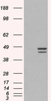
(HEK293 overexpressing Man2A1 (RC220186) and probed (mock transfection in first lane), tested by Origene.)
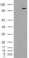
WB (Western Blot)
(HEK293 overexpressing Man2A1 (RC220186) and probed (mock transfection in first lane), tested by Origene.)
MAN2A1, Polyclonal Antibody (Cat# AAA60854)
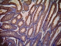 IHC (Immunohiostchemistry)
((1ug/ml) staining of paraffin embedded Mouse Testis. Data kindly provided by Dr. Erwin Goldberg, Northwestern University, Evanston, IL USA.)
IHC (Immunohiostchemistry)
((1ug/ml) staining of paraffin embedded Mouse Testis. Data kindly provided by Dr. Erwin Goldberg, Northwestern University, Evanston, IL USA.)
LDHC, Polyclonal Antibody (Cat# AAA60859)
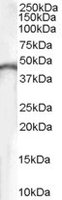
 WB (Western Blot)
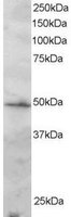
((0. 03ug/ml) staining of Human Placenta lysate (35ug protein in RIPA buffer). Primary incubation was 1 hour. Detected by chemiluminescence.)
WB (Western Blot)
((0. 03ug/ml) staining of Human Placenta lysate (35ug protein in RIPA buffer). Primary incubation was 1 hour. Detected by chemiluminescence.)
Choline acetyltransferase, Polyclonal Antibody (Cat# AAA60863)
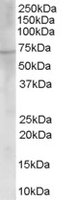
 WB (Western Blot)
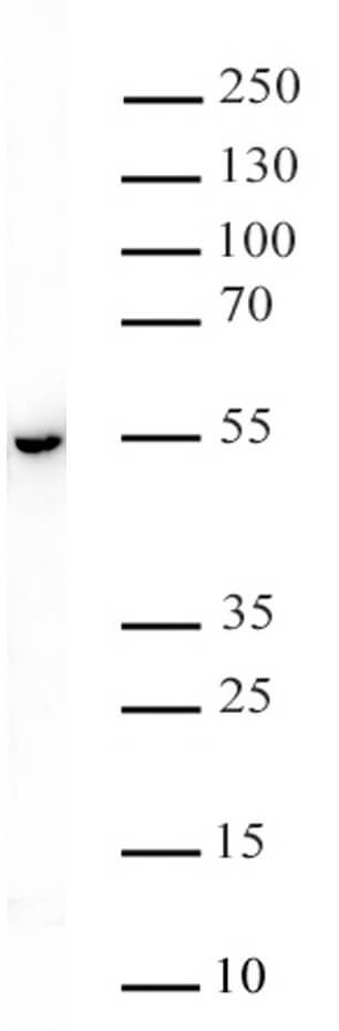
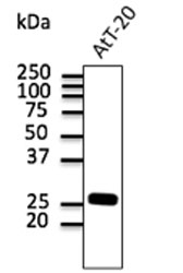
(AAA60866 (0.1ug/ml) staining of human brain lysate (35ug protein in RIPA buffer).Primary incubation was 1 hour. Detected by chemiluminescence.)
WB (Western Blot)
(AAA60866 (0.1ug/ml) staining of human brain lysate (35ug protein in RIPA buffer).Primary incubation was 1 hour. Detected by chemiluminescence.)
CKB/Brain Creatine Kinase, Polyclonal Antibody (Cat# AAA60866)
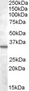 WB (Western Blot)
((2ug/ml) staining of K562 lysate (35ug protein in RIPA buffer). Primary incubation was 1 hour. Detected by chemiluminescence.)
WB (Western Blot)
((2ug/ml) staining of K562 lysate (35ug protein in RIPA buffer). Primary incubation was 1 hour. Detected by chemiluminescence.)
Bradykinin receptor B1, Polyclonal Antibody (Cat# AAA60867)
Her6, Polyclonal Antibody (Cat# AAA60872)
 WB (Western Blot)
(HEK293 overexpressing IRF2 (RC202102) and probed (mock transfection in first lane))
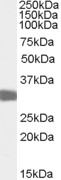
WB (Western Blot)
(HEK293 overexpressing IRF2 (RC202102) and probed (mock transfection in first lane))
IRF2, Polyclonal Antibody (Cat# AAA60878)
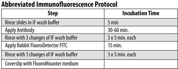
 Application Data
HDAC3 Phospho Ser424, Polyclonal Antibody (Cat# AAA60353)
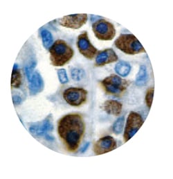
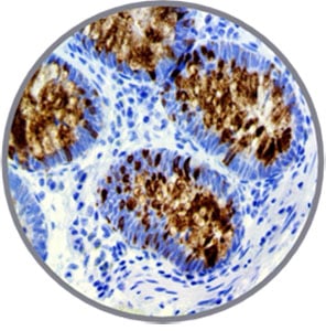
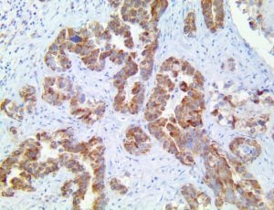 IHC (Immunohiostchemistry)
(CXCR5 Antibody Immunohistochemistry on an FFPE Lung Adenocarcinoma Tissue)
IHC (Immunohiostchemistry)
(CXCR5 Antibody Immunohistochemistry on an FFPE Lung Adenocarcinoma Tissue)
CXCR5/CD185, Polyclonal Antibody (Cat# AAA59365)
 IHC (Immunohiostchemistry)
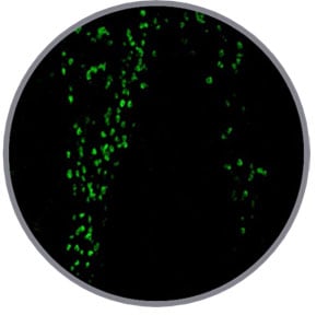
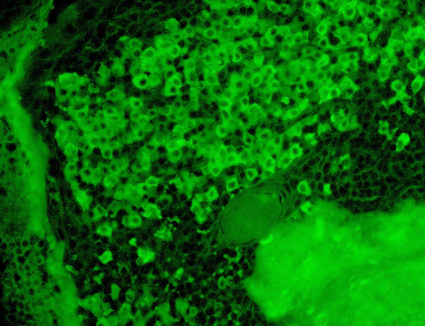 IF (Immunofluorescence)
(IF of Kappa on a FFPE Tonsil Tissue)
IF (Immunofluorescence)
(IF of Kappa on a FFPE Tonsil Tissue)
Kappa, Polyclonal Antibody (Cat# AAA59329)
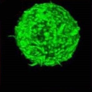 IF (Immunofluorescence)
(IF of IgE)
IF (Immunofluorescence)
(IF of IgE)
IgE, Polyclonal Antibody (Cat# AAA59331)
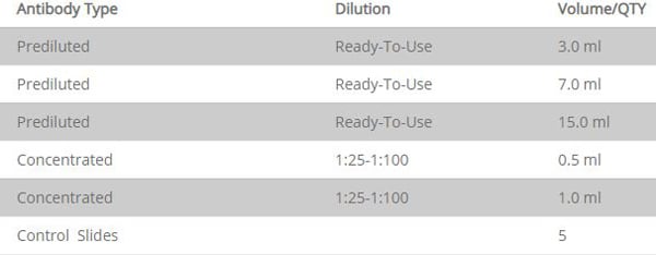
 Application Data
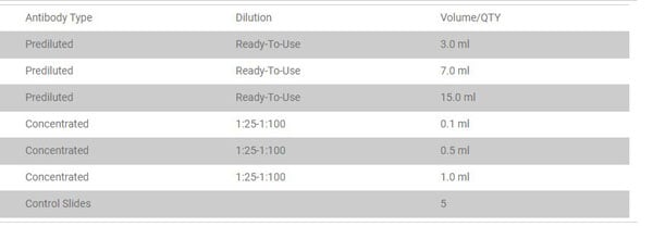
(Suggested Dilution)
Application Data
(Suggested Dilution)
 IF (Immunofluorescence)
(Immunofluorescence - anti-Rab3 Ab in B6 cells at 1/50 dilution; cells were fixed with methanol.)
IF (Immunofluorescence)
(Immunofluorescence - anti-Rab3 Ab in B6 cells at 1/50 dilution; cells were fixed with methanol.)
Rab3, Polyclonal Antibody (Cat# AAA63070)
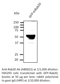 Application Data
Rab20, Polyclonal Antibody (Cat# AAA63082)
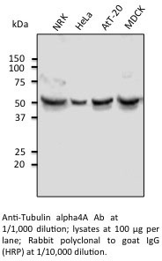 Application Data
TUBA4A, Polyclonal Antibody (Cat# AAA63094)
 Reactivity Data
Rab10, Polyclonal Antibody (Cat# AAA63111)
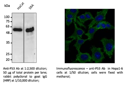
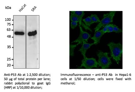 Application Data
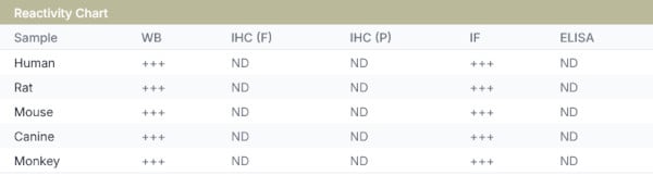
P53, Polyclonal Antibody (Cat# AAA63117)
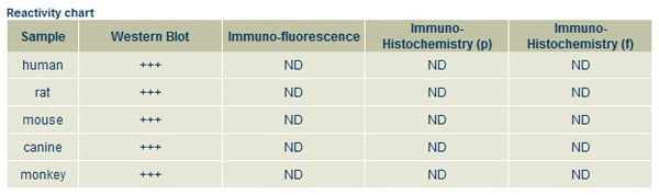
 Reactivity Data
MGP, Polyclonal Antibody (Cat# AAA63124)
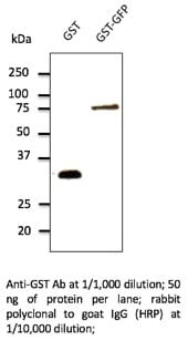
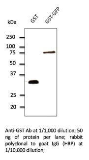 Application Data
GST, Polyclonal Antibody (Cat# AAA63127)
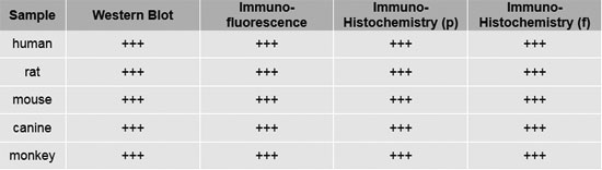
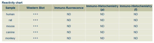
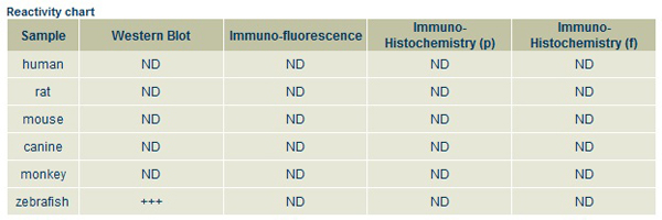
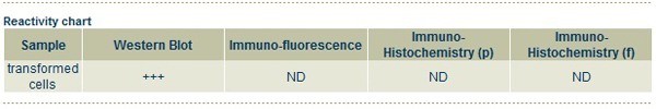
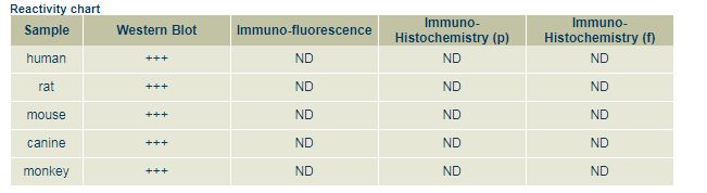
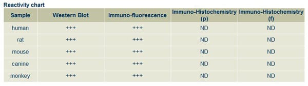
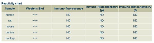
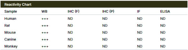 Reactivity Chart
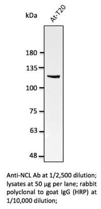
Nucleolin, Polyclonal Antibody (Cat# AAA63145)

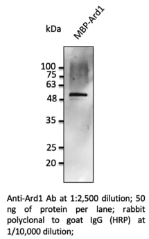 WB (Western Blot)
ARD1, Polyclonal Antibody (Cat# AAA63150)
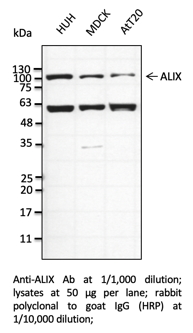 WB (Western Blot)
ALIX, Polyclonal Antibody (Cat# AAA63162)
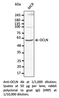 WB (Western Blot)
OCLN, Polyclonal Antibody (Cat# AAA63164)
 Application Data
(Anti-ITGB2 Ab at 1/1,000 dilution; lysates at 50 ug per lane; rabbit polyclonal to goat IgG (HRP) at 1/10,000 dilution;)
Application Data
(Anti-ITGB2 Ab at 1/1,000 dilution; lysates at 50 ug per lane; rabbit polyclonal to goat IgG (HRP) at 1/10,000 dilution;)
ITGB2, Polyclonal Antibody (Cat# AAA63167)
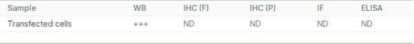
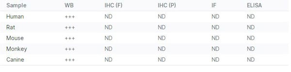
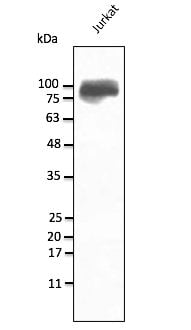
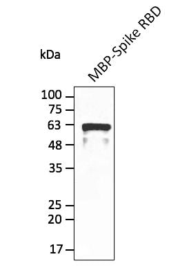 Application Data
(Anti-Spike Ab at 1/2,500 dilution; lane with 30 ng of recombinant fusion protein; rabbit polyclonal to goat IgG (HRP) at 1/10,000 dilution;)
Application Data
(Anti-Spike Ab at 1/2,500 dilution; lane with 30 ng of recombinant fusion protein; rabbit polyclonal to goat IgG (HRP) at 1/10,000 dilution;)
COVID 19 Spike RBD Domain Coronavirus, Polyclonal Antibody (Cat# AAA63174)
 Application Data
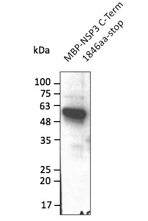
(Anti-NSP3 Ab at 1/2,500 dilution; lane with 30 ng of recombinant fusion protein; rabbit polyclonal to goat IgG (HRP) at 1/10,000 dilution;)
Application Data
(Anti-NSP3 Ab at 1/2,500 dilution; lane with 30 ng of recombinant fusion protein; rabbit polyclonal to goat IgG (HRP) at 1/10,000 dilution;)
COVID 19 NSP3 Coronavirus, Polyclonal Antibody (Cat# AAA63181)
 Application Data
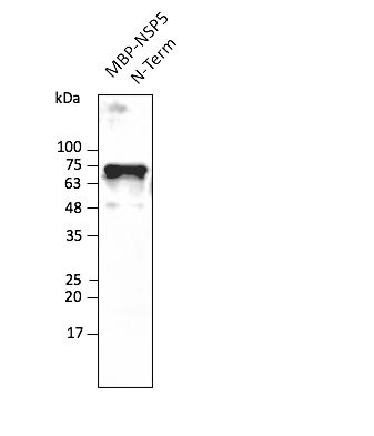
(Anti-NSP5 Ab at 1/2,500 dilution; lane with 30 ng of recombinant fusion protein; rabbit polyclonal to goat IgG (HRP) at 1/10,000 dilution;)
Application Data
(Anti-NSP5 Ab at 1/2,500 dilution; lane with 30 ng of recombinant fusion protein; rabbit polyclonal to goat IgG (HRP) at 1/10,000 dilution;)
COVID 19 NSP5 Coronavirus, Polyclonal Antibody (Cat# AAA63183)
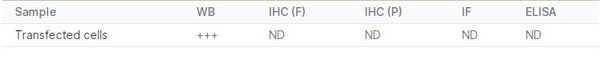
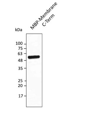 Application Data
(Anti-Membrane protein Ab at 1/2,500 dilution; lane with 30 ng of recombinant fusion protein (149-215 aa); rabbit polyclonal to goat IgG (HRP) at 1/10,000 dilution;)
Application Data
(Anti-Membrane protein Ab at 1/2,500 dilution; lane with 30 ng of recombinant fusion protein (149-215 aa); rabbit polyclonal to goat IgG (HRP) at 1/10,000 dilution;)
COVID 19 Membrane Protein Coronavirus, Polyclonal Antibody (Cat# AAA63197)
 Application Data
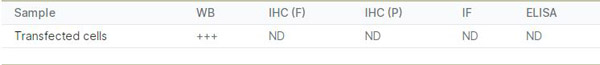
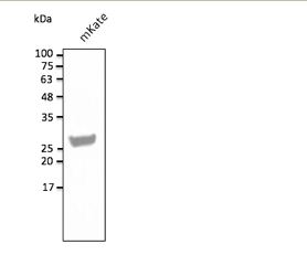
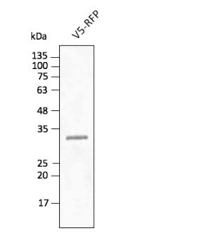
(Anti-mKate Ab at 1/2,500 dilution using HEK293 transfected cell lysates at 50 ug per lane; rabbit polyclonal to goat IgG (HRP) at 1/10,000 dilution;)
Application Data
(Anti-mKate Ab at 1/2,500 dilution using HEK293 transfected cell lysates at 50 ug per lane; rabbit polyclonal to goat IgG (HRP) at 1/10,000 dilution;)
mKate, Polyclonal Antibody (Cat# AAA63198)
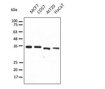

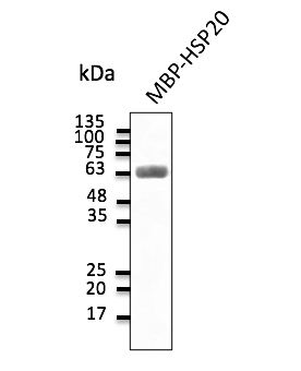 Application Data
(Anti-HSP20 Ab at 1:2,000 dilution; 30 ng of protein per lane; chicken polyclonal to goat IgG conjugated to HRP (AB1125) at 1/10,000 dilution;)
Application Data
(Anti-HSP20 Ab at 1:2,000 dilution; 30 ng of protein per lane; chicken polyclonal to goat IgG conjugated to HRP (AB1125) at 1/10,000 dilution;)
IgG, Polyclonal Secondary Antibody (Cat# AAA63204)
 Application Data
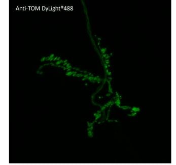
(Anti-tdTomato Ab conjugated to DyLight 488 at 1/2,500 dilution using HEK293 transfected cell lysates at 50 ug per lane;)
Application Data
(Anti-tdTomato Ab conjugated to DyLight 488 at 1/2,500 dilution using HEK293 transfected cell lysates at 50 ug per lane;)
tdTomato, Polyclonal Antibody (Cat# AAA63208)
 Application Data
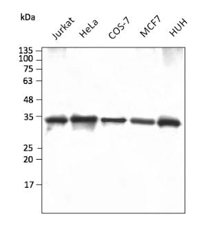
(Anti-GAPDH Ab at 1/2,500 dilution using HEK293 transfected cell lysates at 50 ug per lane; chicken polyclonal to goat IgG conjugated to DyLight 633 (AB200633) at 1/10,000 dilution;)
Application Data
(Anti-GAPDH Ab at 1/2,500 dilution using HEK293 transfected cell lysates at 50 ug per lane; chicken polyclonal to goat IgG conjugated to DyLight 633 (AB200633) at 1/10,000 dilution;)
IgG, Polyclonal Secondary Antibody (Cat# AAA63212)
 IF (Immunofluorescence)
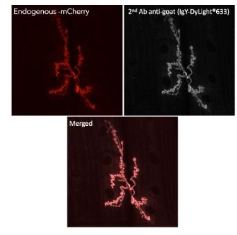
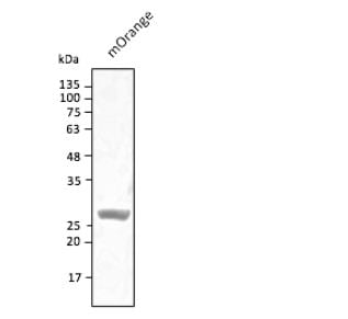
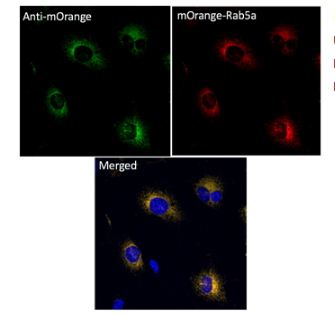
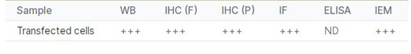
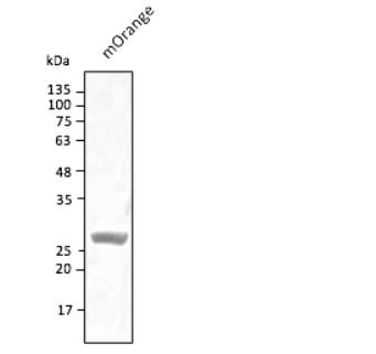
(Immunofluorescence - anti-mOrange Ab using hCEC cells transduced with mOrange-Rab5a; cells were fixed with methanol and anti-mOrange at 1/250;)
IF (Immunofluorescence)
(Immunofluorescence - anti-mOrange Ab using hCEC cells transduced with mOrange-Rab5a; cells were fixed with methanol and anti-mOrange at 1/250;)
mOrange, Polyclonal Antibody (Cat# AAA63215)
 Application Data
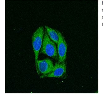
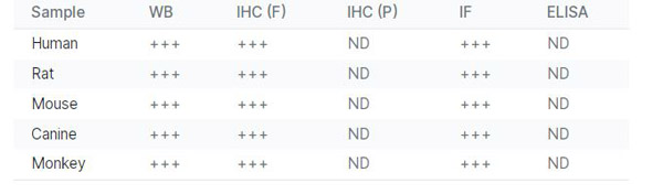
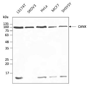
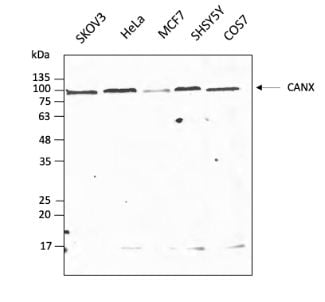
(Anti-CANX Ab conjugated to DyLight@488 at 1/2,000 dilution using cell lysates at 40 ug per lane;)
Application Data
(Anti-CANX Ab conjugated to DyLight@488 at 1/2,000 dilution using cell lysates at 40 ug per lane;)
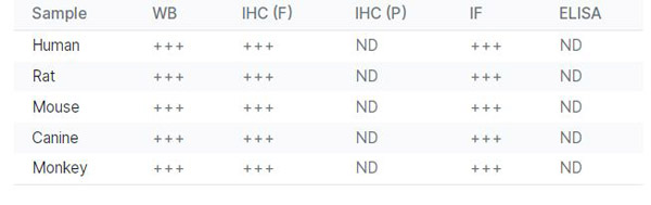
CANX, Polyclonal Antibody (Cat# AAA63231)
 Application Data
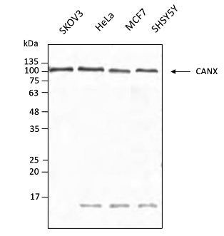
(Anti-CANX Ab conjugated to DyLight@550 at 1/2,000 dilution using cell lysates at 40 ug per lane;)
Application Data
(Anti-CANX Ab conjugated to DyLight@550 at 1/2,000 dilution using cell lysates at 40 ug per lane;)
CANX, Polyclonal Antibody (Cat# AAA63232)
 Application Data
(Anti-CANX Ab conjugated to DyLight@633 at 1/2,000 dilution using cell lysates at 40 ug per lane;)
Application Data
(Anti-CANX Ab conjugated to DyLight@633 at 1/2,000 dilution using cell lysates at 40 ug per lane;)
CANX, Polyclonal Antibody (Cat# AAA63233)
 WB (Western Blot)
c-Jun, Polyclonal Antibody (Cat# AAA63008)
DYKDDDDK (E8), Polyclonal Antibody (Cat# AAA63020)
BSA (FL11), Polyclonal Antibody (Cat# AAA63021)
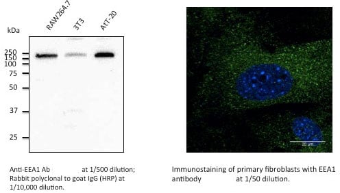 Application Data
EEA1, Polyclonal Antibody (Cat# AAA63042)
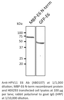 Application Data
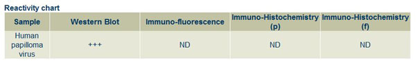
HPV11 E6, Polyclonal Antibody (Cat# AAA63049)
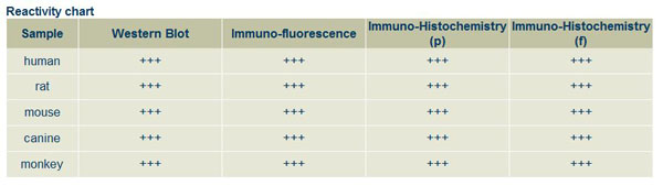
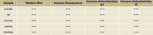 Reactivity Chart
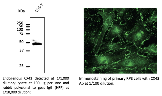
CX43, Polyclonal Antibody (Cat# AAA63051)
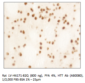 Application Data
HTT, Polyclonal Antibody (Cat# AAA63056)
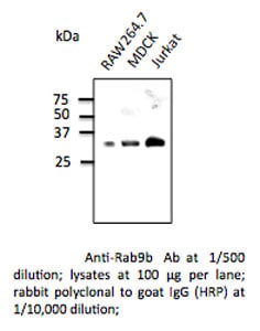 WB (Western Blot)
Rab9b, Polyclonal Antibody (Cat# AAA63058)
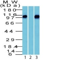 WB (Western Blot)
(Western Blot of beta Catenin (p120) in human brain in 1) absence and 2) presence of immunizing peptide and 3) mouse brain Lysate using beta Catenin (p120) Polyclonal Antibody.)
WB (Western Blot)
(Western Blot of beta Catenin (p120) in human brain in 1) absence and 2) presence of immunizing peptide and 3) mouse brain Lysate using beta Catenin (p120) Polyclonal Antibody.)
beta-Catenin, Polyclonal Antibody (Cat# AAA62578)
 ICC (Immunocytochemistry)
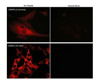
(Immunocytochemical labeling of phosphorylated CRMP2 in mouse C2C12 cells. The cells were probed with CRMP2 (C-terminal region) and CRMP2 (Thr-555) rabbit polyclonal antibodies, then the antibodies were detected using appropriate secondary antibodies conjugated to Cy3. The antibodies were used in the absence (left) or presence (right) of their respective blocking peptide (CX2165 or CX2255).)
ICC (Immunocytochemistry)
(Immunocytochemical labeling of phosphorylated CRMP2 in mouse C2C12 cells. The cells were probed with CRMP2 (C-terminal region) and CRMP2 (Thr-555) rabbit polyclonal antibodies, then the antibodies were detected using appropriate secondary antibodies conjugated to Cy3. The antibodies were used in the absence (left) or presence (right) of their respective blocking peptide (CX2165 or CX2255).)
CRMP2, Polyclonal Antibody (Cat# AAA71603)
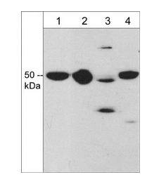
 ICC (Immunocytochemistry)
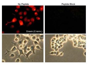
(Immunocytochemical labeling of Draxin in rat PC12 cells differentiated with NGF. The cells were probed with Draxin (C-terminal region) rabbit polyclonal antibody, then the antibody was detected using appropriate secondary antibody conjugated to Cy3. The antibody was used in the absence (left) or presence (right) of blocking peptide (DX3675). Lower images show corresponding phase images.)
ICC (Immunocytochemistry)
(Immunocytochemical labeling of Draxin in rat PC12 cells differentiated with NGF. The cells were probed with Draxin (C-terminal region) rabbit polyclonal antibody, then the antibody was detected using appropriate secondary antibody conjugated to Cy3. The antibody was used in the absence (left) or presence (right) of blocking peptide (DX3675). Lower images show corresponding phase images.)
Draxin, Polyclonal Antibody (Cat# AAA71618)
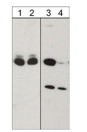
 Application Data
beta-III-Tubulin (C-terminus), Polyclonal Antibody (Cat# AAA71513)
What are Polyclonal Antibodies?
Polyclonal antibodies are antibodies that come from multiple B cell clones of a host animal. The typical hosts used for the majority of polyclonal antibody production are rabbits, goats, sheep, and donkeys. These polyclonal antibodies, once having identified their target, will bind to different epitopes located at different regions or sequences on the same protein/antigen. This ability to bind multiple epitopes is what makes polyclonal antibodies highly sensitive, as explained in our detailed guide on polyclonal antibodies and why they matter.
As a result, they are ideal at locating and binding to the target, even if the target is in very low concentrations (due to many different antibodies being able to bind to the same target molecule, which allows for significant amplification of a downstream signal).
Polyclonal antibodies are typically produced by injecting an antigen into a host animal, which causes the animal’s immune system to attack the foreign antigen by mass generating antibodies against it. After a period of time, serum is collected from the animal and purified using physicochemical fractionation, class-specific affinity purification, and/or antigen-affinity purification.
Key Uses of Polyclonal Antibodies
- Western Blotting: This method is used to find specific proteins in biological samples after separating them by size.
- Immunohistochemistry: IHC helps visualize the location of proteins in tissue sections using various staining techniques.
- ELISA: (Enzyme-Linked Immunosorbent Assay) is typically used to identify specific protein quantities in a sample. ELISAs can be either “Quantitative” or “Qualitative”.
- Flow Cytometry: technique that identifies and measures the specific protein on the surface or inside the cells in a fluid suspension.
- Immunoprecipitation: IP isolates and studies a specific protein from a complex mixture using antibodies.
Why Buy Polyclonal Antibodies from AAA Biotech?
1. Ideal for Various Applications
Our antibodies are generally going to be validated for use in multiple types of assays, including ELISA, Western Blotting, Immunohistochemistry, Immunoprecipitation, amongst others. They are ideal for a wide range of research applications.
2. Rigorous Quality Control
All of the antibodies in our catalog undergo strict quality testing to ensure specificity, sensitivity, and consistent performance. We are confident in the ability of our antibodies to provide you with accurate results.
3. Wide Assortment of Antibodies
Antibodies in our catalog can be found for both common and exotic species, and these antibodies are also available in both conjugated and recombinant forms to suit many diverse experimental needs.
4. Highly Purified
Our antibodies are available in purified forms with over 85% purity, as confirmed by SDS-PAGE. They are also available with tags such as His, Flag, GST, or MBP. We cater to customers worldwide.
FAQ
1. How are polyclonal antibodies produced?
Traditionally, polyclonal antibodies are produced by injecting an antigen into a host animal (such as a rabbit or goat), which then triggers an immune response from the host animal. The animal’s B cells produce antibodies that will recognize different parts of the injected antigen. These antibodies are then collected from the animal’s blood and purified for use.
2. How do polyclonal antibodies differ from monoclonal antibodies?
Polyclonal antibodies are a mix of antibodies that bind to different locations (epitopes) of the same antigen, while monoclonal antibodies are identical and bind to just one specific epitope. This makes polyclonal antibodies more versatile and better at detecting proteins that may be present in low quantities or in altered/modified forms.
3. How should I store polyclonal antibodies?
Polyclonal antibodies should be stored at 4°C for short-term use (up to a few weeks) and at -20°C or -80°C for long-term storage. Avoid repeated freeze-thaw cycles by dividing them into small aliquots. Always check the datasheet for specific storage instructions.