Call us at +1-800-604-9114 for more information about our products or contact us online Need help? +1-800-604-9114 or contact us

Polyclonal Antibodies

At AAA Biotech, we provide a broad range of purified polyclonal antibodies (pAbs) that are able to all be browsed online through our website. Due to their high specificity and strong binding affinity, these antibodies are ideal for wide swathes of research and experimental applications.


Our polyclonal antibodies can easily support your work, whether you use them for Western Blotting, Immunocytochemistry (with or without Immunofluorescence used in conjunction), Immunohistochemistry, Immunoprecipitation, and ELISA tests. We highly encourage you to browse our range of pAbs and choose the one that best suits your experimental model.


Viewing 1750-1800 of 3252 product results


product-image-AAA10683_IHC4.jpg IHC (Immunohistochemistry) (Immunohistochemistry of paraffin-embedded mouse kidney using SPARC antibody at dilution of 1:100 (40x lens).)

SPARC, Polyclonal Antibody (Cat# AAA10683)

Full Name
SPARC Polyclonal Antibody
Gene Names
SPARC; ON
Applications
Immunohistochemistry (IHC)
Purity
Affinity Purification
Pricing
product-image-AAA19902_FCM6.png FCM (Flow Cytometry) (Figure 6. Flow Cytometry analysis of JK cells using anti-MZB1 antibody (AAA19902).Overlay histogram showing JK cells stained with AAA19902 (Blue line). To facilitate intracellular staining, cells were fixed with 4% paraformaldehyde and permeabilized with permeabilization buffer. The cells were blocked with 10% normal goat serum. And then incubated with rabbit anti-MZB1 Antibody (AAA19902, 1ug/1x106 cells) for 30 min at 20 degree C. DyLight488 conjugated goat anti-rabbit IgG was used as secondary antibody for 30 minutes at 20 degree C. Isotype control antibody (Green line) was rabbit IgG (1ug/1x106) used under the same conditions. Unlabelled sample (Red line) was also used as a control.)

MZB1, Polyclonal Antibody (Cat# AAA19902)

Full Name
Anti-MZB1 Antibody Picoband
Gene Names
MZB1; PACAP; pERp1; MEDA-7
Reactivity
Human
Applications
ELISA (EIA), Immunofluorescence (IF), Immunohistochemistry (IHC), Immunocytochemistry (ICC), Western Blot (WB), Flow Cytometry (FC/FACS)
Purity
Immunogen affinity purified.
Pricing
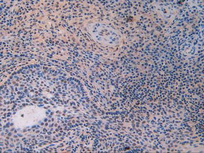
product-image-AAA19141_IHC7.jpg IHC (Immunohistochemistry) (Figure 7. IHC analysis of splicing factor 1 using anti-splicing factor 1 antibody (AAA19141).splicing factor 1 was detected in paraffin-embedded section of rat small intestine tissue. Heat mediated antigen retrieval was performed in citrate buffer (pH6, epitope retrieval solution) for 20 mins. The tissue section was blocked with 10% goat serum. The tissue section was then incubated with 1ug/ml rabbit anti-splicing factor 1 Antibody (AAA19141) overnight at 4 degree C. Biotinylated goat anti-rabbit IgG was used as secondary antibody and incubated for 30 minutes at 37 degree C. The tissue section was developed using Strepavidin-Biotin-Complex (SABC) with DAB as the chromogen.)

splicing factor 1, Polyclonal Antibody (Cat# AAA19141)

Full Name
Anti-splicing factor 1 Picoband antibody
Gene Names
SF1; BBP; MBBP; ZFM1; ZNF162; D11S636; ZCCHC25
Reactivity
Human, Mouse, Rat
No cross reactivity with other proteins.
Applications
ELISA (EIA), Immunohistochemistry (IHC), Western Blot (WB)
Pricing
product-image-AAA29381_IHC6.jpg IHC (Immunohistchemistry) (Dilution: IHC-p 1:50-200, ELISA 1:10000-20000)

IL-8RA, Polyclonal Antibody (Cat# AAA29381)

Full Name
IL-8RA Polyclonal Antibody
Gene Names
CXCR1; C-C; CD128; CD181; CKR-1; IL8R1; IL8RA; CMKAR1; IL8RBA; CDw128a; C-C-CKR-1
Reactivity
Human
Applications
Immunohistochemistry-Paraffin (IHC-P)
Pricing
product-image-AAA30661_IHC8.jpg IHC (Immunohistochemistry) (Immunohistochemistry of paraffin-embedded mouse kidney using LRRK2 at dilution of 1:200 (40x lens).)

LRRK2, Polyclonal Antibody (Cat# AAA30661)

Full Name
LRRK2 Rabbit Polyclonal Antibody
Gene Names
LRRK2; PARK8; RIPK7; ROCO2; AURA17; DARDARIN
Reactivity
Human, Mouse, Rat
Applications
Western Blot (WB), Immunohistochemistry (IHC), Immunofluorescence (IF)
Purity
Affinity purification
Pricing
product-image-AAA31429_APP11.jpg Application Data (At 25 degree C. The primary antibody was diluted at 1/200 and incubated with the sample for 1 hour at 37 degree C. An Alexa Fluor 594 conjugated goat anti-rabbit IgG (H+L) Ab, diluted at 1/600, was used as the secondary antibody.)

p53, Polyclonal Antibody (Cat# AAA31429)

Full Name
Phospho-p53 (Ser392) Antibody
Gene Names
TP53; P53; BCC7; LFS1; TRP53
Reactivity
Human, Mouse
Predicted Reactivity: Pig (88%), Bovine (88%), Sheep (88%), Rabbit (88%)
Applications
Western Blot (WB), Immunohistochemistry (IHC), Immunofluorescence (IF), Immunocytochemistry (ICC), Peptide ELISA (EIA)
Purity
The antibody is from purified rabbit serum by affinity purification via sequential chromatography on phospho-peptide and non-phospho-peptide affinity columns.
Pricing
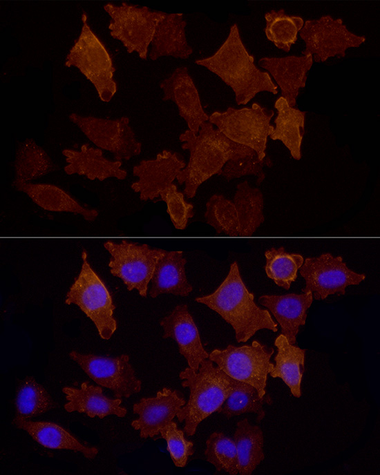
product-image-AAA10694_IF7.jpg IF (Immunofluorescence) (Immunofluorescence analysis of MCF-7 cells using STK11 antibody. Blue: DAPI for nuclear staining.)

STK11, Polyclonal Antibody (Cat# AAA10694)

Full Name
STK11 Polyclonal Antibody
Gene Names
STK11; PJS; LKB1; hLKB1
Reactivity
Human, Mouse, Rat
Applications
Western Blot (WB), Immunohistochemistry (IHC), Immunofluorescence (IF)
Purity
Affinity Purification
Pricing
product-image-AAA14790_APP6.jpg Application Data (C:FGFR2/isolectinB4 (C) and FGFR1/isolectinB4 (D) staining of apparent mesenchymal cells and the subpopulation of endothelial cells. Virtually all other dispersed apparent mesenchymal cells express FGFR1 and FGFR2 (merged image in E). F: FGFR2 (F) and FGFR1 (G) staining in clustered cells of epithelial origin (inferred by morphology here) demonstrating that epithelial cells express both FGFR1 and FGFR2 (merged image with DAPI staining in H).)

FGFR2, Polyclonal Antibody (Cat# AAA14790)

Full Name
FGFR2, NT (FGFR2, BEK, KGFR, KSAM, Fibroblast growth factor receptor 2, K-sam, Keratinocyte growth factor receptor, CD332)
Gene Names
FGFR2; BEK; JWS; BBDS; CEK3; CFD1; ECT1; KGFR; TK14; TK25; BFR-1; CD332; K-SAM
Reactivity
Human, Monkey, Mouse, Rat
Applications
ELISA (EL/EIA), Western Blot (WB), Immunohistochemistry (IHC), Flow Cytometry (FC/FACS), Immunofluorescence (IF)
Purity
Affinity Purified
Purified by Protein A affinity chromatography.
Pricing
product-image-AAA28098_IP8.jpg IP (Immunoprecipitation) (Immunoprecipitation analysis of 200ug extracts of 293T cells using 3ug CTNNA1 antibody. Western blot was performed from the immunoprecipitate using CTNNA1 antibody at a dilition of 1:500.)

CTNNA1, Polyclonal Antibody (Cat# AAA28098)

Full Name
CTNNA1 Polyclonal Antibody
Gene Names
CTNNA1; CAP102
Reactivity
Human, Mouse, Rat
Applications
Western Blot (WB), Immunohistochemistry (IHC), Immunofluorescence (IF), Flow Cytometry (FC/FACS)
Purity
Affinity Purification
Pricing
product-image-AAA28354_IF6.jpg IF (Immunofluorescence) (Immunofluorescence analysis of Raw264.7 cells using eEF1A1 antibody at dilution of 1:100. Blue: DAPI for nuclear staining.)

eEF1A1, Polyclonal Antibody (Cat# AAA28354)

Full Name
eEF1A1 Rabbit pAb
Gene Names
EEF1A1; CCS3; EF1A; PTI1; CCS-3; EE1A1; EEF-1; EEF1A; EF-Tu; LENG7; eEF1A-1; GRAF-1EF; HNGC:16303
Reactivity
Human, Mouse, Rat
Applications
Western Blot (WB), Immunohistochemistry (IHC), Immunofluorescence (IF)
Purity
Affinity purification
Pricing
product-image-AAA29378_IHC6.jpg IHC (Immunohistchemistry) (Dilution: IHC-p 1:50-200, ELISA 1:10000-20000)

Integrin alphaE, Polyclonal Antibody (Cat# AAA29378)

Full Name
Integrin alphaE Polyclonal Antibody
Gene Names
ITGAE; CD103; HUMINAE
Reactivity
Human
Applications
Immunohistochemistry-Paraffin (IHC-P)
Pricing
product-image-AAA30658_IF7.jpg IF (Immunofluorescence) (Immunofluorescence analysis of C6 cells using YAP1 Rabbit pAb.)

YAP1, Polyclonal Antibody (Cat# AAA30658)

Full Name
YAP1 Rabbit Polyclonal Antibody
Gene Names
YAP1; YAP; YKI; YAP2; YAP65
Reactivity
Human, Mouse, Rat
Applications
Western Blot (WB), Immunohistochemistry (IHC), Immunofluorescence (IF)
Purity
Affinity purification
Pricing
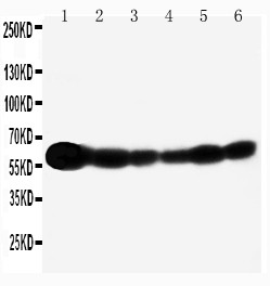
product-image-AAA19907_WB.jpg IHC (Immunohistochemistry) (Figure 10. IHC analysis of ISYNA1 using anti-ISYNA1 antibody (AAA19907).ISYNA1 was detected in a paraffin-embedded section of rat testis tissue. Heat mediated antigen retrieval was performed in EDTA buffer (pH 8.0, epitope retrieval solution). The tissue section was blocked with 10% goat serum. The tissue section was then incubated with 2ug/ml rabbit anti-ISYNA1 Antibody (AAA19907) overnight at 4 degree C. Peroxidase Conjugated Goat Anti-rabbit IgG was used as secondary antibody and incubated for 30 minutes at 37 degree C. The tissue section was developed using HRP Conjugated Rabbit IgG Super Vision Assay Kit ( epitope retrieval solution). The tissue section was blocked with 10% goat serum. The tissue section was then incubated with 2ug/ml rabbit anti-ISYNA1 Antibody (AAA19907) overnight at 4 degree C. Peroxidase Conjugated Goat Anti-rabbit IgG was used as secondary antibody and incubated for 30 minutes at 37 degree C. The tissue section was developed using HRP Conjugated Rabbit IgG Super Vision Assay Kit ( epitope retrieval solution). The tissue section was blocked with 10% goat serum. The tissue section was then incubated with 2ug/ml rabbit anti-ISYNA1 Antibody (AAA19907) overnight at 4 degree C. Peroxidase Conjugated Goat Anti-rabbit IgG was used as secondary antibody and incubated for 30 minutes at 37 degree C. The tissue section was developed using HRP Conjugated Rabbit IgG Super Vision Assay Kit ( epitope retrieval solution). The tissue section was blocked with 10% goat serum. The tissue section was then incubated with 2ug/ml rabbit anti-ISYNA1 Antibody (AAA19907) overnight at 4 degree C. Peroxidase Conjugated Goat Anti-rabbit IgG was used as secondary antibody and incubated for 30 minutes at 37 degree C. The tissue section was developed using HRP Conjugated Rabbit IgG Super Vision Assay Kit ( epitope retrieval solution). The tissue section was blocked with 10% goat serum. The tissue section was then incubated with 2ug/ml rabbit anti-ISYNA1 Antibody (AAA19907) overnight at 4 degree C. Peroxidase Conjugated Goat Anti-rabbit IgG was used as secondary antibody and incubated for 30 minutes at 37 degree C. The tissue section was developed using HRP Conjugated Rabbit IgG Super Vision Assay Kit ( epitope retrieval solution). The tissue section was blocked with 10% goat serum. The tissue section was then incubated with 2ug/ml rabbit anti-ISYNA1 Antibody (AAA19907) overnight at 4 degree C. Peroxidase Conjugated Goat Anti-rabbit IgG was used as secondary antibody and incubated for 30 minutes at 37 degree C. The tissue section was developed using HRP Conjugated Rabbit IgG Super Vision Assay Kit ( epitope retrieval solution). The tissue section was blocked with 10% goat serum. The tissue section was then incubated with 2ug/ml rabbit anti-ISYNA1 Antibody (AAA19907) overnight at 4 degree C. Peroxidase Conjugated Goat Anti-rabbit IgG was used as secondary antibody and incubated for 30 minutes at 37 degree C. The tissue section was developed using HRP Conjugated Rabbit IgG Super Vision Assay Kit ( epitope retrieval solution). The tissue section was blocked with 10% goat serum. The tissue section was then incubated with 2ug/ml rabbit anti-ISYNA1 Antibody (AAA19907) overnight at 4 degree C. Peroxidase Conjugated Goat Anti-rabbit IgG was used as secondary antibody and incubated for 30 minutes at 37 degree C. The tissue section was developed using HRP Conjugated Rabbit IgG Super Vision Assay Kit ( epitope retrieval solution). The tissue section was blocked with 10% goat serum. The tissue section was then incubated with 2ug/ml rabbit anti-ISYNA1 Antibody (AAA19907) overnight at 4 degree C. Peroxidase Conjugated Goat Anti-rabbit IgG was used as secondary antibody and incubated for 30 minutes at 37 degree C. The tissue section was developed using HRP Conjugated Rabbit IgG Super Vision Assay Kit (Lane 2: human K562 whole cell lysates,Lane 3: human 293T whole cell lysates,Lane 4: human HEL whole cell lysates,Lane 5: rat testis tissue lysates,Lane 6: mouse testis tissue lysates.After electrophoresis, proteins were transferred to a nitrocellulose membrane at 150 mA for 50-90 minutes. Blocked the membrane with 5% non-fat milk/TBS for 1.5 hour at RT. The membrane was incubated with rabbit anti-ISYNA1 antigen affinity purified polyclonal antibody (#AAA19907) at 0.25ug/mL overnight at 4 degree C, then washed with TBS-0.1%Tween 3 times with 5 minutes each and probed with a goat anti-rabbit IgG-HRP secondary antibody at a dilution of 1:5000 for 1.5 hour at RT. The signal is developed using an Enhanced Chemiluminescent detection (ECL) kit with Tanon 5200 system. A specific band was detected for ISYNA1 at approximately 61 kDa. The expected band size for ISYNA1 is at 61 kDa.)

ISYNA1, Polyclonal Antibody (Cat# AAA19907)

Full Name
Anti-ISYNA1 Antibody Picoband
Gene Names
ISYNA1; IPS; INO1; INOS; IPS 1; IPS-1
Reactivity
Human, Mouse, Rat
Applications
Western Blot (WB), Immunohistochemistry (IHC), ELISA (EIA)
Purity
Immunogen affinity purified.
Pricing
product-image-AAA30659_IHC10.jpg IHC (Immunohistochemistry) (Immunohistochemistry of paraffin-embedded human gastric cancer using TARDBP at dilution of 1:200 (40x lens).)

TARDBP, Polyclonal Antibody (Cat# AAA30659)

Full Name
TARDBP Rabbit Polyclonal Antibody
Gene Names
TARDBP; ALS10; TDP-43
Reactivity
Human, Mouse, Rat
Applications
Western Blot (WB), Immunohistochemistry (IHC), Immunofluorescence (IF)
Purity
Affinity purification
Pricing
product-image-AAA31427_IHC7.jpg IHC (Immunohistochemistry) (At 1/100 staining Mouse brain tissue by IHC-P. The sample was formaldehyde fixed and a heat mediated antigen retrieval step in citrate buffer was performed. The sample was then blocked and incubated with the primary antibody at 4 degree C overnight. An HRP conjugated anti-Rabbit antibody was used as the secondary antibody.)

NACA, Polyclonal Antibody (Cat# AAA31427)

Full Name
Phospho-NACA (Thr159) Antibody
Gene Names
NACA; HSD48; NACA1; skNAC
Reactivity
Human, Mouse, Rat
Predicted Reactivity: Pig (100%), Zebrafish (100%), Bovine (100%), Sheep (100%), Chicken (100%), Xenopus (100%)
Applications
Western Blot (WB), Immunohistochemistry (IHC), Peptide ELISA (EIA)
Purity
The antibody is from purified rabbit serum by affinity purification via sequential chromatography on phospho-peptide and non-phospho-peptide affinity columns.
Pricing
product-image-AAA10692_IHC7.jpg IHC (Immunohistochemistry) (Immunohistochemistry of paraffin-embedded mouse heart using EDF1 antibody at dilution of 1:100 (40x lens).)

EDF1, Polyclonal Antibody (Cat# AAA10692)

Full Name
EDF1 Polyclonal Antibody
Gene Names
EDF1; MBF1; EDF-1
Reactivity
Human, Mouse, Rat
Applications
Western Blot (WB), Immunohistochemistry (IHC)
Purity
Affinity Purification
Pricing
product-image-AAA29886_ICC7.jpg ICC (Immunocytochemistry) (ICC staining NSUN4 in MCF-7 cells (green). The nuclear counter stain is DAPI (blue). Cells were fixed in paraformaldehyde, permeabilised with 0.25% Triton X100/PBS.)

NSUN4, Polyclonal Antibody (Cat# AAA29886)

Full Name
NSUN4 Antibody
Gene Names
NSUN4; SHTAP
Reactivity
Human, Mouse
Applications
Western Blot (WB), Immunocytochemistry (ICC), Immunohistochemistry (IHC)
Purity
ProA affinity purified
Pricing
product-image-AAA10943_WB9.gif WB (Western Blot) (Figure 9 KD Validation of PUMA (Han et al., 2010)Immunoblot analyses of Tet-induced p53 cells treated with NOXA, Puma, Bim or non-targeting siRNAs that were utilized in this experiment. PUMA protein levels were markedly reduced in PUMA KD cells detected by anti-PUMA antibodies (3041).)

PUMA, Polyclonal Antibody (Cat# AAA10943)

Full Name
PUMA Antibody
Gene Names
BBC3; JFY1; PUMA; JFY-1
Reactivity
Human, Mouse
Applications
ELISA (EIA), Western Blot (WB), Immunocytochemistry (ICC), Immunofluorescence (IF)
Purity
PUMA Antibody is affinity chromatography purified via peptide column.
Pricing
product-image-AAA19135_IHC6.jpg IHC (Immunohistchemistry) (Figure 6. IHC analysis of GALE using anti-GALE antibody (AAA19135).GALE was detected in paraffin-embedded section of rat kidney tissue. Heat mediated antigen retrieval was performed in citrate buffer (pH6, epitope retrieval solution) for 20 mins. The tissue section was blocked with 10% goat serum. The tissue section was then incubated with 1ug/ml rabbit anti-GALE Antibody (AAA19135) overnight at 4 degree C. Biotinylated goat anti-rabbit IgG was used as secondary antibody and incubated for 30 minutes at 37 degree C. The tissue section was developed using Strepavidin-Biotin-Complex (SABC) with DAB as the chromogen.)

GALE, Polyclonal Antibody (Cat# AAA19135)

Full Name
Anti-GALE Picoband antibody
Gene Names
GALE; SDR1E1
Reactivity
Human, Mouse, Rat
No cross reactivity with other proteins.
Applications
ELISA (EIA), Immunohistochemistry (IHC), Western Blot (WB)
Pricing
product-image-AAA19903_FCM6.png FCM (Flow Cytometry) (Figure 6. Flow Cytometry analysis of HepG2 cells using anti-PFKFB1 antibody (AAA19903).Overlay histogram showing HepG2 cells stained with AAA19903 (Blue line). To facilitate intracellular staining, cells were fixed with 4% paraformaldehyde and permeabilized with permeabilization buffer. The cells were blocked with 10% normal goat serum. And then incubated with rabbit anti-PFKFB1 Antibody (AAA19903, 1ug/1x106 cells) for 30 min at 20 degree C. DyLight488 conjugated goat anti-rabbit IgG was used as secondary antibody for 30 minutes at 20 degree C. Isotype control antibody (Green line) was rabbit IgG (1ug/1x106) used under the same conditions. Unlabelled sample (Red line) was also used as a control.)

PFKFB1, Polyclonal Antibody (Cat# AAA19903)

Full Name
Anti-PFKFB1 Antibody Picoband
Gene Names
PFKFB1; F6PK; HL2K; PFRX
Reactivity
Human
Applications
Western Blot (WB), Immunohistochemistry (IHC), Flow Cytometry (FC/FACS), ELISA (EIA)
Purity
Immunogen affinity purified.
Pricing
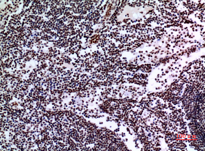
product-image-AAA29375_IHC7.jpg IHC (Immunohistochemistry) (Dilution: IHC-p 1:50-200, ELISA 1:10000-20000)

Napsin A, Polyclonal Antibody (Cat# AAA29375)

Full Name
Napsin A Polyclonal Antibody
Gene Names
NAPSA; KAP; Kdap; NAP1; NAPA; SNAPA
Reactivity
Human
Applications
Immunohistochemistry-Paraffin (IHC-P)
Pricing
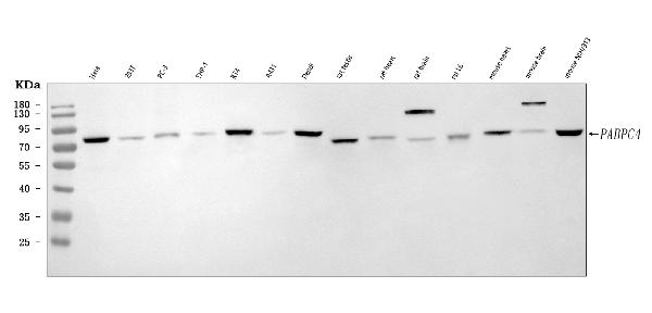
product-image-AAA30655_WB10.jpg WB (Western Blot) (Western blot analysis of extracts of various cell lines, using TOM20 at 1:1000 dilution.)

TOM20, Polyclonal Antibody (Cat# AAA30655)

Full Name
TOM20 Rabbit Polyclonal Antibody
Gene Names
TOMM20; MAS20; MOM19; TOM20
Reactivity
Human, Mouse, Rat
Applications
Western Blot (WB), Immunohistochemistry (IHC), Immunofluorescence (IF)
Purity
Affinity purification
Pricing
product-image-AAA31423_IHC7.jpg IHC (Immunohistochemistry) (At 1/200 staining Human kidney tissue sections by IHC-P. The tissue was formaldehyde fixed and a heat mediated antigen retrieval step in citrate buffer was performed. The tissue was then blocked and incubated with the antibody for 1.5 hours at 22 degree C. An HRP conjugated goat anti-rabbit antibody was used as the secondary antibody.)

CtBP1/2, Polyclonal Antibody (Cat# AAA31423)

Full Name
Phospho-CtBP1/2 (Ser158/Ser164) Antibody
Gene Names
CTBP1; BARS
Reactivity
Human, Mouse, Rat
Predicted Reactivity: Pig (100%), Zebrafish (82%), Bovine (100%), Horse (100%), Dog (100%), Chicken (100%), Xenopus (100%)
Applications
Western Blot (WB), Immunohistochemistry (IHC), Peptide ELISA (EIA)
Purity
The antibody is from purified rabbit serum by affinity purification via sequential chromatography on phospho-peptide and non-phospho-peptide affinity columns.
Pricing
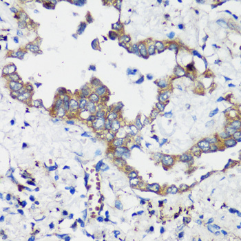
product-image-AAA19136_IHC6.jpg IHC (Immunohistchemistry) (Figure 6. IHC analysis of NSF using anti-NSF antibody (AAA19136).NSF was detected in paraffin-embedded section of human mammary cancer tissue. Heat mediated antigen retrieval was performed in citrate buffer (pH6, epitope retrieval solution) for 20 mins. The tissue section was blocked with 10% goat serum. The tissue section was then incubated with 1ug/ml rabbit anti-NSF Antibody (AAA19136) overnight at 4 degree C. Biotinylated goat anti-rabbit IgG was used as secondary antibody and incubated for 30 minutes at 37 degree C. The tissue section was developed using Strepavidin-Biotin-Complex (SABC) with DAB as the chromogen.)

NSF, Polyclonal Antibody (Cat# AAA19136)

Full Name
Anti-NSF Picoband antibody
Gene Names
NSF; SKD2; SEC18
Reactivity
Human, Mouse, Rat
No cross reactivity with other proteins.
Applications
ELISA (EIA), Immunohistochemistry (IHC), Western Blot (WB)
Pricing
product-image-AAA19904_IF9.jpg IF (Immunofluorescence) (Figure 9. IF analysis of PLEKHG5 using anti-PLEKHG5 antibody (AAA19904).PLEKHG5 was detected in a paraffin-embedded section of human intestinal cancer tissue. Heat mediated antigen retrieval was performed in EDTA buffer (pH 8.0, epitope retrieval solution). The tissue section was blocked with 10% goat serum. The tissue section was then incubated with 5ug/mL rabbit anti-PLEKHG5 Antibody (AAA19904) overnight at 4 degree C. Cy3 Conjugated Goat Anti-Rabbit IgG (BA1032) was used as secondary antibody at 1:500 dilution and incubated for 30 minutes at 37 degree C. The section was counterstained with DAPI. Visualize using a fluorescence microscope and filter sets appropriate for the label used.)

PLEKHG5, Polyclonal Antibody (Cat# AAA19904)

Full Name
Anti-PLEKHG5 Antibody Picoband
Reactivity
Human
Applications
Western Blot (WB), Immunohistochemistry (IHC), Immunocytochemistry (ICC), Immunofluorescence (IF), ELISA (EIA)
Purity
Immunogen affinity purified.
Pricing
product-image-AAA22208_IF7.jpg IF (Immunofluorescence) (Immunofluorescence analysis of U-2 OS cells using TARDBP Polyclonal Antibody at dilution of 1:100 (40x lens). Blue: DAPI for nuclear staining.)

TARDBP, Polyclonal Antibody (Cat# AAA22208)

Full Name
TARDBP Polyclonal Antibody
Gene Names
TARDBP; ALS10; TDP-43
Reactivity
Human, Mouse, Rat
Applications
Immunohistochemistry (IHC), Immunofluorescence (IF)
Purity
Affinity purification
Pricing
product-image-AAA19910_IF6.jpg IF (Immunofluorescence) (Figure 6. IF analysis of F4/80/Adgre1 using anti-F4/80/Adgre1 antibody (AAA19910).F4/80/Adgre1 was detected in a paraffin-embedded section of rat liver tissue. Heat mediated antigen retrieval was performed in EDTA buffer (pH 8.0, epitope retrieval solution). The tissue section was blocked with 10% goat serum. The tissue section was then incubated with 5ug/mL rabbit anti-F4/80/Adgre1 Antibody (AAA19910) overnight at 4 degree C. Cy3 Conjugated Goat Anti-Rabbit IgG (BA1032) was used as secondary antibody at 1:500 dilution and incubated for 30 minutes at 37 degree C. Visualize using a fluorescence microscope and filter sets appropriate for the label used.)

F4/80/Adgre1, Polyclonal Antibody (Cat# AAA19910)

Full Name
Anti-F4/80/Adgre1 Antibody
Gene Names
Emr1; Ly71; F4/80; Gpf480; TM7LN3; DD7A5-7; EGF-TM7
Reactivity
Mouse, Rat
Applications
Immunohistochemistry (IHC), Immunofluorescence (IF)
Purity
Immunogen affinity purified.
Pricing
product-image-AAA30662_IHC9.jpg IHC (Immunohistchemistry) (Immunohistochemistry of paraffin-embedded mouse spleen using DNMT1 at dilution of 1:200 (40x lens).)

DNMT1, Polyclonal Antibody (Cat# AAA30662)

Full Name
DNMT1 Rabbit Polyclonal Antibody
Gene Names
DNMT1; AIM; DNMT; MCMT; CXXC9; HSN1E; ADCADN
Reactivity
Human, Mouse, Rat
Applications
Western Blot (WB), Immunohistochemistry (IHC), Immunofluorescence (IF)
Purity
Affinity purification
Pricing
product-image-AAA31430_APP11.jpg Application Data (At 25 degree C. Samples were then incubated with primary Ab(At 37 degree C. An AlexaFluor594 conjugated goat anti-rabbit IgG(H+L) Ab(Red) and an AlexaFluor488 conjugated goat anti-mouse IgG(H+L) Ab(Green) were used as the secondary antibody.The nuclear counter stain is DAPI(blue).)

PDX1, Polyclonal Antibody (Cat# AAA31430)

Full Name
Phospho-PDX1 (Ser66) Antibody
Gene Names
PDX1; GSF; IPF1; IUF1; IDX-1; MODY4; PDX-1; STF-1
Reactivity
Human, Mouse, Rat
Predicted Reactivity: Pig (88%), Bovine (100%), Horse (100%), Sheep (100%), Rabbit (100%), Dog (100%), Chicken (88%), Xenopus (100%)
Applications
Western Blot (WB), Immunohistochemistry (IHC), Immunofluorescence (IF), Immunocytochemistry (ICC), Peptide ELISA (EIA)
Purity
The antibody is from purified rabbit serum by affinity purification via sequential chromatography on phospho-peptide and non-phospho-peptide affinity columns.
Pricing
product-image-AAA28353_IF7.jpg IF (Immunofluorescence) (Immunofluorescence analysis of mouse pancreas using PNLIPRP1 antibody at dilution of 1:100. Blue: DAPI for nuclear staining.)

PNLIPRP1, Polyclonal Antibody (Cat# AAA28353)

Full Name
PNLIPRP1 Rabbit pAb
Gene Names
PNLIPRP1; PLRP1
Reactivity
Human, Mouse, Rat
Applications
Western Blot (WB), Immunohistochemistry (IHC), Immunofluorescence (IF)
Purity
Affinity purification
Pricing
product-image-AAA29377_IHC6.jpg IHC (Immunohistchemistry) (Dilution: IHC-p 1:50-200, ELISA 1:10000-20000)

CUL-1, Polyclonal Antibody (Cat# AAA29377)

Full Name
CUL-1 Polyclonal Antibody
Reactivity
Human, Mouse, Rat
Applications
Immunohistochemistry-Paraffin (IHC-P)
Pricing
product-image-AAA19138_IHC7.jpg IHC (Immunohistochemistry) (Figure 7. IHC analysis of L1CAM using anti-L1CAM antibody (AAA19138).L1CAM was detected in paraffin-embedded section of human mammary cancer tissue. Heat mediated antigen retrieval was performed in citrate buffer (pH6, epitope retrieval solution) for 20 mins. The tissue section was blocked with 10% goat serum. The tissue section was then incubated with 1ug/ml rabbit anti-L1CAM Antibody (AAA19138) overnight at 4 degree C. Biotinylated goat anti-rabbit IgG was used as secondary antibody and incubated for 30 minutes at 37 degree C. The tissue section was developed using Strepavidin-Biotin-Complex (SABC) with DAB as the chromogen.)

L1CAM, Polyclonal Antibody (Cat# AAA19138)

Full Name
Anti-L1CAM antibody
Gene Names
L1CAM; S10; HSAS; MASA; MIC5; SPG1; CAML1; CD171; HSAS1; N-CAML1; NCAM-L1; N-CAM-L1
Reactivity
Human, Mouse, Rat
No cross reactivity with other proteins.
Applications
Immunohistochemistry (IHC)
Pricing
product-image-AAA22212_IHC6.jpg IHC (Immunohistchemistry) (Immunohistochemistry of paraffin-embedded Human stomach using DEPDC6 Polyclonal Antibody at dilution of 1:200 (40x lens).)

DEPDC6, Polyclonal Antibody (Cat# AAA22212)

Full Name
DEPDC6 Polyclonal Antibody
Gene Names
DEPTOR; DEP.6; DEPDC6
Reactivity
Human, Mouse, Rat
Applications
Immunohistochemistry (IHC)
Purity
Affinity purification
Pricing
product-image-AAA29124_IHC7.jpg IHC (Immunohistochemistry) (Dilution: IF: 1:50-200 Western Blot: 1/500 - 1/2000. Immunohistochemistry: 1/100 - 1/300. ELISA: 1/10000. Not yet tested in other applications.)

Stat1, Polyclonal Antibody (Cat# AAA29124)

Full Name
Stat1 Polyclonal Antibody
Gene Names
STAT1; CANDF7; ISGF-3; STAT91
Reactivity
Human, Mouse, Rat
Applications
Western Blot (WB), Immunohistochemistry-Paraffin (IHC-P), Immunofluorescence (IF)
Pricing
product-image-AAA29380_IHC6.jpg IHC (Immunohistchemistry) (Dilution: IHC-p 1:50-200, ELISA 1:10000-20000)

HLA-DMbeta, Polyclonal Antibody (Cat# AAA29380)

Full Name
HLA-DMbeta Polyclonal Antibody
Gene Names
HLA-DMB; RING7; D6S221E
Reactivity
Human
Applications
Immunohistochemistry-Paraffin (IHC-P)
Pricing
product-image-AAA30660_IHC7.jpg IHC (Immunohistochemistry) (Immunohistochemistry of paraffin-embedded mouse kidney using BRSK1 at dilution of 1:100 (40x lens).)

BRSK1, Polyclonal Antibody (Cat# AAA30660)

Full Name
BRSK1 Rabbit Polyclonal Antibody
Gene Names
BRSK1; hSAD1
Reactivity
Human, Mouse, Rat
Applications
Western Blot (WB), Immunohistochemistry (IHC)
Purity
Affinity purification
Pricing
product-image-AAA29376_IHC6.jpg IHC (Immunohistchemistry) (Dilution: IHC-p 1:50-200, ELISA 1:10000-20000)

HSP 75, Polyclonal Antibody (Cat# AAA29376)

Full Name
HSP 75 Polyclonal Antibody
Gene Names
TRAP1; HSP75; HSP 75; HSP90L; TRAP-1
Reactivity
Human
Applications
Immunohistochemistry-Paraffin (IHC-P)
Pricing
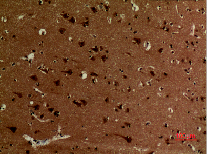
product-image-AAA30656_IHC7.jpg IHC (Immunohistochemistry) (Immunohistochemistry of paraffin-embedded mouse brain using Lamin B1 at dilution of 1:200 (40x lens).)

Lamin B1, Polyclonal Antibody (Cat# AAA30656)

Full Name
Lamin B1 Rabbit Polyclonal Antibody
Gene Names
LMNB1; LMN; ADLD; LMN2; LMNB
Reactivity
Human, Mouse, Rat
Applications
Western Blot (WB), Immunohistochemistry (IHC), Immunofluorescence (IF)
Purity
Affinity purification
Pricing
product-image-AAA19137_FCM8.jpg FCM (Flow Cytometry) (Figure 8. Flow Cytometry analysis of U-87 cells using anti-MVP antibody (AAA19137).Overlay histogram showing U-87 cells stained with AAA19137 (Blue line).The cells were blocked with 10% normal goat serum. And then incubated with rabbit anti-MVP Antibody (AAA19137,1ug/1x10^6 cells) for 30 min at 20 degree C. DyLight®488 conjugated goat anti-rabbit IgG (5-10ug/1x10^6 cells) was used as secondary antibody for 30 minutes at 20 degree C. Isotype control antibody (Green line) was rabbit IgG (1ug/1x106) used under the same conditions. Unlabelled sample (Red line) was also used as a control.)

MVP, Polyclonal Antibody (Cat# AAA19137)

Full Name
Anti-MVP Picoband antibody
Gene Names
MVP; LRP; VAULT1
Reactivity
Human, Mouse, Rat
Applications
ELISA (EIA), Flow Cytometry (FC/FACS), Immunohistochemistry (IHC), Immunocytochemistry (ICC), Western Blot (WB)
Pricing
product-image-AAA23489_WB7.jpg WB (Western Blot) (WB Suggested Anti-HNRPA3 Antibody Titration: 1.25ug/mlPositive Control: Jurkat cell lysateHNRNPA3 is strongly supported by BioGPS gene expression data to be expressed in Human Jurkat cells)

HNRPA3, Polyclonal Antibody (Cat# AAA23489)

Full Name
HNRPA3 antibody - N-terminal region
Gene Names
HNRNPA3; FBRNP; HNRPA3; D10S102; 2610510D13Rik
Reactivity
Cow, Dog, Guinea Pig, Human, Mouse, Rabbit, Rat
Applications
Immunohistochemistry (IHC), Western Blot (WB)
Purity
Protein A purified
Pricing
product-image-AAA19149_IHC7.jpg IHC (Immunohistochemistry) (Figure 7. IHC analysis of Periostin using anti-Periostin antibody (AAA19149).Periostin was detected in paraffin-embedded section of rat small intestine tissue. Heat mediated antigen retrieval was performed in citrate buffer (pH6, epitope retrieval solution) for 20 mins. The tissue section was blocked with 10% goat serum. The tissue section was then incubated with 1ug/ml rabbit anti-Periostin Antibody (AAA19149) overnight at 4 degree C. Biotinylated goat anti-rabbit IgG was used as secondary antibody and incubated for 30 minutes at 37 degree C. The tissue section was developed using Strepavidin-Biotin-Complex (SABC) with DAB as the chromogen.)

Periostin, Polyclonal Antibody (Cat# AAA19149)

Full Name
Anti-Periostin Picoband Antibody
Gene Names
Postn; Plf
Reactivity
Mouse, Rat
No cross reactivity with other proteins.
Applications
ELISA (EIA), Immunohistochemistry (IHC), Western Blot (WB)
Purity
Immunogen affinity purified
Pricing
product-image-AAA29133_IHC7.jpg IHC (Immunohistochemistry) (Dilution: Western Blot: 1/500 - 1/2000. Immunohistochemistry: 1/100 - 1/300. Immunofluorescence: 1/200 - 1/1000. ELISA: 1/40000. Not yet tested in other applications.)

TIMP-2, Polyclonal Antibody (Cat# AAA29133)

Full Name
TIMP-2 Polyclonal Antibody
Gene Names
TIMP2; DDC8; CSC-21K
Reactivity
Human, Mouse, Rat
Applications
Western Blot (WB), Immunohistochemistry-Paraffin (IHC-P), Immunofluorescence (IF)
Pricing
product-image-AAA29645_IF2.jpg IF (Immunofluorescence) (Immunofluorescence analysis of HeLa cell using SPAM1 antibody. Blue: DAPI for nuclear staining.)

SPAM1, Polyclonal Antibody (Cat# AAA29645)

Full Name
SPAM1 Antibody
Gene Names
SPAM1; HYA1; PH20; HYAL1; HYAL3; HYAL5; PH-20; SPAG15; HEL-S-96n
Reactivity
Human, Mouse, Rat
Applications
Western Blot (WB), Immunohistochemistry (IHC), Immunofluorescence (IF)
Purity
Antibodies were purified by affinity purification using immunogen.
Pricing
product-image-AAA30669_IHC7.jpg IHC (Immunohistochemistry) (Immunohistochemistry of paraffin-embedded mouse liver using PSMA7 at dilution of 1:100 (40x lens).)

PSMA7, Polyclonal Antibody (Cat# AAA30669)

Full Name
PSMA7 Rabbit Polyclonal Antibody
Gene Names
PSMA7; C6; HSPC; RC6-1; XAPC7
Reactivity
Human, Mouse, Rat
Applications
Western Blot (WB), Immunohistochemistry (IHC), Immunofluorescence (IF)
Purity
Affinity purification
Pricing
product-image-AAA31437_IHC6.jpg IHC (Immunohistchemistry) (At 1/100 staining Mouse brain tissue by IHC-P. The sample was formaldehyde fixed and a heat mediated antigen retrieval step in citrate buffer was performed. The sample was then blocked and incubated with the primary antibody at 4 degree C overnight. An HRP conjugated anti-Rabbit antibody was used as the secondary antibody.)

USF1, Polyclonal Antibody (Cat# AAA31437)

Full Name
Phospho-USF1 (Thr153) Antibody
Gene Names
USF1; UEF; FCHL; MLTF; FCHL1; MLTFI; HYPLIP1; bHLHb11
Reactivity
Human, Mouse, Rat
Predicted Reactivity: Pig (100%), Bovine (100%), Rabbit (100%), Dog (100%)
Applications
Western Blot (WB), Immunohistochemistry (IHC), Peptide ELISA (EIA)
Purity
The antibody is from purified rabbit serum by affinity purification via sequential chromatography on phospho-peptide and non-phospho-peptide affinity columns.
Pricing
product-image-AAA10958_IF7.gif IF (Immunofluorescence) (Immunofluorescence of Caspase-12 in mouse heart tissue with Caspase-12 antibody at 20 μg/ml.Green: Caspase-12 Antibody (Large) (3195)Blue: DAPI staining)

Caspase-12, Polyclonal Antibody (Cat# AAA10958)

Full Name
Caspase-12 Antibody (Large)
Reactivity
Human, Mouse, Rat
Applications
ELISA (EIA), Western Blot (WB), Immunohistochemistry (IHC), Immunofluorescence (IF)
Purity
Caspase-12 (Large) Antibody is affinity chromatography purified via peptide column.
Pricing
product-image-AAA23499_WB7.jpg WB (Western Blot) (WB Suggested Anti-KRT8 Antibody Titration: 0.2-1 ug/mlPositive Control: HepG2 cell lysateKRT8 is supported by BioGPS gene expression data to be expressed in HepG2)

KRT8, Polyclonal Antibody (Cat# AAA23499)

Full Name
KRT8 antibody - middle region
Gene Names
KRT8; K8; KO; CK8; CK-8; CYK8; K2C8; CARD2
Reactivity
Cow, Dog, Guinea Pig, Horse, Human, Mouse, Pig, Rabbit, Rat
Applications
Western Blot (WB), Immunohistochemistry (IHC)
Purity
Affinity Purified
Pricing
product-image-AAA29131_IHC6.jpg IHC (Immunohistchemistry) (Dilution: IHC-p: 100-300.Western Blot: 1/500 - 1/2000. ELISA: 1/10000. Not yet tested in other applications.)

Tie-1, Polyclonal Antibody (Cat# AAA29131)

Full Name
Tie-1 Polyclonal Antibody
Gene Names
TIE1; TIE; JTK14
Reactivity
Human, Mouse, Rat
Applications
Western Blot (WB), Immunohistochemistry-Paraffin (IHC-P)
Pricing
product-image-AAA30667_IF9.jpg IF (Immunofluorescence) (Confocal immunofluorescence analysis of U2OS cells using LRPPRC Polyclonal at dilution of 1:400. Blue: DAPI for nuclear staining.)

LRPPRC, Polyclonal Antibody (Cat# AAA30667)

Full Name
LRPPRC Rabbit Polyclonal Antibody
Gene Names
LRPPRC; LSFC; GP130; LRP130; CLONE-23970
Reactivity
Human, Mouse, Rat
Applications
Western Blot (WB), Immunohistochemistry (IHC), Immunofluorescence (IF)
Purity
Affinity purification
Pricing
product-image-AAA31435_APP9.jpg Application Data (At 25 degree C. The primary antibody was diluted at 1/200 and incubated with the sample for 1 hour at 37 degree C. An Alexa Fluor 594 conjugated goat anti-rabbit IgG (H+L) Ab, diluted at 1/600, was used as the secondary antibody.)

SRF, Polyclonal Antibody (Cat# AAA31435)

Full Name
Phospho-SRF (Thr159) Antibody
Gene Names
SRF; MCM1
Reactivity
Human, Mouse, Rat
Predicted Reactivity: Pig (100%), Bovine (100%), Horse (100%), Rabbit (100%), Dog (100%), Chicken (100%), Xenopus (100%)
Applications
Western Blot (WB), Immunohistochemistry (IHC), Immunofluorescence (IF), Immunocytochemistry (ICC), Peptide ELISA (EIA)
Purity
The antibody is from purified rabbit serum by affinity purification via sequential chromatography on phospho-peptide and non-phospho-peptide affinity columns.
Pricing

What are Polyclonal Antibodies?


Polyclonal antibodies are antibodies that come from multiple B cell clones of a host animal. The typical hosts used for the majority of polyclonal antibody production are rabbits, goats, sheep, and donkeys. These polyclonal antibodies, once having identified their target, will bind to different epitopes located at different regions or sequences on the same protein/antigen. As a result, they are ideal at locating and binding to the target, even if the target is in very low concentrations (due to many different antibodies being able to bind to the same target molecule, which allows for significant amplification of a downstream signal).


Polyclonal antibodies are typically produced by injecting an antigen into a host animal, which causes the animal’s immune system to attack the foreign antigen by mass generating antibodies against it. After a period of time, serum is collected from the animal and purified using physicochemical fractionation, class-specific affinity purification, and/or antigen-affinity purification.


Key Uses of Polyclonal Antibodies


  • Western Blotting: This method is used to find specific proteins in biological samples after separating them by size.

  • Immunohistochemistry: IHC helps visualize the location of proteins in tissue sections using various staining techniques.

  • ELISA: (Enzyme-Linked Immunosorbent Assay) is typically used to identify specific protein quantities in a sample. ELISAs can be either “Quantitative” or “Qualitative”.

  • Flow Cytometry: technique that identifies and measures the specific protein on the surface or inside the cells in a fluid suspension.

  • Immunoprecipitation: IP isolates and studies a specific protein from a complex mixture using antibodies.


Why Buy Polyclonal Antibodies from AAA Biotech?


1. Ideal for Various Applications


Our antibodies are generally going to be validated for use in multiple types of assays, including ELISA, Western Blotting, Immunohistochemistry, Immunoprecipitation, amongst others. They are ideal for a wide range of research applications.


2. Rigorous Quality Control


All of the antibodies in our catalog undergo strict quality testing to ensure specificity, sensitivity, and consistent performance. We are confident in the ability of our antibodies to provide you with accurate results.


3. Wide Assortment of Antibodies


Antibodies in are catalog can be found for both common and exotic species, and these antibodies are also available in both conjugated and recombinant forms to suit many diverse experimental needs.


4. Highly Purified


Our antibodies are available in purified forms with over 85% purity, as confirmed by SDS-PAGE. They are also available with tags such as His, Flag, GST, or MBP. We cater to customers worldwide.


FAQ


1. How are polyclonal antibodies produced?


Traditionally, polyclonal antibodies are produced by injecting an antigen into a host animal (such as a rabbit or goat), which then triggers an immune response from the host animal. The animal’s B cells produce antibodies that will recognize different parts of the injected antigen. These antibodies are then collected from the animal’s blood and purified for use.


2. How do polyclonal antibodies differ from monoclonal antibodies?


Polyclonal antibodies are a mix of antibodies that bind to different locations (epitopes) of the same antigen, while monoclonal antibodies are identical and bind to just one specific epitope. This makes polyclonal antibodies more versatile and better at detecting proteins that may be present in low quantities or in altered/modified forms.


3. How should I store polyclonal antibodies?


Polyclonal antibodies should be stored at 4°C for short-term use (up to a few weeks) and at -20°C or -80°C for long-term storage. Avoid repeated freeze-thaw cycles by dividing them into small aliquots. Always check the datasheet for specific storage instructions.


Submit a Question

Please complete the form below and a representative will contact you as soon as possible.

Request more Information

Please complete the form below and a representative will contact you as soon as possible.

Request a Manual

Please complete the form below and a representative will contact you as soon as possible.

Request a Quote

Please complete the form below and a representative will contact you as soon as possible.