Filters
▼Clonality
▼Type
▼Reactivity
▼Gene Name
▼Isotype
▼Host
▼Application
▼Clone
▼Polyclonal Antibodies
At AAA Biotech also known as AAA Bio or AAABio, we provide a broad range of purified polyclonal antibodies (pAbs) that are able to all be browsed online through our website. Due to their high specificity and strong binding affinity, these antibodies are ideal for wide swathes of research and experimental applications.
Our polyclonal antibodies can easily support your work, whether you use them for Western Blotting, Immunocytochemistry (with or without Immunofluorescence used in conjunction), Immunohistochemistry, Immunoprecipitation, and ELISA tests. We highly encourage you to browse our range of pAbs and choose the one that best suits your experimental model.
Viewing 3000-3050 of 118597 product results
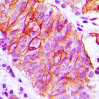
 IHC (Immunohiostchemistry)
(Immunohistochemical analysis of TIM3 staining in human breast cancer formalin fixed paraffin embedded tissue section. The section was pre-treated using heat mediated antigen retrieval with sodium citrate buffer (pH 6.0). The section was then incubated with the antibody at room temperature and detected using an HRP conjugated compact polymer system. DAB was used as the chromogen. The section was then counterstained with haematoxylin and mounted with DPX.)
IHC (Immunohiostchemistry)
(Immunohistochemical analysis of TIM3 staining in human breast cancer formalin fixed paraffin embedded tissue section. The section was pre-treated using heat mediated antigen retrieval with sodium citrate buffer (pH 6.0). The section was then incubated with the antibody at room temperature and detected using an HRP conjugated compact polymer system. DAB was used as the chromogen. The section was then counterstained with haematoxylin and mounted with DPX.)
TIM3, Polyclonal Antibody (Cat# AAA220803)
 IF (Immunofluorescence)
(Immunofluorescent analysis of IFI16 staining in U2OS cells. Formalin-fixed cells were permeabilized with 0.1% Triton X-100 in TBS for 5-10 minutes and blocked with 3% BSA-PBS for 30 minutes at room temperature. Cells were probed with the primary antibody in 3% BSA-PBS and incubated overnight at 4 °C in a humidified chamber. Cells were washed with PBST and incubated with a DyLight 594-conjugated secondary antibody (red) in PBS at room temperature in the dark.)
IF (Immunofluorescence)
(Immunofluorescent analysis of IFI16 staining in U2OS cells. Formalin-fixed cells were permeabilized with 0.1% Triton X-100 in TBS for 5-10 minutes and blocked with 3% BSA-PBS for 30 minutes at room temperature. Cells were probed with the primary antibody in 3% BSA-PBS and incubated overnight at 4 °C in a humidified chamber. Cells were washed with PBST and incubated with a DyLight 594-conjugated secondary antibody (red) in PBS at room temperature in the dark.)
IFI16, Polyclonal Antibody (Cat# AAA220807)
 IF (Immunofluorescence)
(Immunofluorescent analysis of GIF staining in A549 cells. Formalin-fixed cells were permeabilized with 0.1% Triton X-100 in TBS for 5-10 minutes and blocked with 3% BSA-PBS for 30 minutes at room temperature. Cells were probed with the primary antibody in 3% BSA-PBS and incubated overnight at 4 °C in a humidified chamber. Cells were washed with PBST and incubated with a DyLight 594-conjugated secondary antibody (red) in PBS at room temperature in the dark.)
IF (Immunofluorescence)
(Immunofluorescent analysis of GIF staining in A549 cells. Formalin-fixed cells were permeabilized with 0.1% Triton X-100 in TBS for 5-10 minutes and blocked with 3% BSA-PBS for 30 minutes at room temperature. Cells were probed with the primary antibody in 3% BSA-PBS and incubated overnight at 4 °C in a humidified chamber. Cells were washed with PBST and incubated with a DyLight 594-conjugated secondary antibody (red) in PBS at room temperature in the dark.)
GIF, Polyclonal Antibody (Cat# AAA220846)
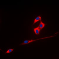
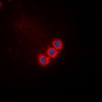
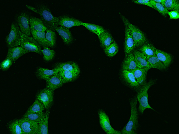
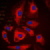
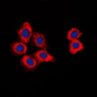 IF (Immunofluorescence)
(Immunofluorescent analysis of CDK15 staining in HepG2 cells. Formalin-fixed cells were permeabilized with 0.1% Triton X-100 in TBS for 5-10 minutes and blocked with 3% BSA-PBS for 30 minutes at room temperature. Cells were probed with the primary antibody in 3% BSA-PBS and incubated overnight at 4 °C in a humidified chamber. Cells were washed with PBST and incubated with a DyLight 594-conjugated secondary antibody (red) in PBS at room temperature in the dark. DAPI was used to stain the cell nuclei (blue).)
IF (Immunofluorescence)
(Immunofluorescent analysis of CDK15 staining in HepG2 cells. Formalin-fixed cells were permeabilized with 0.1% Triton X-100 in TBS for 5-10 minutes and blocked with 3% BSA-PBS for 30 minutes at room temperature. Cells were probed with the primary antibody in 3% BSA-PBS and incubated overnight at 4 °C in a humidified chamber. Cells were washed with PBST and incubated with a DyLight 594-conjugated secondary antibody (red) in PBS at room temperature in the dark. DAPI was used to stain the cell nuclei (blue).)
CDK15, Polyclonal Antibody (Cat# AAA220578)
 IF (Immunofluorescence)
(Immunofluorescent analysis of Adenylate Kinase 6 staining in Jurkat cells. Formalin-fixed cells were permeabilized with 0.1% Triton X-100 in TBS for 5-10 minutes and blocked with 3% BSA-PBS for 30 minutes at room temperature. Cells were probed with the primary antibody in 3% BSA-PBS and incubated overnight at 4 °C in a humidified chamber. Cells were washed with PBST and incubated with a DyLight 594-conjugated secondary antibody (red) in PBS at room temperature in the dark.)
IF (Immunofluorescence)
(Immunofluorescent analysis of Adenylate Kinase 6 staining in Jurkat cells. Formalin-fixed cells were permeabilized with 0.1% Triton X-100 in TBS for 5-10 minutes and blocked with 3% BSA-PBS for 30 minutes at room temperature. Cells were probed with the primary antibody in 3% BSA-PBS and incubated overnight at 4 °C in a humidified chamber. Cells were washed with PBST and incubated with a DyLight 594-conjugated secondary antibody (red) in PBS at room temperature in the dark.)
Adenylate Kinase 6, Polyclonal Antibody (Cat# AAA220583)
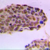
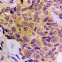
 IHC (Immunohiostchemistry)
(Immunohistochemical analysis of Cytochrome P450 24A1 staining in human breast cancer formalin fixed paraffin embedded tissue section. The section was pre-treated using heat mediated antigen retrieval with sodium citrate buffer (pH 6.0). The section was then incubated with the antibody at room temperature and detected using an HRP conjugated compact polymer system. DAB was used as the chromogen. The section was then counterstained with haematoxylin and mounted with DPX.)
IHC (Immunohiostchemistry)
(Immunohistochemical analysis of Cytochrome P450 24A1 staining in human breast cancer formalin fixed paraffin embedded tissue section. The section was pre-treated using heat mediated antigen retrieval with sodium citrate buffer (pH 6.0). The section was then incubated with the antibody at room temperature and detected using an HRP conjugated compact polymer system. DAB was used as the chromogen. The section was then counterstained with haematoxylin and mounted with DPX.)
Cytochrome P450 24A1, Polyclonal Antibody (Cat# AAA220585)
 IHC (Immunohiostchemistry)
(Immunohistochemical analysis of QSK staining in human breast cancer formalin fixed paraffin embedded tissue section. The section was pre-treated using heat mediated antigen retrieval with sodium citrate buffer (pH 6.0). The section was then incubated with the antibody at room temperature and detected using an HRP conjugated compact polymer system. DAB was used as the chromogen. The section was then counterstained with haematoxylin and mounted with DPX.)
IHC (Immunohiostchemistry)
(Immunohistochemical analysis of QSK staining in human breast cancer formalin fixed paraffin embedded tissue section. The section was pre-treated using heat mediated antigen retrieval with sodium citrate buffer (pH 6.0). The section was then incubated with the antibody at room temperature and detected using an HRP conjugated compact polymer system. DAB was used as the chromogen. The section was then counterstained with haematoxylin and mounted with DPX.)
QSK, Polyclonal Antibody (Cat# AAA220594)
 IF (Immunofluorescence)
(Immunofluorescent analysis of MUC1 staining in HeLa cells. Formalin-fixed cells were permeabilized with 0.1% Triton X-100 in TBS for 5-10 minutes and blocked with 3% BSA-PBS for 30 minutes at room temperature. Cells were probed with the primary antibody in 3% BSA-PBS and incubated overnight at 4 °C in a humidified chamber. Cells were washed with PBST and incubated with a DyLight 594-conjugated secondary antibody (red) in PBS at room temperature in the dark. DAPI was used to stain the cell nuclei (blue).)
IF (Immunofluorescence)
(Immunofluorescent analysis of MUC1 staining in HeLa cells. Formalin-fixed cells were permeabilized with 0.1% Triton X-100 in TBS for 5-10 minutes and blocked with 3% BSA-PBS for 30 minutes at room temperature. Cells were probed with the primary antibody in 3% BSA-PBS and incubated overnight at 4 °C in a humidified chamber. Cells were washed with PBST and incubated with a DyLight 594-conjugated secondary antibody (red) in PBS at room temperature in the dark. DAPI was used to stain the cell nuclei (blue).)
MUC1, Polyclonal Antibody (Cat# AAA220604)
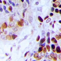
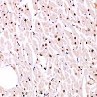 IHC (Immunohiostchemistry)
(Immunohistochemical analysis of MITF (pS180) staining in human heart formalin fixed paraffin embedded tissue section. The section was pre-treated using heat mediated antigen retrieval with sodium citrate buffer (pH 6.0). The section was then incubated with the antibody at room temperature and detected using an HRP conjugated compact polymer system. DAB was used as the chromogen. The section was then counterstained with haematoxylin and mounted with DPX.)
IHC (Immunohiostchemistry)
(Immunohistochemical analysis of MITF (pS180) staining in human heart formalin fixed paraffin embedded tissue section. The section was pre-treated using heat mediated antigen retrieval with sodium citrate buffer (pH 6.0). The section was then incubated with the antibody at room temperature and detected using an HRP conjugated compact polymer system. DAB was used as the chromogen. The section was then counterstained with haematoxylin and mounted with DPX.)
MITF, Polyclonal Antibody (Cat# AAA220625)
 IF (Immunofluorescence)
(Immunofluorescent analysis of NIPA (pS354) staining in HepG2 cells. Formalin-fixed cells were permeabilized with 0.1% Triton X-100 in TBS for 5-10 minutes and blocked with 3% BSA-PBS for 30 minutes at room temperature. Cells were probed with the primary antibody in 3% BSA-PBS and incubated overnight at 4 °C in a humidified chamber. Cells were washed with PBST and incubated with a DyLight 594-conjugated secondary antibody (red) in PBS at room temperature in the dark.)
IF (Immunofluorescence)
(Immunofluorescent analysis of NIPA (pS354) staining in HepG2 cells. Formalin-fixed cells were permeabilized with 0.1% Triton X-100 in TBS for 5-10 minutes and blocked with 3% BSA-PBS for 30 minutes at room temperature. Cells were probed with the primary antibody in 3% BSA-PBS and incubated overnight at 4 °C in a humidified chamber. Cells were washed with PBST and incubated with a DyLight 594-conjugated secondary antibody (red) in PBS at room temperature in the dark.)
NIPA, Polyclonal Antibody (Cat# AAA220999)
 IF (Immunofluorescence)
(Immunofluorescent analysis of MSK1 staining in HEK293T cells. Formalin-fixed cells were permeabilized with 0.1% Triton X-100 in TBS for 5-10 minutes and blocked with 3% BSA-PBS for 30 minutes at room temperature. Cells were probed with the primary antibody in 3% BSA-PBS and incubated overnight at 4 °C in a humidified chamber. Cells were washed with PBST and incubated with a DyLight 594-conjugated secondary antibody (red) in PBS at room temperature in the dark.)
IF (Immunofluorescence)
(Immunofluorescent analysis of MSK1 staining in HEK293T cells. Formalin-fixed cells were permeabilized with 0.1% Triton X-100 in TBS for 5-10 minutes and blocked with 3% BSA-PBS for 30 minutes at room temperature. Cells were probed with the primary antibody in 3% BSA-PBS and incubated overnight at 4 °C in a humidified chamber. Cells were washed with PBST and incubated with a DyLight 594-conjugated secondary antibody (red) in PBS at room temperature in the dark.)
MSK1, Polyclonal Antibody (Cat# AAA221003)
 IF (Immunofluorescence)
(Immunofluorescent analysis of MRG15 staining in K562 cells. Formalin-fixed cells were permeabilized with 0.1% Triton X-100 in TBS for 5-10 minutes and blocked with 3% BSA-PBS for 30 minutes at room temperature. Cells were probed with the primary antibody in 3% BSA-PBS and incubated overnight at 4 °C in a humidified chamber. Cells were washed with PBST and incubated with a DyLight 594-conjugated secondary antibody (red) in PBS at room temperature in the dark.)
IF (Immunofluorescence)
(Immunofluorescent analysis of MRG15 staining in K562 cells. Formalin-fixed cells were permeabilized with 0.1% Triton X-100 in TBS for 5-10 minutes and blocked with 3% BSA-PBS for 30 minutes at room temperature. Cells were probed with the primary antibody in 3% BSA-PBS and incubated overnight at 4 °C in a humidified chamber. Cells were washed with PBST and incubated with a DyLight 594-conjugated secondary antibody (red) in PBS at room temperature in the dark.)
MRG15, Polyclonal Antibody (Cat# AAA221009)
 IF (Immunofluorescence)
(Immunofluorescent analysis of ECHOS1 staining in A431 cells. Formalin-fixed cells were permeabilized with 0.1% Triton X-100 in TBS for 5-10 minutes and blocked with 3% BSA-PBS for 30 minutes at room temperature. Cells were probed with the primary antibody in 3% BSA-PBS and incubated overnight at 4 °C in a humidified chamber. Cells were washed with PBST and incubated with a DyLight 594-conjugated secondary antibody (red) in PBS at room temperature in the dark. DAPI was used to stain the cell nuclei (blue).)
IF (Immunofluorescence)
(Immunofluorescent analysis of ECHOS1 staining in A431 cells. Formalin-fixed cells were permeabilized with 0.1% Triton X-100 in TBS for 5-10 minutes and blocked with 3% BSA-PBS for 30 minutes at room temperature. Cells were probed with the primary antibody in 3% BSA-PBS and incubated overnight at 4 °C in a humidified chamber. Cells were washed with PBST and incubated with a DyLight 594-conjugated secondary antibody (red) in PBS at room temperature in the dark. DAPI was used to stain the cell nuclei (blue).)
ECHOS1, Polyclonal Antibody (Cat# AAA220488)
 IF (Immunofluorescence)
(Immunofluorescent analysis of RACK7 staining in Hela cells. Formalin-fixed cells were permeabilized with 0.1% Triton X-100 in TBS for 5-10 minutes and blocked with 3% BSA-PBS for 30 minutes at room temperature. Cells were probed with the primary antibody in 3% BSA-PBS and incubated overnight at 4 °C in a humidified chamber. Cells were washed with PBST and incubated with a DyLight 594-conjugated secondary antibody (red) in PBS at room temperature in the dark.)
IF (Immunofluorescence)
(Immunofluorescent analysis of RACK7 staining in Hela cells. Formalin-fixed cells were permeabilized with 0.1% Triton X-100 in TBS for 5-10 minutes and blocked with 3% BSA-PBS for 30 minutes at room temperature. Cells were probed with the primary antibody in 3% BSA-PBS and incubated overnight at 4 °C in a humidified chamber. Cells were washed with PBST and incubated with a DyLight 594-conjugated secondary antibody (red) in PBS at room temperature in the dark.)
RACK7, Polyclonal Antibody (Cat# AAA220494)
 IF (Immunofluorescence)
(Immunofluorescent analysis of ATP5L2 staining in HepG2 cells. Formalin-fixed cells were permeabilized with 0.1% Triton X-100 in TBS for 5-10 minutes and blocked with 3% BSA-PBS for 30 minutes at room temperature. Cells were probed with the primary antibody in 3% BSA-PBS and incubated overnight at 4 °C in a humidified chamber. Cells were washed with PBST and incubated with a DyLight 594-conjugated secondary antibody (red) in PBS at room temperature in the dark. DAPI was used to stain the cell nuclei (blue).)
IF (Immunofluorescence)
(Immunofluorescent analysis of ATP5L2 staining in HepG2 cells. Formalin-fixed cells were permeabilized with 0.1% Triton X-100 in TBS for 5-10 minutes and blocked with 3% BSA-PBS for 30 minutes at room temperature. Cells were probed with the primary antibody in 3% BSA-PBS and incubated overnight at 4 °C in a humidified chamber. Cells were washed with PBST and incubated with a DyLight 594-conjugated secondary antibody (red) in PBS at room temperature in the dark. DAPI was used to stain the cell nuclei (blue).)
ATP5L2, Polyclonal Antibody (Cat# AAA220496)
 IHC (Immunohiostchemistry)
(Immunohistochemical analysis of FADD staining in human breast cancer formalin fixed paraffin embedded tissue section. The section was pre-treated using heat mediated antigen retrieval with sodium citrate buffer (pH 6.0). The section was then incubated with the antibody at room temperature and detected using an HRP conjugated compact polymer system. DAB was used as the chromogen. The section was then counterstained with haematoxylin and mounted with DPX.)
IHC (Immunohiostchemistry)
(Immunohistochemical analysis of FADD staining in human breast cancer formalin fixed paraffin embedded tissue section. The section was pre-treated using heat mediated antigen retrieval with sodium citrate buffer (pH 6.0). The section was then incubated with the antibody at room temperature and detected using an HRP conjugated compact polymer system. DAB was used as the chromogen. The section was then counterstained with haematoxylin and mounted with DPX.)
FADD, Polyclonal Antibody (Cat# AAA220525)
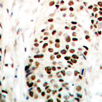
 IHC (Immunohiostchemistry)
(Immunohistochemical analysis of RAD51A (pY315) staining in human breast cancer formalin fixed paraffin embedded tissue section. The section was pre-treated using heat mediated antigen retrieval with sodium citrate buffer (pH 6.0). The section was then incubated with the antibody at room temperature and detected using an HRP conjugated compact polymer system. DAB was used as the chromogen. The section was then counterstained with haematoxylin and mounted with DPX.)
IHC (Immunohiostchemistry)
(Immunohistochemical analysis of RAD51A (pY315) staining in human breast cancer formalin fixed paraffin embedded tissue section. The section was pre-treated using heat mediated antigen retrieval with sodium citrate buffer (pH 6.0). The section was then incubated with the antibody at room temperature and detected using an HRP conjugated compact polymer system. DAB was used as the chromogen. The section was then counterstained with haematoxylin and mounted with DPX.)
RAD51A, Polyclonal Antibody (Cat# AAA220545)
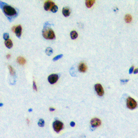
 IHC (Immunohiostchemistry)
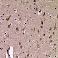
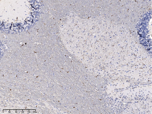
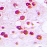
(Immunohistochemical analysis of GLUR4 staining in rat brain formalin fixed paraffin embedded tissue section. The section was pre-treated using heat mediated antigen retrieval with sodium citrate buffer (pH 6.0). The section was then incubated with the antibody at room temperature and detected using an HRP conjugated compact polymer system. DAB was used as the chromogen. The section was then counterstained with haematoxylin and mounted with DPX.)
IHC (Immunohiostchemistry)
(Immunohistochemical analysis of GLUR4 staining in rat brain formalin fixed paraffin embedded tissue section. The section was pre-treated using heat mediated antigen retrieval with sodium citrate buffer (pH 6.0). The section was then incubated with the antibody at room temperature and detected using an HRP conjugated compact polymer system. DAB was used as the chromogen. The section was then counterstained with haematoxylin and mounted with DPX.)
GLUR4, Polyclonal Antibody (Cat# AAA220712)
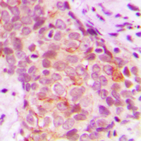
 IHC (Immunohiostchemistry)
(Immunohistochemical analysis of Histone H3 (TriMethyl K18) staining in human breast cancer formalin fixed paraffin embedded tissue section. The section was pre-treated using heat mediated antigen retrieval with sodium citrate buffer (pH 6.0). The section was then incubated with the antibody at room temperature and detected using an HRP conjugated compact polymer system. DAB was used as the chromogen. The section was then counterstained with haematoxylin and mounted with DPX.)
IHC (Immunohiostchemistry)
(Immunohistochemical analysis of Histone H3 (TriMethyl K18) staining in human breast cancer formalin fixed paraffin embedded tissue section. The section was pre-treated using heat mediated antigen retrieval with sodium citrate buffer (pH 6.0). The section was then incubated with the antibody at room temperature and detected using an HRP conjugated compact polymer system. DAB was used as the chromogen. The section was then counterstained with haematoxylin and mounted with DPX.)
Histone H3, Polyclonal Antibody (Cat# AAA220720)
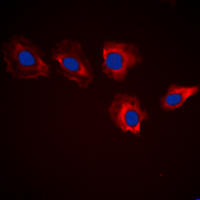
 IF (Immunofluorescence)
(Immunofluorescent analysis of KLK10 staining in A549 cells. Formalin-fixed cells were permeabilized with 0.1% Triton X-100 in TBS for 5-10 minutes and blocked with 3% BSA-PBS for 30 minutes at room temperature. Cells were probed with the primary antibody in 3% BSA-PBS and incubated overnight at 4 °C in a humidified chamber. Cells were washed with PBST and incubated with a DyLight 594-conjugated secondary antibody (red) in PBS at room temperature in the dark.)
IF (Immunofluorescence)
(Immunofluorescent analysis of KLK10 staining in A549 cells. Formalin-fixed cells were permeabilized with 0.1% Triton X-100 in TBS for 5-10 minutes and blocked with 3% BSA-PBS for 30 minutes at room temperature. Cells were probed with the primary antibody in 3% BSA-PBS and incubated overnight at 4 °C in a humidified chamber. Cells were washed with PBST and incubated with a DyLight 594-conjugated secondary antibody (red) in PBS at room temperature in the dark.)
KLK10, Polyclonal Antibody (Cat# AAA220747)
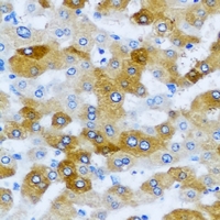
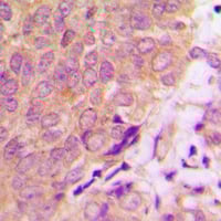
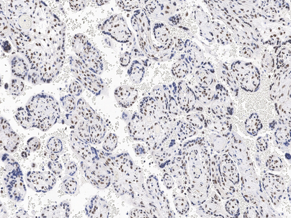 IHC (Immunohistochemisry)
(Immunochemical staining of human Arglu1 in human placenta with rabbit polyclonal antibody at 1:500 dilution, formalin-fixed paraffin embedded sections.)
IHC (Immunohistochemisry)
(Immunochemical staining of human Arglu1 in human placenta with rabbit polyclonal antibody at 1:500 dilution, formalin-fixed paraffin embedded sections.)
Arglu1, Polyclonal Antibody (Cat# AAA258976)
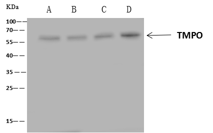
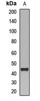
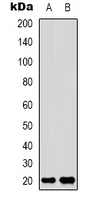
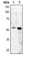
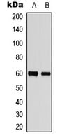
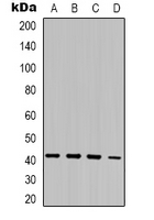
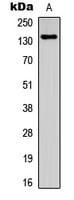
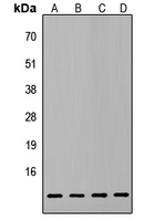
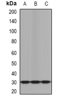
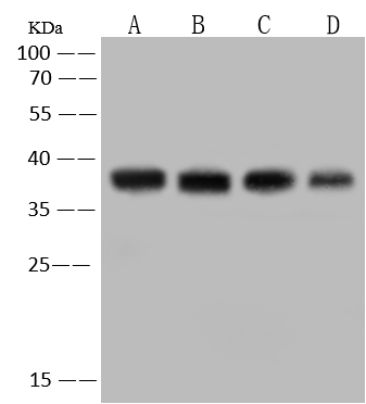
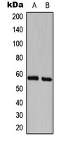
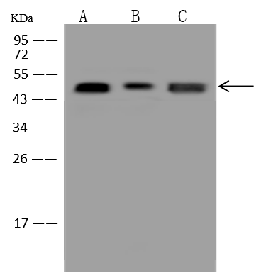
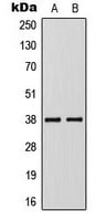
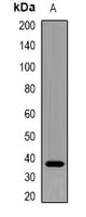
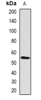
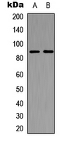
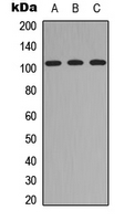
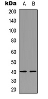
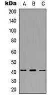
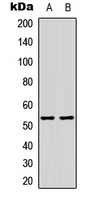
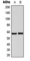
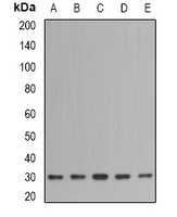
 WB (Western Blot)
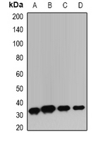
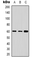
(Anti-TMPO rabbit polyclonal antibody at 1:500 dilutionLane A: HeLa Whole Cell LysateLane B: Jurkat Whole Cell LysateLane C: HepG2 Whole Cell LysateLane D: 293T Whole Cell LysateLysates/proteins at 30 ug per lane.SecondaryGoat Anti-Rabbit IgG (H+L)/HRP at 1/10000 dilution.Developed using the ECL technique.Performed under reducing conditions.Predicted band size:51 kDaObserved band size:57 kDa)
WB (Western Blot)
(Anti-TMPO rabbit polyclonal antibody at 1:500 dilutionLane A: HeLa Whole Cell LysateLane B: Jurkat Whole Cell LysateLane C: HepG2 Whole Cell LysateLane D: 293T Whole Cell LysateLysates/proteins at 30 ug per lane.SecondaryGoat Anti-Rabbit IgG (H+L)/HRP at 1/10000 dilution.Developed using the ECL technique.Performed under reducing conditions.Predicted band size:51 kDaObserved band size:57 kDa)
TMPO, Polyclonal Antibody (Cat# AAA259012)
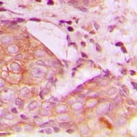
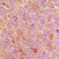
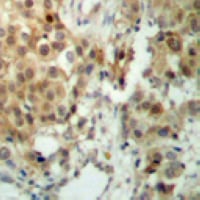
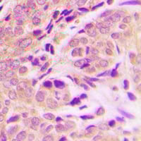
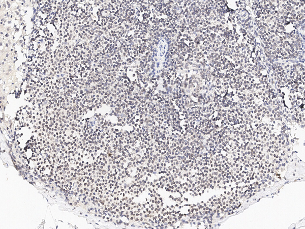
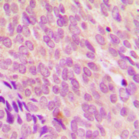
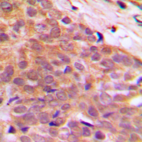
 IHC (Immunohiostchemistry)
(Immunohistochemical analysis of CD147 staining in human prostate cancer formalin fixed paraffin embedded tissue section. The section was pre-treated using heat mediated antigen retrieval with sodium citrate buffer (pH 6.0). The section was then incubated with the antibody at room temperature and detected using an HRP conjugated compact polymer system. DAB was used as the chromogen. The section was then counterstained with haematoxylin and mounted with DPX.)
IHC (Immunohiostchemistry)
(Immunohistochemical analysis of CD147 staining in human prostate cancer formalin fixed paraffin embedded tissue section. The section was pre-treated using heat mediated antigen retrieval with sodium citrate buffer (pH 6.0). The section was then incubated with the antibody at room temperature and detected using an HRP conjugated compact polymer system. DAB was used as the chromogen. The section was then counterstained with haematoxylin and mounted with DPX.)
CD147, Polyclonal Antibody (Cat# AAA259789)
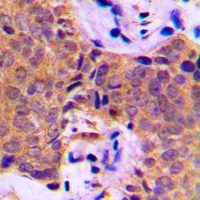
 IHC (Immunohiostchemistry)
(Immunohistochemical analysis of CDK5RAP3 staining in human breast cancer formalin fixed paraffin embedded tissue section. The section was pre-treated using heat mediated antigen retrieval with sodium citrate buffer (pH 6.0). The section was then incubated with the antibody at room temperature and detected using an HRP conjugated compact polymer system. DAB was used as the chromogen. The section was then counterstained with haematoxylin and mounted with DPX.)
IHC (Immunohiostchemistry)
(Immunohistochemical analysis of CDK5RAP3 staining in human breast cancer formalin fixed paraffin embedded tissue section. The section was pre-treated using heat mediated antigen retrieval with sodium citrate buffer (pH 6.0). The section was then incubated with the antibody at room temperature and detected using an HRP conjugated compact polymer system. DAB was used as the chromogen. The section was then counterstained with haematoxylin and mounted with DPX.)
CDK5RAP3, Polyclonal Antibody (Cat# AAA259625)
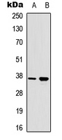
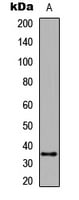
 IP (Immunoprecipitation)
(CHORDC1 was immunoprecipitated using:Lane A:0.5 mg 293 Whole Cell Lysate4 uL anti-CHORDC1 rabbit polyclonal antibody and 60 ug of Immunomagnetic beads Protein A/G.Primary antibody:Anti-CHORDC1 rabbit polyclonal antibody,at 1:100 dilutionSecondary antibody:Goat Anti-Rabbit IgG (H+L)/HRP at 1/10000 dilutionDeveloped using the ECL technique.Performed under reducing conditions.Predicted band size: 37 kDaObserved band size :37 kDa)
IP (Immunoprecipitation)
(CHORDC1 was immunoprecipitated using:Lane A:0.5 mg 293 Whole Cell Lysate4 uL anti-CHORDC1 rabbit polyclonal antibody and 60 ug of Immunomagnetic beads Protein A/G.Primary antibody:Anti-CHORDC1 rabbit polyclonal antibody,at 1:100 dilutionSecondary antibody:Goat Anti-Rabbit IgG (H+L)/HRP at 1/10000 dilutionDeveloped using the ECL technique.Performed under reducing conditions.Predicted band size: 37 kDaObserved band size :37 kDa)
CHORDC1, Polyclonal Antibody (Cat# AAA259093)
 IP (Immunoprecipitation)
(CASP9 was immunoprecipitated using:Lane A:0.5 mg Hela Whole Cell Lysate4 uL anti-CASP9 rabbit polyclonal antibody and 60 ug of Immunomagnetic beads Protein A/G.Primary antibody:Anti-CASP9 rabbit polyclonal antibody,at 1:100 dilutionSecondary antibody:Clean-Blot IP Detection Reagent (HRP) at 1:1000dilutionDeveloped using the ECL technique.Performed under reducing conditions.Predicted band size: 46 kDaObserved band size :46 kDa)
IP (Immunoprecipitation)
(CASP9 was immunoprecipitated using:Lane A:0.5 mg Hela Whole Cell Lysate4 uL anti-CASP9 rabbit polyclonal antibody and 60 ug of Immunomagnetic beads Protein A/G.Primary antibody:Anti-CASP9 rabbit polyclonal antibody,at 1:100 dilutionSecondary antibody:Clean-Blot IP Detection Reagent (HRP) at 1:1000dilutionDeveloped using the ECL technique.Performed under reducing conditions.Predicted band size: 46 kDaObserved band size :46 kDa)
Caspase-9, Polyclonal Antibody (Cat# AAA259106)
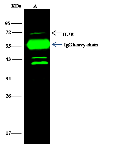 IP (Immunoprecipitation)
(Mouse IL7R was immunoprecipitated using:Lane A:0.5 mg K562 Whole Cell Lysate0.5 uL anti-Mouse IL7R rabbit polyclonal antibody and 15 ul of 50 % Protein G agarose.Primary antibody:Anti-Mouse IL7R rabbit polyclonal antibody,at 1:500 dilutionSecondary antibody:Dylight 800-labeled antibody to rabbit IgG (H+L), at 1:5000 dilutionDeveloped using the odssey technique.Performed under reducing conditions.Predicted band size: 52 kDaObserved band size: 71 kDa)
IP (Immunoprecipitation)
(Mouse IL7R was immunoprecipitated using:Lane A:0.5 mg K562 Whole Cell Lysate0.5 uL anti-Mouse IL7R rabbit polyclonal antibody and 15 ul of 50 % Protein G agarose.Primary antibody:Anti-Mouse IL7R rabbit polyclonal antibody,at 1:500 dilutionSecondary antibody:Dylight 800-labeled antibody to rabbit IgG (H+L), at 1:5000 dilutionDeveloped using the odssey technique.Performed under reducing conditions.Predicted band size: 52 kDaObserved band size: 71 kDa)
IL-7R alpha/CD127, Polyclonal Antibody (Cat# AAA259124)

 IP (Immunoprecipitation)
(mCPB1 was immunoprecipitated using:Lane A:0.5 mg Mouse Spleen Whole Cell Lysate4 uL anti-mCPB1 rabbit polyclonal antibody and 60 ug of Immunomagnetic beads Protein A/G.Primary antibody:Anti-mCPB1 rabbit polyclonal antibody,at 1:100 dilutionSecondary antibody:Goat Anti-Rabbit IgG (H+L)/HRP at 1/10000 dilutionDeveloped using the ECL technique.Performed under reducing conditions.Predicted band size: 40 kDaObserved band size :40 kDa)
IP (Immunoprecipitation)
(mCPB1 was immunoprecipitated using:Lane A:0.5 mg Mouse Spleen Whole Cell Lysate4 uL anti-mCPB1 rabbit polyclonal antibody and 60 ug of Immunomagnetic beads Protein A/G.Primary antibody:Anti-mCPB1 rabbit polyclonal antibody,at 1:100 dilutionSecondary antibody:Goat Anti-Rabbit IgG (H+L)/HRP at 1/10000 dilutionDeveloped using the ECL technique.Performed under reducing conditions.Predicted band size: 40 kDaObserved band size :40 kDa)
Carboxypeptidase B1, Polyclonal Antibody (Cat# AAA259129)
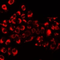
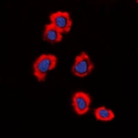
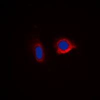 IF (Immunofluorescence)
(Immunofluorescent analysis of Caspase 6 staining in HepG2 cells. Formalin-fixed cells were permeabilized with 0.1% Triton X-100 in TBS for 5-10 minutes and blocked with 3% BSA-PBS for 30 minutes at room temperature. Cells were probed with the primary antibody in 3% BSA-PBS and incubated overnight at 4 °C in a humidified chamber. Cells were washed with PBST and incubated with a DyLight 594-conjugated secondary antibody (red) in PBS at room temperature in the dark. DAPI was used to stain the cell nuclei (blue).)
IF (Immunofluorescence)
(Immunofluorescent analysis of Caspase 6 staining in HepG2 cells. Formalin-fixed cells were permeabilized with 0.1% Triton X-100 in TBS for 5-10 minutes and blocked with 3% BSA-PBS for 30 minutes at room temperature. Cells were probed with the primary antibody in 3% BSA-PBS and incubated overnight at 4 °C in a humidified chamber. Cells were washed with PBST and incubated with a DyLight 594-conjugated secondary antibody (red) in PBS at room temperature in the dark. DAPI was used to stain the cell nuclei (blue).)
Caspase 6, Polyclonal Antibody (Cat# AAA259704)
 IF (Immunofluorescence)
(Immunofluorescent analysis of KLF1/5/7 staining in Jurkat cells. Formalin-fixed cells were permeabilized with 0.1% Triton X-100 in TBS for 5-10 minutes and blocked with 3% BSA-PBS for 30 minutes at room temperature. Cells were probed with the primary antibody in 3% BSA-PBS and incubated overnight at 4 °C in a humidified chamber. Cells were washed with PBST and incubated with a DyLight 594-conjugated secondary antibody (red) in PBS at room temperature in the dark.)
IF (Immunofluorescence)
(Immunofluorescent analysis of KLF1/5/7 staining in Jurkat cells. Formalin-fixed cells were permeabilized with 0.1% Triton X-100 in TBS for 5-10 minutes and blocked with 3% BSA-PBS for 30 minutes at room temperature. Cells were probed with the primary antibody in 3% BSA-PBS and incubated overnight at 4 °C in a humidified chamber. Cells were washed with PBST and incubated with a DyLight 594-conjugated secondary antibody (red) in PBS at room temperature in the dark.)
KLF1/5/7, Polyclonal Antibody (Cat# AAA259718)
 IF (Immunofluorescence)
(Immunofluorescent analysis of HuB staining in K562 cells. Formalin-fixed cells were permeabilized with 0.1% Triton X-100 in TBS for 5-10 minutes and blocked with 3% BSA-PBS for 30 minutes at room temperature. Cells were probed with the primary antibody in 3% BSA-PBS and incubated overnight at 4 °C in a humidified chamber. Cells were washed with PBST and incubated with a DyLight 594-conjugated secondary antibody (red) in PBS at room temperature in the dark.)
IF (Immunofluorescence)
(Immunofluorescent analysis of HuB staining in K562 cells. Formalin-fixed cells were permeabilized with 0.1% Triton X-100 in TBS for 5-10 minutes and blocked with 3% BSA-PBS for 30 minutes at room temperature. Cells were probed with the primary antibody in 3% BSA-PBS and incubated overnight at 4 °C in a humidified chamber. Cells were washed with PBST and incubated with a DyLight 594-conjugated secondary antibody (red) in PBS at room temperature in the dark.)
HuB, Polyclonal Antibody (Cat# AAA259762)
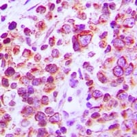 IHC (Immunohiostchemistry)
(Immunohistochemical analysis of CD38 staining in human tonsil formalin fixed paraffin embedded tissue section. The section was pre-treated using heat mediated antigen retrieval with sodium citrate buffer (pH 6.0). The section was then incubated with the antibody at room temperature and detected using an HRP conjugated compact polymer system. DAB was used as the chromogen. The section was then counterstained with haematoxylin and mounted with DPX.)
IHC (Immunohiostchemistry)
(Immunohistochemical analysis of CD38 staining in human tonsil formalin fixed paraffin embedded tissue section. The section was pre-treated using heat mediated antigen retrieval with sodium citrate buffer (pH 6.0). The section was then incubated with the antibody at room temperature and detected using an HRP conjugated compact polymer system. DAB was used as the chromogen. The section was then counterstained with haematoxylin and mounted with DPX.)
CD38, Polyclonal Antibody (Cat# AAA262536)
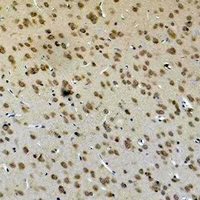
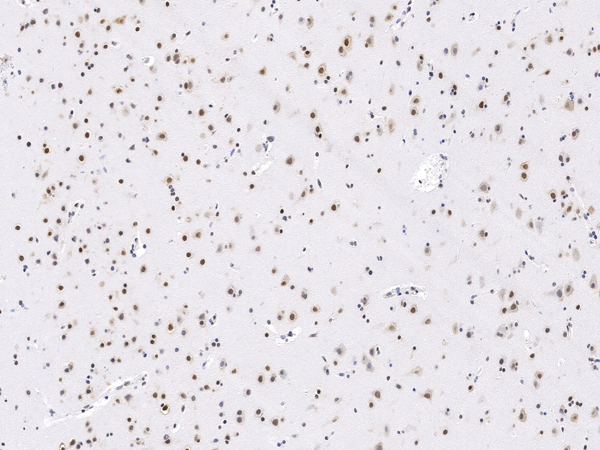
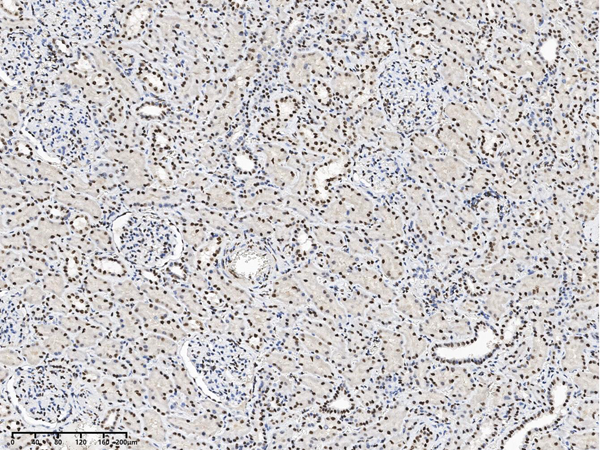
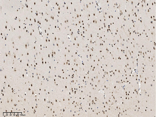
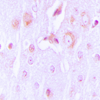
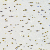 IHC (Immunohistochemisry)
(Immunohistochemical analysis of RBPJ staining in rat brain formalin fixed paraffin embedded tissue section. The section was pre-treated using heat mediated antigen retrieval with sodium citrate buffer (pH 6.0). The section was then incubated with the antibody at room temperature and detected using an HRP conjugated compact polymer system. DAB was used as the chromogen. The section was then counterstained with haematoxylin and mounted with DPX.)
IHC (Immunohistochemisry)
(Immunohistochemical analysis of RBPJ staining in rat brain formalin fixed paraffin embedded tissue section. The section was pre-treated using heat mediated antigen retrieval with sodium citrate buffer (pH 6.0). The section was then incubated with the antibody at room temperature and detected using an HRP conjugated compact polymer system. DAB was used as the chromogen. The section was then counterstained with haematoxylin and mounted with DPX.)
RBPJ, Polyclonal Antibody (Cat# AAA262755)


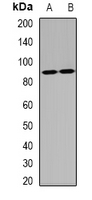
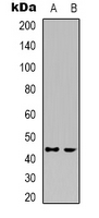
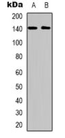
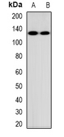
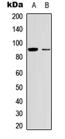
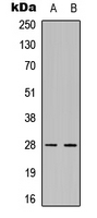
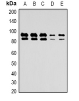
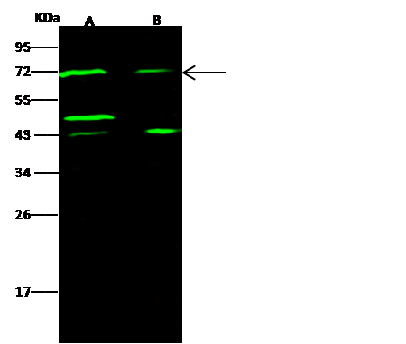
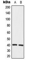
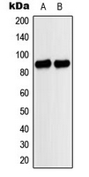
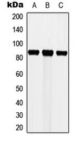
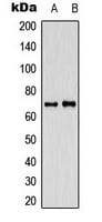
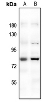
 IP (Immunoprecipitation)
(KAZALD1 was immunoprecipitated using: Lane A:0.5 mg A431 Whole Cell Lysate Lane B:0.5 mg HepG2 Whole Cell Lysate 4 uL anti-KAZALD1 rabbit polyclonal antibody and 60ug of Immunomagnetic beads Protein A/G. Primary antibody: Anti-KAZALD1 rabbit polyclonal antibody,at 1:100 dilution Secondary antibody: Goat Anti-Rabbit IgG (H+L)/HRP at 1/10000 dilution Developed using the ECL technique. Performed under reducing conditions. Predicted band size: 69 kDa Observed band size :69 kDa)
IP (Immunoprecipitation)
(KAZALD1 was immunoprecipitated using: Lane A:0.5 mg A431 Whole Cell Lysate Lane B:0.5 mg HepG2 Whole Cell Lysate 4 uL anti-KAZALD1 rabbit polyclonal antibody and 60ug of Immunomagnetic beads Protein A/G. Primary antibody: Anti-KAZALD1 rabbit polyclonal antibody,at 1:100 dilution Secondary antibody: Goat Anti-Rabbit IgG (H+L)/HRP at 1/10000 dilution Developed using the ECL technique. Performed under reducing conditions. Predicted band size: 69 kDa Observed band size :69 kDa)
DLAT, Polyclonal Antibody (Cat# AAA259412)

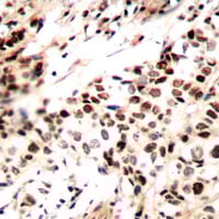
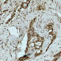
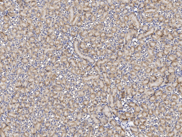
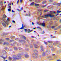
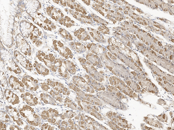
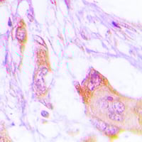
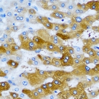
 IHC (Immunohiostchemistry)
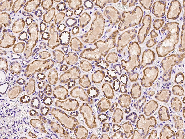
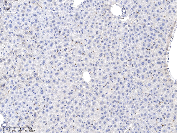
(Immunochemical staining of human HIGD2A in human kidney with rabbit polyclonal antibody at 1:100 dilution, formalin-fixed paraffin embedded sections.)
IHC (Immunohiostchemistry)
(Immunochemical staining of human HIGD2A in human kidney with rabbit polyclonal antibody at 1:100 dilution, formalin-fixed paraffin embedded sections.)
HIGD2A, Polyclonal Antibody (Cat# AAA259451)

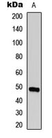
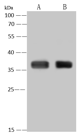
 IP (Immunoprecipitation)
(mCTSS was immunoprecipitated using: Lane A:0.5 mg RAW 264.7 Whole Cell Lysate 4 uL anti-mCTSS rabbit polyclonal antibody and 60ug of Immunomagnetic beads Protein A/G. Primary antibody: Anti-mCTSS rabbit polyclonal antibody,at 1:100 dilution Secondary antibody: Goat Anti-Rabbit IgG (H+L)/HRP at 1/10000 dilution Developed using the ECL technique. Performed under reducing conditions. Predicted band size: 37 kDa Observed band size :37 kDa)
IP (Immunoprecipitation)
(mCTSS was immunoprecipitated using: Lane A:0.5 mg RAW 264.7 Whole Cell Lysate 4 uL anti-mCTSS rabbit polyclonal antibody and 60ug of Immunomagnetic beads Protein A/G. Primary antibody: Anti-mCTSS rabbit polyclonal antibody,at 1:100 dilution Secondary antibody: Goat Anti-Rabbit IgG (H+L)/HRP at 1/10000 dilution Developed using the ECL technique. Performed under reducing conditions. Predicted band size: 37 kDa Observed band size :37 kDa)
Cathepsin S, Polyclonal Antibody (Cat# AAA259469)

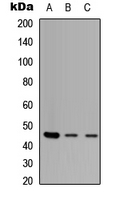
 IP (Immunoprecipitation)
(CCNA2 was immunoprecipitated using: Lane A:0.5 mg U2OS Whole Cell Lysate Lane B:0.5 mg 293T Whole Cell Lysate 4 uL anti-CCNA2 rabbit polyclonal antibody and 60ug of Immunomagnetic beads Protein A/G. Primary antibody: Anti-CCNA2 rabbit polyclonal antibody,at 1:100 dilution Secondary antibody: Clean-Blot IP Detection Reagent (HRP) at 1:1000dilution Developed using the ECL technique. Performed under reducing conditions. Predicted band size: 47 kDa Observed band size :47 kDa)
IP (Immunoprecipitation)
(CCNA2 was immunoprecipitated using: Lane A:0.5 mg U2OS Whole Cell Lysate Lane B:0.5 mg 293T Whole Cell Lysate 4 uL anti-CCNA2 rabbit polyclonal antibody and 60ug of Immunomagnetic beads Protein A/G. Primary antibody: Anti-CCNA2 rabbit polyclonal antibody,at 1:100 dilution Secondary antibody: Clean-Blot IP Detection Reagent (HRP) at 1:1000dilution Developed using the ECL technique. Performed under reducing conditions. Predicted band size: 47 kDa Observed band size :47 kDa)
Cyclin A2/CCNA2, Polyclonal Antibody (Cat# AAA259520)
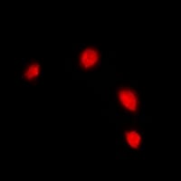
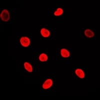
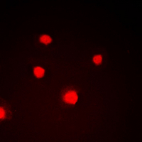
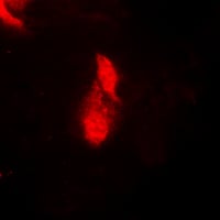
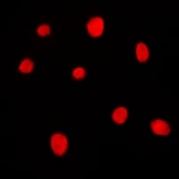
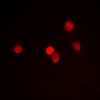
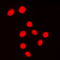
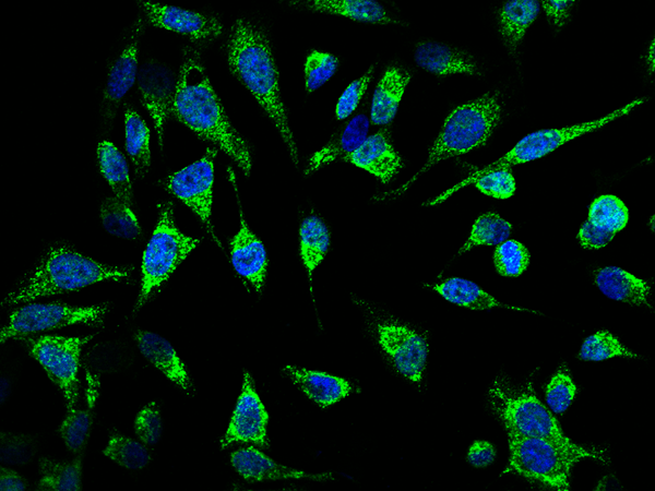
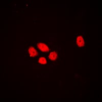
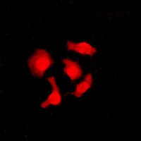
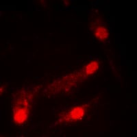
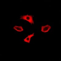
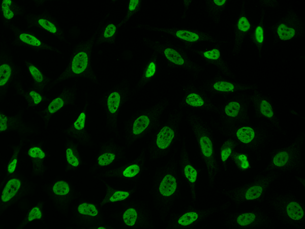 IF (Immunofluorescence)
(Immunofluorescence staining of RALY in HeLa cells. Cells were fixed with 4% PFA, permeabilzed with 0.3% Triton X-100 in PBS,blocked with 10% serum, and incubated with rabbit anti-Human RALY polyclonal antibody (dilution ratio 1:100) at 4 degree C overnight. Then cells were stained with the Alexa Fluor488-conjugated Goat Anti-rabbit IgG secondary antibody (green). Positive staining was localized to Nucleus.)
IF (Immunofluorescence)
(Immunofluorescence staining of RALY in HeLa cells. Cells were fixed with 4% PFA, permeabilzed with 0.3% Triton X-100 in PBS,blocked with 10% serum, and incubated with rabbit anti-Human RALY polyclonal antibody (dilution ratio 1:100) at 4 degree C overnight. Then cells were stained with the Alexa Fluor488-conjugated Goat Anti-rabbit IgG secondary antibody (green). Positive staining was localized to Nucleus.)
RALY, Polyclonal Antibody (Cat# AAA259525)
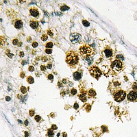
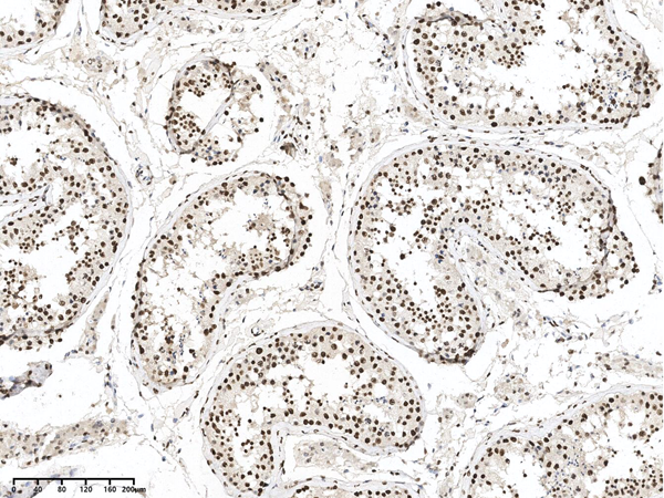
 IHC (Immunohiostchemistry)
(Immunohistochemical analysis of ZNF232 staining in human testis formalin fixed paraffin embedded tissue section. The section was pre-treated using heat mediated antigen retrieval with sodium citrate buffer (pH 6.0). The section was then incubated with the antibody at room temperature and detected using an HRP conjugated compact polymer system. DAB was used as the chromogen. The section was then counterstained with haematoxylin and mounted with DPX.)
IHC (Immunohiostchemistry)
(Immunohistochemical analysis of ZNF232 staining in human testis formalin fixed paraffin embedded tissue section. The section was pre-treated using heat mediated antigen retrieval with sodium citrate buffer (pH 6.0). The section was then incubated with the antibody at room temperature and detected using an HRP conjugated compact polymer system. DAB was used as the chromogen. The section was then counterstained with haematoxylin and mounted with DPX.)
ZNF232, Polyclonal Antibody (Cat# AAA259550)
 IF (Immunofluorescence)
(Immunofluorescent analysis of MARK3 staining in HeLa cells. Formalin-fixed cells were permeabilized with 0.1% Triton X-100 in TBS for 5-10 minutes and blocked with 3% BSA-PBS for 30 minutes at room temperature. Cells were probed with the primary antibody in 3% BSA-PBS and incubated overnight at 4 °C in a humidified chamber. Cells were washed with PBST and incubated with a DyLight 594-conjugated secondary antibody (red) in PBS at room temperature in the dark. DAPI was used to stain the cell nuclei (blue).)
IF (Immunofluorescence)
(Immunofluorescent analysis of MARK3 staining in HeLa cells. Formalin-fixed cells were permeabilized with 0.1% Triton X-100 in TBS for 5-10 minutes and blocked with 3% BSA-PBS for 30 minutes at room temperature. Cells were probed with the primary antibody in 3% BSA-PBS and incubated overnight at 4 °C in a humidified chamber. Cells were washed with PBST and incubated with a DyLight 594-conjugated secondary antibody (red) in PBS at room temperature in the dark. DAPI was used to stain the cell nuclei (blue).)
MARK3, Polyclonal Antibody (Cat# AAA259925)

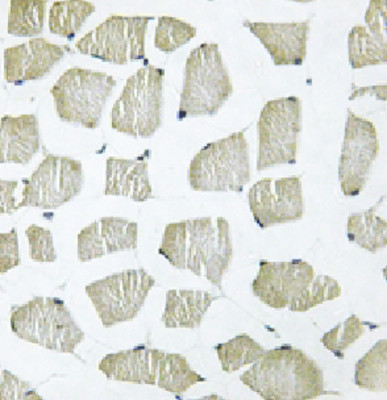 IHC (Immunohistochemisry)
(Immunohistochemical analysis of ITIH4 70k staining in human skeletal muscle formalin fixed paraffin embedded tissue section. The section was pre-treated using heat mediated antigen retrieval with sodium citrate buffer (pH 6.0). The section was then incubated with the antibody at room temperature and detected using an HRP conjugated compact polymer system. DAB was used as the chromogen. The section was then counterstained with haematoxylin and mounted with DPX.)
IHC (Immunohistochemisry)
(Immunohistochemical analysis of ITIH4 70k staining in human skeletal muscle formalin fixed paraffin embedded tissue section. The section was pre-treated using heat mediated antigen retrieval with sodium citrate buffer (pH 6.0). The section was then incubated with the antibody at room temperature and detected using an HRP conjugated compact polymer system. DAB was used as the chromogen. The section was then counterstained with haematoxylin and mounted with DPX.)
ITIH4 70k, Polyclonal Antibody (Cat# AAA259938)
 IF (Immunofluorescence)
(Immunofluorescent analysis of MARK staining in A549 cells. Formalin-fixed cells were permeabilized with 0.1% Triton X-100 in TBS for 5-10 minutes and blocked with 3% BSA-PBS for 30 minutes at room temperature. Cells were probed with the primary antibody in 3% BSA-PBS and incubated overnight at 4 °C in a humidified chamber. Cells were washed with PBST and incubated with a DyLight 594-conjugated secondary antibody (red) in PBS at room temperature in the dark. DAPI was used to stain the cell nuclei (blue).)
IF (Immunofluorescence)
(Immunofluorescent analysis of MARK staining in A549 cells. Formalin-fixed cells were permeabilized with 0.1% Triton X-100 in TBS for 5-10 minutes and blocked with 3% BSA-PBS for 30 minutes at room temperature. Cells were probed with the primary antibody in 3% BSA-PBS and incubated overnight at 4 °C in a humidified chamber. Cells were washed with PBST and incubated with a DyLight 594-conjugated secondary antibody (red) in PBS at room temperature in the dark. DAPI was used to stain the cell nuclei (blue).)
MARK, Polyclonal Antibody (Cat# AAA259962)
 IF (Immunofluorescence)
(Immunofluorescent analysis of DLX3 staining in HEK293T cells. Formalin-fixed cells were permeabilized with 0.1% Triton X-100 in TBS for 5-10 minutes and blocked with 3% BSA-PBS for 30 minutes at room temperature. Cells were probed with the primary antibody in 3% BSA-PBS and incubated overnight at 4 °C in a humidified chamber. Cells were washed with PBST and incubated with a DyLight 594-conjugated secondary antibody (red) in PBS at room temperature in the dark.)
IF (Immunofluorescence)
(Immunofluorescent analysis of DLX3 staining in HEK293T cells. Formalin-fixed cells were permeabilized with 0.1% Triton X-100 in TBS for 5-10 minutes and blocked with 3% BSA-PBS for 30 minutes at room temperature. Cells were probed with the primary antibody in 3% BSA-PBS and incubated overnight at 4 °C in a humidified chamber. Cells were washed with PBST and incubated with a DyLight 594-conjugated secondary antibody (red) in PBS at room temperature in the dark.)
DLX3, Polyclonal Antibody (Cat# AAA259963)
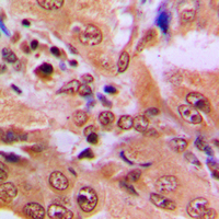
 IHC (Immunohiostchemistry)
(Immunohistochemical analysis of SDCCAG8 staining in human breast cancer formalin fixed paraffin embedded tissue section. The section was pre-treated using heat mediated antigen retrieval with sodium citrate buffer (pH 6.0). The section was then incubated with the antibody at room temperature and detected using an HRP conjugated compact polymer system. DAB was used as the chromogen. The section was then counterstained with haematoxylin and mounted with DPX.)
IHC (Immunohiostchemistry)
(Immunohistochemical analysis of SDCCAG8 staining in human breast cancer formalin fixed paraffin embedded tissue section. The section was pre-treated using heat mediated antigen retrieval with sodium citrate buffer (pH 6.0). The section was then incubated with the antibody at room temperature and detected using an HRP conjugated compact polymer system. DAB was used as the chromogen. The section was then counterstained with haematoxylin and mounted with DPX.)
SDCCAG8, Polyclonal Antibody (Cat# AAA259974)
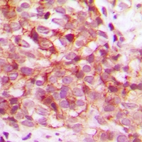
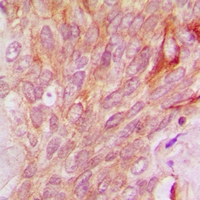
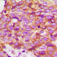
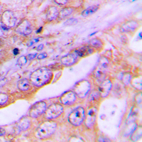
 IHC (Immunohiostchemistry)
(Immunohistochemical analysis of Cortactin staining in human breast cancer formalin fixed paraffin embedded tissue section. The section was pre-treated using heat mediated antigen retrieval with sodium citrate buffer (pH 6.0). The section was then incubated with the antibody at room temperature and detected using an HRP conjugated compact polymer system. DAB was used as the chromogen. The section was then counterstained with haematoxylin and mounted with DPX.)
IHC (Immunohiostchemistry)
(Immunohistochemical analysis of Cortactin staining in human breast cancer formalin fixed paraffin embedded tissue section. The section was pre-treated using heat mediated antigen retrieval with sodium citrate buffer (pH 6.0). The section was then incubated with the antibody at room temperature and detected using an HRP conjugated compact polymer system. DAB was used as the chromogen. The section was then counterstained with haematoxylin and mounted with DPX.)
Cortactin, Polyclonal Antibody (Cat# AAA259981)
 IF (Immunofluorescence)
(Immunofluorescent analysis of CABLES1 staining in SHSY5Y cells. Formalin-fixed cells were permeabilized with 0.1% Triton X-100 in TBS for 5-10 minutes and blocked with 3% BSA-PBS for 30 minutes at room temperature. Cells were probed with the primary antibody in 3% BSA-PBS and incubated overnight at 4 °C in a humidified chamber. Cells were washed with PBST and incubated with a DyLight 594-conjugated secondary antibody (red) in PBS at room temperature in the dark.)
IF (Immunofluorescence)
(Immunofluorescent analysis of CABLES1 staining in SHSY5Y cells. Formalin-fixed cells were permeabilized with 0.1% Triton X-100 in TBS for 5-10 minutes and blocked with 3% BSA-PBS for 30 minutes at room temperature. Cells were probed with the primary antibody in 3% BSA-PBS and incubated overnight at 4 °C in a humidified chamber. Cells were washed with PBST and incubated with a DyLight 594-conjugated secondary antibody (red) in PBS at room temperature in the dark.)
CABLES1, Polyclonal Antibody (Cat# AAA259982)
 IF (Immunofluorescence)
(Immunofluorescent analysis of p53 (AcK386) staining in HeLa cells. Formalin-fixed cells were permeabilized with 0.1% Triton X-100 in TBS for 5-10 minutes and blocked with 3% BSA-PBS for 30 minutes at room temperature. Cells were probed with the primary antibody in 3% BSA-PBS and incubated overnight at 4 °C in a humidified chamber. Cells were washed with PBST and incubated with a DyLight 594-conjugated secondary antibody (red) in PBS at room temperature in the dark.)
IF (Immunofluorescence)
(Immunofluorescent analysis of p53 (AcK386) staining in HeLa cells. Formalin-fixed cells were permeabilized with 0.1% Triton X-100 in TBS for 5-10 minutes and blocked with 3% BSA-PBS for 30 minutes at room temperature. Cells were probed with the primary antibody in 3% BSA-PBS and incubated overnight at 4 °C in a humidified chamber. Cells were washed with PBST and incubated with a DyLight 594-conjugated secondary antibody (red) in PBS at room temperature in the dark.)
p53, Polyclonal Antibody (Cat# AAA259988)
 IF (Immunofluorescence)
(Immunofluorescent analysis of FOXO1 staining in Hela cells. Formalin-fixed cells were permeabilized with 0.1% Triton X-100 in TBS for 5-10 minutes and blocked with 3% BSA-PBS for 30 minutes at room temperature. Cells were probed with the primary antibody in 3% BSA-PBS and incubated overnight at 4 °C in a humidified chamber. Cells were washed with PBST and incubated with a DyLight 594-conjugated secondary antibody (red) in PBS at room temperature in the dark. DAPI was used to stain the cell nuclei (blue).)
IF (Immunofluorescence)
(Immunofluorescent analysis of FOXO1 staining in Hela cells. Formalin-fixed cells were permeabilized with 0.1% Triton X-100 in TBS for 5-10 minutes and blocked with 3% BSA-PBS for 30 minutes at room temperature. Cells were probed with the primary antibody in 3% BSA-PBS and incubated overnight at 4 °C in a humidified chamber. Cells were washed with PBST and incubated with a DyLight 594-conjugated secondary antibody (red) in PBS at room temperature in the dark. DAPI was used to stain the cell nuclei (blue).)
FOXO1, Polyclonal Antibody (Cat# AAA259992)
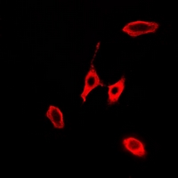
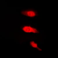
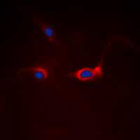
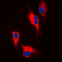 IF (Immunofluorescence)
(Immunofluorescent analysis of Tyrosine Hydroxylase staining in A549 cells. Formalin-fixed cells were permeabilized with 0.1% Triton X-100 in TBS for 5-10 minutes and blocked with 3% BSA-PBS for 30 minutes at room temperature. Cells were probed with the primary antibody in 3% BSA-PBS and incubated overnight at 4 °C in a humidified chamber. Cells were washed with PBST and incubated with a DyLight 594-conjugated secondary antibody (red) in PBS at room temperature in the dark. DAPI was used to stain the cell nuclei (blue).)
IF (Immunofluorescence)
(Immunofluorescent analysis of Tyrosine Hydroxylase staining in A549 cells. Formalin-fixed cells were permeabilized with 0.1% Triton X-100 in TBS for 5-10 minutes and blocked with 3% BSA-PBS for 30 minutes at room temperature. Cells were probed with the primary antibody in 3% BSA-PBS and incubated overnight at 4 °C in a humidified chamber. Cells were washed with PBST and incubated with a DyLight 594-conjugated secondary antibody (red) in PBS at room temperature in the dark. DAPI was used to stain the cell nuclei (blue).)
Tyrosine Hydroxylase, Polyclonal Antibody (Cat# AAA260015)
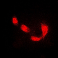
 IF (Immunofluorescence)
(Immunofluorescent analysis of IMPA1 staining in A549 cells. Formalin-fixed cells were permeabilized with 0.1% Triton X-100 in TBS for 5-10 minutes and blocked with 3% BSA-PBS for 30 minutes at room temperature. Cells were probed with the primary antibody in 3% BSA-PBS and incubated overnight at 4 °C in a humidified chamber. Cells were washed with PBST and incubated with a DyLight 594-conjugated secondary antibody (red) in PBS at room temperature in the dark.)
IF (Immunofluorescence)
(Immunofluorescent analysis of IMPA1 staining in A549 cells. Formalin-fixed cells were permeabilized with 0.1% Triton X-100 in TBS for 5-10 minutes and blocked with 3% BSA-PBS for 30 minutes at room temperature. Cells were probed with the primary antibody in 3% BSA-PBS and incubated overnight at 4 °C in a humidified chamber. Cells were washed with PBST and incubated with a DyLight 594-conjugated secondary antibody (red) in PBS at room temperature in the dark.)
IMPA1, Polyclonal Antibody (Cat# AAA262145)
What are Polyclonal Antibodies?
Polyclonal antibodies are antibodies that come from multiple B cell clones of a host animal. The typical hosts used for the majority of polyclonal antibody production are rabbits, goats, sheep, and donkeys. These polyclonal antibodies, once having identified their target, will bind to different epitopes located at different regions or sequences on the same protein/antigen. This ability to bind multiple epitopes is what makes polyclonal antibodies highly sensitive, as explained in our detailed guide on polyclonal antibodies and why they matter.
As a result, they are ideal at locating and binding to the target, even if the target is in very low concentrations (due to many different antibodies being able to bind to the same target molecule, which allows for significant amplification of a downstream signal).
Polyclonal antibodies are typically produced by injecting an antigen into a host animal, which causes the animal’s immune system to attack the foreign antigen by mass generating antibodies against it. After a period of time, serum is collected from the animal and purified using physicochemical fractionation, class-specific affinity purification, and/or antigen-affinity purification.
Key Uses of Polyclonal Antibodies
- Western Blotting: This method is used to find specific proteins in biological samples after separating them by size.
- Immunohistochemistry: IHC helps visualize the location of proteins in tissue sections using various staining techniques.
- ELISA: (Enzyme-Linked Immunosorbent Assay) is typically used to identify specific protein quantities in a sample. ELISAs can be either “Quantitative” or “Qualitative”.
- Flow Cytometry: technique that identifies and measures the specific protein on the surface or inside the cells in a fluid suspension.
- Immunoprecipitation: IP isolates and studies a specific protein from a complex mixture using antibodies.
Why Buy Polyclonal Antibodies from AAA Biotech?
1. Ideal for Various Applications
Our antibodies are generally going to be validated for use in multiple types of assays, including ELISA, Western Blotting, Immunohistochemistry, Immunoprecipitation, amongst others. They are ideal for a wide range of research applications.
2. Rigorous Quality Control
All of the antibodies in our catalog undergo strict quality testing to ensure specificity, sensitivity, and consistent performance. We are confident in the ability of our antibodies to provide you with accurate results.
3. Wide Assortment of Antibodies
Antibodies in our catalog can be found for both common and exotic species, and these antibodies are also available in both conjugated and recombinant forms to suit many diverse experimental needs.
4. Highly Purified
Our antibodies are available in purified forms with over 85% purity, as confirmed by SDS-PAGE. They are also available with tags such as His, Flag, GST, or MBP. We cater to customers worldwide.
FAQ
1. How are polyclonal antibodies produced?
Traditionally, polyclonal antibodies are produced by injecting an antigen into a host animal (such as a rabbit or goat), which then triggers an immune response from the host animal. The animal’s B cells produce antibodies that will recognize different parts of the injected antigen. These antibodies are then collected from the animal’s blood and purified for use.
2. How do polyclonal antibodies differ from monoclonal antibodies?
Polyclonal antibodies are a mix of antibodies that bind to different locations (epitopes) of the same antigen, while monoclonal antibodies are identical and bind to just one specific epitope. This makes polyclonal antibodies more versatile and better at detecting proteins that may be present in low quantities or in altered/modified forms.
3. How should I store polyclonal antibodies?
Polyclonal antibodies should be stored at 4°C for short-term use (up to a few weeks) and at -20°C or -80°C for long-term storage. Avoid repeated freeze-thaw cycles by dividing them into small aliquots. Always check the datasheet for specific storage instructions.