
Filters
Clonality
Type
Reactivity
Gene Name
Isotype
Host
Application
Clone
10000 results for "Recombinant Proteins" - showing 2450-2500
 Bioactivity
(SARS-CoV-2 Spike RBD is coated at 5ug/ml (100 ul/well) can bind ACE-2 (CAT# ATGP3963) in a Functional ELISA assay.)
Bioactivity
(SARS-CoV-2 Spike RBD is coated at 5ug/ml (100 ul/well) can bind ACE-2 (CAT# ATGP3963) in a Functional ELISA assay.)
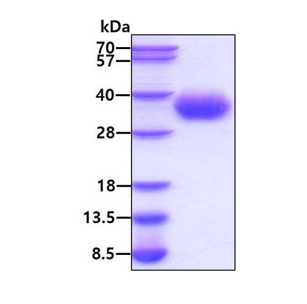
COVID 19 Spike RBD Coronavirus, Active Protein (Cat# AAA48377)
Full Name
Recombinant SARS-CoV-2 (2019-nCoV) Spike RBD protein
Applications
Bioactivity, SDS-PAGE
Purity
>95% by SDS-PAGE
Pricing
 Bioactivity
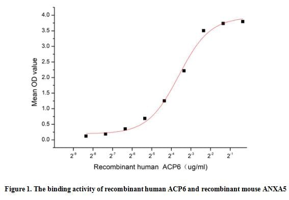
(Lysophosphatidic acid phosphatase type 6 (ACP6), is a 44-47 kDa monomeric member of the histidine acid phosphatase family of proteins. ACP6 is highly expressed in kidney, heart, small intestine, muscle, liver, prostate, testis, ovary and exists as two isoforms as a result of alternative splicing events. It plays a role in regulating lipid metabolism that hydrolyzes LPA to monoacylglycerol (MAG) and phosphate. Annexin V (ANXA5) binds to phospholipid bilayers, forming two-dimensional crystals that block the phospholipids from availability for coagulation enzyme reactions. A functional binding ELISA assay was conducted to detect the interaction of recombinant human ACP6 and recombinant mouse ANXA5. Briefly, ACP6 was diluted serially in PBS with 0.01% BSA (pH 7.4). Duplicate samples of 100 ul were then transferred to ANXA5-coated microtiter wells and incubated for 1h at 37 degree C. Wells were washed with PBST and incubated for 1h with anti-ACP6 pAb, then aspirated and washed 3 times. After incubation with HRP labelled secondary antibody for 1h at 37 degree C, wells were aspirated and washed 5 times. With the addition of substrate solution, wells were incubated 15-25 minutes at 37 degree C. Finally, add 50 uL stop solution to the wells and read at 450/630 nm immediately. The binding activity of recombinant human ACP6 and recombinant mouse ANXA5 was shown in Figure 1, the EC50 for this effect is 0.08 ug/mL.)
Bioactivity
(Lysophosphatidic acid phosphatase type 6 (ACP6), is a 44-47 kDa monomeric member of the histidine acid phosphatase family of proteins. ACP6 is highly expressed in kidney, heart, small intestine, muscle, liver, prostate, testis, ovary and exists as two isoforms as a result of alternative splicing events. It plays a role in regulating lipid metabolism that hydrolyzes LPA to monoacylglycerol (MAG) and phosphate. Annexin V (ANXA5) binds to phospholipid bilayers, forming two-dimensional crystals that block the phospholipids from availability for coagulation enzyme reactions. A functional binding ELISA assay was conducted to detect the interaction of recombinant human ACP6 and recombinant mouse ANXA5. Briefly, ACP6 was diluted serially in PBS with 0.01% BSA (pH 7.4). Duplicate samples of 100 ul were then transferred to ANXA5-coated microtiter wells and incubated for 1h at 37 degree C. Wells were washed with PBST and incubated for 1h with anti-ACP6 pAb, then aspirated and washed 3 times. After incubation with HRP labelled secondary antibody for 1h at 37 degree C, wells were aspirated and washed 5 times. With the addition of substrate solution, wells were incubated 15-25 minutes at 37 degree C. Finally, add 50 uL stop solution to the wells and read at 450/630 nm immediately. The binding activity of recombinant human ACP6 and recombinant mouse ANXA5 was shown in Figure 1, the EC50 for this effect is 0.08 ug/mL.)
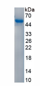
Acid Phosphatase 6, Lysophosphatidic (ACP6), Active Protein (Cat# AAA161908)
Full Name
Active Acid Phosphatase 6, Lysophosphatidic (ACP6)
Gene Names
ACP6; LPAP; ACPL1; PACPL1
Applications
Activity Assay, Cell Culture
Purity
Greater than 90% by SDS-PAGE
Pricing
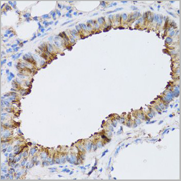
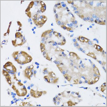
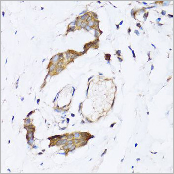
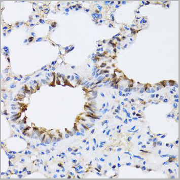
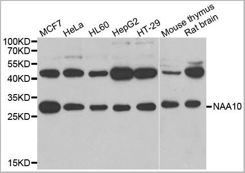
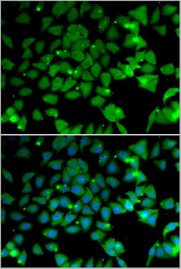 IF (Immunofluorescence)
(Immunofluorescence analysis of U2OS cells using NAA10 antibody. Blue: DAPI for nuclear staining.)
IF (Immunofluorescence)
(Immunofluorescence analysis of U2OS cells using NAA10 antibody. Blue: DAPI for nuclear staining.)
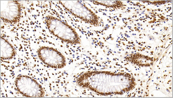
NAA10, Polyclonal Antibody (Cat# AAA28121)
Full Name
NAA10 Polyclonal Antibody
Gene Names
NAA10; TE2; ARD1; NATD; ARD1A; ARD1P; DXS707; MCOPS1
Applications
Western Blot, Immunohistochemistry, Immunofluorescence
Purity
Affinity Purification
Pricing
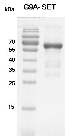
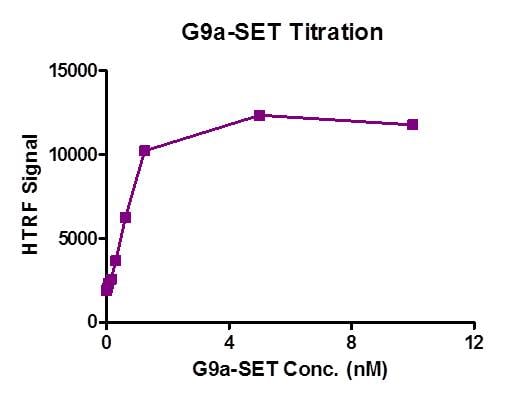 Application Data
(Recombinant EHMT2 (G9A)-SET (913-1193) activity assay. 3.3 uM H3 (1-21) peptide was incubated with EHMT2 (G9A)-SET (913-1193 protein in reaction buffer including 50 mM Tris-HCl, pH 8.6, 0.02% Triton X-100, 2 mM MgCl2, 1 mM TCEP and 50 uM SAM for 3 hours at room temperature. H3K9me2 antibody is used to detect reaction product. )
Application Data
(Recombinant EHMT2 (G9A)-SET (913-1193) activity assay. 3.3 uM H3 (1-21) peptide was incubated with EHMT2 (G9A)-SET (913-1193 protein in reaction buffer including 50 mM Tris-HCl, pH 8.6, 0.02% Triton X-100, 2 mM MgCl2, 1 mM TCEP and 50 uM SAM for 3 hours at room temperature. H3K9me2 antibody is used to detect reaction product. )
EHMT2, Recombinant Protein (Cat# AAA59675)
Full Name
Recombinant EHMT2 (G9A)-SET (913-1193) protein
Gene Names
EHMT2; G9A; BAT8; GAT8; NG36; KMT1C; C6orf30
Pricing
 Bioactivity
(Uracil Phosphoribosyltransferase (UPRT) is an important enzyme involved in the pyrimidine salvage pathway, which is a critical metabolic pathway for the synthesis of nucleotides in various organisms, including humans. This enzyme plays a key role in the conversion of uracil, a pyrimidine base, into uridine-5'-phosphate (UMP), a precursor for the synthesis of RNA and DNA. Besides, Purine Nucleoside Phosphorylase (PNP) has been identified as an interactor of UPRT, thus a functional binding ELISA assay was conducted to detect the interaction of recombinant human UPRT and recombinant mouse PNP. Briefly, UPRT was diluted serially in PBS with 0.01% BSA (pH 7.4). Duplicate samples of 100 ul were then transferred to PNP-coated microtiter wells and incubated for 1h at 37 degree C. Wells were washed with PBST and incubated for 1h with anti-UPRT pAb, then aspirated and washed 3 times. After incubation with HRP labelled secondary antibody for 1h at 37 degree C, wells were aspirated and washed 5 times. With the addition of substrate solution, wells were incubated 15-25 minutes at 37 degree C. Finally, add 50 uL stop solution to the wells and read at 450/630 nm immediately. The binding activity of recombinant human UPRT and recombinant mouse PNP was shown in Figure 1, the EC50 for this effect is 0.017 ug/mL.)
Bioactivity
(Uracil Phosphoribosyltransferase (UPRT) is an important enzyme involved in the pyrimidine salvage pathway, which is a critical metabolic pathway for the synthesis of nucleotides in various organisms, including humans. This enzyme plays a key role in the conversion of uracil, a pyrimidine base, into uridine-5'-phosphate (UMP), a precursor for the synthesis of RNA and DNA. Besides, Purine Nucleoside Phosphorylase (PNP) has been identified as an interactor of UPRT, thus a functional binding ELISA assay was conducted to detect the interaction of recombinant human UPRT and recombinant mouse PNP. Briefly, UPRT was diluted serially in PBS with 0.01% BSA (pH 7.4). Duplicate samples of 100 ul were then transferred to PNP-coated microtiter wells and incubated for 1h at 37 degree C. Wells were washed with PBST and incubated for 1h with anti-UPRT pAb, then aspirated and washed 3 times. After incubation with HRP labelled secondary antibody for 1h at 37 degree C, wells were aspirated and washed 5 times. With the addition of substrate solution, wells were incubated 15-25 minutes at 37 degree C. Finally, add 50 uL stop solution to the wells and read at 450/630 nm immediately. The binding activity of recombinant human UPRT and recombinant mouse PNP was shown in Figure 1, the EC50 for this effect is 0.017 ug/mL.)
Uracil Phosphoribosyltransferase (UPRT), Active Protein (Cat# AAA161941)
Full Name
Active Uracil Phosphoribosyltransferase (UPRT)
Gene Names
UPRT; UPP; FUR1
Applications
Activity Assay, Cell Culture
Purity
Greater than 90% by SDS-PAGE
Pricing
 Bioactivity
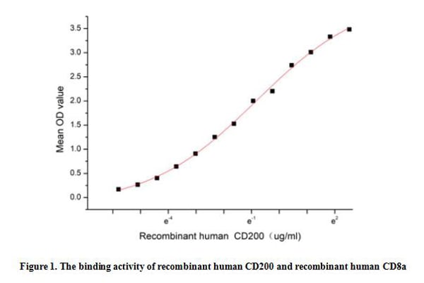
(Cluster Of Differentiation 200 (CD200), also known as OX-2 or MOX1/MOX2, is a membrane glycoprotein belonging to the immunoglobulin superfamily. This protein is widely expressed in various cell types, including neurons, endothelial cells, certain immune cells (such as B cells, dendritic cells, T cell subsets), and various tumor cells. CD200 is a multifaceted protein with important roles in immune regulation, neuronal function, and disease pathogenesis. Besides, Cluster Of Differentiation 8a (CD8a) has been identified as an interactor of CD200, thus a functional binding ELISA assay was conducted to detect the interaction of recombinant human CD200 and recombinant human CD8a. Briefly, CD200 was diluted serially in PBS with 0.01% BSA (pH 7.4). Duplicate samples of 100 ul were then transferred to CD8a-coated microtiter wells and incubated for 1h at 37 degree C. Wells were washed with PBST and incubated for 1h with anti-CD200 pAb, then aspirated and washed 3 times. After incubation with HRP labelled secondary antibody for 1h at 37 degree C, wells were aspirated and washed 5 times. With the addition of substrate solution, wells were incubated 15-25 minutes at 37 degree C. Finally, add 50 uL stop solution to the wells and read at 450/630 nm immediately. The binding activity of recombinant recombinant human CD200 and recombinant human CD8a was shown in Figure 1, the EC50 for this effect is 0.44 ug/mL.)
Bioactivity
(Cluster Of Differentiation 200 (CD200), also known as OX-2 or MOX1/MOX2, is a membrane glycoprotein belonging to the immunoglobulin superfamily. This protein is widely expressed in various cell types, including neurons, endothelial cells, certain immune cells (such as B cells, dendritic cells, T cell subsets), and various tumor cells. CD200 is a multifaceted protein with important roles in immune regulation, neuronal function, and disease pathogenesis. Besides, Cluster Of Differentiation 8a (CD8a) has been identified as an interactor of CD200, thus a functional binding ELISA assay was conducted to detect the interaction of recombinant human CD200 and recombinant human CD8a. Briefly, CD200 was diluted serially in PBS with 0.01% BSA (pH 7.4). Duplicate samples of 100 ul were then transferred to CD8a-coated microtiter wells and incubated for 1h at 37 degree C. Wells were washed with PBST and incubated for 1h with anti-CD200 pAb, then aspirated and washed 3 times. After incubation with HRP labelled secondary antibody for 1h at 37 degree C, wells were aspirated and washed 5 times. With the addition of substrate solution, wells were incubated 15-25 minutes at 37 degree C. Finally, add 50 uL stop solution to the wells and read at 450/630 nm immediately. The binding activity of recombinant recombinant human CD200 and recombinant human CD8a was shown in Figure 1, the EC50 for this effect is 0.44 ug/mL.)
Cluster Of Differentiation 200 (CD200), Active Protein (Cat# AAA161799)
Full Name
Active Cluster Of Differentiation 200 (CD200)
Gene Names
CD200; MRC; MOX1; MOX2; OX-2
Applications
Activity Assay, Cell Culture
Purity
Greater than 90% by SDS-PAGE
Pricing
 Bioactivity
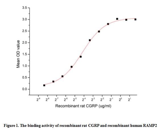
(Calcitonin gene-related peptide (CGRP) is a highly potent vasoactive peptide released from sensory nerves. CGRP is a 37-amino acid and it has two major forms (alpha and beta). It belongs to a group of peptides that all act on an unusual receptor family. These receptors consist of calcitonin receptor-like receptor (CLR) linked to an essential receptor activity modifying protein (RAMP) that is necessary for full functionality. Thus a functional binding ELISA assay was conducted to detect the interaction of recombinant rat CGRP and recombinant human RAMP2. Briefly, CGRP was diluted serially in PBS with 0.01% BSA (pH 7.4). Duplicate samples of 100 ul were then transferred to RAMP2-coated microtiter wells and incubated for 1h at 37 degree C. Wells were washed with PBST and incubated for 1h with anti-CGRP pAb, then aspirated and washed 3 times. After incubation with HRP labelled secondary antibody for 1h at 37 degree C, wells were aspirated and washed 5 times. With the addition of substrate solution, wells were incubated 15-25 minutes at 37 degree C. Finally, add 50 uL stop solution to the wells and read at 450/630 nm immediately. The binding activity of recombinant rat CGRP and recombinant human RAMP2 was shown in Figure 1, the EC50 for this effect is 0.06 ug/mL.)
Bioactivity
(Calcitonin gene-related peptide (CGRP) is a highly potent vasoactive peptide released from sensory nerves. CGRP is a 37-amino acid and it has two major forms (alpha and beta). It belongs to a group of peptides that all act on an unusual receptor family. These receptors consist of calcitonin receptor-like receptor (CLR) linked to an essential receptor activity modifying protein (RAMP) that is necessary for full functionality. Thus a functional binding ELISA assay was conducted to detect the interaction of recombinant rat CGRP and recombinant human RAMP2. Briefly, CGRP was diluted serially in PBS with 0.01% BSA (pH 7.4). Duplicate samples of 100 ul were then transferred to RAMP2-coated microtiter wells and incubated for 1h at 37 degree C. Wells were washed with PBST and incubated for 1h with anti-CGRP pAb, then aspirated and washed 3 times. After incubation with HRP labelled secondary antibody for 1h at 37 degree C, wells were aspirated and washed 5 times. With the addition of substrate solution, wells were incubated 15-25 minutes at 37 degree C. Finally, add 50 uL stop solution to the wells and read at 450/630 nm immediately. The binding activity of recombinant rat CGRP and recombinant human RAMP2 was shown in Figure 1, the EC50 for this effect is 0.06 ug/mL.)
Calcitonin Gene Related Peptide (CGRP), Active Protein (Cat# AAA161798)
Full Name
Active Calcitonin Gene Related Peptide (CGRP)
Applications
Activity Assay, Cell Culture
Purity
Greater than 95% by SDS-PAGE
Pricing
 Bioactivity
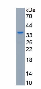
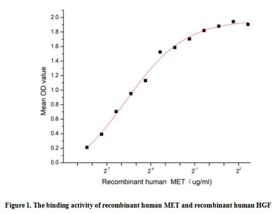
(c-Met is a receptor tyrosine kinase belonging to the MET (MNNG HOS transforming gene) family, and is expressed on the surfaces of various cells. Hepatocyte growth factor (HGF) is the ligand for this receptor. The binding of HGF to c-Met initiates a series of intracellular signals that mediate embryogenesis and wound healing in normal cells. Thus a functional binding ELISA assay was conducted to detect the interaction of recombinant human MET and recombinant human HGF. Briefly, MET was diluted serially in PBS with 0.01% BSA (pH 7.4). Duplicate samples of 100 ul were then transferred to HGF-coated microtiter wells and incubated for 1h at 37 degree C. Wells were washed with PBST and incubated for 1h with anti-MET pAb, then aspirated and washed 3 times. After incubation with HRP labelled secondary antibody for 1h at 37 degree C, wells were aspirated and washed 5 times. With the addition of substrate solution, wells were incubated 15-25 minutes at 37 degree C. Finally, add 50 uL stop solution to the wells and read at 450/630 nm immediately. The binding activity of recombinant human MET and recombinant human HGF was shown in Figure 1, the EC50 for this effect is 0.02 ug/mL.)
Bioactivity
(c-Met is a receptor tyrosine kinase belonging to the MET (MNNG HOS transforming gene) family, and is expressed on the surfaces of various cells. Hepatocyte growth factor (HGF) is the ligand for this receptor. The binding of HGF to c-Met initiates a series of intracellular signals that mediate embryogenesis and wound healing in normal cells. Thus a functional binding ELISA assay was conducted to detect the interaction of recombinant human MET and recombinant human HGF. Briefly, MET was diluted serially in PBS with 0.01% BSA (pH 7.4). Duplicate samples of 100 ul were then transferred to HGF-coated microtiter wells and incubated for 1h at 37 degree C. Wells were washed with PBST and incubated for 1h with anti-MET pAb, then aspirated and washed 3 times. After incubation with HRP labelled secondary antibody for 1h at 37 degree C, wells were aspirated and washed 5 times. With the addition of substrate solution, wells were incubated 15-25 minutes at 37 degree C. Finally, add 50 uL stop solution to the wells and read at 450/630 nm immediately. The binding activity of recombinant human MET and recombinant human HGF was shown in Figure 1, the EC50 for this effect is 0.02 ug/mL.)
C-Met (MET), Active Protein (Cat# AAA161879)
Full Name
Active C-Met (MET)
Gene Names
MET; HGFR; AUTS9; RCCP2; c-Met
Applications
Activity Assay, Cell Culture
Purity
Greater than 80% by SDS-PAGE
Pricing
 Bioactivity
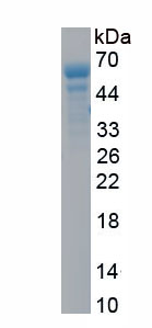
(Glypican-3 (GPC3), a 70 kDa protein, is a member of the glypican family that attaches to the cell surface by a glycosylphosphatidylinositol anchor, is specifically up-regulated in hepatocellular carcinoma (HCC) although rarely or not expressed in normal liver tissues, making it a perfect test and treatment target for HCC. GPC3 is also a negative transcriptional regulator and tumor suppressor that inhibits the growth of breast, ovary, and lung cancer cells. It is reported that GPC3 can form a complex with insulin-like growth factor 2 (IGF2), and might thereby modulate IGF2 action. Thus a functional binding ELISA assay was conducted to detect the interaction of recombinant human GPC3 and recombinant rat IGF2. Briefly, GPC3 was diluted serially in PBS with 0.01% BSA (pH 7.4). Duplicate samples of 100 ul were then transferred to IGF2-coated microtiter wells and incubated for 1h at 37 degree C. Wells were washed with PBST and incubated for 1h with anti-GPC3 pAb, then aspirated and washed 3 times. After incubation with HRP labelled secondary antibody for 1h at 37 degree C, wells were aspirated and washed 5 times. With the addition of substrate solution, wells were incubated 15-25 minutes at 37 degree C. Finally, add 50 uL stop solution to the wells and read at 450/630 nm immediately. The binding activity of recombinant human GPC3 and recombinant rat IGF2 was shown in Figure 1, the EC50 for this effect is 0.08 ug/mL.)
Bioactivity
(Glypican-3 (GPC3), a 70 kDa protein, is a member of the glypican family that attaches to the cell surface by a glycosylphosphatidylinositol anchor, is specifically up-regulated in hepatocellular carcinoma (HCC) although rarely or not expressed in normal liver tissues, making it a perfect test and treatment target for HCC. GPC3 is also a negative transcriptional regulator and tumor suppressor that inhibits the growth of breast, ovary, and lung cancer cells. It is reported that GPC3 can form a complex with insulin-like growth factor 2 (IGF2), and might thereby modulate IGF2 action. Thus a functional binding ELISA assay was conducted to detect the interaction of recombinant human GPC3 and recombinant rat IGF2. Briefly, GPC3 was diluted serially in PBS with 0.01% BSA (pH 7.4). Duplicate samples of 100 ul were then transferred to IGF2-coated microtiter wells and incubated for 1h at 37 degree C. Wells were washed with PBST and incubated for 1h with anti-GPC3 pAb, then aspirated and washed 3 times. After incubation with HRP labelled secondary antibody for 1h at 37 degree C, wells were aspirated and washed 5 times. With the addition of substrate solution, wells were incubated 15-25 minutes at 37 degree C. Finally, add 50 uL stop solution to the wells and read at 450/630 nm immediately. The binding activity of recombinant human GPC3 and recombinant rat IGF2 was shown in Figure 1, the EC50 for this effect is 0.08 ug/mL.)
Glypican 3 (GPC3), Active Protein (Cat# AAA161814)
Full Name
Active Glypican 3 (GPC3)
Gene Names
GPC3; SGB; DGSX; MXR7; SDYS; SGBS; OCI-5; SGBS1; GTR2-2
Applications
Activity Assay, Cell Culture
Purity
Greater than 80% by SDS-PAGE
Pricing
 Application Data
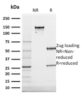
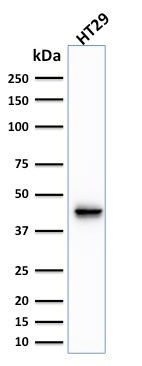
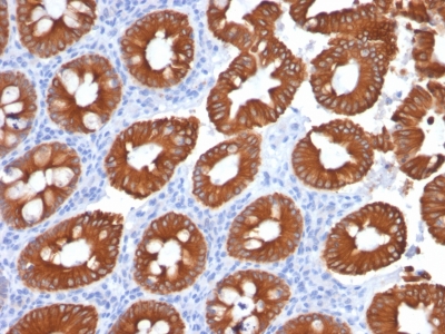
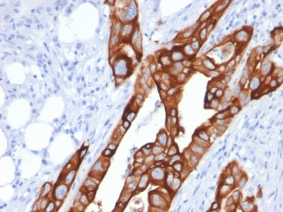
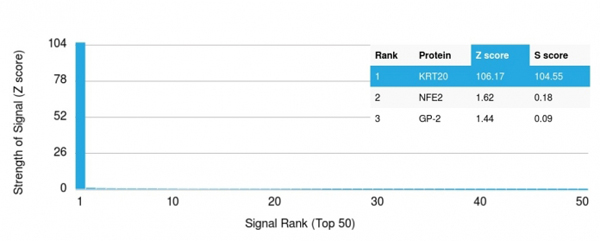
(Analysis of Protein Array containing more than 19,000 full-length human proteins using Cytokeratin 20 (KRT20) Mouse Monoclonal Antibody (KRT20/1991). Z- and S- Score: The Z-score represents the strength of a signal that a monoclonal antibody (MAb) (in combination with a fluorescently-tagged anti-IgG secondary antibody) produces when binding to a particular protein on the HuProtTM array. Z-scores are described in units of standard deviations (SD's) above the mean value of all signals generated on that array. If targets on HuProtTM are arranged in descending order of the Z-score, the S-score is the difference (also in units of SD's) between the Z-score. S-score therefore represents the relative target specificity of a MAb to its intended target. A MAb is considered to specific to its intended target, if the MAb has an S-score of at least 2.5. For example, if a MAb binds to protein X with a Z-score of 43 and to protein Y with a Z-score of 14, then the S-score for the binding of that MAb to protein X is equal to 29.)
Application Data
(Analysis of Protein Array containing more than 19,000 full-length human proteins using Cytokeratin 20 (KRT20) Mouse Monoclonal Antibody (KRT20/1991). Z- and S- Score: The Z-score represents the strength of a signal that a monoclonal antibody (MAb) (in combination with a fluorescently-tagged anti-IgG secondary antibody) produces when binding to a particular protein on the HuProtTM array. Z-scores are described in units of standard deviations (SD's) above the mean value of all signals generated on that array. If targets on HuProtTM are arranged in descending order of the Z-score, the S-score is the difference (also in units of SD's) between the Z-score. S-score therefore represents the relative target specificity of a MAb to its intended target. A MAb is considered to specific to its intended target, if the MAb has an S-score of at least 2.5. For example, if a MAb binds to protein X with a Z-score of 43 and to protein Y with a Z-score of 14, then the S-score for the binding of that MAb to protein X is equal to 29.)
Cytokeratin 20 (KRT20), Monoclonal Antibody (Cat# AAA214794)
Full Name
Cytokeratin 20 (KRT20) (Colorectal Epithelial Marker)
Gene Names
KRT20; K20; CD20; CK20; CK-20; KRT21
Reactivity
Human. Others not known.
Applications
Immunohistochemistry, Western Blot
Purity
Purified Ab with BSA and Azide at 200ug/ml OR Purified Ab WITHOUT BSA and Azide at 1.0mg/ml
Pricing
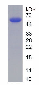
 Bioactivity
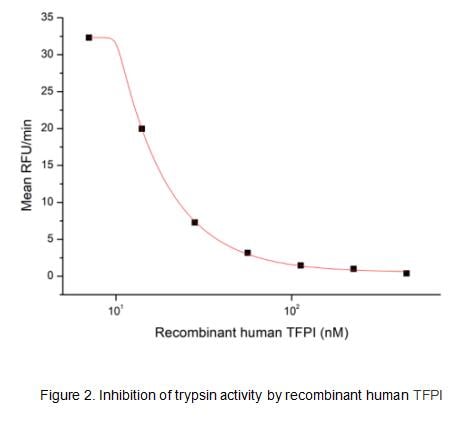
(Human TFPI, also known as lipoprotein-associated coagulation inhibitor (LACI) and extrinsic pathway inhibitor (EPI), is a physiological inhibitor of extrinsic pathway of coagulation and has biological functions of anticoagulation and anti-inflammation. It is a secreted protein with a N-terminal acidic region, three Kunitz (K) domains separated with by two linker regions, and a C-terminal basic region. The activity of recombinant human TFPI was measured by its ability to inhibit trypsin cleavage of a fluorogenic peptide substrate Mca-RPKPVE-Nval-WRK(Dnp)-NH2 in the assay buffer 50 mM Tris, 10 mM CaCl2, 150 mM NaCl, 0.05% (w/v) Brij-35, pH 7.5. Trypsin was diluted to 50 ug/ml in the assay buffer and 20 ul different concentrations of recombinant human TFPI (MW: 55.58 KD) was incubated with 20 ul diluted trypsin at 37 degree C for 15 minutes. Loading 50 uL of the incubated mixtures which were diluted five-fold in assay buffer into empty wells of a plate, and start the reaction by adding 50 uL of 20 uM substrate. Include a substrate blank containing 50 uL of assay buffer and 50 uL of 20 uM substrate. Then read at excitiation and emission wavelengths of 320 nm and 405 nm, respectively, in kinetic mode for 5 minutes. The result was shown in Figure 1 and it was obvious that recombinant human TFPI significantly decreased trypsin activity. The inhibition IC50 was )
Bioactivity
(Human TFPI, also known as lipoprotein-associated coagulation inhibitor (LACI) and extrinsic pathway inhibitor (EPI), is a physiological inhibitor of extrinsic pathway of coagulation and has biological functions of anticoagulation and anti-inflammation. It is a secreted protein with a N-terminal acidic region, three Kunitz (K) domains separated with by two linker regions, and a C-terminal basic region. The activity of recombinant human TFPI was measured by its ability to inhibit trypsin cleavage of a fluorogenic peptide substrate Mca-RPKPVE-Nval-WRK(Dnp)-NH2 in the assay buffer 50 mM Tris, 10 mM CaCl2, 150 mM NaCl, 0.05% (w/v) Brij-35, pH 7.5. Trypsin was diluted to 50 ug/ml in the assay buffer and 20 ul different concentrations of recombinant human TFPI (MW: 55.58 KD) was incubated with 20 ul diluted trypsin at 37 degree C for 15 minutes. Loading 50 uL of the incubated mixtures which were diluted five-fold in assay buffer into empty wells of a plate, and start the reaction by adding 50 uL of 20 uM substrate. Include a substrate blank containing 50 uL of assay buffer and 50 uL of 20 uM substrate. Then read at excitiation and emission wavelengths of 320 nm and 405 nm, respectively, in kinetic mode for 5 minutes. The result was shown in Figure 1 and it was obvious that recombinant human TFPI significantly decreased trypsin activity. The inhibition IC50 was )
Tissue Factor Pathway Inhibitor (TFPI), Active Protein (Cat# AAA161731)
Full Name
Active Tissue Factor Pathway Inhibitor (TFPI)
Gene Names
TFPI; EPI; TFI; LACI; TFPI1
Applications
Activity Assay, Cell Culture
Purity
Greater than 95% by SDS-PAGE
Pricing
 Bioactivity
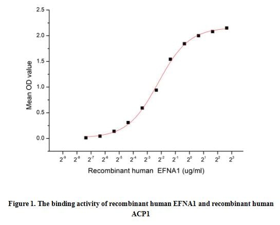
(Acid phosphatase locus 1 (ACP1) is a low molecular weight protein tyrosine phosphatase that has been shown to be an important regulator of insulin receptor signaling. EFNA1 is a kind of receptor tyrosine kinase which can Interact with ACP1. Thus a functional binding ELISA assay was conducted to detect the interaction of recombinant human EFNA1 and recombinant human ACP1. Briefly, EFNA1 was diluted serially in PBS with 0.01% BSA (pH 7.4). Duplicate samples of 100 ul were then transferred to ACP1-coated microtiter wells and incubated for 1h at 37 degree C. Wells were washed with PBST and incubated for 1h with anti-EFNA1 pAb, then aspirated and washed 3 times. After incubation with HRP labelled secondary antibody for 1h at 37 degree C, wells were aspirated and washed 5 times. With the addition of substrate solution, wells were incubated 15-25 minutes at 37 degree C. Finally, add 50 uL stop solution to the wells and read at 450/630 nm immediately. When recombinant human ACP1 is immobilized at 2 ug/ml (100 ul/well), the concentration of EFNA1 that produces 50% optimal binding response is found to be approximately 0.21 ug/ml.)
Bioactivity
(Acid phosphatase locus 1 (ACP1) is a low molecular weight protein tyrosine phosphatase that has been shown to be an important regulator of insulin receptor signaling. EFNA1 is a kind of receptor tyrosine kinase which can Interact with ACP1. Thus a functional binding ELISA assay was conducted to detect the interaction of recombinant human EFNA1 and recombinant human ACP1. Briefly, EFNA1 was diluted serially in PBS with 0.01% BSA (pH 7.4). Duplicate samples of 100 ul were then transferred to ACP1-coated microtiter wells and incubated for 1h at 37 degree C. Wells were washed with PBST and incubated for 1h with anti-EFNA1 pAb, then aspirated and washed 3 times. After incubation with HRP labelled secondary antibody for 1h at 37 degree C, wells were aspirated and washed 5 times. With the addition of substrate solution, wells were incubated 15-25 minutes at 37 degree C. Finally, add 50 uL stop solution to the wells and read at 450/630 nm immediately. When recombinant human ACP1 is immobilized at 2 ug/ml (100 ul/well), the concentration of EFNA1 that produces 50% optimal binding response is found to be approximately 0.21 ug/ml.)
Acid Phosphatase 1 (ACP1), Active Protein (Cat# AAA161820)
Full Name
Active Acid Phosphatase 1 (ACP1)
Gene Names
ACP1; HAAP
Applications
Activity Assay, Cell Culture
Purity
Greater than 90% by SDS-PAGE
Pricing
 Application Data
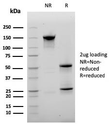
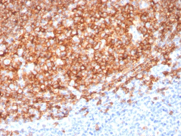
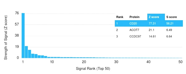
(Analysis of Protein Array containing more than 19,000 full-length human proteins using CD20 Mouse Recombinant Monoclonal Antibody (MS4A1/3410). Z- and S- Score: The Z-score represents the strength of a signal that a monoclonal antibody (Monoclonal Antibody) (in combination with a fluorescently-tagged anti-IgG secondary antibody) produces when binding to a particular protein on the HuProtTM array. Z-scores are described in units of standard deviations (SD’s) above the mean value of all signals generated on that array. If targets on HuProtTM are arranged in descending order of the Z-score, the S-score is the difference (also in units of SD’s) between the Z-score. S-score therefore represents the relative target specificity of a Monoclonal Antibody to its intended target. A Monoclonal Antibody is considered to specific to its intended target, if the Monoclonal Antibody has an S-score of at least 2.5. For example, if a Monoclonal Antibody binds to protein X with a Z-score of 43 and to protein Y with a Z-score of 14, then the S-score for the binding of that Monoclonal Antibody to protein X is equal to 29.)
Application Data
(Analysis of Protein Array containing more than 19,000 full-length human proteins using CD20 Mouse Recombinant Monoclonal Antibody (MS4A1/3410). Z- and S- Score: The Z-score represents the strength of a signal that a monoclonal antibody (Monoclonal Antibody) (in combination with a fluorescently-tagged anti-IgG secondary antibody) produces when binding to a particular protein on the HuProtTM array. Z-scores are described in units of standard deviations (SD’s) above the mean value of all signals generated on that array. If targets on HuProtTM are arranged in descending order of the Z-score, the S-score is the difference (also in units of SD’s) between the Z-score. S-score therefore represents the relative target specificity of a Monoclonal Antibody to its intended target. A Monoclonal Antibody is considered to specific to its intended target, if the Monoclonal Antibody has an S-score of at least 2.5. For example, if a Monoclonal Antibody binds to protein X with a Z-score of 43 and to protein Y with a Z-score of 14, then the S-score for the binding of that Monoclonal Antibody to protein X is equal to 29.)
CD20/MS4A1, Monoclonal Antibody (Cat# AAA215294)
Full Name
CD20/MS4A1 (B-Cell Marker)
Gene Names
MS4A1; B1; S7; Bp35; CD20; CVID5; MS4A2; LEU-16
Reactivity
Human
Applications
Immunohistochemistry
Purity
Purified Ab with BSA and Azide at 200ug/ml OR Purified Ab WITHOUT BSA and Azide at 1.0mg/ml
Pricing
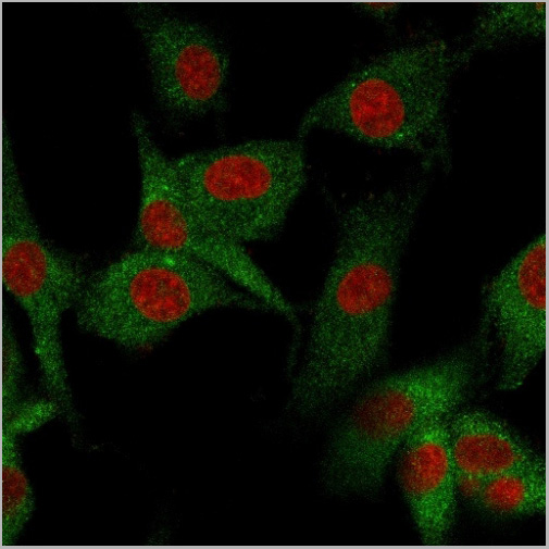
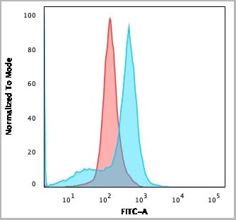
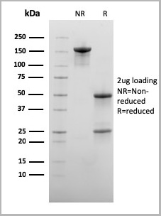
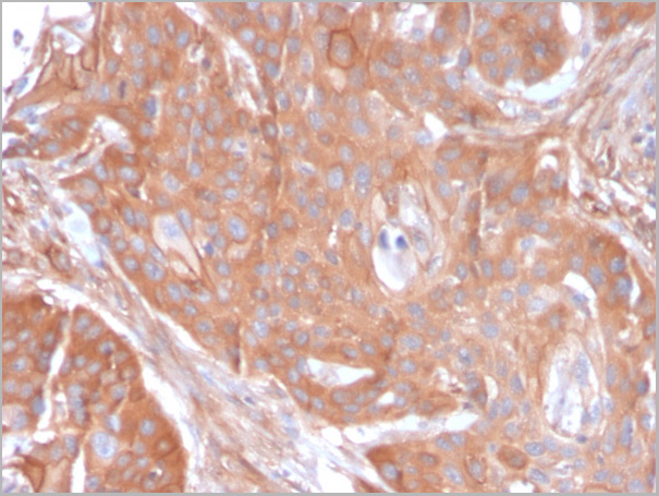
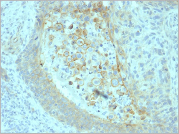
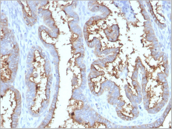
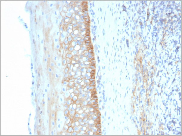
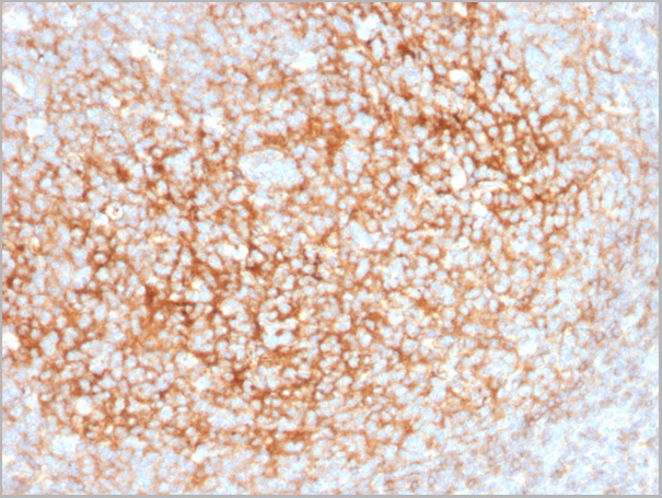
 Application Data
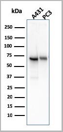
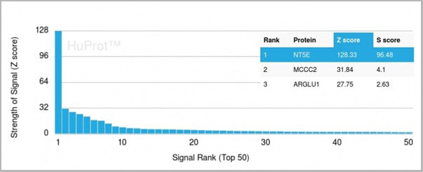
(Analysis of Protein Array containing more than 19,000 full-length human proteins using CD73 Mouse Recombinant Monoclonal Antibody (NT5E/2545) Z- and S- Score: The Z-score represents the strength of a signal that a monoclonal antibody (MAb) (in combination with a fluorescently-tagged anti-IgG secondary antibody) produces when binding to a particular protein on the HuProtTM array. Z-scores are described in units of standard deviations (SD’s) above the mean value of all signals generated on that array. If targets on HuProtTM are arranged in descending order of the Z-score, the S-score is the difference (also in units of SD’s) between the Z-score. S-score therefore represents the relative target specificity of a MAb to its intended target. A MAb is considered to specific to its intended target, if the MAb has an S-score of at least 2.5. For example, if a MAb binds to protein X with a Z-score of 43 and to protein Y with a Z-score of 14, then the S-score for the binding of that MAb to protein X is equal to 29.)
Application Data
(Analysis of Protein Array containing more than 19,000 full-length human proteins using CD73 Mouse Recombinant Monoclonal Antibody (NT5E/2545) Z- and S- Score: The Z-score represents the strength of a signal that a monoclonal antibody (MAb) (in combination with a fluorescently-tagged anti-IgG secondary antibody) produces when binding to a particular protein on the HuProtTM array. Z-scores are described in units of standard deviations (SD’s) above the mean value of all signals generated on that array. If targets on HuProtTM are arranged in descending order of the Z-score, the S-score is the difference (also in units of SD’s) between the Z-score. S-score therefore represents the relative target specificity of a MAb to its intended target. A MAb is considered to specific to its intended target, if the MAb has an S-score of at least 2.5. For example, if a MAb binds to protein X with a Z-score of 43 and to protein Y with a Z-score of 14, then the S-score for the binding of that MAb to protein X is equal to 29.)
CD73, Monoclonal Antibody (Cat# AAA23928)
Full Name
CD73 (Immuno-Oncology Target)
Gene Names
NT5E; NT; eN; NT5; NTE; eNT; CD73; E5NT; CALJA
Reactivity
Human
Applications
Western Blot, Flow Cytometry, Immunofluorescence, Immunohistochemistry
Purity
Purified Ab with BSA and Azide at 200ug/ml
Pricing
 Bioactivity
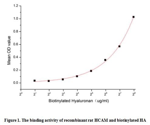
(Homing Associated Cell Adhesion Molecule (HCAM), also known as CD44, is a ubiquitous multistructural and multifunctional cells surface adhesion molecule involved in cell-cell and cell-matrix interactions. CD44 is broadly expressed, including in the membranes of B cells, granulocytes, monocytes, and erythrocytes as well as on many thymocytes and mature T cells, besides it is highly expressed in many cancers and regulates metastasis via recruitment of CD44 to the cell surface. This protein is a receptor for hyaluronic acid (HA) and can also interact with other ligands, such as osteopontin, collagens, and matrix metalloproteinases (MMPs). Thus a functional binding ELISA assay was conducted to detect the interaction of recombinant rat HCAM and biotinylated hyaluronan (HA). Briefly, biotin-linked HA were diluted serially in PBS, with 0.01% BSA (pH 7.4). Duplicate samples of 100 ul were then transferred to HCAM-coated microtiter wells and incubated for 2h at 37 degree C. Wells were washed with PBST 3 times and incubation with Streptavidin-HRP for 1 hour, then wells were aspirated and washed 5 times. With the addition of substrate solution, wells were incubated 15-25 minutes at 37 degree C. Finally, add 50 ul stop solution to the wells and read at 450/630 nm immediately. The binding activity of recombinant rat HCAM and biotinylated HA was shown in Figure 1, and this effect was in a dose dependent manner.)
Bioactivity
(Homing Associated Cell Adhesion Molecule (HCAM), also known as CD44, is a ubiquitous multistructural and multifunctional cells surface adhesion molecule involved in cell-cell and cell-matrix interactions. CD44 is broadly expressed, including in the membranes of B cells, granulocytes, monocytes, and erythrocytes as well as on many thymocytes and mature T cells, besides it is highly expressed in many cancers and regulates metastasis via recruitment of CD44 to the cell surface. This protein is a receptor for hyaluronic acid (HA) and can also interact with other ligands, such as osteopontin, collagens, and matrix metalloproteinases (MMPs). Thus a functional binding ELISA assay was conducted to detect the interaction of recombinant rat HCAM and biotinylated hyaluronan (HA). Briefly, biotin-linked HA were diluted serially in PBS, with 0.01% BSA (pH 7.4). Duplicate samples of 100 ul were then transferred to HCAM-coated microtiter wells and incubated for 2h at 37 degree C. Wells were washed with PBST 3 times and incubation with Streptavidin-HRP for 1 hour, then wells were aspirated and washed 5 times. With the addition of substrate solution, wells were incubated 15-25 minutes at 37 degree C. Finally, add 50 ul stop solution to the wells and read at 450/630 nm immediately. The binding activity of recombinant rat HCAM and biotinylated HA was shown in Figure 1, and this effect was in a dose dependent manner.)
Homing Associated Cell Adhesion Molecule (HCAM), Active Protein (Cat# AAA161766)
Full Name
Active Homing Associated Cell Adhesion Molecule (HCAM)
Gene Names
Cd44; CD44A; METAA; RHAMM
Applications
Activity Assay, Cell Culture
Purity
Greater than 95% by SDS-PAGE
Pricing
 WB (Western Blot)
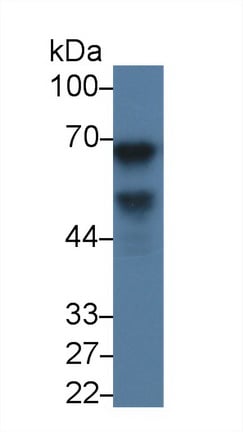
(Western Blot; Sample: Human Lung lysate; Primary Ab: 3ug/ml Mouse AntiHuman aFP AntibodySecond Ab: 0.2ug/mL HRPLinked Caprine AntiMouse IgG Polyclonal Antibody(Catalog: SAA544Mu19))
WB (Western Blot)
(Western Blot; Sample: Human Lung lysate; Primary Ab: 3ug/ml Mouse AntiHuman aFP AntibodySecond Ab: 0.2ug/mL HRPLinked Caprine AntiMouse IgG Polyclonal Antibody(Catalog: SAA544Mu19))
AlphaFetoprotein (AFP), Monoclonal Antibody (Cat# AAA151493)
Full Name
Monoclonal Antibody to AlphaFetoprotein (AFP)
Gene Names
AFP; FETA; HPAFP
Reactivity
Human
Applications
Immunoprecipitation, Immunocytochemistry, Immunohistochemistry, Western Blot
Purity
Protein A + Protein G affinity chromatography
Pricing


 WB (Western Blot)
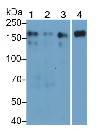
(Western Blot; Sample: Lane1: Hela cell lysate; Lane2: MCF7 cell lysate; Lane3: A431 cell lysate; Lane4: Human Placenta lysate Primary Ab: 0.04ug/ml Mouse AntiHuman EGFR Antibody Second Ab: 0.2ug/mL HRPLinked Caprine AntiMouse IgG Polyclonal Antibody (Catalog: SAA544Mu19))
WB (Western Blot)
(Western Blot; Sample: Lane1: Hela cell lysate; Lane2: MCF7 cell lysate; Lane3: A431 cell lysate; Lane4: Human Placenta lysate Primary Ab: 0.04ug/ml Mouse AntiHuman EGFR Antibody Second Ab: 0.2ug/mL HRPLinked Caprine AntiMouse IgG Polyclonal Antibody (Catalog: SAA544Mu19))
Epidermal Growth Factor Receptor (EGFR), Monoclonal Antibody (Cat# AAA151620)
Full Name
Monoclonal Antibody to Epidermal Growth Factor Receptor (EGFR)
Gene Names
EGFR; ERBB; HER1; mENA; ERBB1; PIG61
Reactivity
Human
Applications
Immunoprecipitation, Immunocytochemistry, Immunohistochemistry, Western Blot
Purity
Protein A + Protein G affinity chromatography
Pricing
 Bioactivity
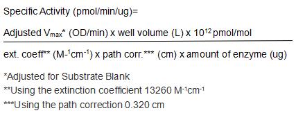
(The classical complement pathway plays a major role in innate immunity against infection. This pathway is triggered by C1, a multimolecular complex composed of the recognition protein C1q and two serine proteases, C1r and C1s. Following the C1q recognition, C1r is autoactivated, and in turn activates C1s, which cleaves C4 and C2, the C1 substrates. Both C1r and C1s activation involve cleavage of a specific Arg-Ile bond, converting single-chain proenzymes into active proteases of disulfide bond-linked chains (A and B). The full-length (amino acid residues 1-707) of mouse C1r was expressed which activity was measured by its ability to cleaves a thioester substrate Z-Lys-SBzl•HCl. The reaction was performed in 50 mM Tris, pH 7.5 (Assay Buffer), initiated by addition 50 uL of various concentrations of C1r (diluted by Assay Buffer) to 50 ul substrate mixture of 0.2mM Z-Lys-SBzl•HCl and 0.2 mM DTNB. The final well serves as a negative control with no C1r, replaced with 50 ul assay buffer. Then read in kinetic mode for 5 minutes at an absorbance of 405 nm. The specific activity of recombinant mouse C1r is > 500 pmol/min/ug.)
Bioactivity
(The classical complement pathway plays a major role in innate immunity against infection. This pathway is triggered by C1, a multimolecular complex composed of the recognition protein C1q and two serine proteases, C1r and C1s. Following the C1q recognition, C1r is autoactivated, and in turn activates C1s, which cleaves C4 and C2, the C1 substrates. Both C1r and C1s activation involve cleavage of a specific Arg-Ile bond, converting single-chain proenzymes into active proteases of disulfide bond-linked chains (A and B). The full-length (amino acid residues 1-707) of mouse C1r was expressed which activity was measured by its ability to cleaves a thioester substrate Z-Lys-SBzl•HCl. The reaction was performed in 50 mM Tris, pH 7.5 (Assay Buffer), initiated by addition 50 uL of various concentrations of C1r (diluted by Assay Buffer) to 50 ul substrate mixture of 0.2mM Z-Lys-SBzl•HCl and 0.2 mM DTNB. The final well serves as a negative control with no C1r, replaced with 50 ul assay buffer. Then read in kinetic mode for 5 minutes at an absorbance of 405 nm. The specific activity of recombinant mouse C1r is > 500 pmol/min/ug.)
Complement Component 1, R Subcomponent (C1r), Active Protein (Cat# AAA161831)
Full Name
Active Complement Component 1, R Subcomponent (C1r)
Gene Names
C1ra; C1r; mC1rA; AI132558
Applications
Activity Assay, Cell Culture
Purity
Greater than 90% by SDS-PAGE
Pricing
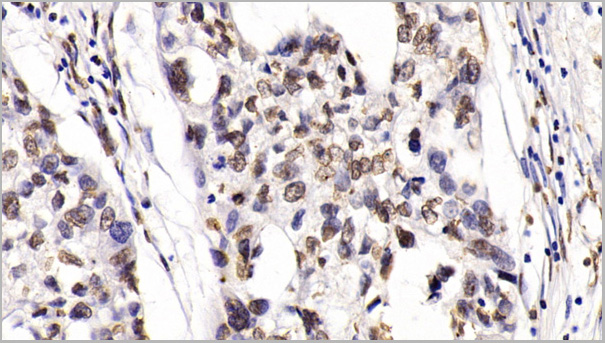
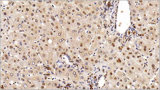
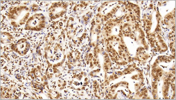
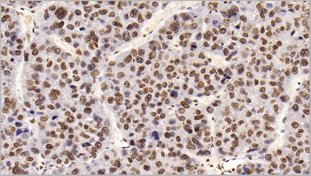
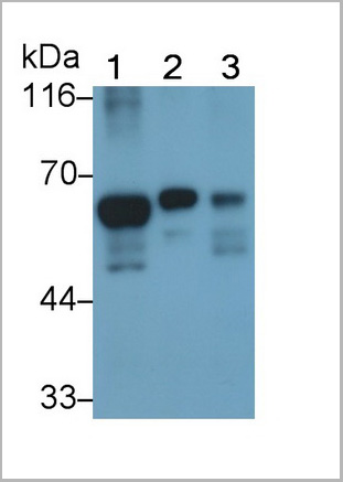 WB (Western Blot)
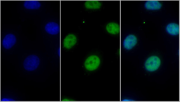
(Western Blot; Sample: Lane1: Human Lung lysate; Lane2: A549 cell lysate; Lane3: HepG2 cell lysate Primary Ab: 0.2ug/ml Mouse AntiHuman HDAC1 Antibody Second Ab: 0.2ug/mL HRPLinked Rabbit AntiMouse IgG Polyclonal Antibody (Catalog: SAA544Mu19))
WB (Western Blot)
(Western Blot; Sample: Lane1: Human Lung lysate; Lane2: A549 cell lysate; Lane3: HepG2 cell lysate Primary Ab: 0.2ug/ml Mouse AntiHuman HDAC1 Antibody Second Ab: 0.2ug/mL HRPLinked Rabbit AntiMouse IgG Polyclonal Antibody (Catalog: SAA544Mu19))
Histone Deacetylase 1 (HDAC1), Monoclonal Antibody (Cat# AAA21203)
Full Name
Monoclonal Antibody to Histone Deacetylase 1 (HDAC1)
Gene Names
HDAC1; HD1; RPD3; GON-10; RPD3L1
Reactivity
Human
Applications
Western Blot, Immunohistochemistry, Immunocytochemistry, Immunoprecipitation
Purity
Protein A + Protein G affinity chromatography
Pricing
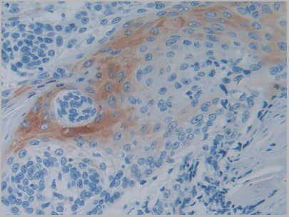
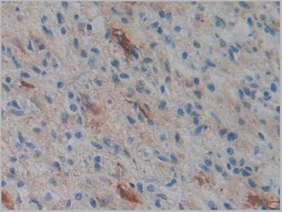
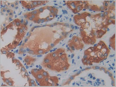
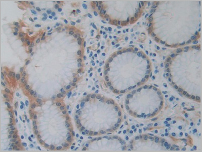
 WB (Western Blot)
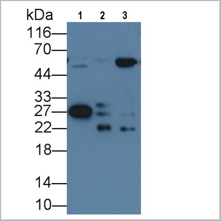
(Western Blot; Sample: Lane1: Human Lung lysate; Lane2: Porcine Skeletal muscle lysate; Lane3: Porcine Heart lysate Primary Ab: 2ug/ml Mouse AntiHuman Hsp27 Antibody Second Ab: 0.2ug/mL HRPLinked Caprine AntiMouse IgG Polyclonal Antibody (Catalog: SAA544Mu19))
WB (Western Blot)
(Western Blot; Sample: Lane1: Human Lung lysate; Lane2: Porcine Skeletal muscle lysate; Lane3: Porcine Heart lysate Primary Ab: 2ug/ml Mouse AntiHuman Hsp27 Antibody Second Ab: 0.2ug/mL HRPLinked Caprine AntiMouse IgG Polyclonal Antibody (Catalog: SAA544Mu19))
Heat Shock Protein 27 (Hsp27), Monoclonal Antibody (Cat# AAA21185)
Full Name
Monoclonal Antibody to Heat Shock Protein 27 (Hsp27)
Gene Names
HSPB1; CMT2F; HMN2B; HSP27; HSP28; Hsp25; SRP27; HS.76067
Reactivity
Human, Pig
Applications
Western Blot, Immunohistochemistry, Immunocytochemistry, Immunoprecipitation
Purity
Protein A + Protein G affinity chromatography
Pricing
 FCM/FACS (Flow Cytometry)
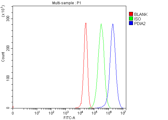
(Figure 2. Flow Cytometry analysis of 293T cells using anti-PDIP/PDIA2 antibody (AAA126295).Overlay histogram showing 293T cells stained with AAA126295 (Blue line). The cells were blocked with 10% normal goat serum. And then incubated with rabbit anti-PDIP/PDIA2 Antibody (AAA126295, 1 ug/1x10^6 cells) for 30 min at 20 degree C. DyLight488 conjugated goat anti-rabbit IgG was used as secondary antibody for 30 minutes at 20 degree C. Isotype control antibody (Green line) was rabbit IgG (1 ug/1x10^6) used under the same conditions. Unlabelled sample (Red line) was also used as a control.)
FCM/FACS (Flow Cytometry)
(Figure 2. Flow Cytometry analysis of 293T cells using anti-PDIP/PDIA2 antibody (AAA126295).Overlay histogram showing 293T cells stained with AAA126295 (Blue line). The cells were blocked with 10% normal goat serum. And then incubated with rabbit anti-PDIP/PDIA2 Antibody (AAA126295, 1 ug/1x10^6 cells) for 30 min at 20 degree C. DyLight488 conjugated goat anti-rabbit IgG was used as secondary antibody for 30 minutes at 20 degree C. Isotype control antibody (Green line) was rabbit IgG (1 ug/1x10^6) used under the same conditions. Unlabelled sample (Red line) was also used as a control.)
PDIP/PDIA2, Polyclonal Antibody (Cat# AAA126295)
Full Name
Anti-PDIP/PDIA2 Antibody Picoband
Gene Names
PDIA2; PDI; PDA2; PDIP; PDIR
Reactivity
Human
Applications
Flow Cytometry, Western Blot
Purity
Immunogen affinity purified.
Pricing
 FCM/FACS (Flow Cytometry)
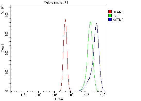
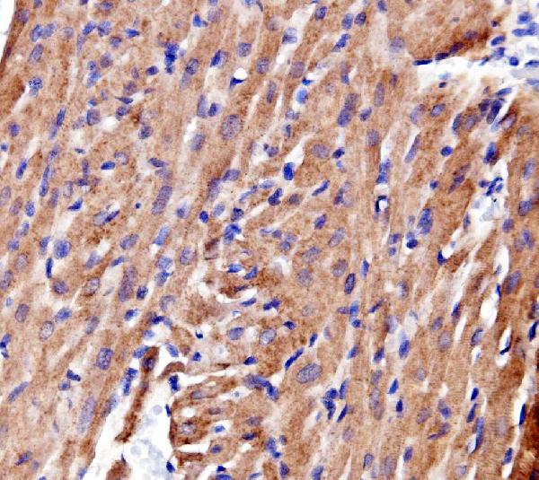
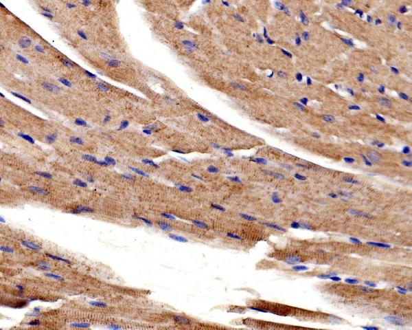
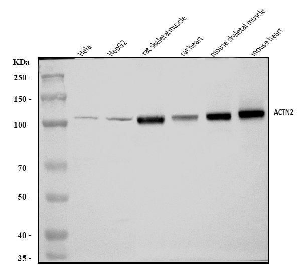
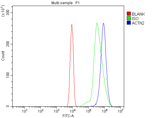
(Figure 5. Flow Cytometry analysis of ANA-1 cells using anti-ACTN2 antibody (AAA126331).Overlay histogram showing ANA-1 cells stained with AAA126331 (Blue line). The cells were blocked with 10% normal goat serum. And then incubated with rabbit anti-ACTN2 Antibody (AAA126331, 1 ug/1x10^6 cells) for 30 min at 20 degree C. DyLight488 conjugated goat anti-rabbit IgG was used as secondary antibody for 30 minutes at 20 degree C. Isotype control antibody (Green line) was rabbit IgG (1 ug/1x10^6) used under the same conditions. Unlabelled sample (Red line) was also used as a control.)
FCM/FACS (Flow Cytometry)
(Figure 5. Flow Cytometry analysis of ANA-1 cells using anti-ACTN2 antibody (AAA126331).Overlay histogram showing ANA-1 cells stained with AAA126331 (Blue line). The cells were blocked with 10% normal goat serum. And then incubated with rabbit anti-ACTN2 Antibody (AAA126331, 1 ug/1x10^6 cells) for 30 min at 20 degree C. DyLight488 conjugated goat anti-rabbit IgG was used as secondary antibody for 30 minutes at 20 degree C. Isotype control antibody (Green line) was rabbit IgG (1 ug/1x10^6) used under the same conditions. Unlabelled sample (Red line) was also used as a control.)
ACTN2, Polyclonal Antibody (Cat# AAA126331)
Full Name
Anti-ACTN2 Antibody Picoband
Gene Names
ACTN2; CMD1AA
Reactivity
Human, Mouse, Rat
Applications
Flow Cytometry, Immunohistochemistry, Western Blot
Purity
Immunogen affinity purified.
Pricing
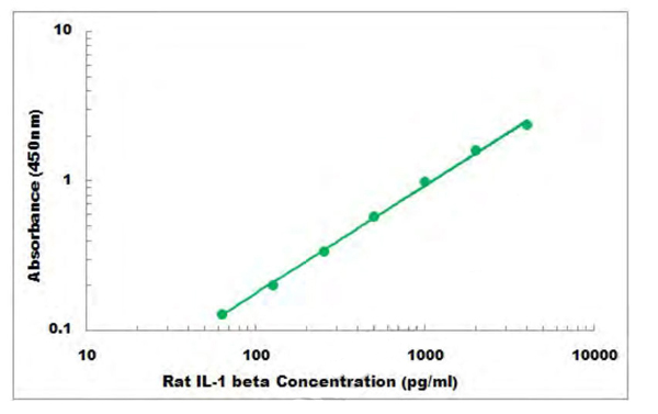 Standard Curve (Sample)
IL-1 beta, ELISA Kit (Cat# AAA105950)
Full Name
Rat IL-1 beta ELISA Kit
Reactivity
Rat
Applications
Sandwich ELISA
Pricing
 SDS-PAGE
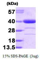
Alcohol dehydrogenase, Recombinant Protein (Cat# AAA48613)
Full Name
Alcohol dehydrogenase, 1-325aa, Human, E Coli
Gene Names
AKR1A1; ALR; ARM; DD3; ALDR1; HEL-S-6
Applications
SDS-PAGE
Purity
> 90% by SDS-PAGE
Pricing
 Application Data
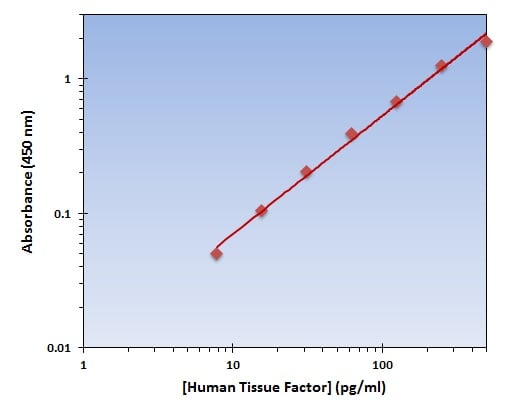
(The OmniKine Human Tissue Factor ELISA Kit allows for the detection and quantification of endogenous levels of natural and/or recombinant Human Tissue Factor proteins within the range of 8-500 pg/ml.)
Application Data
(The OmniKine Human Tissue Factor ELISA Kit allows for the detection and quantification of endogenous levels of natural and/or recombinant Human Tissue Factor proteins within the range of 8-500 pg/ml.)
Tissue Factor, ELISA Kit (Cat# AAA315796)
Full Name
Tissue Factor (Human) OmniKine ELISA Kit
Gene Names
F3; TF; TFA; CD142
Reactivity
Human
Pricing
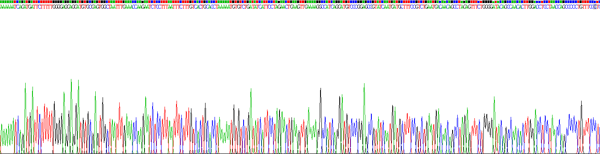
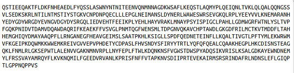
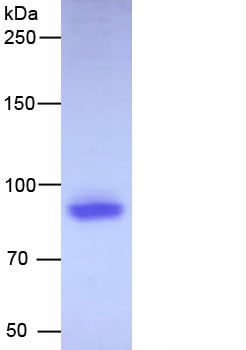 SDS-PAGE
(Figure. SDS-PAGE)
SDS-PAGE
(Figure. SDS-PAGE)
Angiotensin I Converting Enzyme 2 (ACE2), Recombinant Protein (Cat# AAA150696)
Full Name
Recombinant Angiotensin I Converting Enzyme 2 (ACE2)
Gene Names
ACE2; ACEH
Applications
Western Blot
Purity
>90%
Pricing
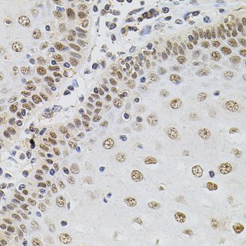
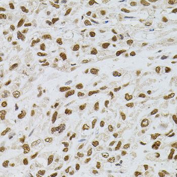
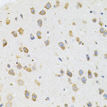
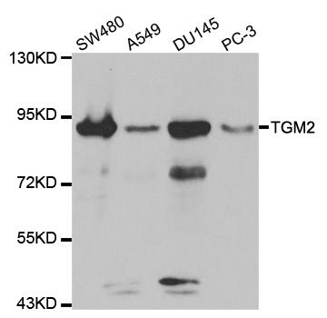
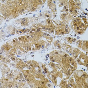 IHC (Immunohistochemistry)
(Immunohistochemistry of paraffin-embedded human stomach using TGM2 antibody (40x lens).)
IHC (Immunohistochemistry)
(Immunohistochemistry of paraffin-embedded human stomach using TGM2 antibody (40x lens).)
TGM2, Polyclonal Antibody (Cat# AAA37335)
Full Name
TGM2 Polyclonal Antibody
Gene Names
TGM2; TG2; TGC; GNAH; HEL-S-45; G-ALPHA-h
Reactivity
Human, Mouse
Applications
Immunohistochemistry, Western Blot
Purity
Affinity Purification
Pricing
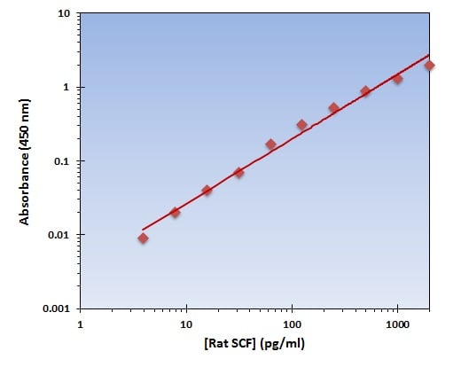 Application Data
(The OmniKine Rat SCF ELISA Kit allows for the detection and quantification of endogenous levels of natural and/or recombinant Rat SCF proteins within the range of 16-2000 pg/ml.)
Application Data
(The OmniKine Rat SCF ELISA Kit allows for the detection and quantification of endogenous levels of natural and/or recombinant Rat SCF proteins within the range of 16-2000 pg/ml.)
SCF, ELISA Kit (Cat# AAA315703)
Full Name
SCF (Rat) OmniKine ELISA Kit
Gene Names
Kitlg; Mgf; SCF; Kitl
Reactivity
Rat
Pricing
 SDS-PAGE
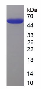
(SDS-PAGE)
SDS-PAGE
(SDS-PAGE)
Neurofilament, Light Polypeptide (NEFL), Recombinant Protein (Cat# AAA152354)
Full Name
Recombinant Neurofilament, Light Polypeptide (NEFL)
Gene Names
Nefl; Nfl; NF-L; NF68; CMT2E; AI847934
Applications
Western Blot
Purity
Greater than 95% by SDS-PAGE
Pricing
 Application Data
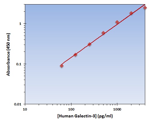
(The OmniKine Human Galectin-3 ELISA Kit allows for the detection and quantification of endogenous levels of natural and/or recombinant Human Galectin-3 proteins within the range of 63-4000 pg/ml.)
Application Data
(The OmniKine Human Galectin-3 ELISA Kit allows for the detection and quantification of endogenous levels of natural and/or recombinant Human Galectin-3 proteins within the range of 63-4000 pg/ml.)
Galectin-3, ELISA Kit (Cat# AAA315810)
Full Name
Galectin-3 (Human) OmniKine ELISA Kit
Gene Names
LGALS3; L31; GAL3; MAC2; CBP35; GALBP; GALIG; LGALS2
Reactivity
Human
Pricing
 Amino Acid Info
(Amino Acid info)
Amino Acid Info
(Amino Acid info)
Annexin A2 (ANXA2), Recombinant Protein (Cat# AAA153497)
Full Name
Recombinant Annexin A2 (ANXA2)
Gene Names
Anxa2; Cal1h; AW215814
Applications
Western Blot
Purity
Greater than 90% by SDS-PAGE
Pricing
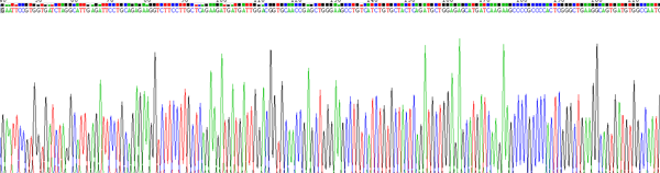
 Amino Acid Info
Pyruvate kinase isozymes M2 (PKM2), Recombinant Protein (Cat# AAA153363)
Full Name
Recombinant Pyruvate kinase isozymes M2 (PKM2)
Gene Names
Pkm; Pk3; Pk-2; Pk-3; Pkm2; AA414905; AL024370; AL024424
Applications
Western Blot
Purity
Greater than 97% by SDS-PAGE
Pricing
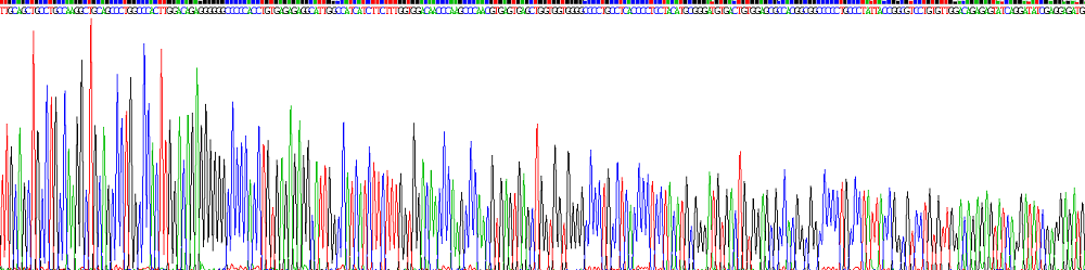
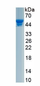 SDS-PAGE
(Figure. SDS-PAGE)
SDS-PAGE
(Figure. SDS-PAGE)
Amine Oxidase, Copper Containing 3 (AOC3), Recombinant Protein (Cat# AAA161993)
Full Name
Recombinant Amine Oxidase, Copper Containing 3 (AOC3)
Gene Names
Aoc3; SSAO; VAP1
Applications
Western Blot
Purity
Greater than 90% by SDS-PAGE
Pricing
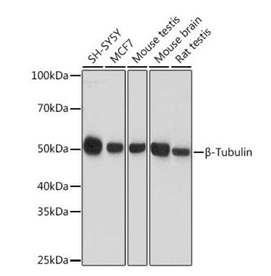 WB (Western Blot)
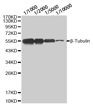
(Western blot analysis of extracts of various cell lines, using ?-Tubulin antibody (AAA36659) at 1:10000 dilution. Secondary antibody: HRP Goat Anti-Rabbit IgG (H+L) at 1:10000 dilution.Lysates/proteins: 25ug per lane.Blocking buffer: 3% nonfat dry milk in TBST.Detection: ECL Basic Kit.Exposure time: 10s.)
WB (Western Blot)
(Western blot analysis of extracts of various cell lines, using ?-Tubulin antibody (AAA36659) at 1:10000 dilution. Secondary antibody: HRP Goat Anti-Rabbit IgG (H+L) at 1:10000 dilution.Lysates/proteins: 25ug per lane.Blocking buffer: 3% nonfat dry milk in TBST.Detection: ECL Basic Kit.Exposure time: 10s.)
beta-Tubulin, Antibody (Cat# AAA36659)
Full Name
beta-Tubulin Polyclonal Antibody
Gene Names
TUBB3; CDCBM; FEOM3; TUBB4; CDCBM1; CFEOM3; beta-4; CFEOM3A
Applications
Immunohistochemistry, Western Blot
Purity
Affinity purification
Pricing
 WB (Western Blot)
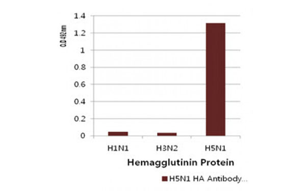
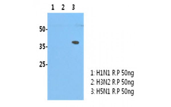
(H5N1/HA1 recombinant protein (50 ng) was resolved by SDS-PAGE, transferred to PVDF membrane and probed with H5N1/HA1 antibody (1:3000). Proteins were visualized using a goat anti-mouse secondary antibody conjugated to HRP and an ECL detection system.)
WB (Western Blot)
(H5N1/HA1 recombinant protein (50 ng) was resolved by SDS-PAGE, transferred to PVDF membrane and probed with H5N1/HA1 antibody (1:3000). Proteins were visualized using a goat anti-mouse secondary antibody conjugated to HRP and an ECL detection system.)
Influenza A H5N1 HA1, Monoclonal Antibody (Cat# AAA107166)
Full Name
Influenza A H5N1 HA1 antibody
Applications
Western Blot
Purity
Influenza A HA1 antibody was purified by protein G affinity chromatography
Pricing
 ELISA
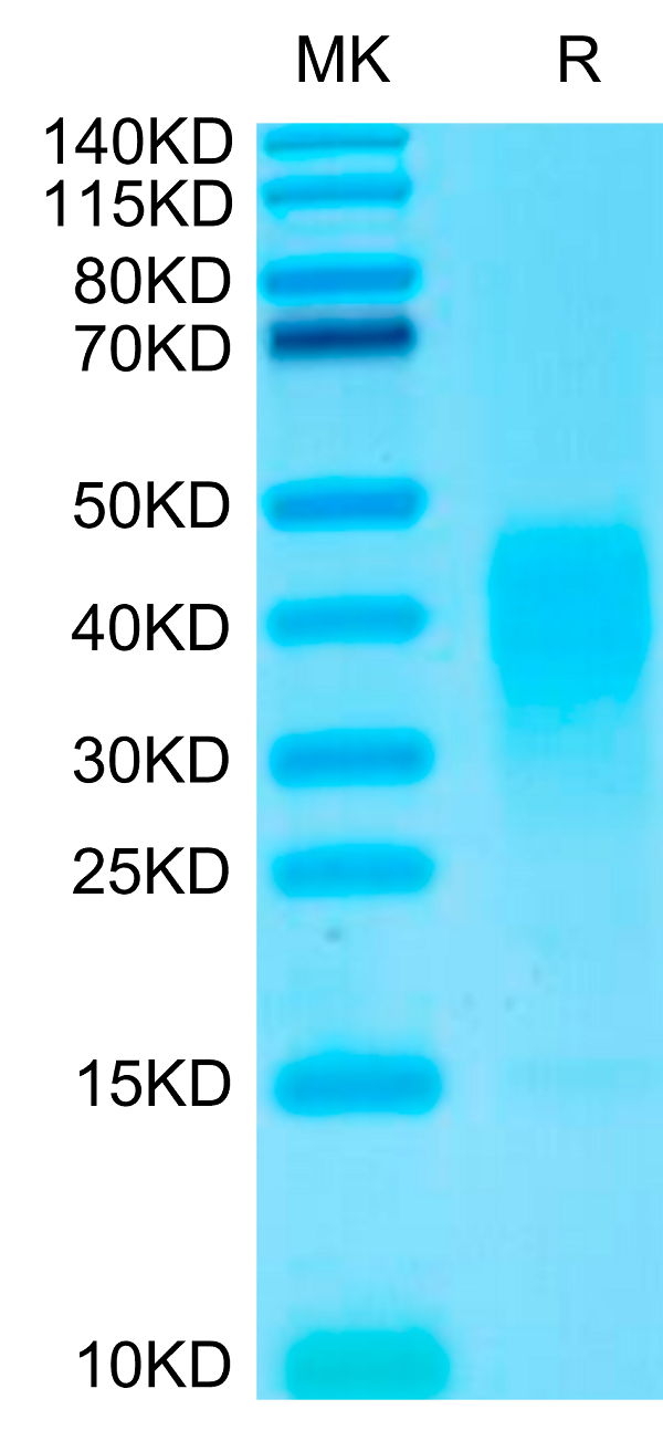
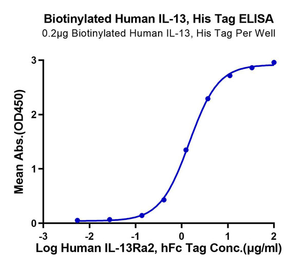
(Biotinylated Human IL-13 Protein ELISA DataImmobilized Biotinylated Human IL-13, His Tag at 2?g/ml (100?l/well) on the streptavidin (5?g/ml) precoated plate. Dose response curve for Human IL-13Ra2, hFc Tag with the EC50 of 1.4?g/ml determined by ELISA.)
ELISA
(Biotinylated Human IL-13 Protein ELISA DataImmobilized Biotinylated Human IL-13, His Tag at 2?g/ml (100?l/well) on the streptavidin (5?g/ml) precoated plate. Dose response curve for Human IL-13Ra2, hFc Tag with the EC50 of 1.4?g/ml determined by ELISA.)
IL-13, Recombinant Protein (Cat# AAA76178)
Full Name
Biotinylated Human IL-13 Protein Recombinant
Purity
>95%, as determined by SDS-PAGE and HPLC
Pricing
 Bioactivity
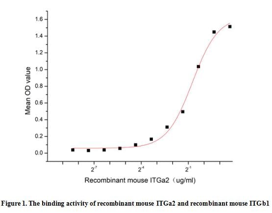
(Integrin Alpha 2 (ITGa2), also known as CD49b, is one of twelve integrin family alpha subunits which is pivotal to sustain the integrity of the cytoskeletal-extracellular matrix linkage among all cell adhesion receptors. Some research had shown that ITGa2 is a cell transmembrane receptor that assists the adhesion of other several cells to the extracellular matrix. In addition, changes of ITGa2 expression affect the immune microenvironment and immunogenicity of tumors. Integrin Beta 1 (ITGb1) has been identified as an interactor of ITGa2, thus a functional binding ELISA assay was conducted to detect the interaction of recombinant mouse ITGa2 and recombinant mouse ITGb1. Briefly, ITGa2 was diluted serially in PBS with 0.01% BSA (pH 7.4). Duplicate samples of 100 ul were then transferred to ITGb1-coated microtiter wells and incubated for 1h at 37 degree C. Wells were washed with PBST and incubated for 1h with anti-ITGa2 pAb, then aspirated and washed 3 times. After incubation with HRP labelled secondary antibody for 1h at 37 degree C, wells were aspirated and washed 5 times. With the addition of substrate solution, wells were incubated 15-25 minutes at 37 degree C. Finally, add 50 uL stop solution to the wells and read at 450/630 nm immediately. The binding activity of recombinant mouse ITGa2 and recombinant mouse ITGb1 was shown in Figure 1, the EC50 for this effect is 0.6 ug/mL.)
Bioactivity
(Integrin Alpha 2 (ITGa2), also known as CD49b, is one of twelve integrin family alpha subunits which is pivotal to sustain the integrity of the cytoskeletal-extracellular matrix linkage among all cell adhesion receptors. Some research had shown that ITGa2 is a cell transmembrane receptor that assists the adhesion of other several cells to the extracellular matrix. In addition, changes of ITGa2 expression affect the immune microenvironment and immunogenicity of tumors. Integrin Beta 1 (ITGb1) has been identified as an interactor of ITGa2, thus a functional binding ELISA assay was conducted to detect the interaction of recombinant mouse ITGa2 and recombinant mouse ITGb1. Briefly, ITGa2 was diluted serially in PBS with 0.01% BSA (pH 7.4). Duplicate samples of 100 ul were then transferred to ITGb1-coated microtiter wells and incubated for 1h at 37 degree C. Wells were washed with PBST and incubated for 1h with anti-ITGa2 pAb, then aspirated and washed 3 times. After incubation with HRP labelled secondary antibody for 1h at 37 degree C, wells were aspirated and washed 5 times. With the addition of substrate solution, wells were incubated 15-25 minutes at 37 degree C. Finally, add 50 uL stop solution to the wells and read at 450/630 nm immediately. The binding activity of recombinant mouse ITGa2 and recombinant mouse ITGb1 was shown in Figure 1, the EC50 for this effect is 0.6 ug/mL.)
Integrin Alpha 2 (ITGa2), Active Protein (Cat# AAA161836)
Full Name
Active Integrin Alpha 2 (ITGa2)
Gene Names
Itga2; DX5; CD49B
Applications
Activity Assay, Cell Culture
Purity
Greater than 95% by SDS-PAGE
Pricing
 WB (Western Blot)
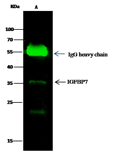
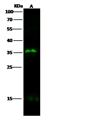
(Anti-IGFBP7 rabbit monoclonal antibody at 1:500 dilutionLane A: THP1 Whole Cell LysateLysates/proteins at 30 ug per lane.SecondaryGoat Anti-Rabbit IgG H&L (Dylight800) at 1/10000 dilution.Developed using the Odyssey technique.Performed under reducing conditions.Predicted band size:29 kDaObserved band size:35 kDa)
WB (Western Blot)
(Anti-IGFBP7 rabbit monoclonal antibody at 1:500 dilutionLane A: THP1 Whole Cell LysateLysates/proteins at 30 ug per lane.SecondaryGoat Anti-Rabbit IgG H&L (Dylight800) at 1/10000 dilution.Developed using the Odyssey technique.Performed under reducing conditions.Predicted band size:29 kDaObserved band size:35 kDa)
IGFBP7, Monoclonal Antibody (Cat# AAA255634)
Full Name
Recombinant Anti-IGFBP7 Antibody, Rabbit Monoclonal
Gene Names
IGFBP7; AGM; PSF; TAF; FSTL2; IBP-7; MAC25; IGFBP-7; RAMSVPS; IGFBP-7v; IGFBPRP1
Reactivity
Human
Applications
Immunoprecipitation, Western Blot
Purity
Protein A
Pricing
 Application Data
S100 Calcium Binding Protein B (S100B), Active Protein (Cat# AAA148146)
Full Name
Active S100 Calcium Binding Protein B (S100B)
Gene Names
S100b; S100P
Reactivity
Rat
Applications
Activity Assay, Cell Culture
Purity
> 95%
Pricing
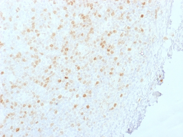
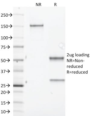 SDS-PAGE
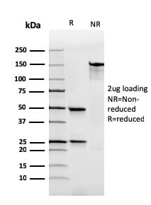
(SDS-PAGE Analysis Purified Aurora B Mouse Monoclonal Antibody (AURKB/1845). Confirmation of Integrity and Purity of Antibody.)
SDS-PAGE
(SDS-PAGE Analysis Purified Aurora B Mouse Monoclonal Antibody (AURKB/1845). Confirmation of Integrity and Purity of Antibody.)
Aurora B, Monoclonal Antibody (Cat# AAA215287)
Full Name
Aurora B (Proliferation Marker)
Gene Names
AURKB; AIK2; AIM1; ARK2; AurB; IPL1; STK5; AIM-1; ARK-2; STK-1; STK12; PPP1R48; aurkb-sv1; aurkb-sv2
Reactivity
Human
Applications
Immunohistochemistry
Purity
Purified Ab with BSA and Azide at 200ug/ml OR Purified Ab WITHOUT BSA and Azide at 1.0mg/ml
Pricing
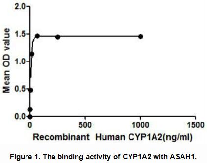
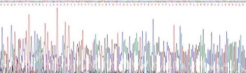 Application Data
Cytochrome P450 1A2 (CYP1A2), Active Protein (Cat# AAA148207)
Full Name
Active Cytochrome P450 1A2 (CYP1A2)
Gene Names
CYP1A2; CP12; CYPIA2; P3-450; P450(PA)
Reactivity
Human
Applications
Activity Assay, Cell Culture
Purity
> 95%
Pricing
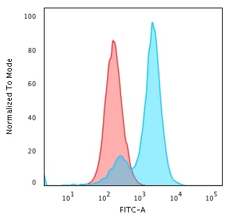 FCM/FACS (Flow Cytometry)
(Flow Cytometric Analysis of PFA-fixed HeLa cells using Collagen VII Mouse Monoclonal Antibody (LH7.2) followed by Goat anti-Mouse IgG-CF488 (Blue); Isotype Control (Red))
FCM/FACS (Flow Cytometry)
(Flow Cytometric Analysis of PFA-fixed HeLa cells using Collagen VII Mouse Monoclonal Antibody (LH7.2) followed by Goat anti-Mouse IgG-CF488 (Blue); Isotype Control (Red))
Collagen VII, Monoclonal Antibody (Cat# AAA214899)
Full Name
Collagen VII
Gene Names
COL7A1; EBD1; EBR1; EBDCT; NDNC8
Reactivity
Human, Sheep, Goat, Horse, Guinea Pig, Cow
Applications
Immunohistochemistry, Immunofluorescence, Flow Cytometry, Western Blot
Purity
Purified Ab with BSA and Azide at 200ug/ml OR Purified Ab WITHOUT BSA and Azide at 1.0mg/ml
Pricing
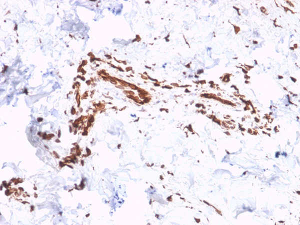
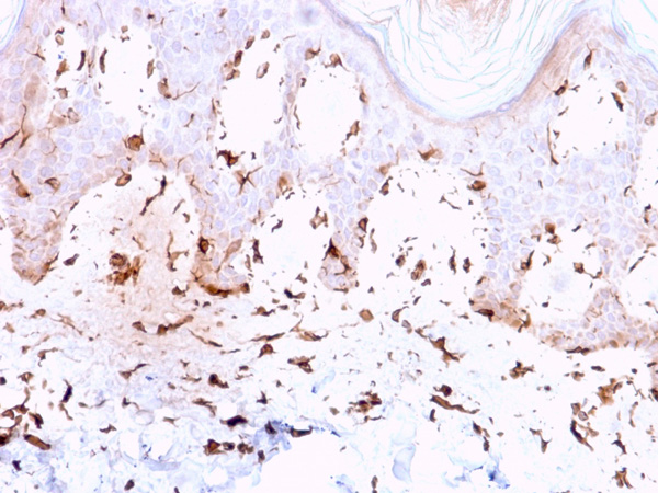
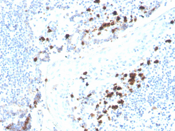 IHC (Immunohiostchemistry)
(Formalin-fixed, paraffin-embedded human tonsil stained with IgA Recombinant Mouse Monoclonal Antibody (rHISA43).)
IHC (Immunohiostchemistry)
(Formalin-fixed, paraffin-embedded human tonsil stained with IgA Recombinant Mouse Monoclonal Antibody (rHISA43).)
Immunoglobulin Alpha (IgA) Heavy Chain, Antibody (Cat# AAA215019)
Full Name
Immunoglobulin Alpha (IgA) Heavy Chain
Gene Names
IGHA1; IgA1
Reactivity
Human
Applications
Immunohistochemistry
Purity
Purified Ab with BSA and Azide at 200ug/ml OR Purified Ab WITHOUT BSA and Azide at 1.0mg/ml
Pricing
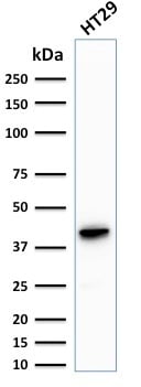
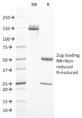
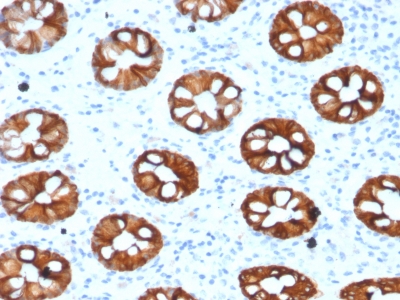
 Application Data
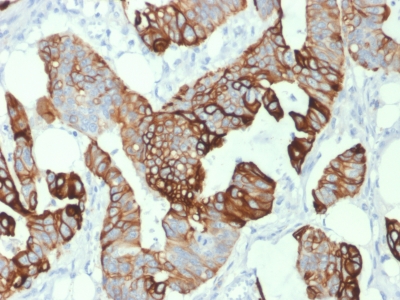
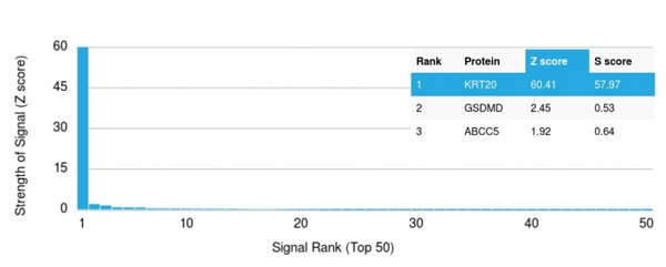
(Analysis of Protein Array containing more than 19,000 full-length human proteins using Cytokeratin 20 (KRT20) Mouse Monoclonal Antibody (KRT20/1993). Z- and S- Score: The Z-score represents the strength of a signal that a monoclonal antibody (MAb) (in combination with a fluorescently-tagged anti-IgG secondary antibody) produces when binding to a particular protein on the HuProtTM array. Z-scores are described in units of standard deviations (SD's) above the mean value of all signals generated on that array. If targets on HuProtTM are arranged in descending order of the Z-score, the S-score is the difference (also in units of SD's) between the Z-score. S-score therefore represents the relative target specificity of a MAb to its intended target. A MAb is considered to specific to its intended target, if the MAb has an S-score of at least 2.5. For example, if a MAb binds to protein X with a Z-score of 43 and to protein Y with a Z-score of 14, then the S-score for the binding of that MAb to protein X is equal to 29.)
Application Data
(Analysis of Protein Array containing more than 19,000 full-length human proteins using Cytokeratin 20 (KRT20) Mouse Monoclonal Antibody (KRT20/1993). Z- and S- Score: The Z-score represents the strength of a signal that a monoclonal antibody (MAb) (in combination with a fluorescently-tagged anti-IgG secondary antibody) produces when binding to a particular protein on the HuProtTM array. Z-scores are described in units of standard deviations (SD's) above the mean value of all signals generated on that array. If targets on HuProtTM are arranged in descending order of the Z-score, the S-score is the difference (also in units of SD's) between the Z-score. S-score therefore represents the relative target specificity of a MAb to its intended target. A MAb is considered to specific to its intended target, if the MAb has an S-score of at least 2.5. For example, if a MAb binds to protein X with a Z-score of 43 and to protein Y with a Z-score of 14, then the S-score for the binding of that MAb to protein X is equal to 29.)
Cytokeratin 20 (KRT20), Monoclonal Antibody (Cat# AAA214795)
Full Name
Cytokeratin 20 (KRT20) (Colorectal Epithelial Marker)
Gene Names
KRT20; K20; CD20; CK20; CK-20; KRT21
Reactivity
Human. Others not known.
Applications
Immunohistochemistry, Western Blot
Purity
Purified Ab with BSA and Azide at 200ug/ml OR Purified Ab WITHOUT BSA and Azide at 1.0mg/ml
Pricing
 Bioactivity
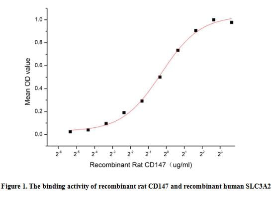
(Extracellular matrix metalloproteinase (MMP) inducer (EMMPRIN), also known as basigin and CD147, is a 4466 kDa, variably N and Oglycosylated, type I transmembrane protein that belongs to the immunoglobulin superfamily. EMMPRIN is 269 amino acids (aa) in length and contains a 24 aa signal sequence, a 183 aa extracellular domain (ECD), a 21 aa transmembrane (TM) segment and a 41 aa cytoplasmic tail. The ECD contains one C2type and one Vtype Iglike domain. EMMPRIN is expressed in areas of tissue remodeling, including endometrium, placenta, skin, and regions undergoing angiogenesis. It is also expressed on cells with high metabolic activity, such as lymphoblasts, macrophages and particularly tumor cells. A functional ELISA assay was conducted to detect the interaction of recombinant rat EMMPRIN/CD147 and recombinant human SLC3A2. Briefly, CD147 was diluted serially in PBS with 0.01% BSA (pH 7.4). Duplicate samples of 100 ul were then transferred to SLC3A2-coated microtiter wells and incubated for 1h at 37 degree C. Wells were washed with PBST and incubated for 1h with anti-CD147 pAb, then aspirated and washed 3 times. After incubation with HRP labelled secondary antibody for 1h at 37 degree C, wells were aspirated and washed 5 times. With the addition of substrate solution, wells were incubated 15-25 minutes at 37 degree C. Finally, add 50 uL stop solution to the wells and read at 450/630nm immediately. The binding activity of recombinant rat CD147 and recombinant human SLC3A2 was shown in Figure 1, the EC50 for this effect is 0.82 ug/mL.)
Bioactivity
(Extracellular matrix metalloproteinase (MMP) inducer (EMMPRIN), also known as basigin and CD147, is a 4466 kDa, variably N and Oglycosylated, type I transmembrane protein that belongs to the immunoglobulin superfamily. EMMPRIN is 269 amino acids (aa) in length and contains a 24 aa signal sequence, a 183 aa extracellular domain (ECD), a 21 aa transmembrane (TM) segment and a 41 aa cytoplasmic tail. The ECD contains one C2type and one Vtype Iglike domain. EMMPRIN is expressed in areas of tissue remodeling, including endometrium, placenta, skin, and regions undergoing angiogenesis. It is also expressed on cells with high metabolic activity, such as lymphoblasts, macrophages and particularly tumor cells. A functional ELISA assay was conducted to detect the interaction of recombinant rat EMMPRIN/CD147 and recombinant human SLC3A2. Briefly, CD147 was diluted serially in PBS with 0.01% BSA (pH 7.4). Duplicate samples of 100 ul were then transferred to SLC3A2-coated microtiter wells and incubated for 1h at 37 degree C. Wells were washed with PBST and incubated for 1h with anti-CD147 pAb, then aspirated and washed 3 times. After incubation with HRP labelled secondary antibody for 1h at 37 degree C, wells were aspirated and washed 5 times. With the addition of substrate solution, wells were incubated 15-25 minutes at 37 degree C. Finally, add 50 uL stop solution to the wells and read at 450/630nm immediately. The binding activity of recombinant rat CD147 and recombinant human SLC3A2 was shown in Figure 1, the EC50 for this effect is 0.82 ug/mL.)
Cluster Of Differentiation 147 (CD147), Active Protein (Cat# AAA161852)
Full Name
Active Cluster Of Differentiation 147 (CD147)
Gene Names
Bsg; CE9; HT7; 5A11; Ox47R; EMMPRIN; basignin
Applications
Activity Assay, Cell Culture
Purity
Greater than 95% by SDS-PAGE
Pricing
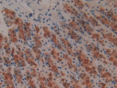

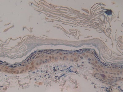 IHC (Immunohistochemisry)
(DAB staining on IHCP;Sample: Rat Skin Tissue; Primary Ab: 30ug/ml Mouse AntiRat IL1b AntibodySecond Ab: 2ug/mL HRPLinked Caprine AntiMouse IgG Polyclonal Antibody(Catalog: SAA544Mu19))
IHC (Immunohistochemisry)
(DAB staining on IHCP;Sample: Rat Skin Tissue; Primary Ab: 30ug/ml Mouse AntiRat IL1b AntibodySecond Ab: 2ug/mL HRPLinked Caprine AntiMouse IgG Polyclonal Antibody(Catalog: SAA544Mu19))
Interleukin 1 Beta (IL1b), Monoclonal Antibody (Cat# AAA151569)
Full Name
Monoclonal Antibody to Interleukin 1 Beta (IL1b)
Reactivity
Rat
Applications
Immunoprecipitation, Immunocytochemistry, Immunohistochemistry, Western Blot
Purity
Protein A + Protein G affinity chromatography
Pricing
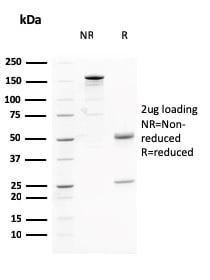
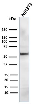
 Application Data
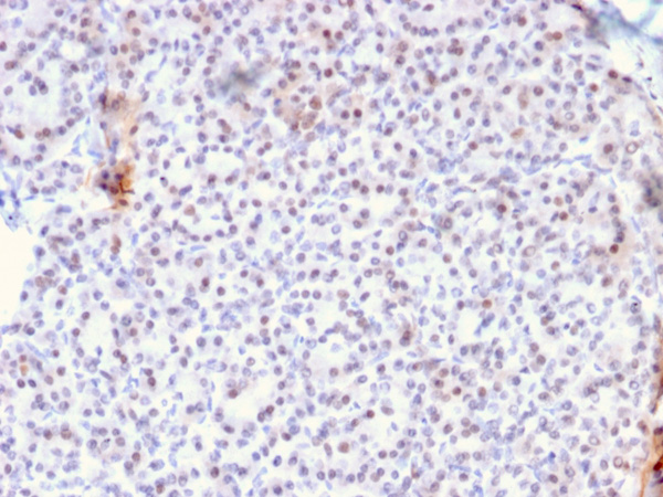
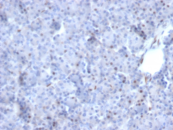
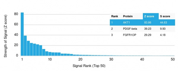
(Analysis of Protein Array containing more than 19,000 full-length human proteins using AKT1 Mouse Monoclonal Antibody (AKT1/2784).Z- and S- Score: The Z-score represents the strength of a signal that a monoclonal antibody (MAb) (in combination with a fluorescently-tagged anti-IgG secondary antibody) produces when binding to a particular protein on the HuProtTM array. Z-scores are described in units of standard deviations (SD's) above the mean value of all signals generated on that array. If targets on HuProtTM are arranged in descending order of the Z-score, the S-score is the difference (also in units of SD's) between the Z-score. S-score therefore represents the relative target specificity of a MAb to its intended target. A MAb is considered to specific to its intended target, if the MAb has an S-score of at least 2.5. For example, if a MAb binds to protein X with a Z-score of 43 and to protein Y with a Z-score of 14, then the S-score for the binding of that MAb to protein X is equal to 29.)
Application Data
(Analysis of Protein Array containing more than 19,000 full-length human proteins using AKT1 Mouse Monoclonal Antibody (AKT1/2784).Z- and S- Score: The Z-score represents the strength of a signal that a monoclonal antibody (MAb) (in combination with a fluorescently-tagged anti-IgG secondary antibody) produces when binding to a particular protein on the HuProtTM array. Z-scores are described in units of standard deviations (SD's) above the mean value of all signals generated on that array. If targets on HuProtTM are arranged in descending order of the Z-score, the S-score is the difference (also in units of SD's) between the Z-score. S-score therefore represents the relative target specificity of a MAb to its intended target. A MAb is considered to specific to its intended target, if the MAb has an S-score of at least 2.5. For example, if a MAb binds to protein X with a Z-score of 43 and to protein Y with a Z-score of 14, then the S-score for the binding of that MAb to protein X is equal to 29.)
AKT1, Monoclonal Antibody (Cat# AAA214951)
Full Name
AKT1 (Prognostic Marker for Neuroendocrine Tumors)
Gene Names
AKT1; AKT; PKB; RAC; CWS6; PRKBA; PKB-ALPHA; RAC-ALPHA
Reactivity
Human, Mouse
Applications
Immunohistochemistry, Western Blot
Purity
Purified Ab with BSA and Azide at 200ug/ml OR Purified Ab WITHOUT BSA and Azide at 1.0mg/ml
Pricing
 FCM/FACS (Flow Cytometry)
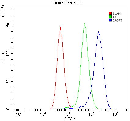
(Figure 2. Flow Cytometry analysis of mouse spleen tissues using anti-Caspase-9 p35/Casp9 antibody (AAA125515).Overlay histogram showing mouse spleen tissues stained with AAA125515 (Blue line). The tissues were blocked with 10% normal goat serum. And then incubated with rabbit anti-Caspase-9 p35/Casp9 Antibody (AAA125515, 1μg/1x106 cells) for 30 min at 20 degree C. DyLight®488 conjugated goat anti-rabbit IgG (5-10μg/1x106 cells) was used as secondary antibody for 30 minutes at 20 degree C. Isotype control antibody (Green line) was rabbit IgG (1μg/1x106) used under the same conditions. Unlabelled sample (Red line) was also used as a control.)
FCM/FACS (Flow Cytometry)
(Figure 2. Flow Cytometry analysis of mouse spleen tissues using anti-Caspase-9 p35/Casp9 antibody (AAA125515).Overlay histogram showing mouse spleen tissues stained with AAA125515 (Blue line). The tissues were blocked with 10% normal goat serum. And then incubated with rabbit anti-Caspase-9 p35/Casp9 Antibody (AAA125515, 1μg/1x106 cells) for 30 min at 20 degree C. DyLight®488 conjugated goat anti-rabbit IgG (5-10μg/1x106 cells) was used as secondary antibody for 30 minutes at 20 degree C. Isotype control antibody (Green line) was rabbit IgG (1μg/1x106) used under the same conditions. Unlabelled sample (Red line) was also used as a control.)
Caspase-9 p35/Casp9, Polyclonal Antibody (Cat# AAA125515)
Full Name
Anti-Caspase-9 p35/Casp9 Antibody
Gene Names
Casp9; Mch6; APAF-3; CASP-9; AI115399; AW493809; ICE-LAP6; Caspase-9
Reactivity
Mouse, Rat
Applications
Direct ELISA, Flow Cytometry, Western Blot
Purity
Immunogen affinity purified.
Pricing
 WB (Western Blot)
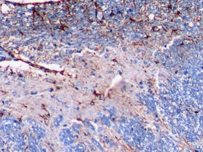
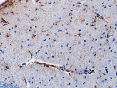
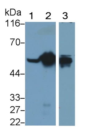
(Western Blot; Sample: Lane1: Rat Cerebrum lysate; Lane2: Rat Cerebellum lysate; Lane3: U87MG cell lysate Primary Ab: 0.1ug/ml Mouse AntiRat GFAP Antibody Second Ab: 0.2ug/mL HRPLinked Caprine AntiMouse IgG Polyclonal Antibody (Catalog: SAA544Mu19))
WB (Western Blot)
(Western Blot; Sample: Lane1: Rat Cerebrum lysate; Lane2: Rat Cerebellum lysate; Lane3: U87MG cell lysate Primary Ab: 0.1ug/ml Mouse AntiRat GFAP Antibody Second Ab: 0.2ug/mL HRPLinked Caprine AntiMouse IgG Polyclonal Antibody (Catalog: SAA544Mu19))
Glial Fibrillary Acidic Protein (GFAP), Monoclonal Antibody (Cat# AAA151456)
Full Name
Monoclonal Antibody to Glial Fibrillary Acidic Protein (GFAP)
Reactivity
Human, Rat
Applications
Immunoprecipitation, Immunocytochemistry, Immunohistochemistry, Western Blot
Purity
Protein A + Protein G affinity chromatography
Pricing
 Application Data
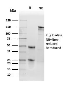
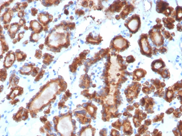
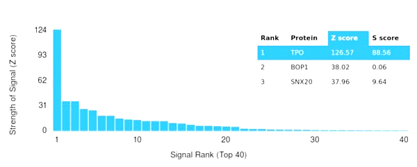
(Analysis of Protein Array containing more than 19,000 full-length human proteins using Thyroid Peroxidase Mouse Monoclonal Antibody (TPO/3698). Z- and S- Score: The Z-score represents the strength of a signal that a monoclonal antibody (MAb) (in combination with a fluorescently-tagged anti-IgG secondary antibody) produces when binding to a particular protein on the HuProtTM array. Z-scores are described in units of standard deviations (SD's) above the mean value of all signals generated on that array. If targets on HuProtTM are arranged in descending order of the Z-score, the S-score is the difference (also in units of SD's) between the Z-score. S-score therefore represents the relative target specificity of a MAb to its intended target. A MAb is considered to specific to its intended target, if the MAb has an S-score of at least 2.5. For example, if a MAb binds to protein X with a Z-score of 43 and to protein Y with a Z-score of 14, then the S-score for the binding of that MAb to protein X is equal to 29.)
Application Data
(Analysis of Protein Array containing more than 19,000 full-length human proteins using Thyroid Peroxidase Mouse Monoclonal Antibody (TPO/3698). Z- and S- Score: The Z-score represents the strength of a signal that a monoclonal antibody (MAb) (in combination with a fluorescently-tagged anti-IgG secondary antibody) produces when binding to a particular protein on the HuProtTM array. Z-scores are described in units of standard deviations (SD's) above the mean value of all signals generated on that array. If targets on HuProtTM are arranged in descending order of the Z-score, the S-score is the difference (also in units of SD's) between the Z-score. S-score therefore represents the relative target specificity of a MAb to its intended target. A MAb is considered to specific to its intended target, if the MAb has an S-score of at least 2.5. For example, if a MAb binds to protein X with a Z-score of 43 and to protein Y with a Z-score of 14, then the S-score for the binding of that MAb to protein X is equal to 29.)
TPO (Thyroid Peroxidase), Monoclonal Antibody (Cat# AAA215469)
Full Name
TPO (Thyroid Peroxidase) (Thyroid Marker)
Gene Names
TPO; MSA; TPX; TDH2A
Reactivity
Human
Applications
Immunohistochemistry
Purity
Purified Ab with BSA and Azide at 200ug/ml or Purified Ab with BSA and Azide at 200ug/ml or Purified Ab WITHOUT BSA and Azide at 1.0mg/ml
Pricing