Filters
▼Clonality
▼Type
▼Reactivity
▼Gene Name
▼Isotype
▼Host
▼Application
▼Clone
▼Active Proteins
AAA Biotech also known as AAA Bio or AAABio provides a variety of high-quality recombinant and natural/native proteins that are proven to work in a wide range of experiments. Explore our products to find the active protein that best fits your needs or experimental model.
Viewing 1-50 of 2875 product results
Factor X, Active Protein (Cat# AAA71837)
GST Yb2, Active Protein (Cat# AAA71846)
Interferon alpha-2b, Active Protein (Cat# AAA62203)
 Bioactivity
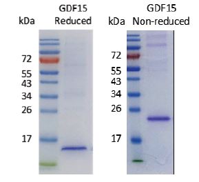
Growth Differentiation Factor 15 (hGDF15), Active Protein (Cat# AAA60569)
prolactin soluble receptor, Active Protein (Cat# AAA60571)
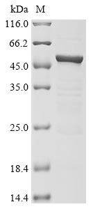
(a) Analysis by RP-HPLC.
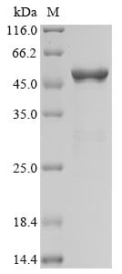
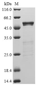
(b) Analysis by SDS-PAGE.
(c) Gel filtration at pH 8 under non denaturative conditions.
Factor V, Active Protein (Cat# AAA71836)
Purification: Immunoaffinity chromatography
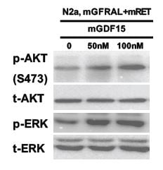
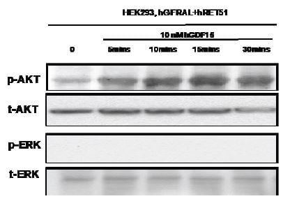 Application Data
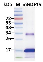
Growth Differentiation Factor 15 (hGDF15), Active Protein (Cat# AAA60567)
IGF-I, Active Protein (Cat# AAA60570)
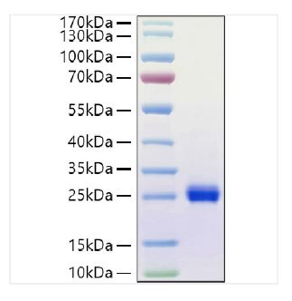
(a) Analysis by reducing and non-reducing SDS-PAGE gel.
(b) Gel-filtration chromatography under non denaturing conditions.
super leptin antagonist, Active Protein (Cat# AAA60573)
(a) Gel filtration analysis.
(b) Analysis by reducing and non-reducing SDS-PAGE gel.
GST A3-3, Active Protein (Cat# AAA71844)
GST P1-1, Active Protein (Cat# AAA71845)
Chorionic Gonadotropin, Active Protein (Cat# AAA59624)
Luteinizing Hormone, Active Protein (Cat# AAA59626)
Interleukin 4, Active Protein (Cat# AAA62204)
Bovine Type I Collagen Substrate, Active Protein (Cat# AAA60516)
 Bioactivity
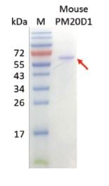
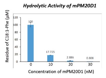
Peptidase M20 Domain-containing Protein 1 (mPM20D1), Active Protein (Cat# AAA60568)
interleukin 22 receptor antagonist, Active Protein (Cat# AAA60572)
(a) Analysis by reducing and non-reducing SDS-PAGE Silver Stained gel.
(b) Gel filtration chromatography under non-denaturing conditions.
Platelet-derived growth factor, Active Protein (Cat# AAA62063)
Prostate Specific Antigen, Active Protein (Cat# AAA59627)
Gamma-Glutamyl Transpeptidase, Active Protein (Cat# AAA224516)
C. Albicans eno1, Active Protein (Cat# AAA224573)
 Bioactivity
COVID 19 Spike glycoprotein (S) Coronavirus, Active Protein (Cat# AAA278918)
Influenza A, Active Protein (Cat# AAA224715)
COVID 19 Spike RBD Coronavirus, Active Protein (Cat# AAA281531)
Coxsackievirus B1 VP1, Active Protein (Cat# AAA224547)
 Bioactivity
COVID 19 Nucleocapsid (NP) Coronavirus, Active Protein (Cat# AAA278894)
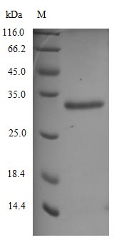
 Bioactivity
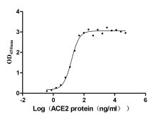
COVID 19 Spike glycoprotein (S) Coronavirus, Active Protein (Cat# AAA278916)
 Bioactivity
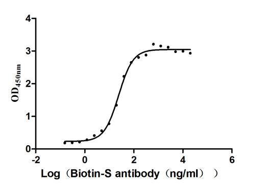
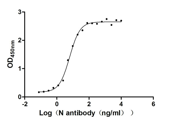
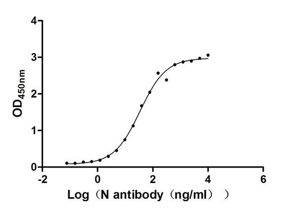
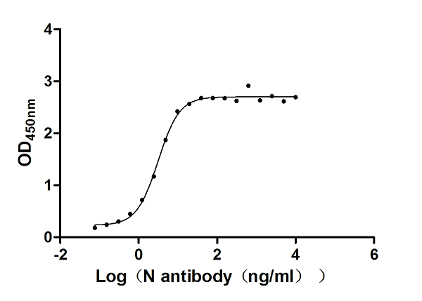
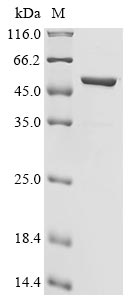
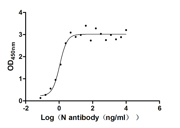
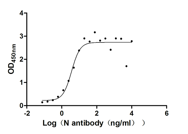
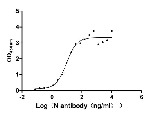
COVID 19 Nucleocapsid (NP) Coronavirus, Active Protein (Cat# AAA278892)
 Bioactivity
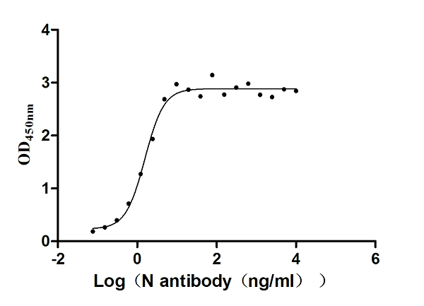
(Measured by its binding ability in a functional ELISA. Immobilized Human CD69 at 2?g/mL can bind Anti-CD69 recombinant antibody, the EC50 is 23.17-26.04 ng/mL.)
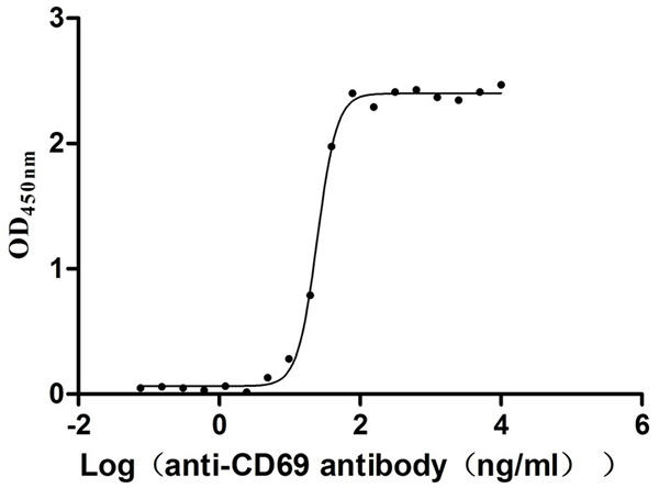
Bioactivity
(Measured by its binding ability in a functional ELISA. Immobilized Human CD69 at 2?g/mL can bind Anti-CD69 recombinant antibody, the EC50 is 23.17-26.04 ng/mL.)
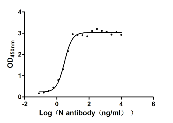
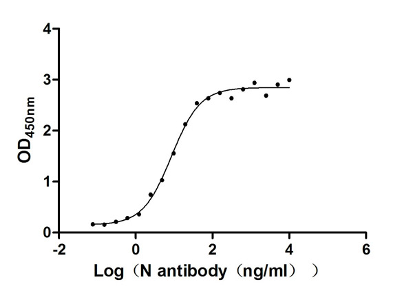
Early activation antigen CD69 (CD69), Active Protein (Cat# AAA279197)
 Bioactivity
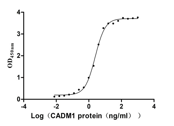
(Measured by its binding ability in a functional ELISA. Immobilized Human CRTAM at 2?g/mL can bind Human CADM1, the EC50 is 2.277-2.649 ng/mL)
Bioactivity
(Measured by its binding ability in a functional ELISA. Immobilized Human CRTAM at 2?g/mL can bind Human CADM1, the EC50 is 2.277-2.649 ng/mL)
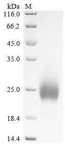
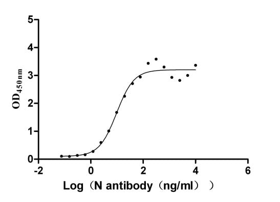
Cytotoxic and regulatory T-cell molecule (CRTAM), Active Protein (Cat# AAA279205)
 Bioactivity
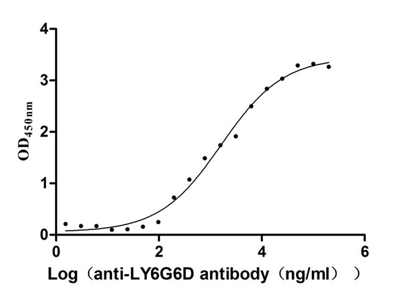
(Measured by its binding ability in a functional ELISA. Immobilized Mouse Ly6g6d at 2 ?g/mL can bind Anti-LY6G6D recombinant antibody . The EC50 is 1.159-2.305 ?g/mL.)
Bioactivity
(Measured by its binding ability in a functional ELISA. Immobilized Mouse Ly6g6d at 2 ?g/mL can bind Anti-LY6G6D recombinant antibody . The EC50 is 1.159-2.305 ?g/mL.)
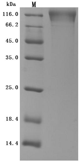
Lymphocyte antigen 6 complex locus protein G6d (Ly6g6d), Active Protein (Cat# AAA279248)
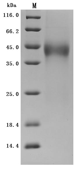
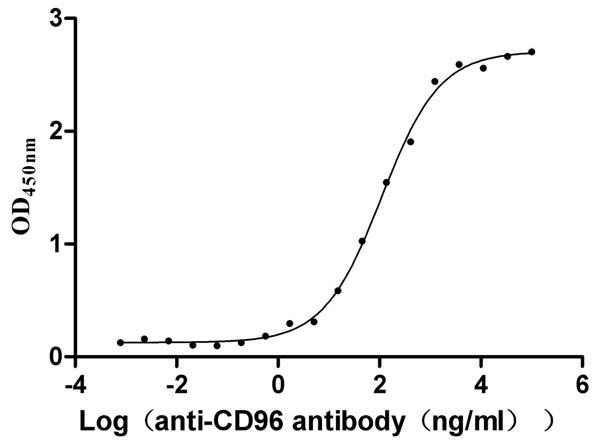 Bioactivity
T-cell surface protein tactile (CD96), Active Protein (Cat# AAA278902)
Creatine Kinase, Active Protein (Cat# AAA224710)
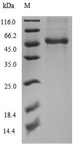
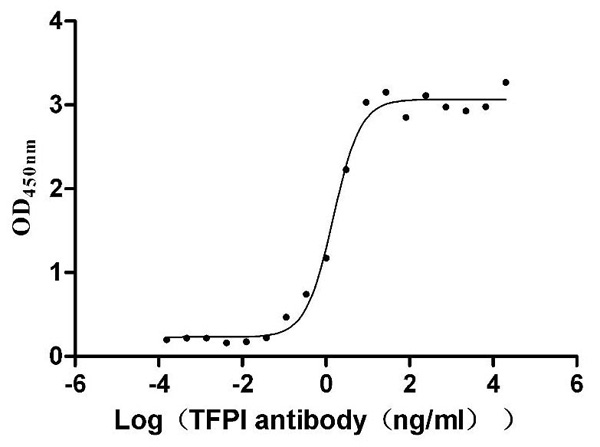 Bioactivity
(Measured by its binding ability in a functional ELISA. Immobilized Human TFPI at 1 ?g/ml can bind Anti-TFPI recombinant antibody, the EC50 is 1.242-1.788 ng/mL.)
Bioactivity
(Measured by its binding ability in a functional ELISA. Immobilized Human TFPI at 1 ?g/ml can bind Anti-TFPI recombinant antibody, the EC50 is 1.242-1.788 ng/mL.)
Tissue factor pathway inhibitor (TFPI), Active Protein (Cat# AAA279229)
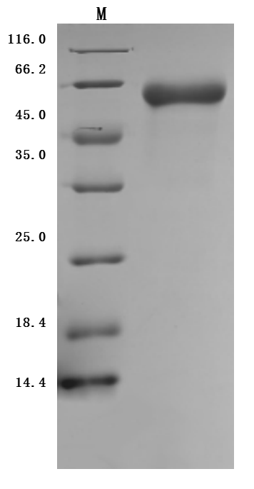
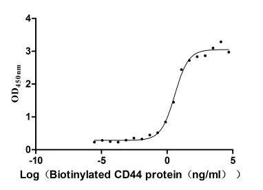 Bioactivity
CD44 antigen (CD44), Active Protein (Cat# AAA278900)
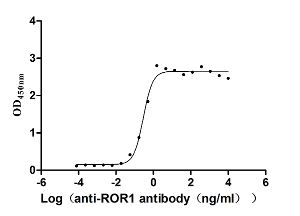 Bioactivity
Inactive tyrosine-protein kinase transmembrane receptor ROR1 (ROR1), Active Protein (Cat# AAA278913)
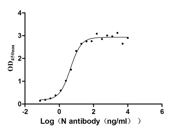
 Bioactivity
COVID 19 Nucleocapsid (NP) Coronavirus, Active Protein (Cat# AAA278893)
 Application Data
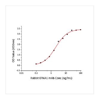
(Immobilized Human EFNA1 at 1 ug/mL (100 uL/well) can bind Human Ephrin A1 Rabbit mAb with a linear range of 0.098-2.066 ng/mL.)
Application Data
(Immobilized Human EFNA1 at 1 ug/mL (100 uL/well) can bind Human Ephrin A1 Rabbit mAb with a linear range of 0.098-2.066 ng/mL.)
Ephrin-A1/EFNA1, Active Protein (Cat# AAA282509)
 Bioactivity
COVID 19 Nucleocapsid (NP) Coronavirus, Active Protein (Cat# AAA278888)
 Bioactivity
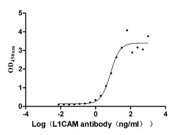
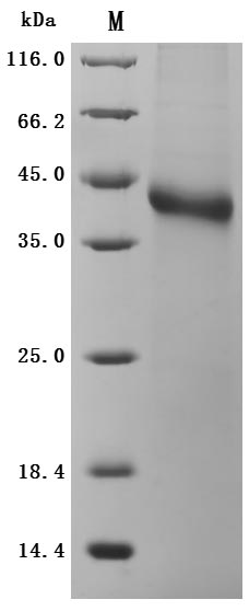
Neural cell adhesion molecule L1 (L1CAM), Active Protein (Cat# AAA278906)
 Bioactivity
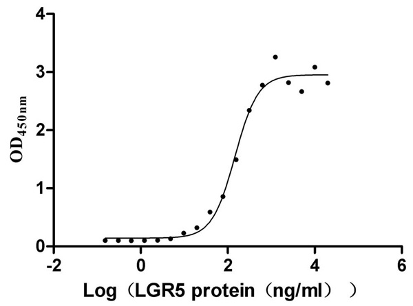
(Measured by its binding ability in a functional ELISA. Immobilized Human RSPO1 at 2 ?g/ml can bind Human LGR5, the EC50 is 124.0-174.1 ng/mL.)
Bioactivity
(Measured by its binding ability in a functional ELISA. Immobilized Human RSPO1 at 2 ?g/ml can bind Human LGR5, the EC50 is 124.0-174.1 ng/mL.)
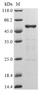
R-spondin-1 (RSPO1), Active Protein (Cat# AAA279250)

 Bioactivity
COVID 19 Nucleocapsid (NP) Coronavirus, Active Protein (Cat# AAA278890)
 Bioactivity
COVID 19 Nucleocapsid (NP) Coronavirus, Active Protein (Cat# AAA278899)
 Bioactivity
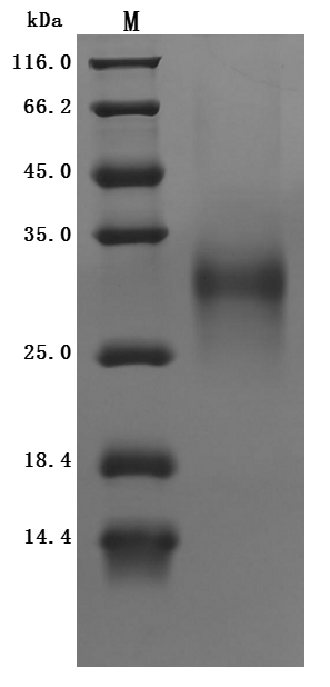
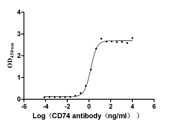
HLA class II histocompatibility antigen gamma chain (CD74), Active Protein (Cat# AAA278901)
 Bioactivity
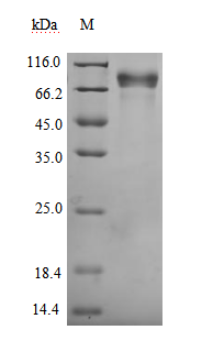
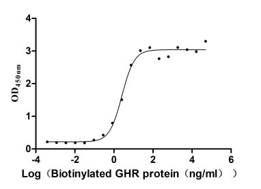
Growth hormone receptor (GHR), Active Protein (Cat# AAA278905)
 Bioactivity
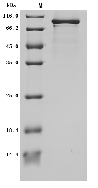
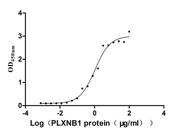
Plexin-B1 (PLXNB1), Active Protein (Cat# AAA278910)
What Are Active Proteins?
Proteins are large molecules made up of long chains of amino acids. They will typically fold into a very particular 3-dimensional shape/conformation, that is sometimes referred to as their “native” form, which allows them to work properly in the body. For the purposes of product categorization, AAA Biotech will typically refer to proteins purified from their original animal host as being “native” proteins (this is to signify their difference compared to their recombinant proteins or “synthetic” protein counterparts).
If a protein successfully folds into the correct shape, it will typically display high fidelity characteristics to its original protein in its original animal host and be classified as an active protein, as it will be able to function “normally” in most enzymatic or binding capacities. If it loses this shape, due to factors such as heat or strong chemicals (such as detergents), it becomes inactive and is no longer able to perform its basic functions.
All of the proteins in this category are made under strict quality control, and they are active, pure, low in contaminants, and stable. Most are stored as freeze-dried powders and come without extra tags, so they’re very close to the actual natural/native form.
Learn more in our guide “How active proteins work”.
Key Applications of Active Proteins
1. Scientific Research
- Aid in the study of how proteins function in the body
- Aid in understanding various disease processes
2. Drug Development
- Powerful tools to investigate how potential drugs interact with specific proteins
- Ideal for identifying drug targets
3. Cell Culture
- Are routinely utilized to support cell growth and function (e.g., using exogenous growth factors)
- Can be used to promote cellular development into specific types (differentiation)
4. Diagnostics
- Regularly utilized in tests to detect diseases or infections (e.g., COVID-19, cancer)
- Note: All products are strictly for research-use only (RUO).
5. Therapeutics
- Some active proteins are used directly as treatments (e.g., insulin, enzymes)
- Note: All products are strictly for research-use only (RUO).
6. Vaccine Development
- Used to create or test vaccines by mimicking parts of viruses or bacteria
7. Biochemical Assays
- They can facilitate the characterization of enzyme activity, binding strength, or protein interactions in lab tests
Why Buy Active Proteins from AAA Biotech?
- High biological activity – Verified to perform as expected or indicated on datasheet
- Strict quality control – We are confident in our active proteins’ reliability and consistency
- High purity & low endotoxin – Ideal for applications involving sensitive or precious samples/components
- Freeze-dried for stability – Long shelf life and straightforward storage
- Mostly tag-free – Closer to natural/native protein form
FAQ
1. What are active proteins used for in research?
Active proteins are used primarily in the study of how proteins function, in characterizing/discovering drug interactions, supporting cell growth, running biochemical assays, and in development of diagnostics or therapeutics.
2. How are AAA Biotech's active proteins validated?
AAA Biotech’s active proteins are validated through strict quality control and functional assays to ensure they are properly folded and active. “Active”, though, can be an ambiguous term, so if a specific “activity” or “binding” capability of a protein is of crucial interest to you, please inquire with us prior to purchase, and we will provide further details on how the “Active” modifier was determined to be applicable.
3. Are these proteins tested for biological activity?
Yes, all active proteins from AAA Biotech are tested to confirm they have the expected biological activity before being offered for use. Though, said “biological activity” can be either “enzymatic”, “binding”, or both.