Filters
▼Clonality
▼Type
▼Reactivity
▼Gene Name
▼Isotype
▼Host
▼Application
▼Clone
▼Monoclonal Antibodies
Get accurate results in your research with our Monoclonal Antibodies, which are specially made to target exactly what you require for your research, and will produce consistent, reliable performance in lab tests.
Viewing 450-500 of 27645 product results
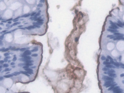
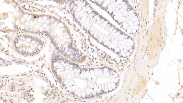
 IHC (Immunohiostchemistry)
(DAB staining on IHCP;Sample: Human Colon Tissue; Primary Ab: 30ug/ml Mouse AntiHuman NB1 AntibodySecond Ab: 2ug/mL HRPLinked Caprine AntiMouse IgG Polyclonal Antibody(Catalog: SAA544Mu19))
IHC (Immunohiostchemistry)
(DAB staining on IHCP;Sample: Human Colon Tissue; Primary Ab: 30ug/ml Mouse AntiHuman NB1 AntibodySecond Ab: 2ug/mL HRPLinked Caprine AntiMouse IgG Polyclonal Antibody(Catalog: SAA544Mu19))
Neutrophil Specific Antigen 1 (NB1), Monoclonal Antibody (Cat# AAA151658)
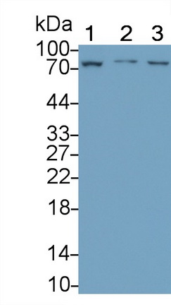
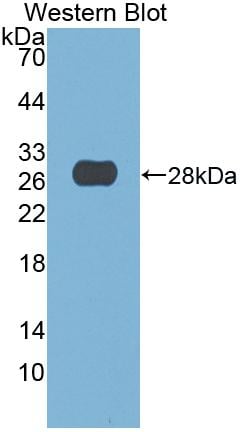
 WB (Western Blot)
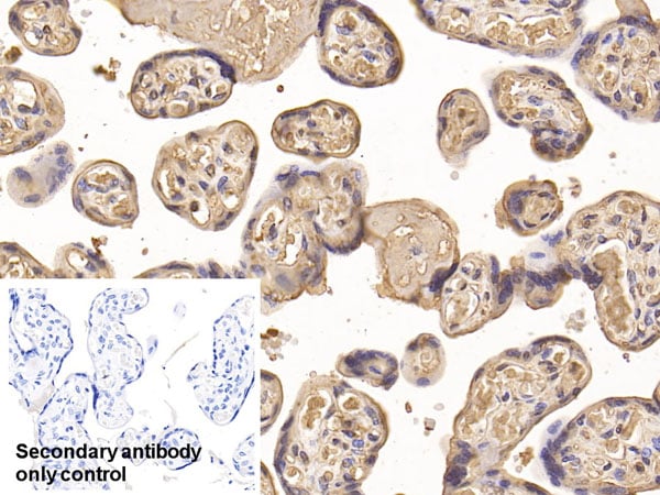
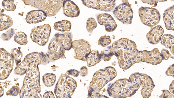
(Western Blot; Sample: human Placenta lysate Primary Ab: 1.5ug/ml Mouse Anti-human IGFBP1 Antibody Second Ab: 0.2ug/mL HRP-Linked Caprine Anti-Mouse IgG Polyclonal Antibody)
WB (Western Blot)
(Western Blot; Sample: human Placenta lysate Primary Ab: 1.5ug/ml Mouse Anti-human IGFBP1 Antibody Second Ab: 0.2ug/mL HRP-Linked Caprine Anti-Mouse IgG Polyclonal Antibody)
Inulin Like Growth Factor Binding Protein 1 (IGFBP1), Monoclonal Antibody (Cat# AAA152667)
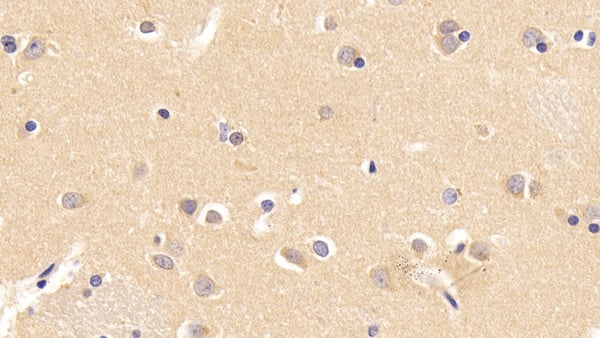
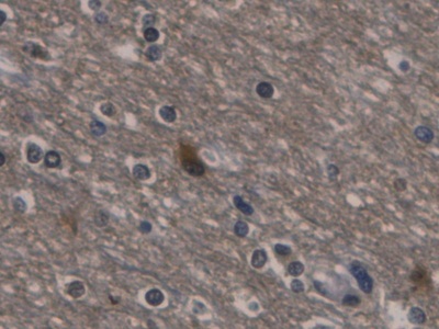
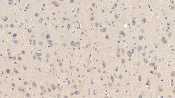 IHC (Immunohistochemistry)
(DAB staining on IHCP;Sample: Porcine Cerebrum Tissue; Primary Ab: 10ug/ml Mouse AntiHuman IFNa/bR1 AntibodySecond Ab: 2ug/mL HRPLinked Caprine AntiMouse IgG Polyclonal Antibody(Catalog: SAA544Mu19))
IHC (Immunohistochemistry)
(DAB staining on IHCP;Sample: Porcine Cerebrum Tissue; Primary Ab: 10ug/ml Mouse AntiHuman IFNa/bR1 AntibodySecond Ab: 2ug/mL HRPLinked Caprine AntiMouse IgG Polyclonal Antibody(Catalog: SAA544Mu19))
Interferon Alpha/Beta Receptor 1 (IFNa/bR1), Monoclonal Antibody (Cat# AAA151711)
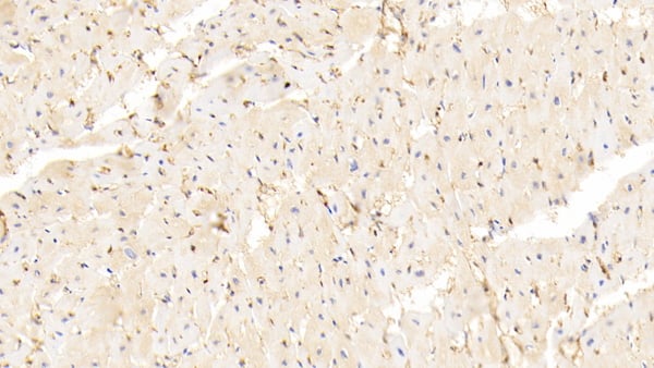
 IHC (Immunohiostchemistry)
(DAB staining on IHCP;Sample: Human Skeletal muscle Tissue; Primary Ab: 30ug/ml Mouse AntiHuman AchE AntibodySecond Ab: 2ug/mL HRPLinked Caprine AntiMouse IgG Polyclonal Antibody(Catalog: SAA544Mu19))
IHC (Immunohiostchemistry)
(DAB staining on IHCP;Sample: Human Skeletal muscle Tissue; Primary Ab: 30ug/ml Mouse AntiHuman AchE AntibodySecond Ab: 2ug/mL HRPLinked Caprine AntiMouse IgG Polyclonal Antibody(Catalog: SAA544Mu19))
Acetylcholinesterase (ACHE), Monoclonal Antibody (Cat# AAA151713)
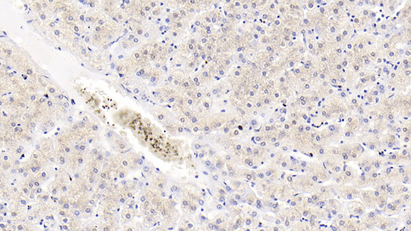
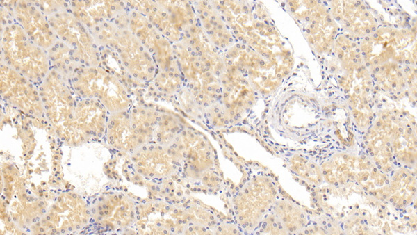
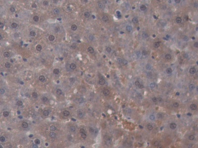
 IHC (Immunohiostchemistry)
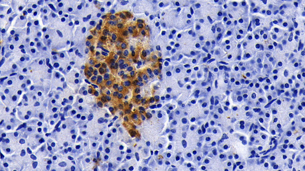
(DAB staining on IHCP;Sample: Human Liver Tissue; Primary Ab: 30ug/ml Mouse AntiHuman AchE AntibodySecond Ab: 2ug/mL HRPLinked Caprine AntiMouse IgG Polyclonal Antibody(Catalog: SAA544Mu19))
IHC (Immunohiostchemistry)
(DAB staining on IHCP;Sample: Human Liver Tissue; Primary Ab: 30ug/ml Mouse AntiHuman AchE AntibodySecond Ab: 2ug/mL HRPLinked Caprine AntiMouse IgG Polyclonal Antibody(Catalog: SAA544Mu19))
Acetylcholinesterase (ACHE), Monoclonal Antibody (Cat# AAA151714)
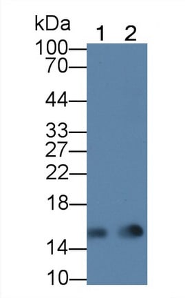
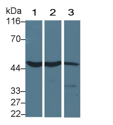
 WB (Western Blot)
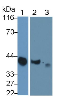
(Western Blot; Sample: Lane1: Human Saliva; Lane2: Rat Pancreas lysate; Lane3: Mouse Pancreas lysate Primary Ab: 0.3ug/ml Mouse AntiHuman AMY1 Antibody Second Ab: 0.2ug/mL HRPLinked Caprine AntiMouse IgG Polyclonal Antibody (Catalog: SAA544Mu19))
WB (Western Blot)
(Western Blot; Sample: Lane1: Human Saliva; Lane2: Rat Pancreas lysate; Lane3: Mouse Pancreas lysate Primary Ab: 0.3ug/ml Mouse AntiHuman AMY1 Antibody Second Ab: 0.2ug/mL HRPLinked Caprine AntiMouse IgG Polyclonal Antibody (Catalog: SAA544Mu19))
Salivary Alpha Amylase (AMY1A), Monoclonal Antibody (Cat# AAA151719)
 IHC (Immunohiostchemistry)
(DAB staining on IHCP;Sample: Rat Colon Tissue; Primary Ab: 20ug/ml Mouse AntiMultispecies LPS AntibodySecond Ab: 2ug/mL HRPLinked Caprine AntiMouse IgG Polyclonal Antibody(Catalog: SAA544Mu19))
IHC (Immunohiostchemistry)
(DAB staining on IHCP;Sample: Rat Colon Tissue; Primary Ab: 20ug/ml Mouse AntiMultispecies LPS AntibodySecond Ab: 2ug/mL HRPLinked Caprine AntiMouse IgG Polyclonal Antibody(Catalog: SAA544Mu19))
Lipopolysaccharide (LPS), Monoclonal Antibody (Cat# AAA151721)
 IHC (Immunohiostchemistry)
(DAB staining on IHCP;Sample: Rat Small intestine Tissue; Primary Ab: 20ug/ml Mouse AntiMultispecies LPS AntibodySecond Ab: 2ug/mL HRPLinked Caprine AntiMouse IgG Polyclonal Antibody(Catalog: SAA544Mu19))
IHC (Immunohiostchemistry)
(DAB staining on IHCP;Sample: Rat Small intestine Tissue; Primary Ab: 20ug/ml Mouse AntiMultispecies LPS AntibodySecond Ab: 2ug/mL HRPLinked Caprine AntiMouse IgG Polyclonal Antibody(Catalog: SAA544Mu19))
Lipopolysaccharide (LPS), Monoclonal Antibody (Cat# AAA151722)
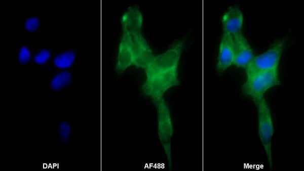
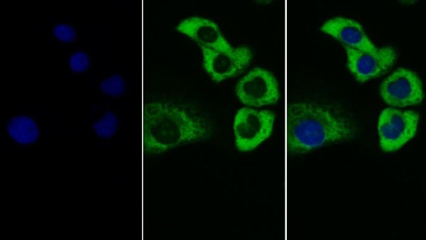
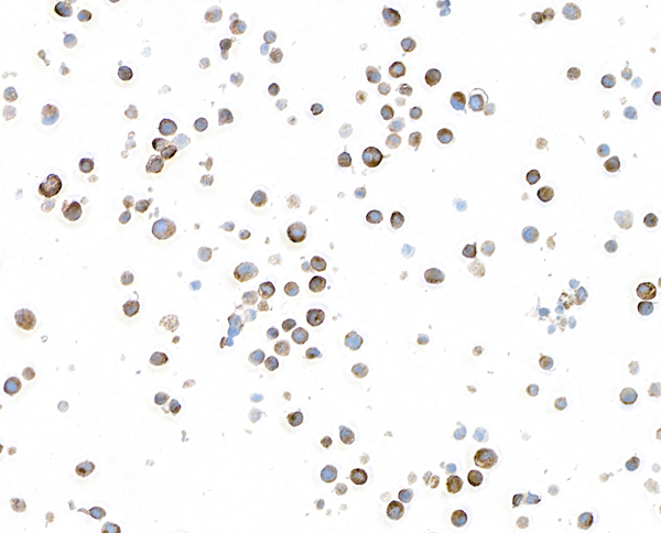
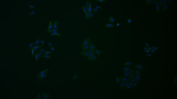 IF (Immunofluorescence)
(FITC staining on IF; Samples: Human HepG2 cell; Primary Ab: 20ug/ml Mouse AntiHuman LAG3 Antibody Second Ab: 3ug/ml FITCLinked Caprine AntiMouse IgG Polyclonal Antibody (Catalog: SAA544Mu18))
IF (Immunofluorescence)
(FITC staining on IF; Samples: Human HepG2 cell; Primary Ab: 20ug/ml Mouse AntiHuman LAG3 Antibody Second Ab: 3ug/ml FITCLinked Caprine AntiMouse IgG Polyclonal Antibody (Catalog: SAA544Mu18))
Lymphocyte Activation Gene 3 (LAG3), Monoclonal Antibody (Cat# AAA151729)
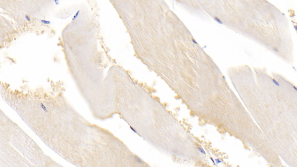
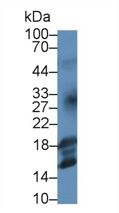
 WB (Western Blot)
(Western Blot; Sample: Lane1: Rat Heart lysate; Lane2: Rat Kidney lysate Primary Ab: 2ug/ml Mouse AntiHuman FABP4 Antibody Second Ab: 0.2ug/mL HRPLinked Caprine AntiMouse IgG Polyclonal Antibody (Catalog: SAA544Mu19))
WB (Western Blot)
(Western Blot; Sample: Lane1: Rat Heart lysate; Lane2: Rat Kidney lysate Primary Ab: 2ug/ml Mouse AntiHuman FABP4 Antibody Second Ab: 0.2ug/mL HRPLinked Caprine AntiMouse IgG Polyclonal Antibody (Catalog: SAA544Mu19))
Fatty Acid Binding Protein 4 (FABP4), Monoclonal Antibody (Cat# AAA151732)
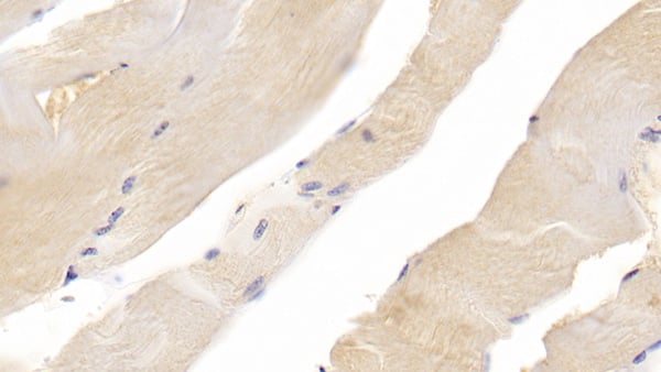
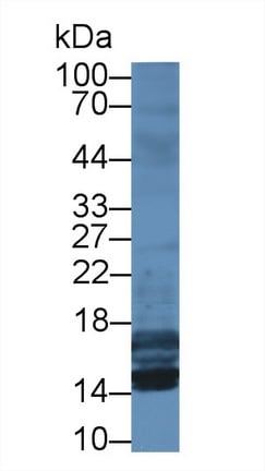
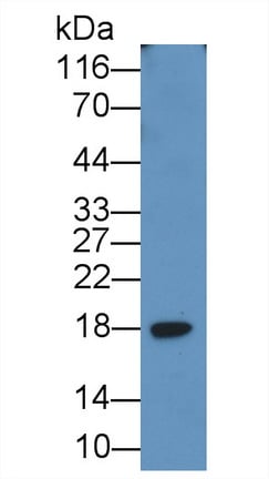
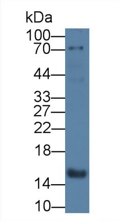 WB (Western Blot)
(Western Blot; Sample: Rat Heart lysate Primary Ab: 2ug/ml Mouse AntiHuman FABP4 Antibody Second Ab: 0.2ug/mL HRPLinked Caprine AntiMouse IgG Polyclonal Antibody (Catalog: SAA544Mu19))
WB (Western Blot)
(Western Blot; Sample: Rat Heart lysate Primary Ab: 2ug/ml Mouse AntiHuman FABP4 Antibody Second Ab: 0.2ug/mL HRPLinked Caprine AntiMouse IgG Polyclonal Antibody (Catalog: SAA544Mu19))
Fatty Acid Binding Protein 4 (FABP4), Monoclonal Antibody (Cat# AAA151733)
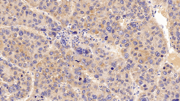
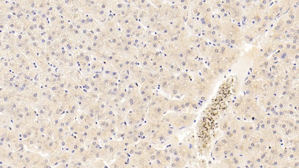
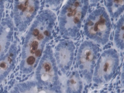
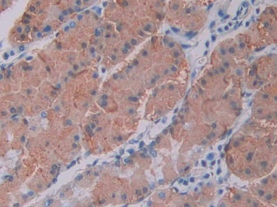
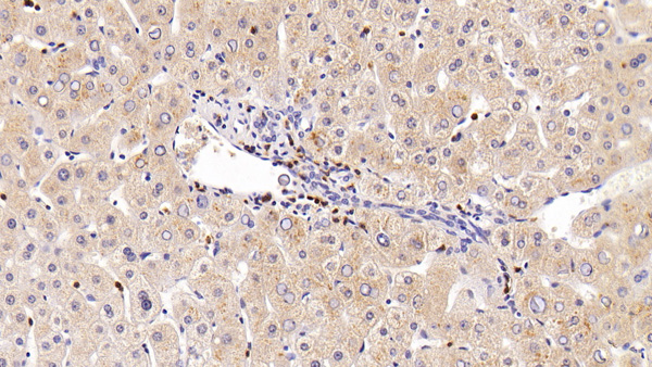
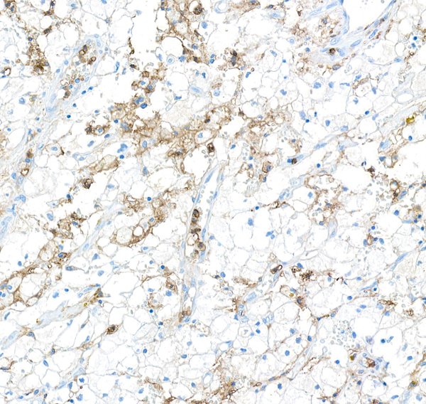
 IHC (Immunohiostchemistry)
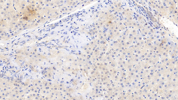
(DAB staining on IHCP;Sample: Human Liver Tissue; Primary Ab: 30ug/ml Mouse AntiHuman CCK8 AntibodySecond Ab: 2ug/mL HRPLinked Caprine AntiMouse IgG Polyclonal Antibody(Catalog: SAA544Mu19))
IHC (Immunohiostchemistry)
(DAB staining on IHCP;Sample: Human Liver Tissue; Primary Ab: 30ug/ml Mouse AntiHuman CCK8 AntibodySecond Ab: 2ug/mL HRPLinked Caprine AntiMouse IgG Polyclonal Antibody(Catalog: SAA544Mu19))
Cholecystokinin 8 (CCK8), Monoclonal Antibody (Cat# AAA151674)
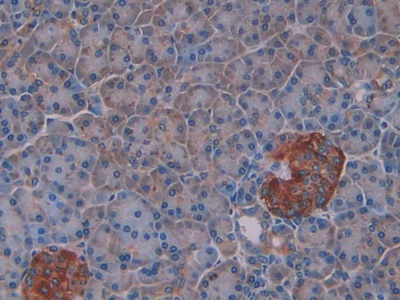
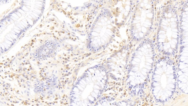
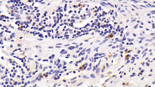
 IHC (Immunohistochemisry)
(DAB staining on IHCP;Sample: Human Stomach cancer Tissue; Primary Ab: 40ug/ml Mouse AntiHuman CCK8 AntibodySecond Ab: 2ug/mL HRPLinked Caprine AntiMouse IgG Polyclonal Antibody(Catalog: SAA544Mu19))
IHC (Immunohistochemisry)
(DAB staining on IHCP;Sample: Human Stomach cancer Tissue; Primary Ab: 40ug/ml Mouse AntiHuman CCK8 AntibodySecond Ab: 2ug/mL HRPLinked Caprine AntiMouse IgG Polyclonal Antibody(Catalog: SAA544Mu19))
Cholecystokinin 8 (CCK8), Monoclonal Antibody (Cat# AAA151675)
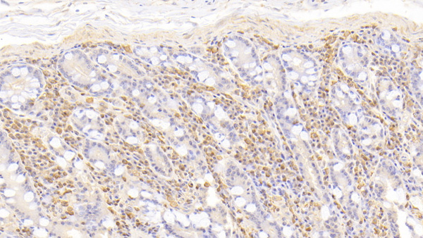
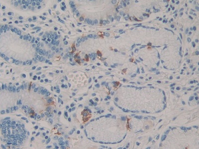
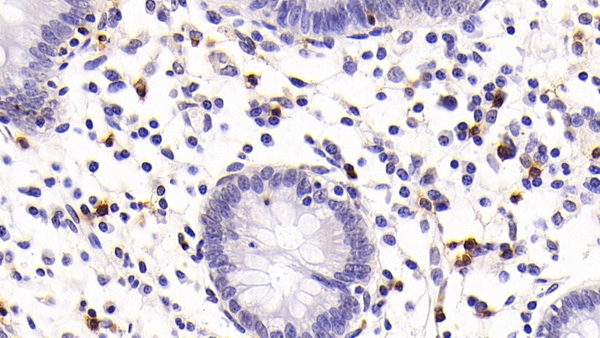
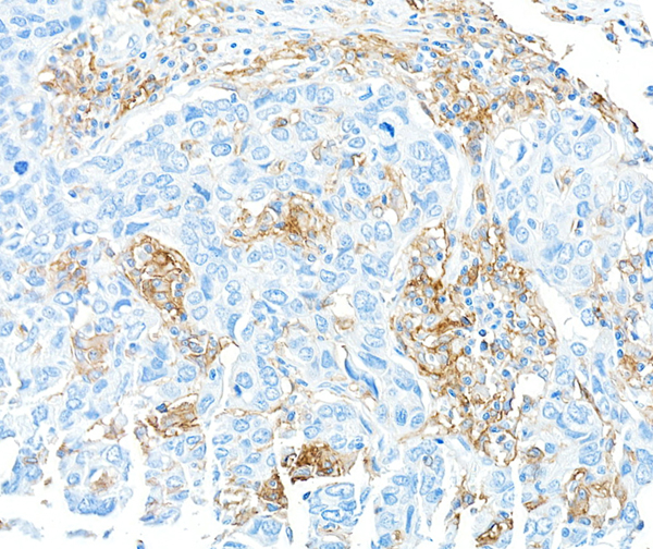
 IHC (Immunohiostchemistry)
(DAB staining on IHCP;Sample: Human Stomach Tissue; Primary Ab: 30ug/ml Mouse AntiHuman CCK8 AntibodySecond Ab: 2ug/mL HRPLinked Caprine AntiMouse IgG Polyclonal Antibody(Catalog: SAA544Mu19))
IHC (Immunohiostchemistry)
(DAB staining on IHCP;Sample: Human Stomach Tissue; Primary Ab: 30ug/ml Mouse AntiHuman CCK8 AntibodySecond Ab: 2ug/mL HRPLinked Caprine AntiMouse IgG Polyclonal Antibody(Catalog: SAA544Mu19))
Cholecystokinin 8 (CCK8), Monoclonal Antibody (Cat# AAA151676)
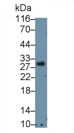 WB (Western Blot)
(Western Blot; Sample: Human PBMC lysate Primary Ab: 1ug/ml Mouse AntiHuman CD8a Antibody Second Ab: 0.2ug/mL HRPLinked Caprine AntiMouse IgG Polyclonal Antibody (Catalog: SAA544Mu19))
WB (Western Blot)
(Western Blot; Sample: Human PBMC lysate Primary Ab: 1ug/ml Mouse AntiHuman CD8a Antibody Second Ab: 0.2ug/mL HRPLinked Caprine AntiMouse IgG Polyclonal Antibody (Catalog: SAA544Mu19))
Cluster Of Differentiation 8a (CD8a), Monoclonal Antibody (Cat# AAA151679)
 WB (Western Blot)
(Western Blot; Sample: Lane1: Porcine Serum; Lane2: Porcine Lung lysate; Lane3: Porcine Cerebrum lysate Primary Ab: 2ug/ml Mouse AntiPorcine CLU Antibody Second Ab: 0.2ug/mL HRPLinked Caprine AntiMouse IgG Polyclonal Antibody (Catalog: SAA544Mu19))
WB (Western Blot)
(Western Blot; Sample: Lane1: Porcine Serum; Lane2: Porcine Lung lysate; Lane3: Porcine Cerebrum lysate Primary Ab: 2ug/ml Mouse AntiPorcine CLU Antibody Second Ab: 0.2ug/mL HRPLinked Caprine AntiMouse IgG Polyclonal Antibody (Catalog: SAA544Mu19))
Clusterin (CLU), Monoclonal Antibody (Cat# AAA151688)
 WB (Western Blot)
(Western Blot; Sample: Lane1: Human Serum; Lane2: Human Urine; Lane3: Human Placenta lysate Primary Ab: 3ug/ml Mouse AntiHuman FGb Antibody Second Ab: 0.2ug/mL HRPLinked Caprine AntiMouse IgG Polyclonal Antibody (Catalog: SAA544Mu19))
WB (Western Blot)
(Western Blot; Sample: Lane1: Human Serum; Lane2: Human Urine; Lane3: Human Placenta lysate Primary Ab: 3ug/ml Mouse AntiHuman FGb Antibody Second Ab: 0.2ug/mL HRPLinked Caprine AntiMouse IgG Polyclonal Antibody (Catalog: SAA544Mu19))
Fibrinogen Beta Chain (FGB), Monoclonal Antibody (Cat# AAA151693)
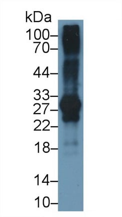
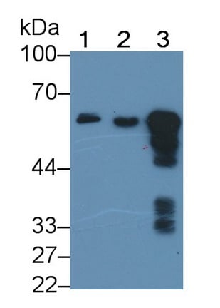
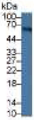
 WB (Western Blot)
(Western Blot; Sample: Lane1: Porcine CerebuM lysate; Lane2: Rat CerebuM lysate; Lane3: Hela cell lysate Primary Ab: 5ug/ml Mouse Anti-human NSE Antibody Second Ab: 0.2ug/mL HRP-Linked Caprine Anti-Mouse IgG Polyclonal Antibody)
WB (Western Blot)
(Western Blot; Sample: Lane1: Porcine CerebuM lysate; Lane2: Rat CerebuM lysate; Lane3: Hela cell lysate Primary Ab: 5ug/ml Mouse Anti-human NSE Antibody Second Ab: 0.2ug/mL HRP-Linked Caprine Anti-Mouse IgG Polyclonal Antibody)
Enolase, Neuron Specific (NSE), Monoclonal Antibody (Cat# AAA152606)
 WB (Western Blot)
(Western Blot; Sample: Porcine Heart lysatePrimary Ab: 0.2ug/ml Mouse Anti-human IGF1 AntibodySecond Ab: 0.2ug/mL HRP-Linked Caprine Anti-Mouse IgG Polyclonal Antibody)
WB (Western Blot)
(Western Blot; Sample: Porcine Heart lysatePrimary Ab: 0.2ug/ml Mouse Anti-human IGF1 AntibodySecond Ab: 0.2ug/mL HRP-Linked Caprine Anti-Mouse IgG Polyclonal Antibody)
Inulin Like Growth Factor 1 (IGF1), Monoclonal Antibody (Cat# AAA152611)
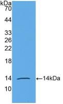
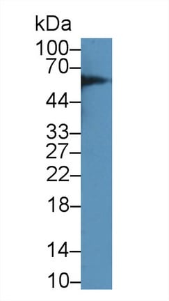
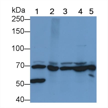 WB (Western Blot)
(Western Blot; Sample: Lane1: Bovine CerebuM lysate; Lane2: Bovine Testis lysate; Lane3: Bovine Liver lysate; Lane4: MCF7 cell lysate; Lane5: A431 cell lysate Primary Ab: 2 ug/ml Mouse Anti-Bovine HSPA1B Antibody Second Ab: 0.2ug/mL HRP-Linked Caprine Anti-Mouse IgG Polyclonal AntibodyA5)
WB (Western Blot)
(Western Blot; Sample: Lane1: Bovine CerebuM lysate; Lane2: Bovine Testis lysate; Lane3: Bovine Liver lysate; Lane4: MCF7 cell lysate; Lane5: A431 cell lysate Primary Ab: 2 ug/ml Mouse Anti-Bovine HSPA1B Antibody Second Ab: 0.2ug/mL HRP-Linked Caprine Anti-Mouse IgG Polyclonal AntibodyA5)
Heat Shock 70kDa Protein 1B (HSPA1B), Monoclonal Antibody (Cat# AAA152634)
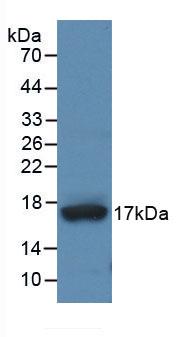
 WB (Western Blot)
(Western Blot; Sample: Lane1: Mouse Skin lysate; Lane2: Rat Skin lysate Primary Ab: 2ug/ml Mouse Anti-human KRT17 Antibody Second Ab: 0.2ug/mL HRP-Linked Caprine Anti-Mouse IgG Polyclonal Antibody)
WB (Western Blot)
(Western Blot; Sample: Lane1: Mouse Skin lysate; Lane2: Rat Skin lysate Primary Ab: 2ug/ml Mouse Anti-human KRT17 Antibody Second Ab: 0.2ug/mL HRP-Linked Caprine Anti-Mouse IgG Polyclonal Antibody)
Cytokeratin 17 (CK17), Monoclonal Antibody (Cat# AAA152637)
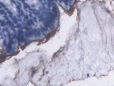
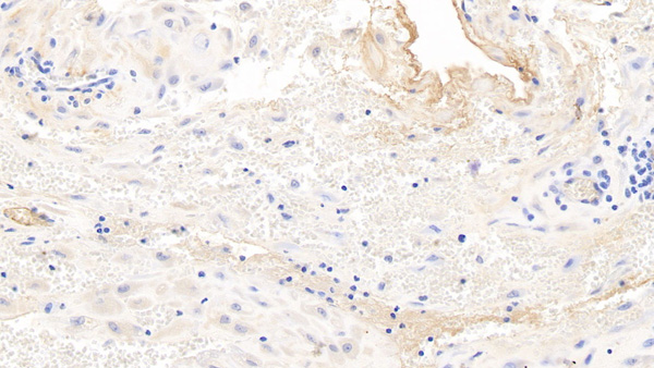
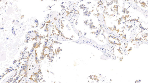
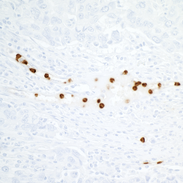
 IHC (Immunohiostchemistry)
(DAB staining on IHC-P; Samples: Rat Liver Tissue))
IHC (Immunohiostchemistry)
(DAB staining on IHC-P; Samples: Rat Liver Tissue))
Sex Hormone Binding Globulin (SHBG), Monoclonal Antibody (Cat# AAA130623)
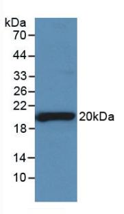
 WB (Western Blot)
(Western Blot:Sample: Recombinant DNASE1, Human.)
WB (Western Blot)
(Western Blot:Sample: Recombinant DNASE1, Human.)
Deoxyribonuclease I (DNASE1), Monoclonal Antibody (Cat# AAA130628)
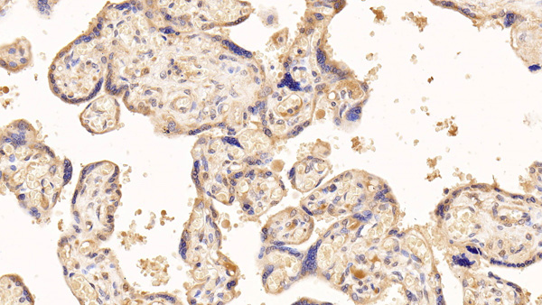
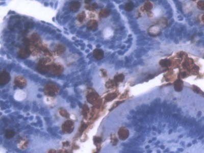
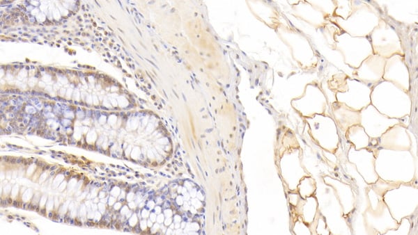
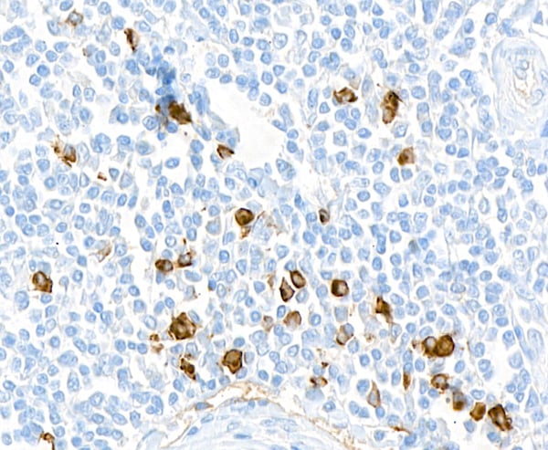
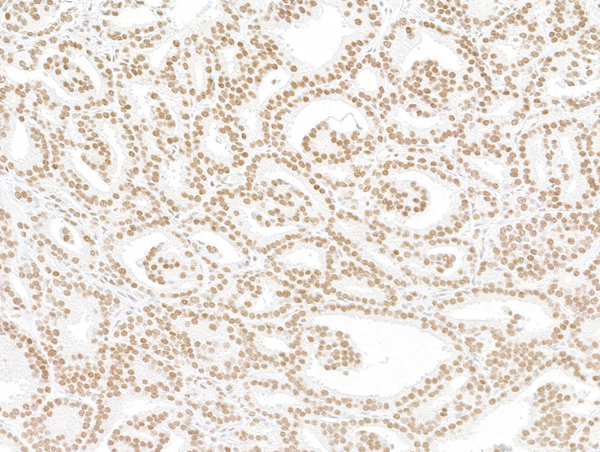
 IHC (Immunohistochemisry)
(DAB staining on IHC-P;Samples: Human Pancreas Tissue;Primary Ab: 40ug/ml Mouse Anti-Human AMH AntibodySecond Ab: 2ug/mL HRP-Linked Caprine Anti-Mouse IgG Polyclonal Antibody)
IHC (Immunohistochemisry)
(DAB staining on IHC-P;Samples: Human Pancreas Tissue;Primary Ab: 40ug/ml Mouse Anti-Human AMH AntibodySecond Ab: 2ug/mL HRP-Linked Caprine Anti-Mouse IgG Polyclonal Antibody)
Anti-Mullerian Hormone (AMH), Monoclonal Antibody (Cat# AAA130629)
 WB (Western Blot)
(Western Blot: Sample: Recombinant ADAMTS4, Human.)
WB (Western Blot)
(Western Blot: Sample: Recombinant ADAMTS4, Human.)
A Disintegrin And Metalloproteinase With Thrombospondin 4 (ADAMTS4), Monoclonal Antibody (Cat# AAA130642)
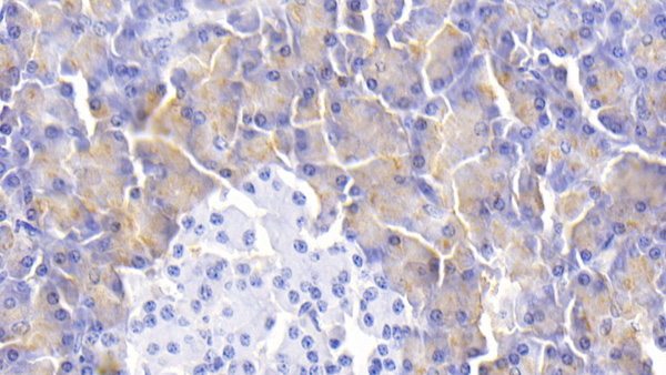
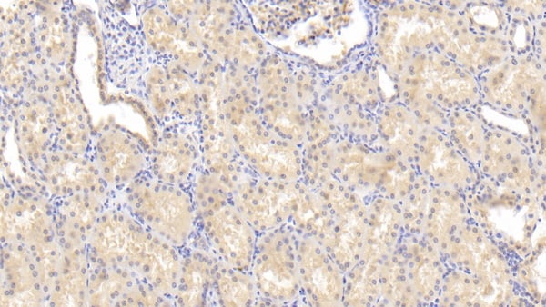
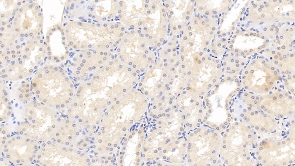
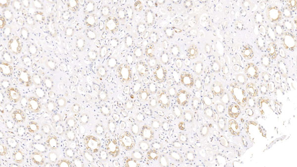
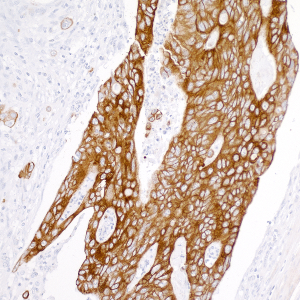
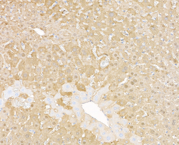
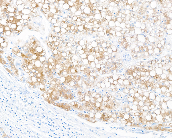
 IHC (Immunohistochemisry)
(DAB staining on IHC-P;Samples: Human Liver Tissue;Primary Ab: 30ug/ml Mouse Anti-Human AT AntibodySecond Ab: 2ug/mL HRP-Linked Caprine Anti-Mouse IgG Polyclonal Antibody)
IHC (Immunohistochemisry)
(DAB staining on IHC-P;Samples: Human Liver Tissue;Primary Ab: 30ug/ml Mouse Anti-Human AT AntibodySecond Ab: 2ug/mL HRP-Linked Caprine Anti-Mouse IgG Polyclonal Antibody)
Antithrombin (AT), Monoclonal Antibody (Cat# AAA130643)
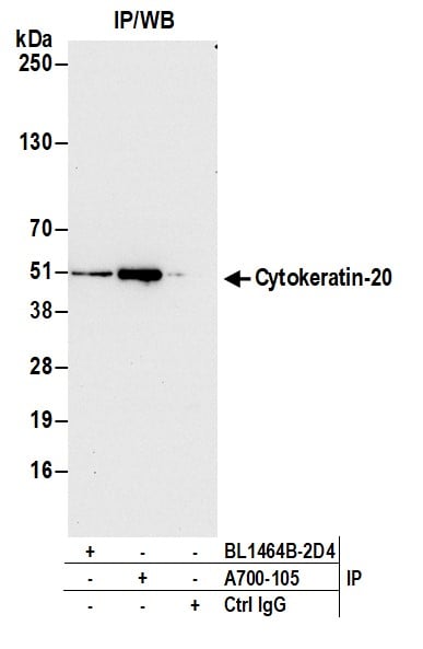
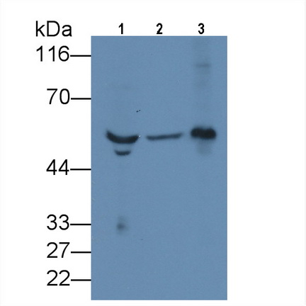
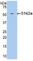
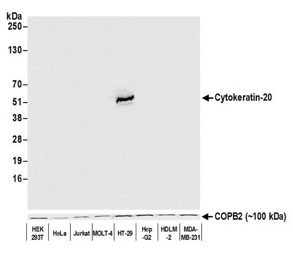 WB (Western Blot)
(Detection of human Cytokeratin-20 by western blot. Samples: Whole cell lysate (50 ug) from HEK293T, HeLa, Jurkat, MOLT-4, HT-29, Hep-G2, HDLM-2, and MDA-MB-231 cells prepared using NETN lysis buffer. Antibody: Rabbit anti-Cytokeratin-20 recombinant monoclonal antibody (AAA213580 lot 1) used at 1:1000. Secondary: HRP-conjugated goat anti-rabbit IgG . Detection: Chemiluminescence with an exposure time of 1 second. Lower Panel: Rabbit anti-COPB2 antibody .)
WB (Western Blot)
(Detection of human Cytokeratin-20 by western blot. Samples: Whole cell lysate (50 ug) from HEK293T, HeLa, Jurkat, MOLT-4, HT-29, Hep-G2, HDLM-2, and MDA-MB-231 cells prepared using NETN lysis buffer. Antibody: Rabbit anti-Cytokeratin-20 recombinant monoclonal antibody (AAA213580 lot 1) used at 1:1000. Secondary: HRP-conjugated goat anti-rabbit IgG . Detection: Chemiluminescence with an exposure time of 1 second. Lower Panel: Rabbit anti-COPB2 antibody .)
Cytokeratin 20, Monoclonal Recombinant Antibody (Cat# AAA213580)
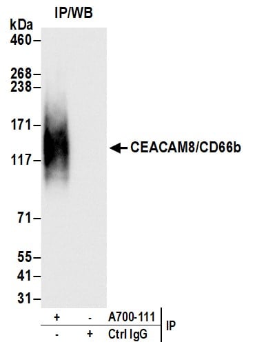
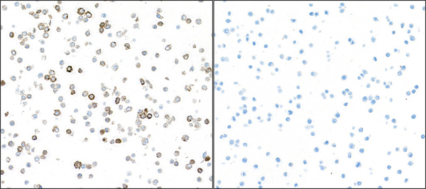
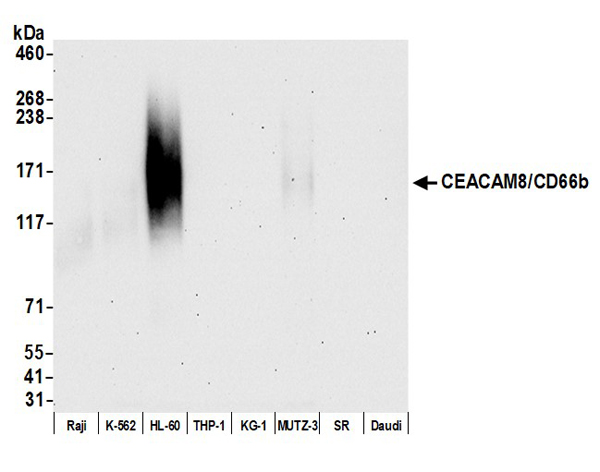 WB (Western Blot)
(Detection of human CEACAM8/CD66b by western blot. Samples: Whole cell lysate (10 ug) from Raji, K-562, HL-60, THP-1, KG-1, MUTZ-3, SR, and Daudi cells prepared using NETN lysis buffer. Antibody: Rabbit anti-CEACAM8/CD66b recombinant monoclonal antibody (AAA213582 lot 1) used at 1:1000. Secondary: HRP-conjugated goat anti-rabbit IgG . Detection: Chemiluminescence with an exposure time of 30 seconds.)
WB (Western Blot)
(Detection of human CEACAM8/CD66b by western blot. Samples: Whole cell lysate (10 ug) from Raji, K-562, HL-60, THP-1, KG-1, MUTZ-3, SR, and Daudi cells prepared using NETN lysis buffer. Antibody: Rabbit anti-CEACAM8/CD66b recombinant monoclonal antibody (AAA213582 lot 1) used at 1:1000. Secondary: HRP-conjugated goat anti-rabbit IgG . Detection: Chemiluminescence with an exposure time of 30 seconds.)
CEACAM8/CD66b, Monoclonal Recombinant Antibody (Cat# AAA213582)
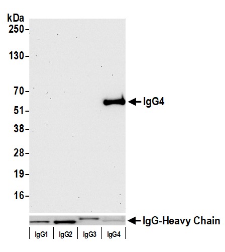 WB (Western Blot)
(Detection of human IgG4 by western blot. Samples: Purified human IgG1 (0.1 ug), IgG2 (0.5 ug), IgG3 (0.5 ug), and IgG4 (0.5 ug). Antibody: Rabbit anti-Human IgG4 recombinant monoclonal antibody (AAA213584 lot 1) used at 1:1000. Secondary: HRP-conjugated goat anti-rabbit IgG . Detection: Chemiluminescence with an exposure time of 30 seconds. Lower Panel: Goat anti-Human IgG-heavy and light chain antibody .)
WB (Western Blot)
(Detection of human IgG4 by western blot. Samples: Purified human IgG1 (0.1 ug), IgG2 (0.5 ug), IgG3 (0.5 ug), and IgG4 (0.5 ug). Antibody: Rabbit anti-Human IgG4 recombinant monoclonal antibody (AAA213584 lot 1) used at 1:1000. Secondary: HRP-conjugated goat anti-rabbit IgG . Detection: Chemiluminescence with an exposure time of 30 seconds. Lower Panel: Goat anti-Human IgG-heavy and light chain antibody .)
IgG4, Monoclonal Recombinant Antibody (Cat# AAA213584)
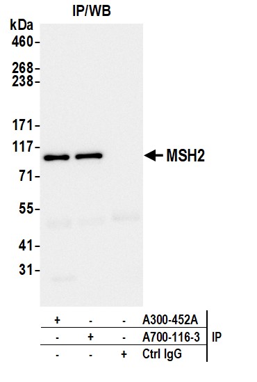
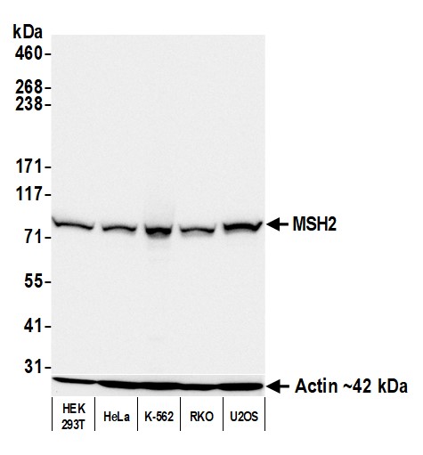 WB (Western Blot)
(Detection of human MSH2 by western blot. Samples: Whole cell lysate (50 ug) from HEK293T, HeLa, K-562, RKO, and U2OS cells prepared using NETN lysis buffer. Antibody: Rabbit anti-MSH2 recombinant monoclonal antibody (AAA213587 lot 3) used at 1:1000. Secondary: HRP-conjugated goat anti-rabbit IgG . Detection: Chemiluminescence with an exposure time of 3 seconds. Lower Panel: Rabbit anti-Actin recombinant monoclonal antibody .)
WB (Western Blot)
(Detection of human MSH2 by western blot. Samples: Whole cell lysate (50 ug) from HEK293T, HeLa, K-562, RKO, and U2OS cells prepared using NETN lysis buffer. Antibody: Rabbit anti-MSH2 recombinant monoclonal antibody (AAA213587 lot 3) used at 1:1000. Secondary: HRP-conjugated goat anti-rabbit IgG . Detection: Chemiluminescence with an exposure time of 3 seconds. Lower Panel: Rabbit anti-Actin recombinant monoclonal antibody .)
MSH2, Monoclonal Recombinant Antibody (Cat# AAA213587)
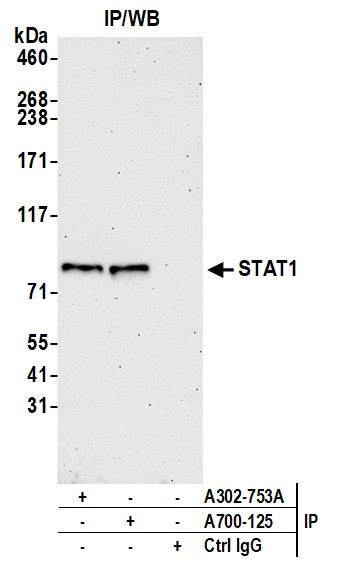
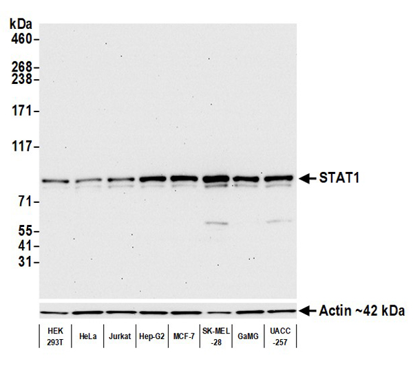 WB (Western Blot)
(Detection of human STAT1 by western blot. Samples: Whole cell lysate (5 to 10 ug) from HEK293T, HeLa, Jurkat, Hep-G2, MCF-7, SK-MEL-28, GaMG, and UACC-257 cells prepared using NETN lysis buffer. Antibody: Rabbit anti-STAT1 recombinant monoclonal antibody (AAA213591 lot 1) used at 1:1000. Secondary: HRP-conjugated goat anti-rabbit IgG . Detection: Chemiluminescence with an exposure time of 30 seconds. Lower Panel: Rabbit anti-Actin recombinant monoclonal antibody .)
WB (Western Blot)
(Detection of human STAT1 by western blot. Samples: Whole cell lysate (5 to 10 ug) from HEK293T, HeLa, Jurkat, Hep-G2, MCF-7, SK-MEL-28, GaMG, and UACC-257 cells prepared using NETN lysis buffer. Antibody: Rabbit anti-STAT1 recombinant monoclonal antibody (AAA213591 lot 1) used at 1:1000. Secondary: HRP-conjugated goat anti-rabbit IgG . Detection: Chemiluminescence with an exposure time of 30 seconds. Lower Panel: Rabbit anti-Actin recombinant monoclonal antibody .)
STAT1, Monoclonal Recombinant Antibody (Cat# AAA213591)
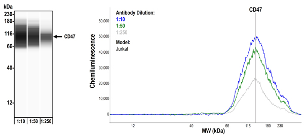
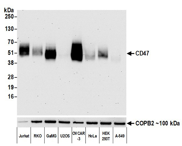 WB (Western Blot)
(Detection of human CD47 by western blot. Samples: Whole cell lysate (2 ug) from Jurkat, RKO, GaMG, U2OS, OVCAR-3, HeLa, HEK293T, and A-549 cells prepared using NETN lysis buffer. Antibody: Rabbit anti-CD47 recombinant monoclonal antibody (AAA213595 Lot 1) used at 1:1000. Secondary: HRP-conjugated goat anti-rabbit IgG . Detection: Chemiluminescence with an exposure time of 75 seconds. Lower Panel: Rabbit anti-COPB2 antibody .)
WB (Western Blot)
(Detection of human CD47 by western blot. Samples: Whole cell lysate (2 ug) from Jurkat, RKO, GaMG, U2OS, OVCAR-3, HeLa, HEK293T, and A-549 cells prepared using NETN lysis buffer. Antibody: Rabbit anti-CD47 recombinant monoclonal antibody (AAA213595 Lot 1) used at 1:1000. Secondary: HRP-conjugated goat anti-rabbit IgG . Detection: Chemiluminescence with an exposure time of 75 seconds. Lower Panel: Rabbit anti-COPB2 antibody .)
CD47, Monoclonal Recombinant Antibody (Cat# AAA213595)
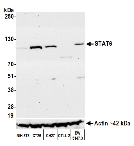
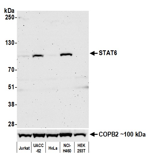 WB (Western Blot)
(Detection of human STAT6 by western blot. Samples: Whole cell lysate (50 ug) from Jurkat, UACC-62, HeLa, NCI-H460, and HEK293T cells prepared using NETN lysis buffer. Antibody: Rabbit anti-STAT6 recombinant monoclonal antibody (AAA213606 lot 1) used at 1:1000. Secondary: HRP-conjugated goat anti-rabbit IgG . Detection: Chemiluminescence with an exposure time of 3 minutes. Lower Panel: Rabbit anti-COPB2 antibody .)
WB (Western Blot)
(Detection of human STAT6 by western blot. Samples: Whole cell lysate (50 ug) from Jurkat, UACC-62, HeLa, NCI-H460, and HEK293T cells prepared using NETN lysis buffer. Antibody: Rabbit anti-STAT6 recombinant monoclonal antibody (AAA213606 lot 1) used at 1:1000. Secondary: HRP-conjugated goat anti-rabbit IgG . Detection: Chemiluminescence with an exposure time of 3 minutes. Lower Panel: Rabbit anti-COPB2 antibody .)
STAT6, Monoclonal Recombinant Antibody (Cat# AAA213606)
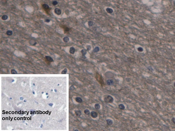
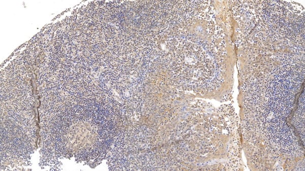
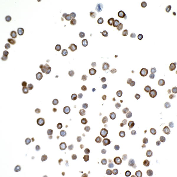
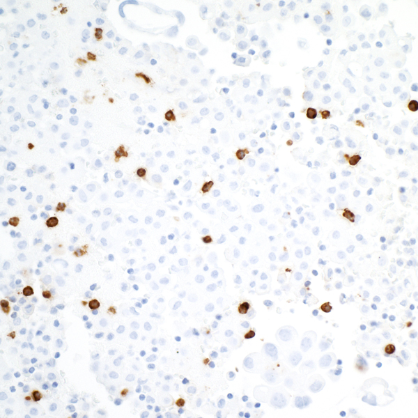
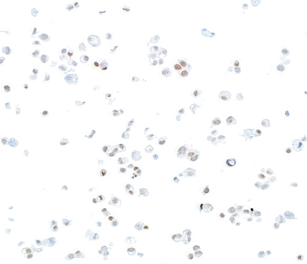
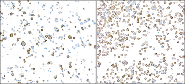
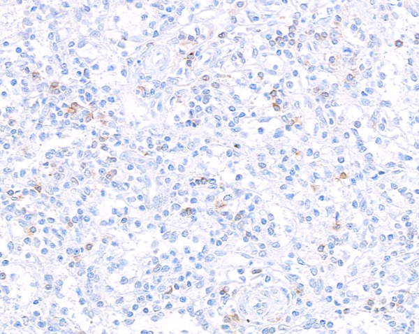
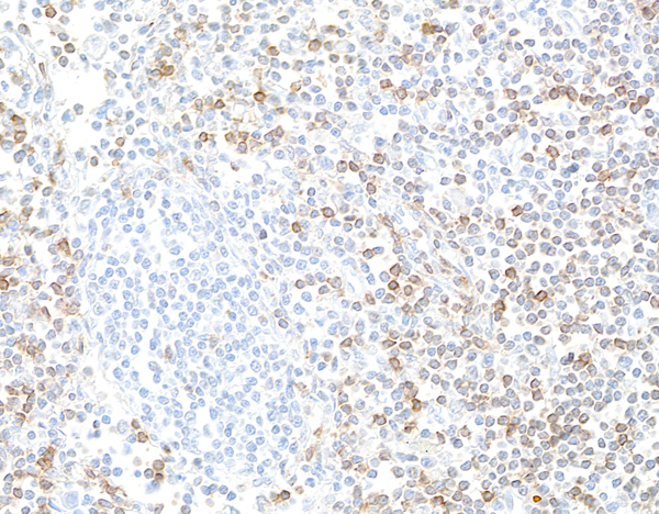
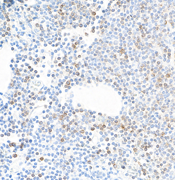
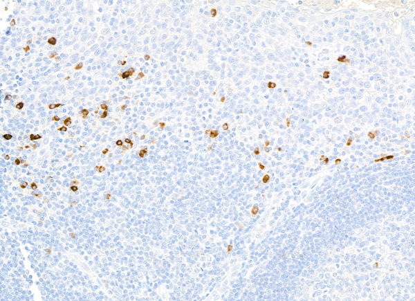
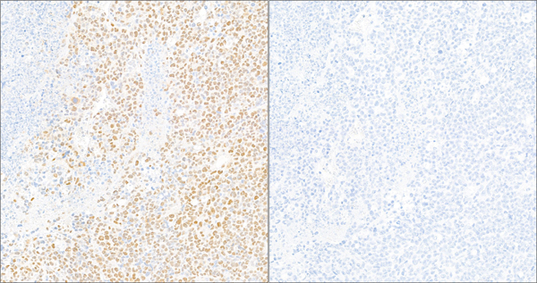
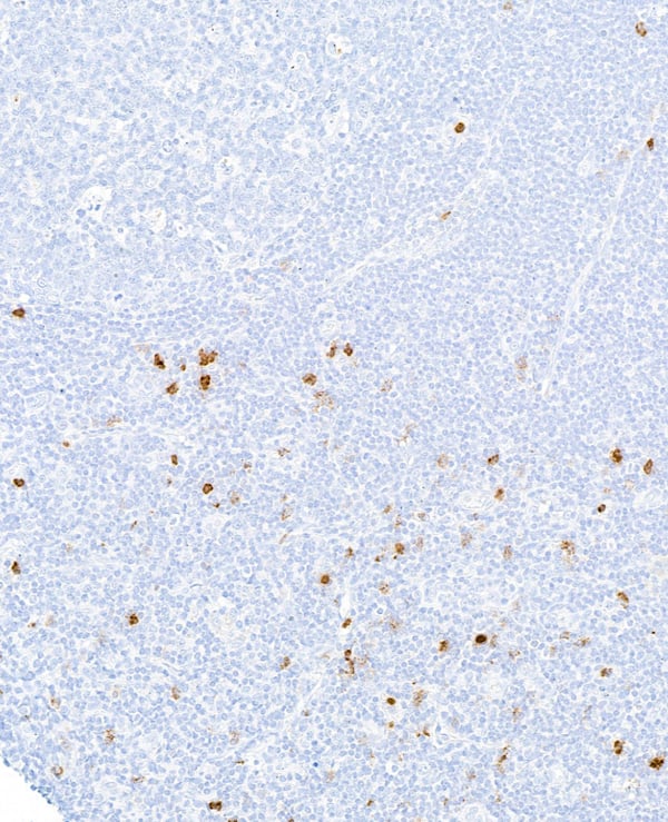 IHC (Immunohistochemisry)
(Detection of human CD25/IL-2R alpha in FFPE tonsil by immunohistochemistry. Antibody: Rabbit anti-CD25/IL-2R alpha recombinant monoclonal antibody (AAA213615 lot 1). Secondary: HRP-conjugated goat anti-rabbit IgG . Substrate: DAB.)
IHC (Immunohistochemisry)
(Detection of human CD25/IL-2R alpha in FFPE tonsil by immunohistochemistry. Antibody: Rabbit anti-CD25/IL-2R alpha recombinant monoclonal antibody (AAA213615 lot 1). Secondary: HRP-conjugated goat anti-rabbit IgG . Substrate: DAB.)
CD25/IL-2R alpha, Monoclonal Recombinant Antibody (Cat# AAA213615)
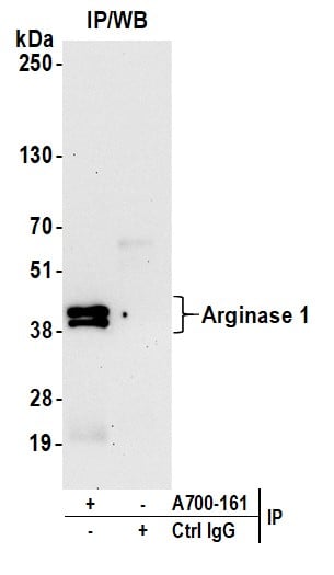
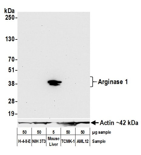 WB (Western Blot)
(Detection of mouse Arginase 1 by western blot. Samples: Lysate from from H-4-II-E, NIH 3T3, mouse liver, TCMK-1, and AML12 cells. Antibody: Rabbit anti-Arginase-1 recombinant monoclonal antibody (AAA213618 lot 1) used at 1:1000. Secondary: HRP-conjugated goat anti-rabbit IgG . Detection: Chemiluminescence with an exposure time of 30 seconds. Lower Panel: Rabbit anti-Actin recombinant monoclonal antibody .)
WB (Western Blot)
(Detection of mouse Arginase 1 by western blot. Samples: Lysate from from H-4-II-E, NIH 3T3, mouse liver, TCMK-1, and AML12 cells. Antibody: Rabbit anti-Arginase-1 recombinant monoclonal antibody (AAA213618 lot 1) used at 1:1000. Secondary: HRP-conjugated goat anti-rabbit IgG . Detection: Chemiluminescence with an exposure time of 30 seconds. Lower Panel: Rabbit anti-Actin recombinant monoclonal antibody .)
Arginase 1, Monoclonal Recombinant Antibody (Cat# AAA213618)

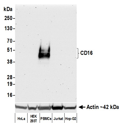 WB (Western Blot)
(Detection of human CD16 by western blot. Samples: Whole cell lysate (50 ug) from HeLa, HEK293T, PBMCs, Jurkat, and Hep-G2 cells prepared using NETN lysis buffer. Antibody: Rabbit anti-CD16 recombinant monoclonal antibody (AAA213620 lot 1) used at 1:1000. Secondary: HRP-conjugated goat anti-rabbit IgG . Detection: Chemiluminescence with an exposure time of 75 seconds. Lower Panel: Rabbit anti-Actin recombinant monoclonal antibody .)
WB (Western Blot)
(Detection of human CD16 by western blot. Samples: Whole cell lysate (50 ug) from HeLa, HEK293T, PBMCs, Jurkat, and Hep-G2 cells prepared using NETN lysis buffer. Antibody: Rabbit anti-CD16 recombinant monoclonal antibody (AAA213620 lot 1) used at 1:1000. Secondary: HRP-conjugated goat anti-rabbit IgG . Detection: Chemiluminescence with an exposure time of 75 seconds. Lower Panel: Rabbit anti-Actin recombinant monoclonal antibody .)
CD16, Monoclonal Recombinant Antibody (Cat# AAA213620)
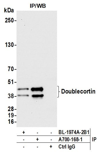
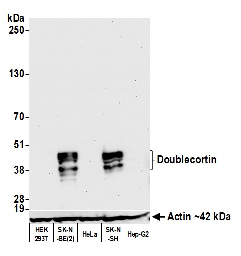 WB (Western Blot)
(Detection of human Doublecortin by western blot. Samples: Whole cell lysate (50 ug) from HEK293T, SK-N-BE(2), HeLa, SK-N-SH, and Hep-G2 cells prepared using NETN lysis buffer. Antibody: Rabbit anti-Doublecortin recombinant monoclonal antibody (AAA213622 lot 1) used at 1:1000. Secondary: HRP-conjugated goat anti-rabbit IgG . Detection: Chemiluminescence with an exposure time of 30 seconds.)
WB (Western Blot)
(Detection of human Doublecortin by western blot. Samples: Whole cell lysate (50 ug) from HEK293T, SK-N-BE(2), HeLa, SK-N-SH, and Hep-G2 cells prepared using NETN lysis buffer. Antibody: Rabbit anti-Doublecortin recombinant monoclonal antibody (AAA213622 lot 1) used at 1:1000. Secondary: HRP-conjugated goat anti-rabbit IgG . Detection: Chemiluminescence with an exposure time of 30 seconds.)
Doublecortin, Monoclonal Recombinant Antibody (Cat# AAA213622)
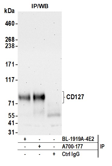
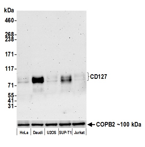 WB (Western Blot)
(Detection of human CD127/IL-7R alpha by western blot. Samples: Whole cell lysate (25 ug) from HeLa, Daudi, U2OS, SUP-T1, and Jurkat cells prepared using NETN lysis buffer. Antibody: Rabbit anti-CD127 recombinant monoclonal antibody (AAA213628 lot 1) used at 1:1000. Secondary: HRP-conjugated goat anti-rabbit IgG . Detection: Chemiluminescence with an exposure time of 3 minutes. Lower Panel: Rabbit anti-COPB2 antibody .)
WB (Western Blot)
(Detection of human CD127/IL-7R alpha by western blot. Samples: Whole cell lysate (25 ug) from HeLa, Daudi, U2OS, SUP-T1, and Jurkat cells prepared using NETN lysis buffer. Antibody: Rabbit anti-CD127 recombinant monoclonal antibody (AAA213628 lot 1) used at 1:1000. Secondary: HRP-conjugated goat anti-rabbit IgG . Detection: Chemiluminescence with an exposure time of 3 minutes. Lower Panel: Rabbit anti-COPB2 antibody .)
CD127/IL-7R alpha, Monoclonal Recombinant Antibody (Cat# AAA213628)
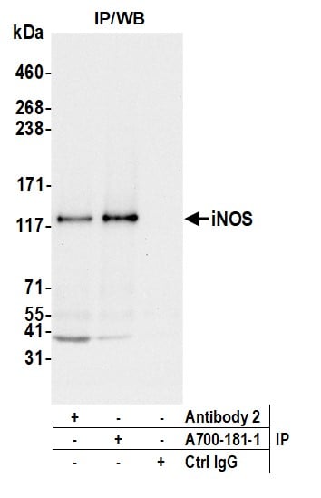
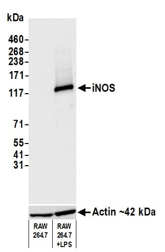 WB (Western Blot)
(Detection of mouse iNOS by western blot. Samples: Whole cell lysate (10 ug) from RAW 264.7 and LPS-treated RAW 264.7 cells prepared using NETN lysis buffer. Antibody: Rabbit anti-iNOS recombinant monoclonal antibody (AAA213630 lot 1) used at 1:1000. Secondary: HRP-conjugated goat anti-rabbit IgG . Detection: Chemiluminescence with an exposure time of 3 seconds. Lower Panel: Rabbit anti-Actin recombinant monoclonal antibody .)
WB (Western Blot)
(Detection of mouse iNOS by western blot. Samples: Whole cell lysate (10 ug) from RAW 264.7 and LPS-treated RAW 264.7 cells prepared using NETN lysis buffer. Antibody: Rabbit anti-iNOS recombinant monoclonal antibody (AAA213630 lot 1) used at 1:1000. Secondary: HRP-conjugated goat anti-rabbit IgG . Detection: Chemiluminescence with an exposure time of 3 seconds. Lower Panel: Rabbit anti-Actin recombinant monoclonal antibody .)
iNOS, Monoclonal Recombinant Antibody (Cat# AAA213630)
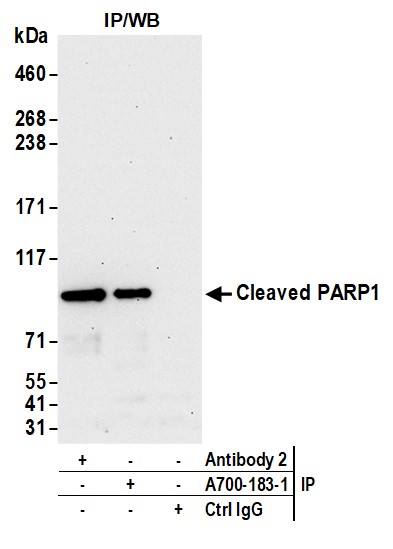
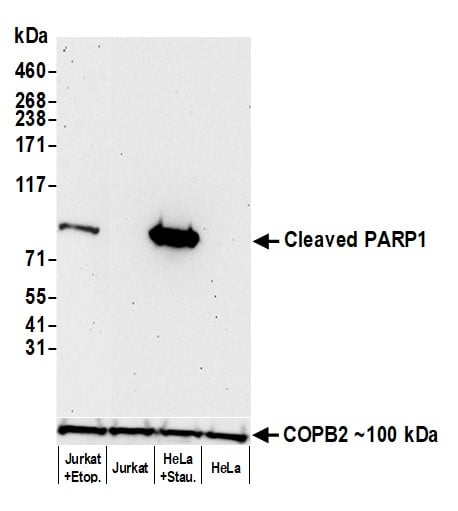 WB (Western Blot)
(Detection of human Cleaved PARP1 by western blot. Samples: Whole cell lysate (50 ug) from etoposide-treated Jurkat cells, untreated Jurkat cells, 1 uM staurosporine-treated HeLa cells, and untreated HeLa cells prepared using NETN lysis buffer. Antibody: Rabbit anti-Cleaved PARP1 recombinant monoclonal antibody (AAA213631 lot 1) used at 1:1000. Secondary: HRP-conjugated goat anti-rabbit IgG . Detection: Chemiluminescence with an exposure time of 3 minutes. Lower Panel: Rabbit anti-COPB2 antibody .)
WB (Western Blot)
(Detection of human Cleaved PARP1 by western blot. Samples: Whole cell lysate (50 ug) from etoposide-treated Jurkat cells, untreated Jurkat cells, 1 uM staurosporine-treated HeLa cells, and untreated HeLa cells prepared using NETN lysis buffer. Antibody: Rabbit anti-Cleaved PARP1 recombinant monoclonal antibody (AAA213631 lot 1) used at 1:1000. Secondary: HRP-conjugated goat anti-rabbit IgG . Detection: Chemiluminescence with an exposure time of 3 minutes. Lower Panel: Rabbit anti-COPB2 antibody .)
Cleaved PARP1, Monoclonal Recombinant Antibody (Cat# AAA213631)
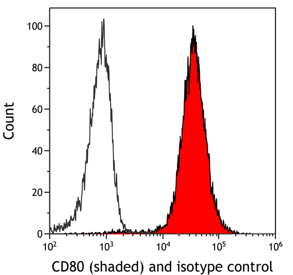
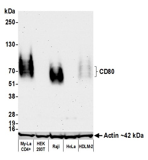 WB (Western Blot)
(Detection of human CD80 by western blot. Samples: Whole cell lysate (50 ug) from My-La CD4+, HEK293T, Raji, HeLa, and HDLM-2 cells prepared using NETN lysis buffer. Antibody: Rabbit anti-CD80 recombinant monoclonal antibody (AAA213639 lot 1) used at 1:1000. Secondary: HRP-conjugated goat anti-rabbit IgG . Detection: Chemiluminescence with an exposure time of 75 seconds. Lower Panel: Rabbit anti-Actin recombinant monoclonal antibody .)
WB (Western Blot)
(Detection of human CD80 by western blot. Samples: Whole cell lysate (50 ug) from My-La CD4+, HEK293T, Raji, HeLa, and HDLM-2 cells prepared using NETN lysis buffer. Antibody: Rabbit anti-CD80 recombinant monoclonal antibody (AAA213639 lot 1) used at 1:1000. Secondary: HRP-conjugated goat anti-rabbit IgG . Detection: Chemiluminescence with an exposure time of 75 seconds. Lower Panel: Rabbit anti-Actin recombinant monoclonal antibody .)
CD80, Monoclonal Recombinant Antibody (Cat# AAA213639)
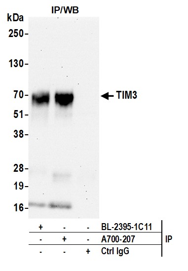
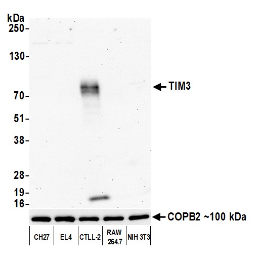 WB (Western Blot)
(Detection of mouse TIM3 by western blot. Samples: Whole cell lysate (10 ug) from CH27, EL4, CTLL-2, RAW 264.7, and NIH 3T3 cells prepared using NETN lysis buffer. Antibody: Rabbit anti-TIM3 recombinant monoclonal antibody (AAA213640 lot 1) used at 1:1000. Secondary: HRP-conjugated goat anti-rabbit IgG . Detection: Chemiluminescence with an exposure time of 30 seconds. Lower Panel: Rabbit anti-COPB2 antibody .)
WB (Western Blot)
(Detection of mouse TIM3 by western blot. Samples: Whole cell lysate (10 ug) from CH27, EL4, CTLL-2, RAW 264.7, and NIH 3T3 cells prepared using NETN lysis buffer. Antibody: Rabbit anti-TIM3 recombinant monoclonal antibody (AAA213640 lot 1) used at 1:1000. Secondary: HRP-conjugated goat anti-rabbit IgG . Detection: Chemiluminescence with an exposure time of 30 seconds. Lower Panel: Rabbit anti-COPB2 antibody .)
TIM3, Monoclonal Recombinant Antibody (Cat# AAA213640)
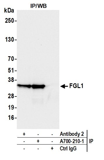
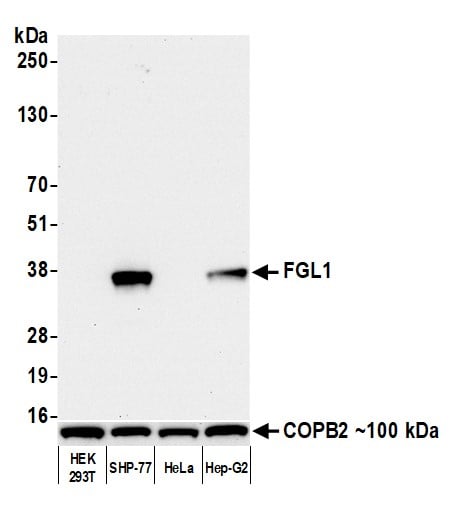 WB (Western Blot)
(Detection of human Hepassocin/FGL1 by western blot. Samples: Whole cell lysate (10 ug) from HEK293T, SHP-77, HeLa, and Hep-G2 cells prepared using NETN lysis buffer. Antibody: Rabbit anti-Hepassocin/FGL1 recombinant monoclonal antibody (AAA213642 lot 1) used at 1:1000. Secondary: HRP-conjugated goat anti-rabbit IgG . Detection: Chemiluminescence with an exposure time of 10 seconds. Lower Panel: Rabbit anti-COPB2 antibody .)
WB (Western Blot)
(Detection of human Hepassocin/FGL1 by western blot. Samples: Whole cell lysate (10 ug) from HEK293T, SHP-77, HeLa, and Hep-G2 cells prepared using NETN lysis buffer. Antibody: Rabbit anti-Hepassocin/FGL1 recombinant monoclonal antibody (AAA213642 lot 1) used at 1:1000. Secondary: HRP-conjugated goat anti-rabbit IgG . Detection: Chemiluminescence with an exposure time of 10 seconds. Lower Panel: Rabbit anti-COPB2 antibody .)
Hepassocin/FGL1, Monoclonal Recombinant Antibody (Cat# AAA213642)


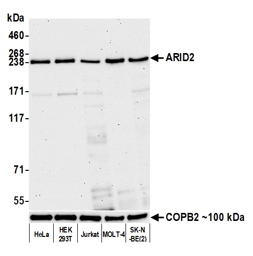 WB (Western Blot)
(Detection of human ARID2 by western blot. Samples: Whole cell lysate (10 ug) from HeLa, HEK293T, Jurkat, MOLT-4, and SK-N-BE(2) cells prepared using NETN lysis buffer. Antibody: Rabbit anti-ARID2 recombinant monoclonal antibody (AAA213643 lot 1) used at 1:1000. Secondary: HRP-conjugated goat anti-rabbit IgG . Detection: Chemiluminescence with an exposure time of 75 seconds. Lower Panel: Rabbit anti-COPB2 antibody .)
WB (Western Blot)
(Detection of human ARID2 by western blot. Samples: Whole cell lysate (10 ug) from HeLa, HEK293T, Jurkat, MOLT-4, and SK-N-BE(2) cells prepared using NETN lysis buffer. Antibody: Rabbit anti-ARID2 recombinant monoclonal antibody (AAA213643 lot 1) used at 1:1000. Secondary: HRP-conjugated goat anti-rabbit IgG . Detection: Chemiluminescence with an exposure time of 75 seconds. Lower Panel: Rabbit anti-COPB2 antibody .)
ARID2, Monoclonal Recombinant Antibody (Cat# AAA213643)
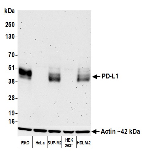 WB (Western Blot)
(Detection of human PD-L1 by western blot. Samples: Whole cell lysate (25 ug) from RKO, HeLa, SUP-M2, HEK293T, and HDLM-2 cells prepared using NETN lysis buffer. Antibody: Rabbit anti-PD-L1 recombinant monoclonal antibody (AAA213649 lot 1) used at 1:1000. Secondary: HRP-conjugated goat anti-rabbit IgG . Detection: Chemiluminescence with an exposure time of 3 minutes. Lower Panel: Rabbit anti-Actin recombinant monoclonal antibody .)
WB (Western Blot)
(Detection of human PD-L1 by western blot. Samples: Whole cell lysate (25 ug) from RKO, HeLa, SUP-M2, HEK293T, and HDLM-2 cells prepared using NETN lysis buffer. Antibody: Rabbit anti-PD-L1 recombinant monoclonal antibody (AAA213649 lot 1) used at 1:1000. Secondary: HRP-conjugated goat anti-rabbit IgG . Detection: Chemiluminescence with an exposure time of 3 minutes. Lower Panel: Rabbit anti-Actin recombinant monoclonal antibody .)
PD-L1, Monoclonal Recombinant Antibody (Cat# AAA213649)
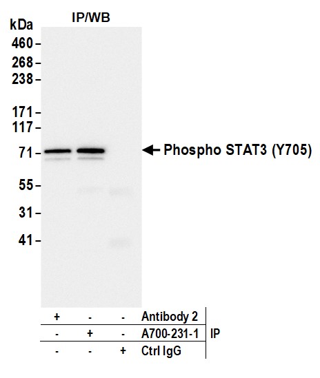
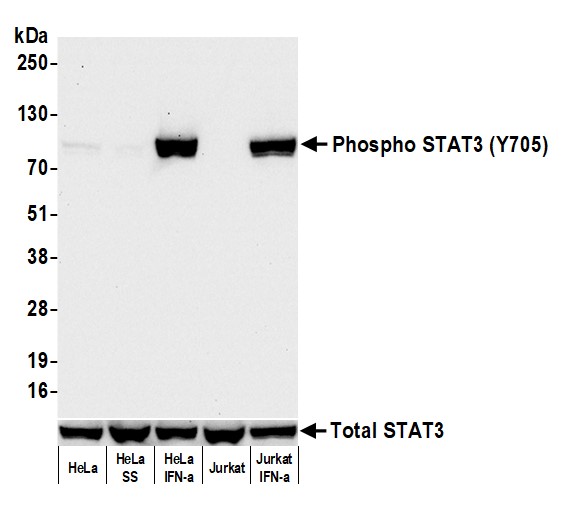 WB (Western Blot)
(Detection of human Phospho STAT3 (Y705) by western blot. Samples: Whole cell lysate (50 ug) from HeLa, HeLa (serum starved), HeLa (IFN-alpha), Jurkat, and Jurkat (IFN-alpha) cells prepared using NETN lysis buffer. Antibody: Rabbit anti-Phospho STAT3 (Y705) recombinant monoclonal antibody (AAA213650 lot 1) used at 1:1000. Secondary: HRP-conjugated goat anti-rabbit IgG . Detection: Chemiluminescence with an exposure time of 10 seconds. Lower Panel: Rabbit anti-STAT3 recombinant monoclonal antibody .)
WB (Western Blot)
(Detection of human Phospho STAT3 (Y705) by western blot. Samples: Whole cell lysate (50 ug) from HeLa, HeLa (serum starved), HeLa (IFN-alpha), Jurkat, and Jurkat (IFN-alpha) cells prepared using NETN lysis buffer. Antibody: Rabbit anti-Phospho STAT3 (Y705) recombinant monoclonal antibody (AAA213650 lot 1) used at 1:1000. Secondary: HRP-conjugated goat anti-rabbit IgG . Detection: Chemiluminescence with an exposure time of 10 seconds. Lower Panel: Rabbit anti-STAT3 recombinant monoclonal antibody .)
STAT3-pY705, Monoclonal Recombinant Antibody (Cat# AAA213650)
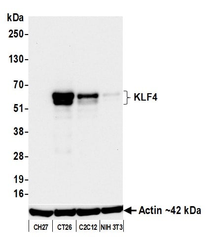
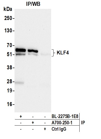
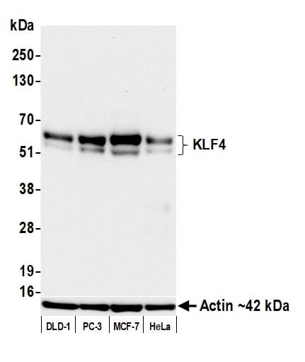 WB (Western Blot)
(Detection of human KLF4 by western blot. Samples: Whole cell lysate (25 ug) from DLD-1, PC-3, MCF-7, and HeLa cells prepared using NETN lysis buffer. Antibody: Rabbit anti-KLF4 recombinant monoclonal antibody (AAA213663 lot 1) used at 1:1000. Secondary: HRP-conjugated goat anti-rabbit IgG . Detection: Chemiluminescence with an exposure time of 30 seconds. Lower Panel: Rabbit anti-Actin recombinant monoclonal antibody .)
WB (Western Blot)
(Detection of human KLF4 by western blot. Samples: Whole cell lysate (25 ug) from DLD-1, PC-3, MCF-7, and HeLa cells prepared using NETN lysis buffer. Antibody: Rabbit anti-KLF4 recombinant monoclonal antibody (AAA213663 lot 1) used at 1:1000. Secondary: HRP-conjugated goat anti-rabbit IgG . Detection: Chemiluminescence with an exposure time of 30 seconds. Lower Panel: Rabbit anti-Actin recombinant monoclonal antibody .)
KLF4, Monoclonal Recombinant Antibody (Cat# AAA213663)
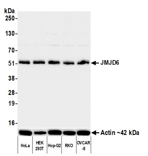
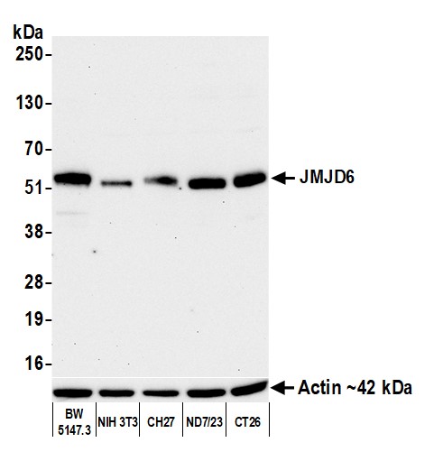 WB (Western Blot)
(Detection of mouse JMJD6 by western blot. Samples: Whole cell lysate (10 ug) from BW5147.3, NIH 3T3, CH27, ND7/23, and CT26 cells prepared using NETN lysis buffer. Antibody: Rabbit anti-JMJD6 recombinant monoclonal antibody (AAA213667 lot 1) used at 1:1000. Secondary: HRP-conjugated goat anti-rabbit IgG . Detection: Chemiluminescence with an exposure time of 75 seconds. Lower Panel: Rabbit anti-Actin recombinant monoclonal antibody .)
WB (Western Blot)
(Detection of mouse JMJD6 by western blot. Samples: Whole cell lysate (10 ug) from BW5147.3, NIH 3T3, CH27, ND7/23, and CT26 cells prepared using NETN lysis buffer. Antibody: Rabbit anti-JMJD6 recombinant monoclonal antibody (AAA213667 lot 1) used at 1:1000. Secondary: HRP-conjugated goat anti-rabbit IgG . Detection: Chemiluminescence with an exposure time of 75 seconds. Lower Panel: Rabbit anti-Actin recombinant monoclonal antibody .)
JMJD6, Monoclonal Recombinant Antibody (Cat# AAA213667)
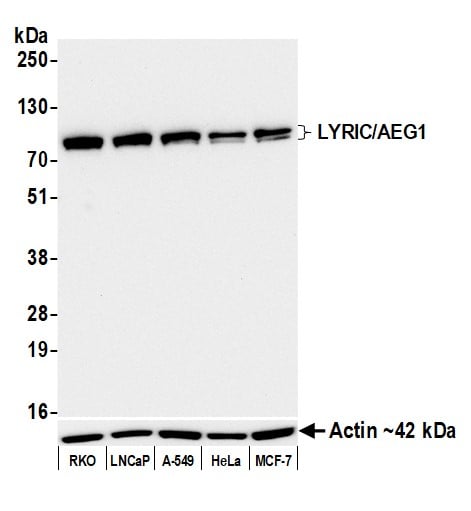
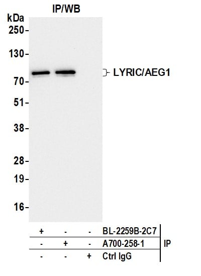
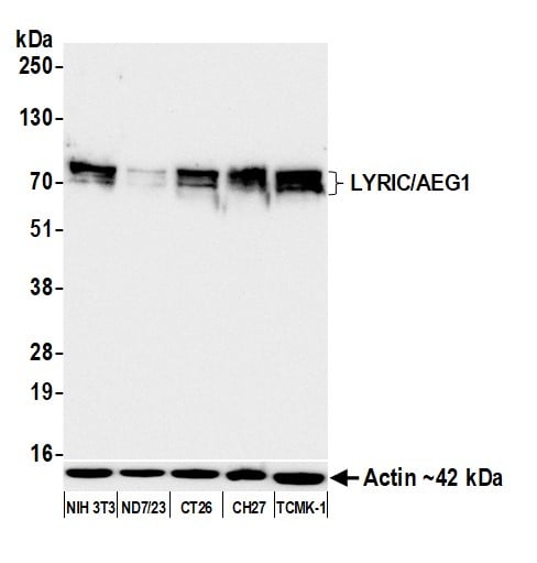 WB (Western Blot)
(Detection of mouse LYRIC/AEG1 by western blot. Samples: Whole cell lysate (25 ug) from NIH 3T3, ND7/23, CT26, CH27, and TCMK-1 cells prepared using NETN lysis buffer. Antibody: Rabbit anti-LYRIC/AEG1 recombinant monoclonal antibody (AAA213670 lot 1) used at 1:1000. Secondary: HRP-conjugated goat anti-rabbit IgG . Detection: Chemiluminescence with an exposure time of 10 seconds. Lower Panel: Rabbit anti-Actin recombinant monoclonal antibody .)
WB (Western Blot)
(Detection of mouse LYRIC/AEG1 by western blot. Samples: Whole cell lysate (25 ug) from NIH 3T3, ND7/23, CT26, CH27, and TCMK-1 cells prepared using NETN lysis buffer. Antibody: Rabbit anti-LYRIC/AEG1 recombinant monoclonal antibody (AAA213670 lot 1) used at 1:1000. Secondary: HRP-conjugated goat anti-rabbit IgG . Detection: Chemiluminescence with an exposure time of 10 seconds. Lower Panel: Rabbit anti-Actin recombinant monoclonal antibody .)
LYRIC/AEG1, Monoclonal Recombinant Antibody (Cat# AAA213670)
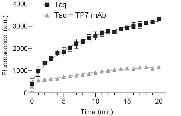
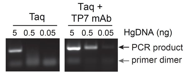 Application Data
(Anti-Taq [TP7] decreases primer dimer formation and increases PCR sensitivity. Antibody: Mouse anti-Taq Polymerase Recombinant Monoclonal Antibody [TP7] (AAA213680 lot 1). Amplification of a 306 basepair region of the human Numb gene was performed with forward and reverse primers that are prone to low temperature annealing due to complementarity at their 3' ends (Kubu. Biotechniques. 2008). At ambient temperatures, Taq DNA polymerase will extend the annealed primer dimers and create a low molecular weight product that prevents proper amplification of the target sequence. At low template abundance ()
Application Data
(Anti-Taq [TP7] decreases primer dimer formation and increases PCR sensitivity. Antibody: Mouse anti-Taq Polymerase Recombinant Monoclonal Antibody [TP7] (AAA213680 lot 1). Amplification of a 306 basepair region of the human Numb gene was performed with forward and reverse primers that are prone to low temperature annealing due to complementarity at their 3' ends (Kubu. Biotechniques. 2008). At ambient temperatures, Taq DNA polymerase will extend the annealed primer dimers and create a low molecular weight product that prevents proper amplification of the target sequence. At low template abundance ()
Taq Polymerase, Monoclonal Recombinant Antibody (Cat# AAA213680)
What are Monoclonal Antibodies?
Monoclonal antibodies are specialized laboratory-produced proteins developed for binding to specific biological antigens or other molecular targets. Since they come from a single cell (or clone), they are especially consistent and accurate in the data they are involved in producing.
This type of antibody material has been shown to be a powerful tool in finding and subsequently destroying harmful cells in an organism, such as those found in cancers or various autoimmune diseases. This makes them excellent aids in medical testing and research, which is why they are so widely used.
AAA Biotech offers a comprehensive range of high-quality monoclonal antibodies that perform effectively in various laboratory tests, including (amongst others) ELISA, western blotting, immunohistochemistry, and flow cytometry. All of the products in our catalog are thoroughly quality tested to make sure that they are reliable and will consistently perform well in your research.
What Are The Uses of Monoclonal Antibodies
Monoclonal antibodies are used in many lab tests, including (amongst others) ELISA, western blotting, immunohistochemistry, and flow cytometry.
ELISA is a test that helps detect a specific substance/analyte in a sample. It uses antibodies (often monoclonal) bound to a solid surface (such as the well of a microplate) to “capture” the substance/analyte in the sample and immobilize it so that the detection antibody component can then bind to it and produce a signal, which can then be measured.
Western blotting identifies specific proteins in a sample. The sample is first separated on a gel, and then antibodies are applied that will typically bind to the target, which will all be localized to a single band in a lane.
Immunohistochemistry helps locate specific proteins in cells or tissue samples using antibodies.
Flow cytometry looks at and sorts cells. It uses antibodies that are conjugated to reporter molecules called “fluorophores”, which, under special lights, emit light themselves, which can then be measured by a detector instrument. For a deeper understanding of these techniques, explore our complete guide to monoclonal antibodies and their benefits.
How Monoclonal Antibodies Are Used as Medicine?
Please note that all of the products listed in AAA Biotech’s also known as AAA Bio or AAABio catalog are strictly for research-use only (RUO).
Monoclonal antibodies can also be used as therapeutic/medical treatments, particularly in the context of cancers. They are designed to find and bind to specific cells or proteins, helping the immune system recognize and attack the cancer. These treatments work in different ways, such as:
- Radioimmunotherapy attaches a small amount of radioactive molecule to the antibody, so it delivers the radiation directly to the cancer cells that the antibody is specifically binding to.
- Antibody-directed enzyme prodrug therapy uses antibodies that are specifically bound to special enzymes. These enzymes activate a harmless drug in the body and turn it into a cancer-killing drug only near the cancer cells—this helps avoid harming healthy cells.
- Immunoliposomes are tiny “bubbles” filled with medicine/drug and coated with antibodies. They carry the drug straight to the cancer cells.
Why Buy Monoclonal Antibodies From Us?
At AAA Biotech, we provide high-performance monoclonal antibodies designed to support a wide range of research needs.
1. Validated for Versatile Applications
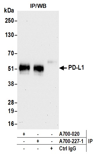
The antibodies in our catalog are extensively validated and compatible with multiple techniques, including (but not limited to) ELISA, flow cytometry (FC), immunocytochemistry (ICC), immunofluorescence (IF), immunohistochemistry (IHC), immunoprecipitation (IP), and western blotting (WB).
2. Wide Selection & Specialized Options
We offer antibodies for common and rare species, that are available in various conjugated forms, and also in recombinant formats. Essentially, there is almost anything one might need to meet their experimental model’s requirements.
3. High-Quality Proteins
Our proteins meet high purity standards—90% or more as confirmed by SDS-PAGE. Many are available with tags like His, Flag, GST, or MBP, and we also supply native and biologically active proteins for functional studies.
Frequently Asked Questions
1. Are your monoclonal antibodies validated for specific applications?
Yes, our antibodies are tested and validated for use in methods such as ELISA, western blot, IHC, flow cytometry, and more. Refer to specific product pages or datasheets for individual product information.
2. How do I choose the right monoclonal antibody for my application?
Review the product details directly for application validation, species reactivity, and target information. You may also contact our support team at any time for help.
3. How quickly can I receive my order?
Most orders are processed and shipped within 1–3 business days, depending on product availability and your shipping location.