Filters
▼Clonality
▼Type
▼Reactivity
▼Gene Name
▼Isotype
▼Host
▼Application
▼Clone
▼Monoclonal Antibodies
Get accurate results in your research with our Monoclonal Antibodies, which are specially made to target exactly what you require for your research, and will produce consistent, reliable performance in lab tests.
Viewing 500-550 of 27645 product results
 WB (Western Blot)
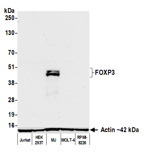
(Detection of human FOXP3 by western blot. Samples: Whole cell lysate (50 ug) from Jurkat, HEK293T, MJ, MOLT-4, and RPMI-8226 cells prepared using NETN lysis buffer. Antibody: Rabbit anti-FOXP3 recombinant monoclonal antibody (AAA213684 lot 1) used at 1:1000. Secondary: HRP-conjugated goat anti-rabbit IgG . Detection: Chemiluminescence with an exposure time of 30 seconds. Lower Panel: Rabbit anti-Actin recombinant monoclonal antibody .)
WB (Western Blot)
(Detection of human FOXP3 by western blot. Samples: Whole cell lysate (50 ug) from Jurkat, HEK293T, MJ, MOLT-4, and RPMI-8226 cells prepared using NETN lysis buffer. Antibody: Rabbit anti-FOXP3 recombinant monoclonal antibody (AAA213684 lot 1) used at 1:1000. Secondary: HRP-conjugated goat anti-rabbit IgG . Detection: Chemiluminescence with an exposure time of 30 seconds. Lower Panel: Rabbit anti-Actin recombinant monoclonal antibody .)
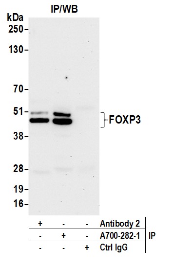
FOXP3, Monoclonal Recombinant Antibody (Cat# AAA213684)
 WB (Western Blot)
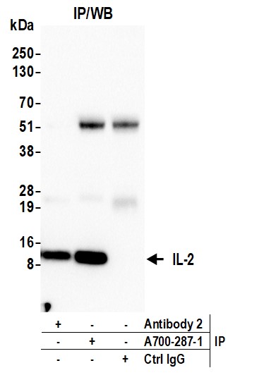
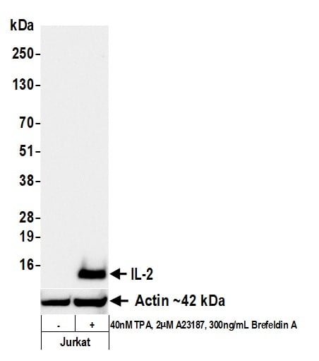
(Detection of human IL-2 by western blot. Samples: Whole cell lysate (50 ug) from Jurkat cells untreated (-) or treated (+) with 40nM TPA, 2uM A23187, and 300ng/mL Brefeldin A. Antibody: Rabbit anti-IL-2 recombinant monoclonal antibody (AAA213687 lot 1) used at 1:1000. Secondary: HRP-conjugated goat anti-rabbit IgG . Detection: Chemiluminescence with an exposure time of 30 seconds. Lower Panel: Rabbit anti-Actin recombinant monoclonal antibody .)
WB (Western Blot)
(Detection of human IL-2 by western blot. Samples: Whole cell lysate (50 ug) from Jurkat cells untreated (-) or treated (+) with 40nM TPA, 2uM A23187, and 300ng/mL Brefeldin A. Antibody: Rabbit anti-IL-2 recombinant monoclonal antibody (AAA213687 lot 1) used at 1:1000. Secondary: HRP-conjugated goat anti-rabbit IgG . Detection: Chemiluminescence with an exposure time of 30 seconds. Lower Panel: Rabbit anti-Actin recombinant monoclonal antibody .)
IL-2, Monoclonal Recombinant Antibody (Cat# AAA213687)
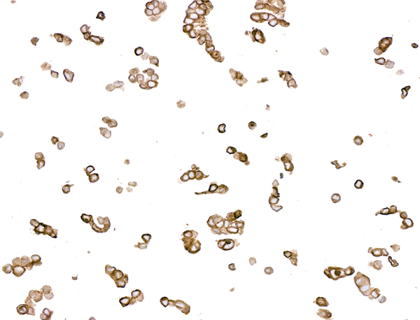
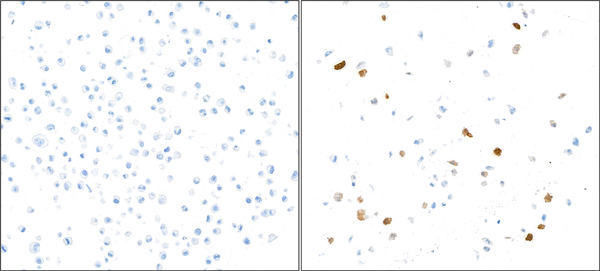
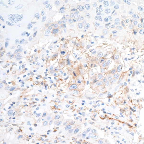
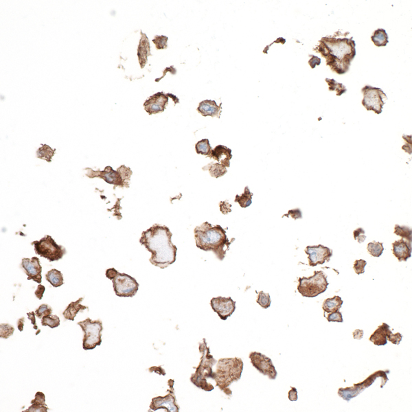
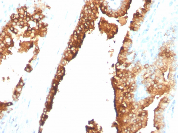
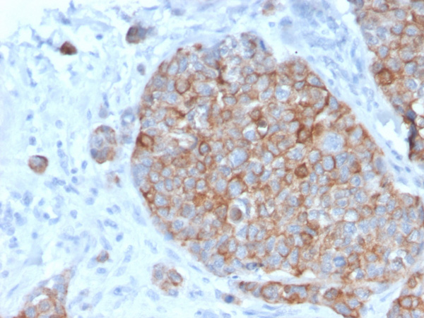
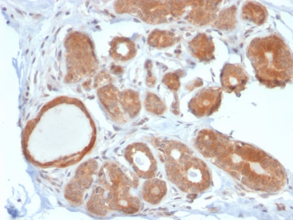
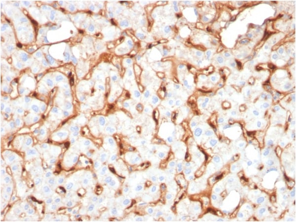
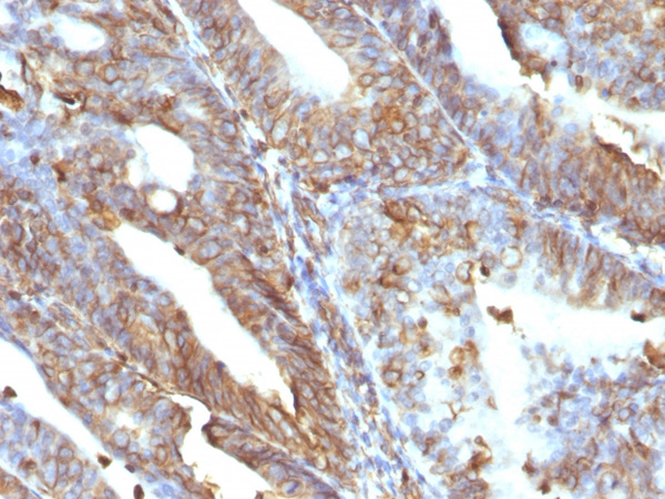
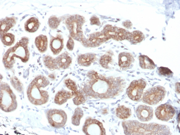
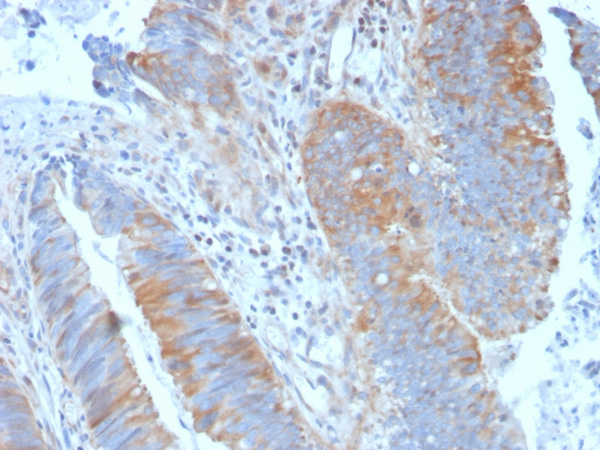
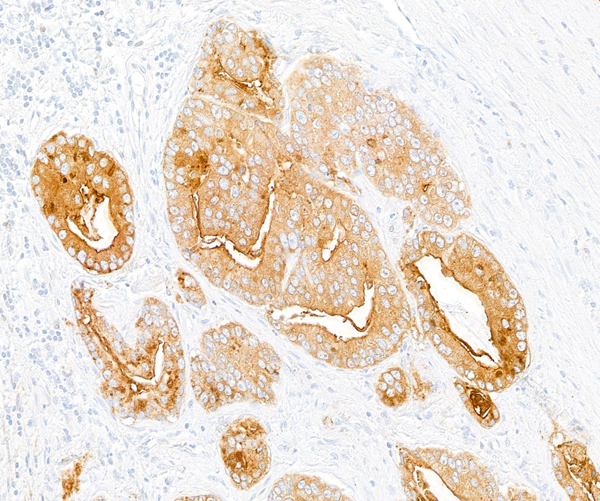 IHC (Immunohistochemistry)
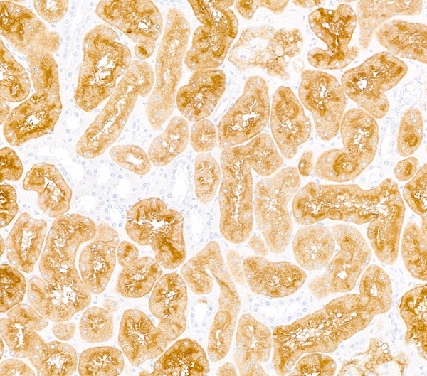
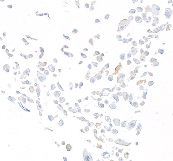
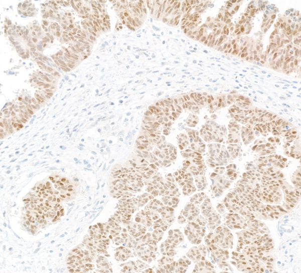
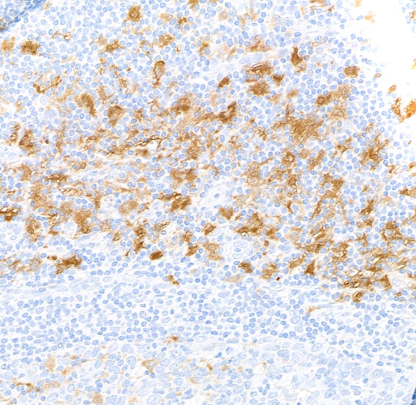
(Detection of human DPP4/CD26 by immunohistochemistry. Sample: FFPE section of prostate carcinoma. Antibody: Rabbit anti-DPP4/CD26 recombinant monoclonal antibody [BLR288L) (AAA213688). Secondary: HRP-conjugated goat anti-rabbit IgG .)
IHC (Immunohistochemistry)
(Detection of human DPP4/CD26 by immunohistochemistry. Sample: FFPE section of prostate carcinoma. Antibody: Rabbit anti-DPP4/CD26 recombinant monoclonal antibody [BLR288L) (AAA213688). Secondary: HRP-conjugated goat anti-rabbit IgG .)
DPP4/CD26, Monoclonal Recombinant Antibody (Cat# AAA213688)
 WB (Western Blot)
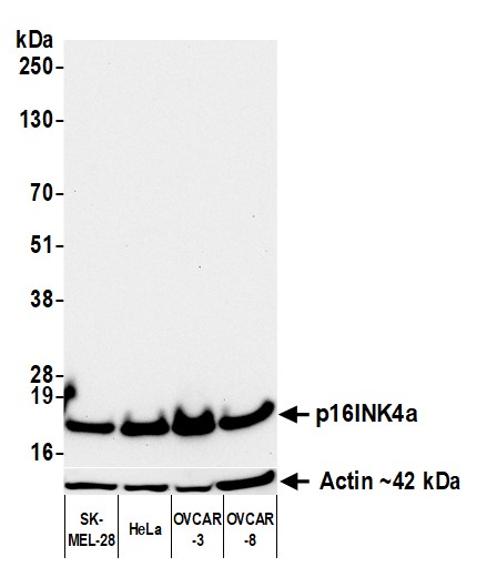
(Detection of human p16INK4a by western blot. Samples: Whole cell lysate (50 ug) from SK-MEL-28, HeLa, OVCAR-3, and OVCAR-8 cells prepared using NETN lysis buffer. Antibody: Rabbit anti-p16INK4a recombinant monoclonal antibody [BC42] (AAA213691 lot 1) used at 1:1000. Secondary: HRP-conjugated goat anti-rabbit IgG . Detection: Chemiluminescence with an exposure time of 30 seconds. Lower Panel: Rabbit anti-Actin recombinant monoclonal antibody .)
WB (Western Blot)
(Detection of human p16INK4a by western blot. Samples: Whole cell lysate (50 ug) from SK-MEL-28, HeLa, OVCAR-3, and OVCAR-8 cells prepared using NETN lysis buffer. Antibody: Rabbit anti-p16INK4a recombinant monoclonal antibody [BC42] (AAA213691 lot 1) used at 1:1000. Secondary: HRP-conjugated goat anti-rabbit IgG . Detection: Chemiluminescence with an exposure time of 30 seconds. Lower Panel: Rabbit anti-Actin recombinant monoclonal antibody .)
p16INK4a, Monoclonal Recombinant Antibody (Cat# AAA213691)
 WB (Western Blot)
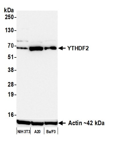
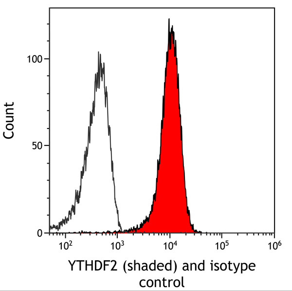
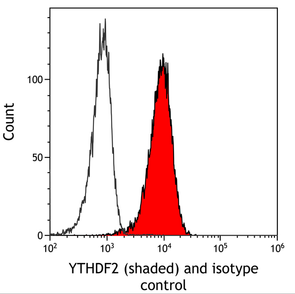
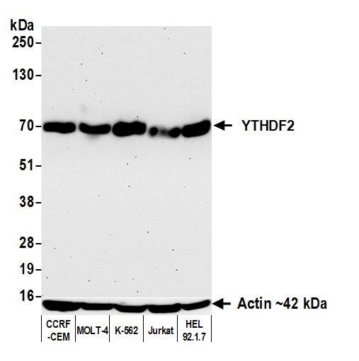
(Detection of human YTHDF2 by western blot. Samples: Whole cell lysate (50 ug) from CCRF-CEM, MOLT-4, K-562, Jurkat, and HEL 92.1.7 cells prepared using NETN lysis buffer. Antibody: Rabbit anti-YTHDF2 recombinant monoclonal antibody (AAA213695 lot 1) used at 1:1000. Secondary: HRP-conjugated goat anti-rabbit IgG . Detection: Chemiluminescence with an exposure time of 30 seconds. Lower Panel: Rabbit anti-Actin recombinant monoclonal antibody .)
WB (Western Blot)
(Detection of human YTHDF2 by western blot. Samples: Whole cell lysate (50 ug) from CCRF-CEM, MOLT-4, K-562, Jurkat, and HEL 92.1.7 cells prepared using NETN lysis buffer. Antibody: Rabbit anti-YTHDF2 recombinant monoclonal antibody (AAA213695 lot 1) used at 1:1000. Secondary: HRP-conjugated goat anti-rabbit IgG . Detection: Chemiluminescence with an exposure time of 30 seconds. Lower Panel: Rabbit anti-Actin recombinant monoclonal antibody .)
YTHDF2, Monoclonal Recombinant Antibody (Cat# AAA213695)
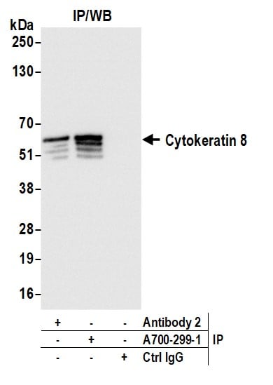
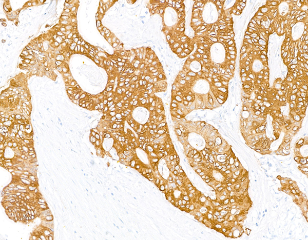

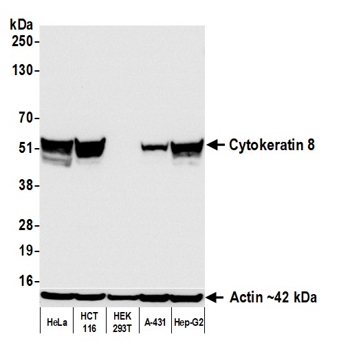 WB (Western Blot)
(Detection of human Cytokeratin 8 by western blot. Samples: Whole cell lysate (50 ug) from HeLa, HCT 116, HEK293T, A-431, and Hep-G2 cells prepared using NETN lysis buffer. Antibody: Rabbit anti-Cytokeratin 8 recombinant monoclonal antibody (AAA213697 lot 1) used at 1:1000. Secondary: HRP-conjugated goat anti-rabbit IgG . Detection: Chemiluminescence with an exposure time of 1 second. Lower Panel: Rabbit anti-Actin recombinant monoclonal antibody .)
WB (Western Blot)
(Detection of human Cytokeratin 8 by western blot. Samples: Whole cell lysate (50 ug) from HeLa, HCT 116, HEK293T, A-431, and Hep-G2 cells prepared using NETN lysis buffer. Antibody: Rabbit anti-Cytokeratin 8 recombinant monoclonal antibody (AAA213697 lot 1) used at 1:1000. Secondary: HRP-conjugated goat anti-rabbit IgG . Detection: Chemiluminescence with an exposure time of 1 second. Lower Panel: Rabbit anti-Actin recombinant monoclonal antibody .)
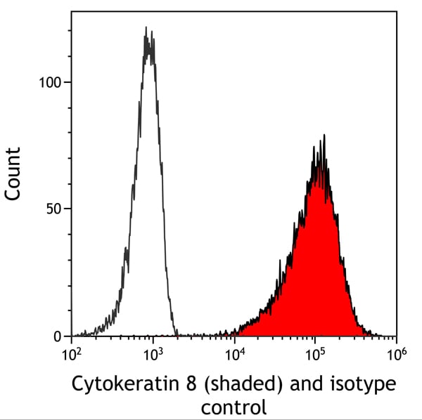
Cytokeratin 8, Monoclonal Recombinant Antibody (Cat# AAA213697)
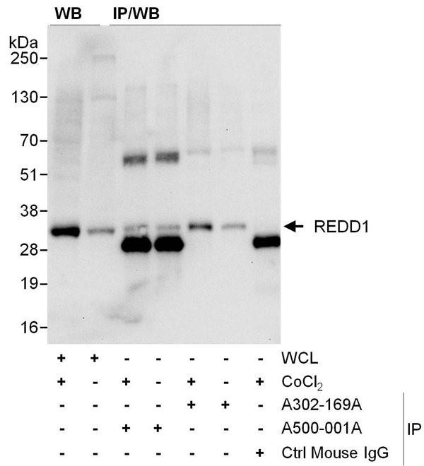 WB (Western Blot)
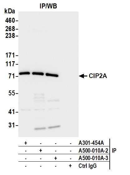
(Detection of human REDD1 by western blot and immunoprecipitation. Samples: Whole cell lysate (WCL) [50 ug for WB; 1mg for IP, 20% of IP loaded] from HeLa cells. Lysate was prepared from untreated (-) cells or cells treated (+) with CoCl2. Antibodies: Mouse monoclonal anti-REDD1 antibody [1G11] (AAA213493 lot 1) was used at 1:1000 for WB and 3 ul/mg of lysate for IP. REDD1 was also immunoprecipitated (lanes 5&6) by rabbit anti-REDD1 antibody . Secondary: HRP-conjugated goat anti-mouse IgG . Detection: Chemiluminescence with an exposure time of 10 seconds.)
WB (Western Blot)
(Detection of human REDD1 by western blot and immunoprecipitation. Samples: Whole cell lysate (WCL) [50 ug for WB; 1mg for IP, 20% of IP loaded] from HeLa cells. Lysate was prepared from untreated (-) cells or cells treated (+) with CoCl2. Antibodies: Mouse monoclonal anti-REDD1 antibody [1G11] (AAA213493 lot 1) was used at 1:1000 for WB and 3 ul/mg of lysate for IP. REDD1 was also immunoprecipitated (lanes 5&6) by rabbit anti-REDD1 antibody . Secondary: HRP-conjugated goat anti-mouse IgG . Detection: Chemiluminescence with an exposure time of 10 seconds.)
REDD1, Monoclonal Antibody (Cat# AAA213493)
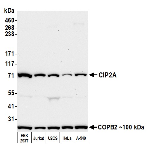
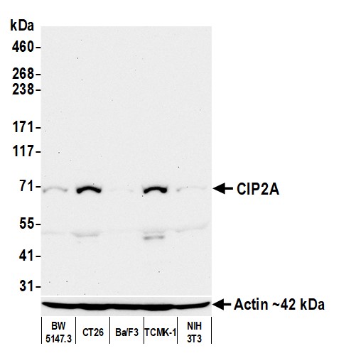 WB (Western Blot)
(Detection of mouse CIP2A by western blot. Samples: Whole cell lysate (50 ug) from BW5147.3, CT26, Ba/F3, TCMK-1, and NIH 3T3 cells prepared using NETN lysis buffer. Antibody: Mouse anti-CIP2A monoclonal antibody [2A1-9A4] (AAA213496 lot 3) used at 1:1000. Secondary: HRP-conjugated goat anti-mouse IgG . Detection: Chemiluminescence with an exposure time of 75 seconds. Lower Panel: Rabbit anti-Actin recombinant monoclonal antibody .)
WB (Western Blot)
(Detection of mouse CIP2A by western blot. Samples: Whole cell lysate (50 ug) from BW5147.3, CT26, Ba/F3, TCMK-1, and NIH 3T3 cells prepared using NETN lysis buffer. Antibody: Mouse anti-CIP2A monoclonal antibody [2A1-9A4] (AAA213496 lot 3) used at 1:1000. Secondary: HRP-conjugated goat anti-mouse IgG . Detection: Chemiluminescence with an exposure time of 75 seconds. Lower Panel: Rabbit anti-Actin recombinant monoclonal antibody .)
CIP2A, Monoclonal Antibody (Cat# AAA213496)
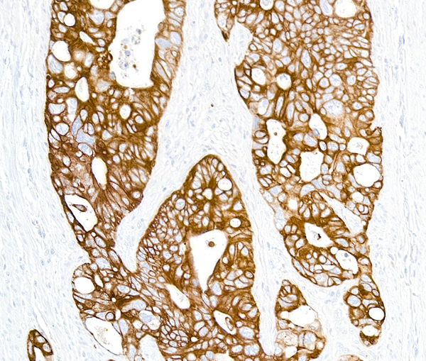
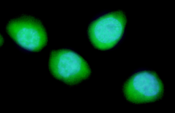
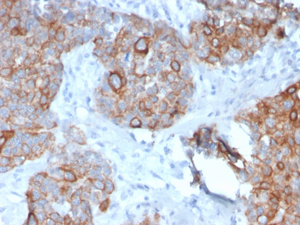
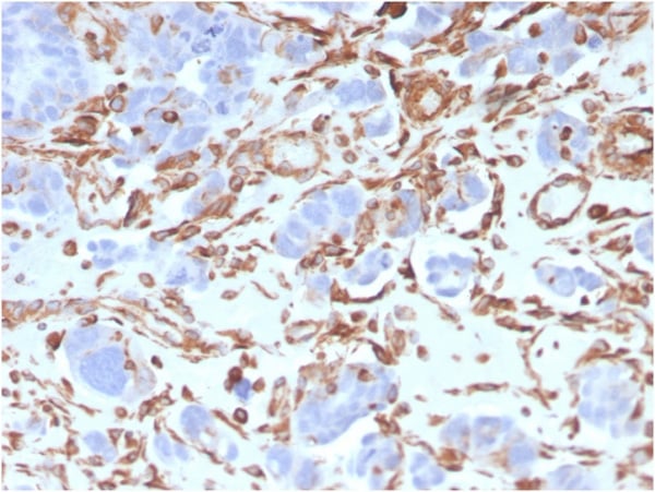
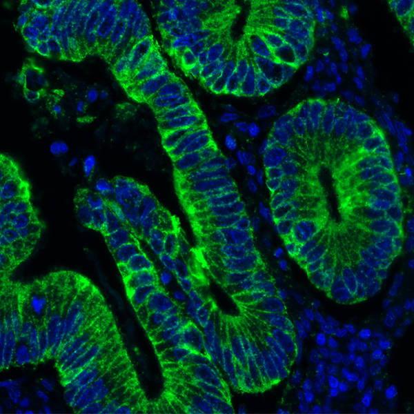 IHC (Immunohistochemistry)
(Detection of human Cytokeratin (green) by immunohistochemistry. Sample: FFPE section of human colon carcinoma. Antibody: Mouse anti-Cytokeratin monoclonal antibody [AE1/AE3] (AAA213503) used at 1:500. Secondary: HRP-conjugated goat anti-mouse IgG . Substrate: Opal. Counterstain: DAPI (blue).)
IHC (Immunohistochemistry)
(Detection of human Cytokeratin (green) by immunohistochemistry. Sample: FFPE section of human colon carcinoma. Antibody: Mouse anti-Cytokeratin monoclonal antibody [AE1/AE3] (AAA213503) used at 1:500. Secondary: HRP-conjugated goat anti-mouse IgG . Substrate: Opal. Counterstain: DAPI (blue).)
Cytokeratin, Monoclonal Antibody (Cat# AAA213503)
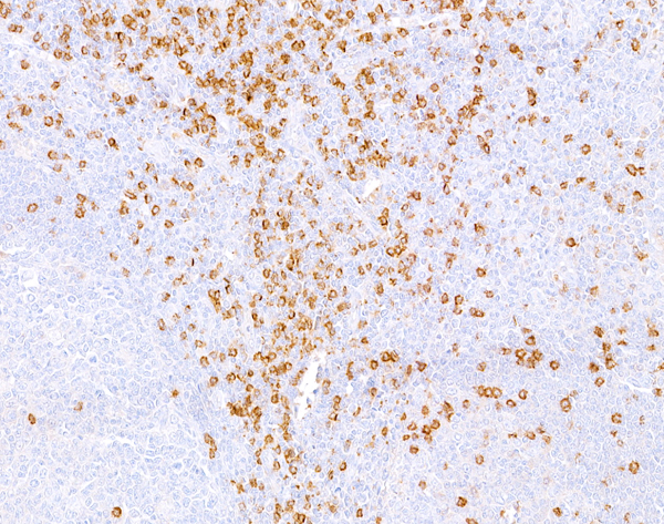
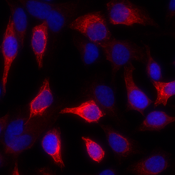
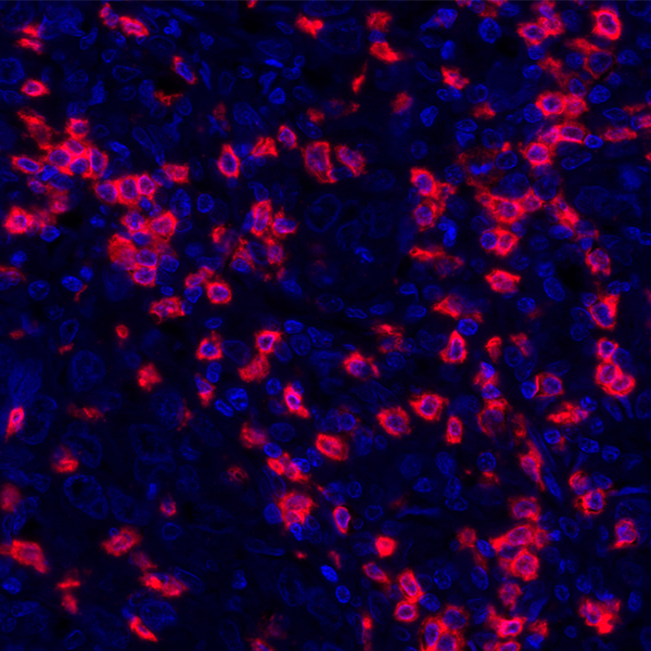
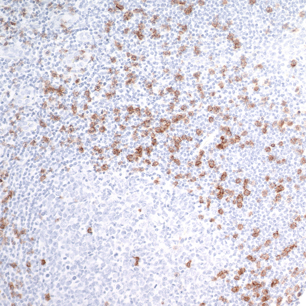
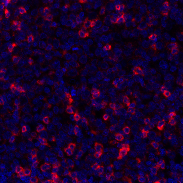
 IHC (Immunohistochemisry)
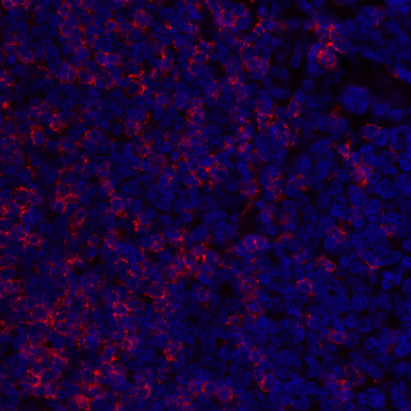
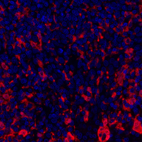
(Detection of human CD8 alpha by immunhistochemistry. Sample: FFPE section of human tonsil. Antibody: Mouse monoclonal anti-CD8 alpha antibody [C8/144B] (AAA213505 lot 1) used at 1:100. Secondary: DyLight 594-conjugated goat anti-mouse IgG .)
IHC (Immunohistochemisry)
(Detection of human CD8 alpha by immunhistochemistry. Sample: FFPE section of human tonsil. Antibody: Mouse monoclonal anti-CD8 alpha antibody [C8/144B] (AAA213505 lot 1) used at 1:100. Secondary: DyLight 594-conjugated goat anti-mouse IgG .)
CD8 alpha, Monoclonal Antibody (Cat# AAA213505)
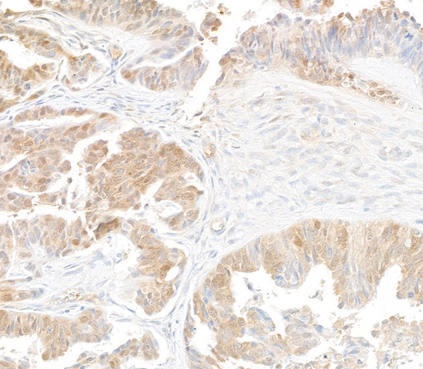
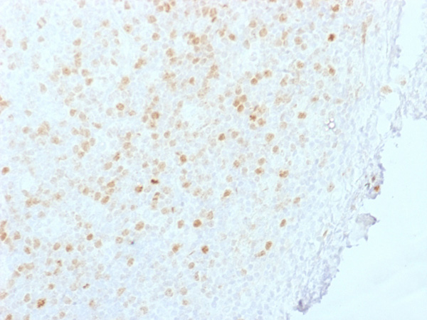
 IHC (Immunohistochemisry)
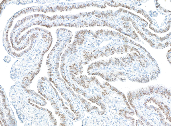
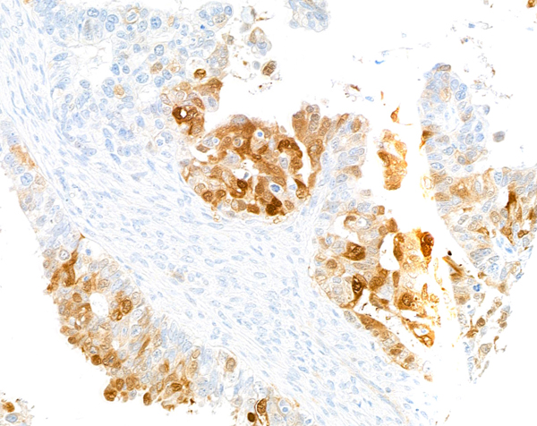
(Detection of human PAX8 by immunohistochemistry. Sample: FFPE section of ovarian carcinoma. Antibody: Mouse anti-PAX8 monoclonal antibody [BC12] (AAA213521-1). Detection:Biocare Medical MACH 4 mouse probe/HRP Polymer.)
IHC (Immunohistochemisry)
(Detection of human PAX8 by immunohistochemistry. Sample: FFPE section of ovarian carcinoma. Antibody: Mouse anti-PAX8 monoclonal antibody [BC12] (AAA213521-1). Detection:Biocare Medical MACH 4 mouse probe/HRP Polymer.)
PAX8, Monoclonal Antibody (Cat# AAA213521)
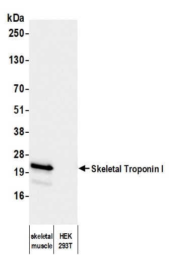

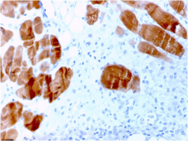
 WB (Western Blot)
(Detection of mouse Skeletal Troponin I by western blot. Samples: Whole tissue lysate (50 ug) from mouse liver and skeletal muscle. Antibody: Mouse anti-Skeletal Troponin I monoclonal antibody [FI-23] (AAA213524 lot 1) used at 1:1000. Secondary: HRP-conjugated goat anti-mouse IgG . Detection: Chemiluminescence with an exposure time of 1 second.)
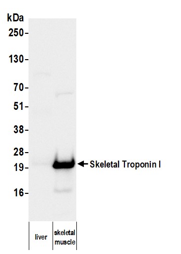
WB (Western Blot)
(Detection of mouse Skeletal Troponin I by western blot. Samples: Whole tissue lysate (50 ug) from mouse liver and skeletal muscle. Antibody: Mouse anti-Skeletal Troponin I monoclonal antibody [FI-23] (AAA213524 lot 1) used at 1:1000. Secondary: HRP-conjugated goat anti-mouse IgG . Detection: Chemiluminescence with an exposure time of 1 second.)
Skeletal Troponin I, Monoclonal Antibody (Cat# AAA213524)
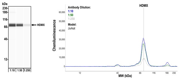
 WB (Western Blot)
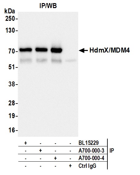
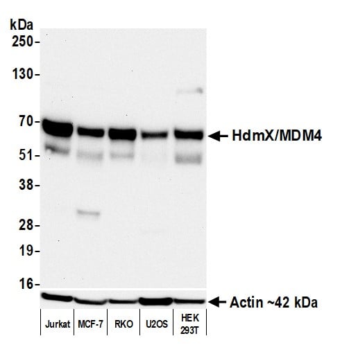
(Detection of human HdmX/MDM4 by western blot. Samples: Whole cell lysate (50 ug) from Jurkat, MCF-7, RKO, U2OS, and HEK293T cells prepared using NETN lysis buffer. Antibody: Rabbit anti-HdmX/MDM4 recombinant monoclonal antibody [BL-3-2F2] (AAA213529 lot 4) used at 1:1000. Secondary: HRP-conjugated goat anti-rabbit IgG . Detection: Chemiluminescence with an exposure time of 75 seconds. Lower Panel: Rabbit anti-Actin recombinant monoclonal antibody .)
WB (Western Blot)
(Detection of human HdmX/MDM4 by western blot. Samples: Whole cell lysate (50 ug) from Jurkat, MCF-7, RKO, U2OS, and HEK293T cells prepared using NETN lysis buffer. Antibody: Rabbit anti-HdmX/MDM4 recombinant monoclonal antibody [BL-3-2F2] (AAA213529 lot 4) used at 1:1000. Secondary: HRP-conjugated goat anti-rabbit IgG . Detection: Chemiluminescence with an exposure time of 75 seconds. Lower Panel: Rabbit anti-Actin recombinant monoclonal antibody .)
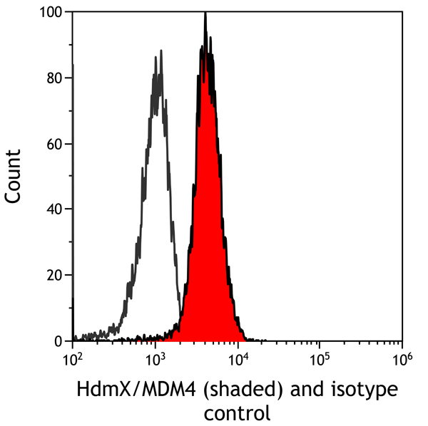
HdmX/MDM4, Monoclonal Recombinant Antibody (Cat# AAA213529)
 WB (Western Blot)
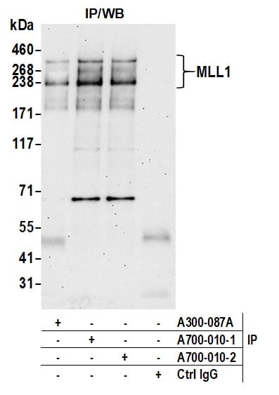
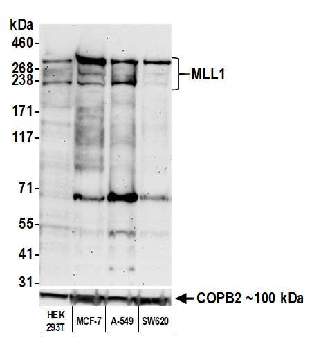
(Detection of human MLL1 by western blot. Samples: Whole cell lysate (50 ug) from HEK293T, MCF-7, A-549, and SW620 cells prepared using NETN lysis buffer. Antibody: Rabbit anti-MLL1 recombinant monoclonal antibody [BL-175-7E8] (AAA213534 lot 2) used at 1:1000. Secondary: HRP-conjugated goat anti-rabbit IgG . Detection: Chemiluminescence with an exposure time of 75 seconds. Lower Panel: Rabbit anti-COPB2 antibody .)
WB (Western Blot)
(Detection of human MLL1 by western blot. Samples: Whole cell lysate (50 ug) from HEK293T, MCF-7, A-549, and SW620 cells prepared using NETN lysis buffer. Antibody: Rabbit anti-MLL1 recombinant monoclonal antibody [BL-175-7E8] (AAA213534 lot 2) used at 1:1000. Secondary: HRP-conjugated goat anti-rabbit IgG . Detection: Chemiluminescence with an exposure time of 75 seconds. Lower Panel: Rabbit anti-COPB2 antibody .)
MLL1, Monoclonal Recombinant Antibody (Cat# AAA213534)
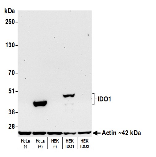 WB (Western Blot)
(Detection of human IDO1 by western blot. Samples: Whole cell lysate (50 ug) from HeLa treated with IFN-gamma (+) or mock treated (-), untransfected HEK, IDO1 over-expressing HEK, and IDO2 over-expressing HEK cells prepared using NETN lysis buffer. Antibody: Rabbit anti-IDO1 recombinant monoclonal antibody (AAA213540 lot 2) used at 1:1000. Secondary: HRP-conjugated goat anti-rabbit IgG . Detection: Chemiluminescence with an exposure time of 30 seconds. Lower Panel: Rabbit anti-Actin recombinant monoclonal antibody .)
WB (Western Blot)
(Detection of human IDO1 by western blot. Samples: Whole cell lysate (50 ug) from HeLa treated with IFN-gamma (+) or mock treated (-), untransfected HEK, IDO1 over-expressing HEK, and IDO2 over-expressing HEK cells prepared using NETN lysis buffer. Antibody: Rabbit anti-IDO1 recombinant monoclonal antibody (AAA213540 lot 2) used at 1:1000. Secondary: HRP-conjugated goat anti-rabbit IgG . Detection: Chemiluminescence with an exposure time of 30 seconds. Lower Panel: Rabbit anti-Actin recombinant monoclonal antibody .)
IDO1, Monoclonal Recombinant Antibody (Cat# AAA213540)
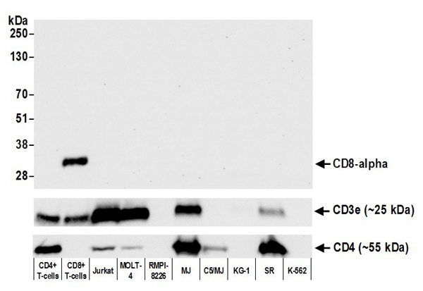 WB (Western Blot)
(Detection of human CD8 alpha by western blot. Samples: Whole cell lysate (50 ug) from CD4+ T-cells, CD8+ T-cells, Jurkat, MOLT-4, RPMI-8226, MJ, C5/MJ, KG-1, SR, and K-562 cells prepared using NETN lysis buffer. Antibody: Rabbit anti-CD8 alpha recombinant monoclonal antibody (AAA213544 lot 1) used at 1:1000. Secondary: HRP-conjugated goat anti-rabbit IgG . Detection: Chemiluminescence with an exposure time of 30 seconds. Lower panel: Recombinant monoclonal antibodies to CD3e and CD4 .)
WB (Western Blot)
(Detection of human CD8 alpha by western blot. Samples: Whole cell lysate (50 ug) from CD4+ T-cells, CD8+ T-cells, Jurkat, MOLT-4, RPMI-8226, MJ, C5/MJ, KG-1, SR, and K-562 cells prepared using NETN lysis buffer. Antibody: Rabbit anti-CD8 alpha recombinant monoclonal antibody (AAA213544 lot 1) used at 1:1000. Secondary: HRP-conjugated goat anti-rabbit IgG . Detection: Chemiluminescence with an exposure time of 30 seconds. Lower panel: Recombinant monoclonal antibodies to CD3e and CD4 .)
CD8 alpha, Monoclonal Recombinant Antibody (Cat# AAA213544)
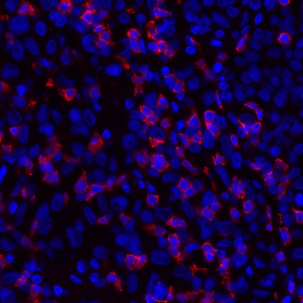
 WB (Western Blot)
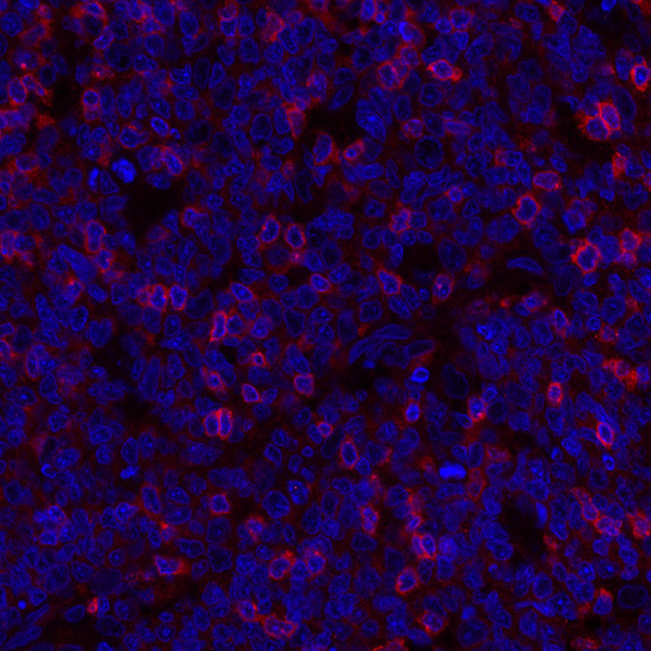
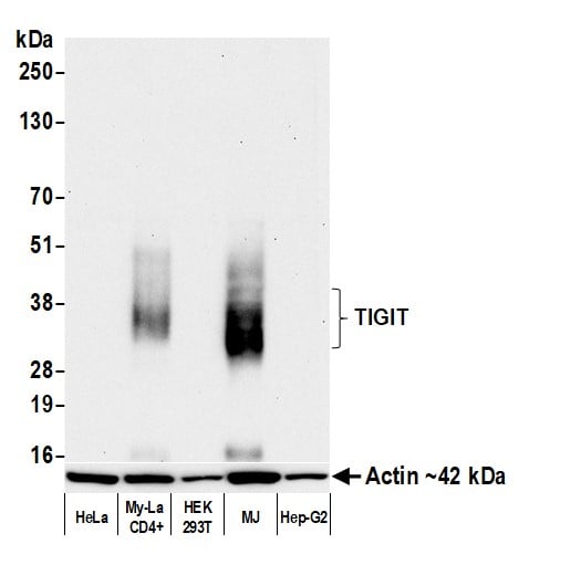
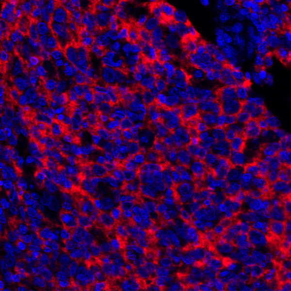
(Detection of human TIGIT by western blot. Samples: Whole cell lysate (10 ug) from HeLa, My-La CD4+, HEK293T, MJ, and Hep-G2 cells prepared using NETN lysis buffer. Antibody: Rabbit anti-TIGIT recombinant monoclonal antibody (AAA213546 lot 6) used at 1:1000. Secondary: HRP-conjugated goat anti-rabbit IgG . Detection: Chemiluminescence with an exposure time of 75 seconds. Lower Panel: Rabbit anti-Actin recombinant monoclonal antibody .)
WB (Western Blot)
(Detection of human TIGIT by western blot. Samples: Whole cell lysate (10 ug) from HeLa, My-La CD4+, HEK293T, MJ, and Hep-G2 cells prepared using NETN lysis buffer. Antibody: Rabbit anti-TIGIT recombinant monoclonal antibody (AAA213546 lot 6) used at 1:1000. Secondary: HRP-conjugated goat anti-rabbit IgG . Detection: Chemiluminescence with an exposure time of 75 seconds. Lower Panel: Rabbit anti-Actin recombinant monoclonal antibody .)
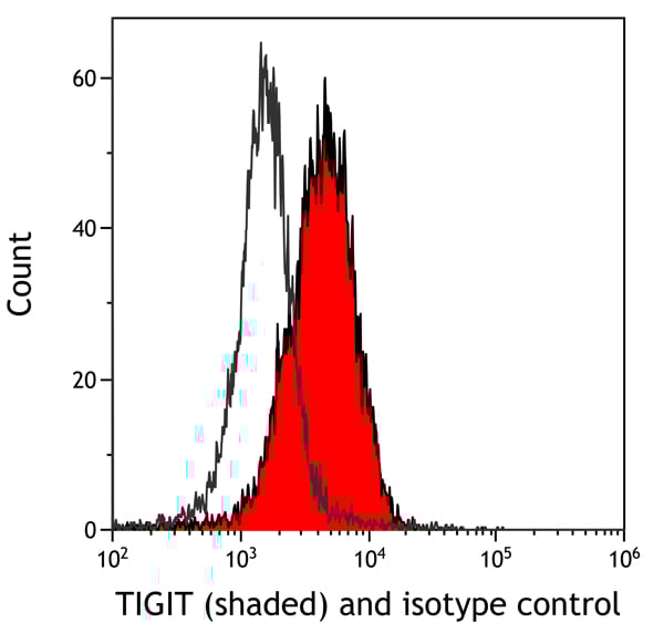
TIGIT, Monoclonal Recombinant Antibody (Cat# AAA213546)
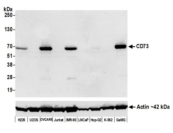 WB (Western Blot)
(Detection of human CD73 by western blot. Samples: Whole cell lysate (50 ug) from NCI-H226, U2OS, OVCAR-8, Jurkat, IMR-90, LNCaP, Hep-G2, K-562, and GaMG cells prepared using NETN lysis buffer. Antibody: Rabbit anti-CD73 recombinant monoclonal antibody (AAA213551 lot 1) used at 1:1000. Secondary: HRP-conjugated goat anti-rabbit IgG . Detection: Chemiluminescence with an exposure time of 30 seconds. Lower Panel: Rabbit anti-Actin recombinant monoclonal .)
WB (Western Blot)
(Detection of human CD73 by western blot. Samples: Whole cell lysate (50 ug) from NCI-H226, U2OS, OVCAR-8, Jurkat, IMR-90, LNCaP, Hep-G2, K-562, and GaMG cells prepared using NETN lysis buffer. Antibody: Rabbit anti-CD73 recombinant monoclonal antibody (AAA213551 lot 1) used at 1:1000. Secondary: HRP-conjugated goat anti-rabbit IgG . Detection: Chemiluminescence with an exposure time of 30 seconds. Lower Panel: Rabbit anti-Actin recombinant monoclonal .)
CD73, Monoclonal Recombinant Antibody (Cat# AAA213551)
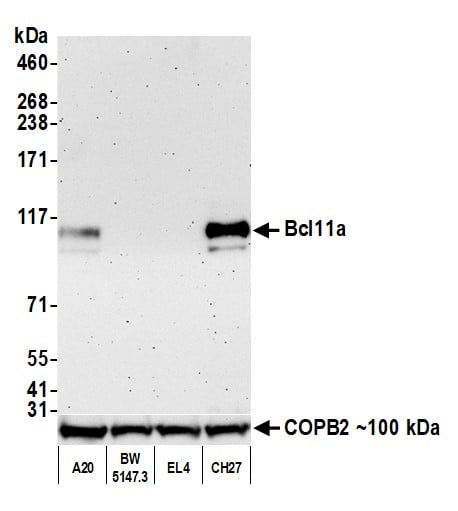
 WB (Western Blot)
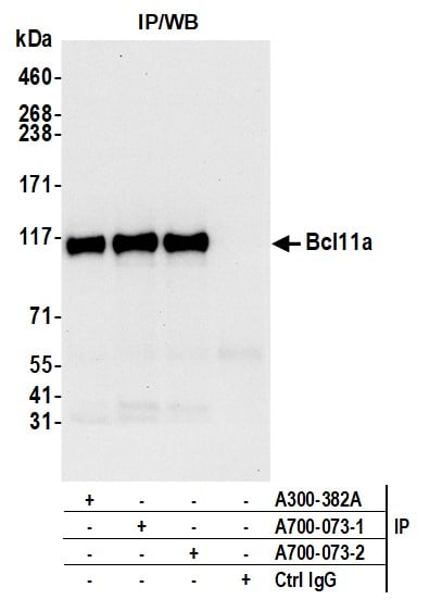
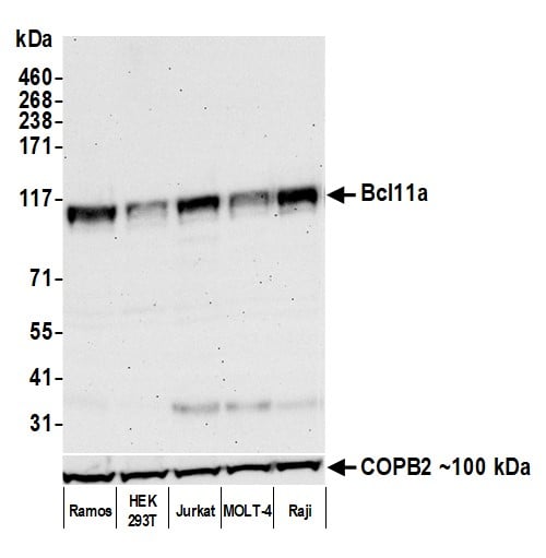
(Detection of human Bcl11a by western blot. Samples: Whole cell lysate (50 ug) from Ramos, HEK293T, Jurkat, MOLT-4, and Raji cells prepared using NETN lysis buffer. Antibody: Rabbit anti-Bcl11a recombinant monoclonal antibody (AAA213561 lot 2) used at 1:1000. Secondary: HRP-conjugated goat anti-rabbit IgG . Detection: Chemiluminescence with an exposure time of 75 seconds. Lower Panel: Rabbit anti-COPB2 antibody .)
WB (Western Blot)
(Detection of human Bcl11a by western blot. Samples: Whole cell lysate (50 ug) from Ramos, HEK293T, Jurkat, MOLT-4, and Raji cells prepared using NETN lysis buffer. Antibody: Rabbit anti-Bcl11a recombinant monoclonal antibody (AAA213561 lot 2) used at 1:1000. Secondary: HRP-conjugated goat anti-rabbit IgG . Detection: Chemiluminescence with an exposure time of 75 seconds. Lower Panel: Rabbit anti-COPB2 antibody .)
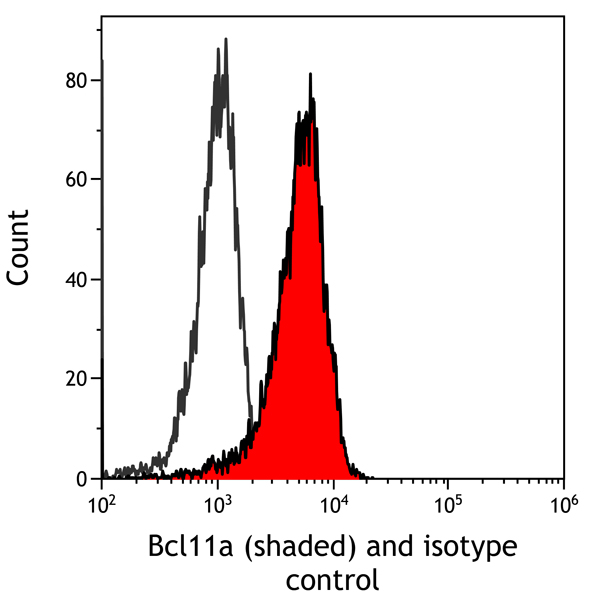
Bcl11a, Monoclonal Recombinant Antibody (Cat# AAA213561)
IgG4, Monoclonal Antibody (Cat# AAA214174)
CD44, Monoclonal Antibody (Cat# AAA214176)
 Application Data
Histone H4, Monoclonal Antibody (Cat# AAA214203)
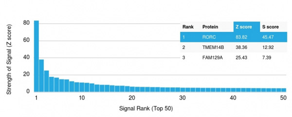 Application Data
(Analysis of Protein Array containing more than 19,000 full-length human proteins using ROR-gamma/RORC Mouse Monoclonal Antibody (RORC/2942). Z- and S- Score: The Z-score represents the strength of a signal that a monoclonal antibody (MAb) (in combination with a fluorescently-tagged anti-IgG secondary antibody) produces when binding to a particular protein on the HuProtTM array. Z-scores are described in units of standard deviations (SD’s) above the mean value of all signals generated on that array. If targets on HuProtTM are arranged in descending order of the Z-score, the S-score is the difference (also in units of SD’s) between the Z-score. S-score therefore represents the relative target specificity of a MAb to its intended target. A MAb is considered to specific to its intended target, if the MAb has an S-score of at least 2.5. For example, if a MAb binds to protein X with a Z-score of 43 and to protein Y with a Z-score of 14, then the S-score for the binding of that MAb to protein X is equal to 29.)
Application Data
(Analysis of Protein Array containing more than 19,000 full-length human proteins using ROR-gamma/RORC Mouse Monoclonal Antibody (RORC/2942). Z- and S- Score: The Z-score represents the strength of a signal that a monoclonal antibody (MAb) (in combination with a fluorescently-tagged anti-IgG secondary antibody) produces when binding to a particular protein on the HuProtTM array. Z-scores are described in units of standard deviations (SD’s) above the mean value of all signals generated on that array. If targets on HuProtTM are arranged in descending order of the Z-score, the S-score is the difference (also in units of SD’s) between the Z-score. S-score therefore represents the relative target specificity of a MAb to its intended target. A MAb is considered to specific to its intended target, if the MAb has an S-score of at least 2.5. For example, if a MAb binds to protein X with a Z-score of 43 and to protein Y with a Z-score of 14, then the S-score for the binding of that MAb to protein X is equal to 29.)
ROR-gamma/RORC, Monoclonal Antibody (Cat# AAA215186)

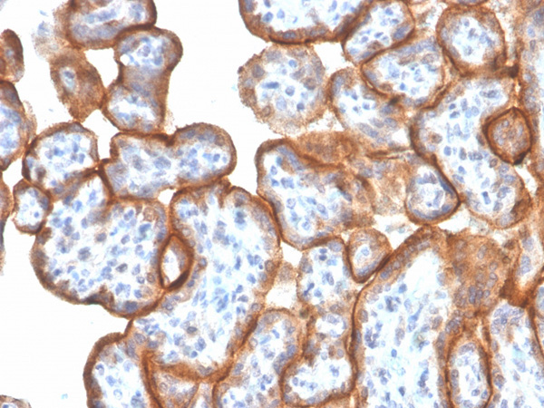
 IF (Immunofluorescence)
(Confocal Immunofluorescent analysis of A2058 cells using CF488-labeled Isotype Control Monoclonal Antibody (IgG2a) (Green). F-actin filaments were labeled with Dylight 554 Phalloidin (red). DAPI was used to stain the cell nuclei (blue). (Negative Control))
IF (Immunofluorescence)
(Confocal Immunofluorescent analysis of A2058 cells using CF488-labeled Isotype Control Monoclonal Antibody (IgG2a) (Green). F-actin filaments were labeled with Dylight 554 Phalloidin (red). DAPI was used to stain the cell nuclei (blue). (Negative Control))
S100B, Monoclonal Antibody (Cat# AAA215189)
 Application Data
(Analysis of Protein Array containing more than 19,000 full-length human proteins using SDHB Mouse Monoclonal Antibody (SDHB/2382). Z- and S- Score: The Z-score represents the strength of a signal that a monoclonal antibody (Monoclonal Antibody) (in combination with a fluorescently-tagged anti-IgG secondary antibody) produces when binding to a particular protein on the HuProtTM array. Z-scores are described in units of standard deviations (SD's) above the mean value of all signals generated on that array. If targets on HuProtTM are arranged in descending order of the Z-score, the S-score is the difference (also in units of SD's) between the Z-score. S-score therefore represents the relative target specificity of a Monoclonal Antibody to its intended target. A Monoclonal Antibody is considered to specific to its intended target, if the Monoclonal Antibody has an S-score of at least 2.5. For example, if a Monoclonal Antibody binds to protein X with a Z-score of 43 and to protein Y with a Z-score of 14, then the S-score for the binding of that Monoclonal Antibody to protein X is equal to 29.)
Application Data
(Analysis of Protein Array containing more than 19,000 full-length human proteins using SDHB Mouse Monoclonal Antibody (SDHB/2382). Z- and S- Score: The Z-score represents the strength of a signal that a monoclonal antibody (Monoclonal Antibody) (in combination with a fluorescently-tagged anti-IgG secondary antibody) produces when binding to a particular protein on the HuProtTM array. Z-scores are described in units of standard deviations (SD's) above the mean value of all signals generated on that array. If targets on HuProtTM are arranged in descending order of the Z-score, the S-score is the difference (also in units of SD's) between the Z-score. S-score therefore represents the relative target specificity of a Monoclonal Antibody to its intended target. A Monoclonal Antibody is considered to specific to its intended target, if the Monoclonal Antibody has an S-score of at least 2.5. For example, if a Monoclonal Antibody binds to protein X with a Z-score of 43 and to protein Y with a Z-score of 14, then the S-score for the binding of that Monoclonal Antibody to protein X is equal to 29.)
SDHB (Succinate Dehydrogenase B), Monoclonal Antibody (Cat# AAA215191)
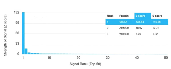
 Application Data
(Analysis of Protein Array containing more than 19,000 full-length human proteins using Monospecific Mouse Monoclonal Antibody to VISTA (VISTA/2865). Z- and S- Score: The Z-score represents the strength of a signal that a monoclonal antibody (Monoclonal Antibody) (in combination with a fluorescently-tagged anti-IgG secondary antibody) produces when binding to a particular protein on the HuProtTM array. Z-scores are described in units of standard deviations (SD’s) above the mean value of all signals generated on that array. If targets on HuProtTM are arranged in descending order of the Z-score, the S-score is the difference (also in units of SD’s) between the Z-score. S-score therefore represents the relative target specificity of a Monoclonal Antibody to its intended target. A Monoclonal Antibody is considered to specific to its intended target, if the Monoclonal Antibody has an S-score of at least 2.5. For example, if a Monoclonal Antibody binds to protein X with a Z-score of 43 and to protein Y with a Z-score of 14, then the S-score for the binding of that Monoclonal Antibody to protein X is equal to 29.)
Application Data
(Analysis of Protein Array containing more than 19,000 full-length human proteins using Monospecific Mouse Monoclonal Antibody to VISTA (VISTA/2865). Z- and S- Score: The Z-score represents the strength of a signal that a monoclonal antibody (Monoclonal Antibody) (in combination with a fluorescently-tagged anti-IgG secondary antibody) produces when binding to a particular protein on the HuProtTM array. Z-scores are described in units of standard deviations (SD’s) above the mean value of all signals generated on that array. If targets on HuProtTM are arranged in descending order of the Z-score, the S-score is the difference (also in units of SD’s) between the Z-score. S-score therefore represents the relative target specificity of a Monoclonal Antibody to its intended target. A Monoclonal Antibody is considered to specific to its intended target, if the Monoclonal Antibody has an S-score of at least 2.5. For example, if a Monoclonal Antibody binds to protein X with a Z-score of 43 and to protein Y with a Z-score of 14, then the S-score for the binding of that Monoclonal Antibody to protein X is equal to 29.)
VISTA/GI24, Monoclonal Antibody (Cat# AAA215194)
 Application Data
(Analysis of Protein Array containing more than 19,000 full-length human proteins using Monospecific Mouse Monoclonal Antibody to VISTA (VISTA/3006). Z- and S- Score: The Z-score represents the strength of a signal that a monoclonal antibody (Monoclonal Antibody) (in combination with a fluorescently-tagged anti-IgG secondary antibody) produces when binding to a particular protein on the HuProtTM array. Z-scores are described in units of standard deviations (SD’s) above the mean value of all signals generated on that array. If targets on HuProtTM are arranged in descending order of the Z-score, the S-score is the difference (also in units of SD’s) between the Z-score. S-score therefore represents the relative target specificity of a Monoclonal Antibody to its intended target. A Monoclonal Antibody is considered to specific to its intended target, if the Monoclonal Antibody has an S-score of at least 2.5. For example, if a Monoclonal Antibody binds to protein X with a Z-score of 43 and to protein Y with a Z-score of 14, then the S-score for the binding of that Monoclonal Antibody to protein X is equal to 29.)
Application Data
(Analysis of Protein Array containing more than 19,000 full-length human proteins using Monospecific Mouse Monoclonal Antibody to VISTA (VISTA/3006). Z- and S- Score: The Z-score represents the strength of a signal that a monoclonal antibody (Monoclonal Antibody) (in combination with a fluorescently-tagged anti-IgG secondary antibody) produces when binding to a particular protein on the HuProtTM array. Z-scores are described in units of standard deviations (SD’s) above the mean value of all signals generated on that array. If targets on HuProtTM are arranged in descending order of the Z-score, the S-score is the difference (also in units of SD’s) between the Z-score. S-score therefore represents the relative target specificity of a Monoclonal Antibody to its intended target. A Monoclonal Antibody is considered to specific to its intended target, if the Monoclonal Antibody has an S-score of at least 2.5. For example, if a Monoclonal Antibody binds to protein X with a Z-score of 43 and to protein Y with a Z-score of 14, then the S-score for the binding of that Monoclonal Antibody to protein X is equal to 29.)
VISTA/GI24, Monoclonal Antibody (Cat# AAA215195)
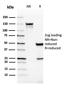
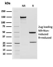
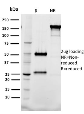
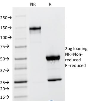
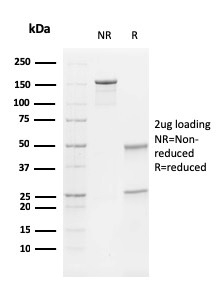
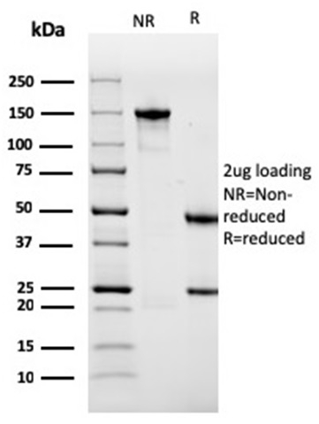
 SDS-PAGE
(SDS-PAGE Analysis Purified CDw75 Mouse Monoclonal Antibody (ZB55).Confirmation of Purity and Integrity of Antibody.)
SDS-PAGE
(SDS-PAGE Analysis Purified CDw75 Mouse Monoclonal Antibody (ZB55).Confirmation of Purity and Integrity of Antibody.)
CDw75, Monoclonal Antibody (Cat# AAA215197)
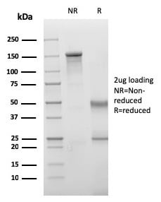
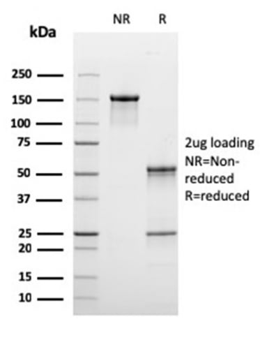
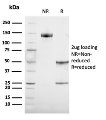
 SDS-PAGE
(SDS-PAGE Analysis Purified STAT2 Mouse Monoclonal Antibody (STAT2/2650). Confirmation of Purity and Integrity of Antibody.)
SDS-PAGE
(SDS-PAGE Analysis Purified STAT2 Mouse Monoclonal Antibody (STAT2/2650). Confirmation of Purity and Integrity of Antibody.)
STAT2, Monoclonal Antibody (Cat# AAA215209)
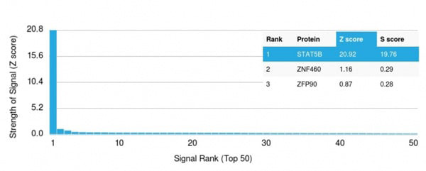 Application Data
(Analysis of Protein Array containing >19,000 full-length human proteins using STAT5B Mouse Monoclonal Antibody (STAT5B/2611) Z- and S- Score: The Z-score represents the strength of a signal that a monoclonal antibody (Monoclonal Antibody) (in combination with a fluorescently-tagged anti-IgG secondary antibody) produces when binding to a particular protein on the HuProtTM array. Z-scores are described in units of standard deviations (SD's) above the mean value of all signals generated on that array. If targets on HuProtTM are arranged in descending order of the Z-score, the S-score is the difference (also in units of SD's) between the Z-score. S-score therefore represents the relative target specificity of a Monoclonal Antibody to its intended target. A Monoclonal Antibody is considered to specific to its intended target, if the Monoclonal Antibody has an S-score of at least 2.5. For example, if a Monoclonal Antibody binds to protein X with a Z-score of 43 and to protein Y with a Z-score of 14, then the S-score for the binding of that Monoclonal Antibody to protein X is equal to 29.)
Application Data
(Analysis of Protein Array containing >19,000 full-length human proteins using STAT5B Mouse Monoclonal Antibody (STAT5B/2611) Z- and S- Score: The Z-score represents the strength of a signal that a monoclonal antibody (Monoclonal Antibody) (in combination with a fluorescently-tagged anti-IgG secondary antibody) produces when binding to a particular protein on the HuProtTM array. Z-scores are described in units of standard deviations (SD's) above the mean value of all signals generated on that array. If targets on HuProtTM are arranged in descending order of the Z-score, the S-score is the difference (also in units of SD's) between the Z-score. S-score therefore represents the relative target specificity of a Monoclonal Antibody to its intended target. A Monoclonal Antibody is considered to specific to its intended target, if the Monoclonal Antibody has an S-score of at least 2.5. For example, if a Monoclonal Antibody binds to protein X with a Z-score of 43 and to protein Y with a Z-score of 14, then the S-score for the binding of that Monoclonal Antibody to protein X is equal to 29.)
STAT5B, Monoclonal Antibody (Cat# AAA215210)
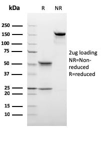
 SDS-PAGE
(SDS-PAGE Analysis Purified Tal1 Mouse Monoclonal Antibody (BTL73). Confirmation of Integrity and Purity of Antibody.)
SDS-PAGE
(SDS-PAGE Analysis Purified Tal1 Mouse Monoclonal Antibody (BTL73). Confirmation of Integrity and Purity of Antibody.)
Tal1, Monoclonal Antibody (Cat# AAA215213)
 Application Data
(Analysis of Protein Array containing more than 19,000 full-length human proteins using C1QB Mouse Monoclonal Antibody (C1QB/2965).Z- and S- Score: The Z-score represents the strength of a signal that a monoclonal antibody (Monoclonal Antibody) (in combination with a fluorescently-tagged anti-IgG secondary antibody) produces when binding to a particular protein on the HuProtTM array. Z-scores are described in units of standard deviations (SD's) above the mean value of all signals generated on that array. If targets on HuProtTM are arranged in descending order of the Z-score, the S-score is the difference (also in units of SD's) between the Z-score. S-score therefore represents the relative target specificity of a Monoclonal Antibody to its intended target. A Monoclonal Antibody is considered to specific to its intended target, if the Monoclonal Antibody has an S-score of at least 2.5. For example, if a Monoclonal Antibody binds to protein X with a Z-score of 43 and to protein Y with a Z-score of 14, then the S-score for the binding of that Monoclonal Antibody to protein X is equal to 29.)
Application Data
(Analysis of Protein Array containing more than 19,000 full-length human proteins using C1QB Mouse Monoclonal Antibody (C1QB/2965).Z- and S- Score: The Z-score represents the strength of a signal that a monoclonal antibody (Monoclonal Antibody) (in combination with a fluorescently-tagged anti-IgG secondary antibody) produces when binding to a particular protein on the HuProtTM array. Z-scores are described in units of standard deviations (SD's) above the mean value of all signals generated on that array. If targets on HuProtTM are arranged in descending order of the Z-score, the S-score is the difference (also in units of SD's) between the Z-score. S-score therefore represents the relative target specificity of a Monoclonal Antibody to its intended target. A Monoclonal Antibody is considered to specific to its intended target, if the Monoclonal Antibody has an S-score of at least 2.5. For example, if a Monoclonal Antibody binds to protein X with a Z-score of 43 and to protein Y with a Z-score of 14, then the S-score for the binding of that Monoclonal Antibody to protein X is equal to 29.)
C1QB/Complement C1q B-Chain, Monoclonal Antibody (Cat# AAA215226)
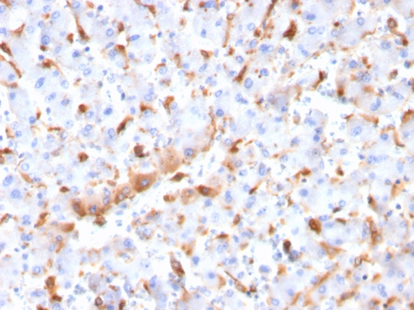
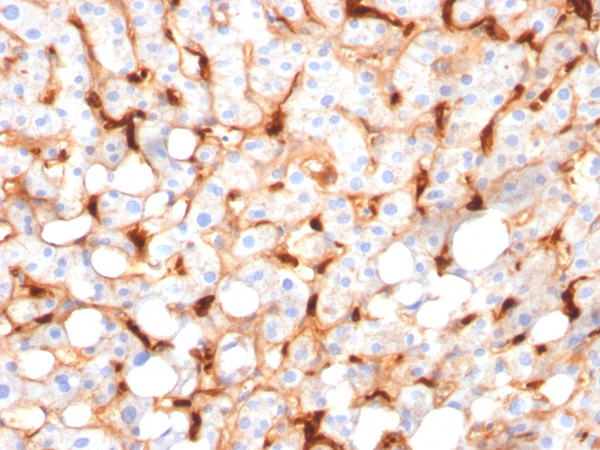
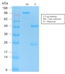
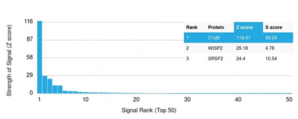 Application Data
(Analysis of Protein Array containing more than 19,000 full-length human proteins using C1QB Mouse Monoclonal Antibody (C1QB/2966).Z- and S- Score: The Z-score represents the strength of a signal that a monoclonal antibody (MAb) (in combination with a fluorescently-tagged anti-IgG secondary antibody) produces when binding to a particular protein on the HuProtTM array. Z-scores are described in units of standard deviations (SD's) above the mean value of all signals generated on that array. If targets on HuProtTM are arranged in descending order of the Z-score, the S-score is the difference (also in units of SD's) between the Z-score. S-score therefore represents the relative target specificity of a MAb to its intended target. A MAb is considered to specific to its intended target, if the MAb has an S-score of at least 2.5. For example, if a MAb binds to protein X with a Z-score of 43 and to protein Y with a Z-score of 14, then the S-score for the binding of that MAb to protein X is equal to 29.)
Application Data
(Analysis of Protein Array containing more than 19,000 full-length human proteins using C1QB Mouse Monoclonal Antibody (C1QB/2966).Z- and S- Score: The Z-score represents the strength of a signal that a monoclonal antibody (MAb) (in combination with a fluorescently-tagged anti-IgG secondary antibody) produces when binding to a particular protein on the HuProtTM array. Z-scores are described in units of standard deviations (SD's) above the mean value of all signals generated on that array. If targets on HuProtTM are arranged in descending order of the Z-score, the S-score is the difference (also in units of SD's) between the Z-score. S-score therefore represents the relative target specificity of a MAb to its intended target. A MAb is considered to specific to its intended target, if the MAb has an S-score of at least 2.5. For example, if a MAb binds to protein X with a Z-score of 43 and to protein Y with a Z-score of 14, then the S-score for the binding of that MAb to protein X is equal to 29.)
C1QB/Complement C1q B-Chain, Monoclonal Antibody (Cat# AAA215227)
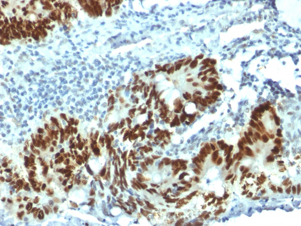
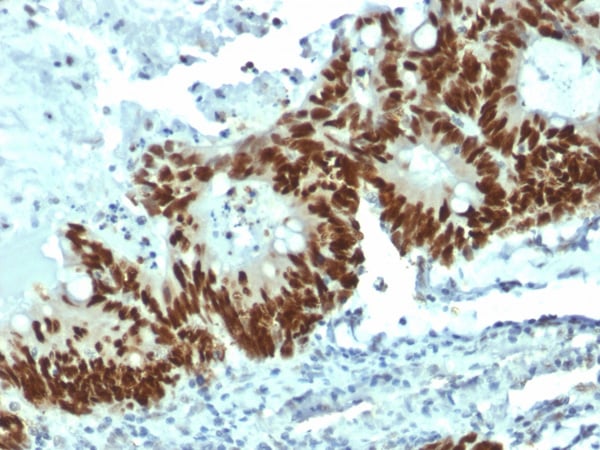
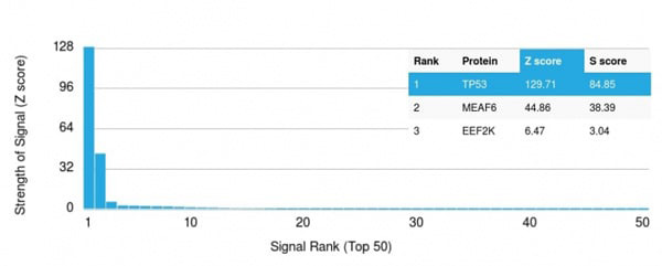
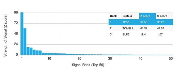 Application Data
(Analysis of Protein Array containing more than 19,000 full-length human proteins using p53 Mouse Monoclonal Antibody (PAb1801) Z- and S- Score: The Z-score represents the strength of a signal that a monoclonal antibody (Monoclonal Antibody) (in combination with a fluorescently-tagged anti-IgG secondary antibody) produces when binding to a particular protein on the HuProtTM array. Z-scores are described in units of standard deviations (SD's) above the mean value of all signals generated on that array. If targets on HuProtTM are arranged in descending order of the Z-score, the S-score is the difference (also in units of SD's) between the Z-score. S-score therefore represents the relative target specificity of a Monoclonal Antibody to its intended target. A Monoclonal Antibody is considered to specific to its intended target, if the Monoclonal Antibody has an S-score of at least 2.5. For example, if a Monoclonal Antibody binds to protein X with a Z-score of 43 and to protein Y with a Z-score of 14, then the S-score for the binding of that Monoclonal Antibody to protein X is equal to 29.)
Application Data
(Analysis of Protein Array containing more than 19,000 full-length human proteins using p53 Mouse Monoclonal Antibody (PAb1801) Z- and S- Score: The Z-score represents the strength of a signal that a monoclonal antibody (Monoclonal Antibody) (in combination with a fluorescently-tagged anti-IgG secondary antibody) produces when binding to a particular protein on the HuProtTM array. Z-scores are described in units of standard deviations (SD's) above the mean value of all signals generated on that array. If targets on HuProtTM are arranged in descending order of the Z-score, the S-score is the difference (also in units of SD's) between the Z-score. S-score therefore represents the relative target specificity of a Monoclonal Antibody to its intended target. A Monoclonal Antibody is considered to specific to its intended target, if the Monoclonal Antibody has an S-score of at least 2.5. For example, if a Monoclonal Antibody binds to protein X with a Z-score of 43 and to protein Y with a Z-score of 14, then the S-score for the binding of that Monoclonal Antibody to protein X is equal to 29.)
p53 Tumor Suppressor Protein, Monoclonal Antibody (Cat# AAA215228)
Does not react with Mouse or Rat.
 Application Data
(Analysis of Protein Array containing more than 19,000 full-length human proteins using p53 Mouse Monoclonal Antibody (DO-1) Z- and S- Score: The Z-score represents the strength of a signal that a monoclonal antibody (MAb) (in combination with a fluorescently-tagged anti-IgG secondary antibody) produces when binding to a particular protein on the HuProtTM array. Z-scores are described in units of standard deviations (SD's) above the mean value of all signals generated on that array. If targets on HuProtTM are arranged in descending order of the Z-score, the S-score is the difference (also in units of SD's) between the Z-score. S-score therefore represents the relative target specificity of a MAb to its intended target. A MAb is considered to specific to its intended target, if the MAb has an S-score of at least 2.5. For example, if a MAb binds to protein X with a Z-score of 43 and to protein Y with a Z-score of 14, then the S-score for the binding of that MAb to protein X is equal to 29.)
Application Data
(Analysis of Protein Array containing more than 19,000 full-length human proteins using p53 Mouse Monoclonal Antibody (DO-1) Z- and S- Score: The Z-score represents the strength of a signal that a monoclonal antibody (MAb) (in combination with a fluorescently-tagged anti-IgG secondary antibody) produces when binding to a particular protein on the HuProtTM array. Z-scores are described in units of standard deviations (SD's) above the mean value of all signals generated on that array. If targets on HuProtTM are arranged in descending order of the Z-score, the S-score is the difference (also in units of SD's) between the Z-score. S-score therefore represents the relative target specificity of a MAb to its intended target. A MAb is considered to specific to its intended target, if the MAb has an S-score of at least 2.5. For example, if a MAb binds to protein X with a Z-score of 43 and to protein Y with a Z-score of 14, then the S-score for the binding of that MAb to protein X is equal to 29.)
p53 Tumor Suppressor Protein, Monoclonal Antibody (Cat# AAA215231)
Does not react with Monkey or Rat.
 Application Data
(Analysis of Protein Array containing more than 19,000 full-length human proteins using TRAF1 Mouse Monoclonal Antibody (TRAF1/3298) Z- and S- Score: The Z-score represents the strength of a signal that a monoclonal antibody (Monoclonal Antibody) (in combination with a fluorescently-tagged anti-IgG secondary antibody) produces when binding to a particular protein on the HuProtTM array. Z-scores are described in units of standard deviations (SD’s) above the mean value of all signals generated on that array. If targets on HuProtTM are arranged in descending order of the Z-score, the S-score is the difference (also in units of SD’s) between the Z-score. S-score therefore represents the relative target specificity of a Monoclonal Antibody to its intended target. A Monoclonal Antibody is considered to specific to its intended target, if the Monoclonal Antibody has an S-score of at least 2.5. For example, if a Monoclonal Antibody binds to protein X with a Z-score of 43 and to protein Y with a Z-score of 14, then the S-score for the binding of that Monoclonal Antibody to protein X is equal to 29.)
Application Data
(Analysis of Protein Array containing more than 19,000 full-length human proteins using TRAF1 Mouse Monoclonal Antibody (TRAF1/3298) Z- and S- Score: The Z-score represents the strength of a signal that a monoclonal antibody (Monoclonal Antibody) (in combination with a fluorescently-tagged anti-IgG secondary antibody) produces when binding to a particular protein on the HuProtTM array. Z-scores are described in units of standard deviations (SD’s) above the mean value of all signals generated on that array. If targets on HuProtTM are arranged in descending order of the Z-score, the S-score is the difference (also in units of SD’s) between the Z-score. S-score therefore represents the relative target specificity of a Monoclonal Antibody to its intended target. A Monoclonal Antibody is considered to specific to its intended target, if the Monoclonal Antibody has an S-score of at least 2.5. For example, if a Monoclonal Antibody binds to protein X with a Z-score of 43 and to protein Y with a Z-score of 14, then the S-score for the binding of that Monoclonal Antibody to protein X is equal to 29.)
TRAF1 (TNFR-Associated Factor 1), Monoclonal Antibody (Cat# AAA215237)
 SDS-PAGE
(SDS-PAGE Analysis Purified TSH beta Mouse Monoclonal Antibody (TSHb/1317). Confirmation of Purity and Integrity of Antibody.)
SDS-PAGE
(SDS-PAGE Analysis Purified TSH beta Mouse Monoclonal Antibody (TSHb/1317). Confirmation of Purity and Integrity of Antibody.)
Thyroid Stimulating Hormone, beta (TSH beta), Monoclonal Antibody (Cat# AAA215239)
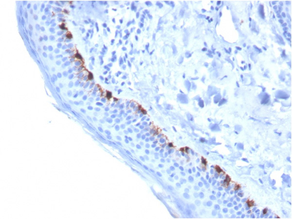
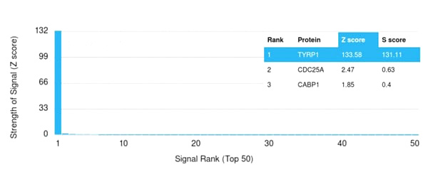 Application Data
(Analysis of Protein Array containing more than 19,000 full-length human proteins using TYRP1-Monospecific Mouse Monoclonal Antibody (TYRP1/3284) Z- and S- Score: The Z-score represents the strength of a signal that a monoclonal antibody (Monoclonal Antibody) (in combination with a fluorescently-tagged anti-IgG secondary antibody) produces when binding to a particular protein on the HuProtTM array. Z-scores are described in units of standard deviations (SD’s) above the mean value of all signals generated on that array. If targets on HuProtTM are arranged in descending order of the Z-score, the S-score is the difference (also in units of SD’s) between the Z-score. S-score therefore represents the relative target specificity of a Monoclonal Antibody to its intended target. A Monoclonal Antibody is considered to specific to its intended target, if the Monoclonal Antibody has an S-score of at least 2.5. For example, if a Monoclonal Antibody binds to protein X with a Z-score of 43 and to protein Y with a Z-score of 14, then the S-score for the binding of that Monoclonal Antibody to protein X is equal to 29.)
Application Data
(Analysis of Protein Array containing more than 19,000 full-length human proteins using TYRP1-Monospecific Mouse Monoclonal Antibody (TYRP1/3284) Z- and S- Score: The Z-score represents the strength of a signal that a monoclonal antibody (Monoclonal Antibody) (in combination with a fluorescently-tagged anti-IgG secondary antibody) produces when binding to a particular protein on the HuProtTM array. Z-scores are described in units of standard deviations (SD’s) above the mean value of all signals generated on that array. If targets on HuProtTM are arranged in descending order of the Z-score, the S-score is the difference (also in units of SD’s) between the Z-score. S-score therefore represents the relative target specificity of a Monoclonal Antibody to its intended target. A Monoclonal Antibody is considered to specific to its intended target, if the Monoclonal Antibody has an S-score of at least 2.5. For example, if a Monoclonal Antibody binds to protein X with a Z-score of 43 and to protein Y with a Z-score of 14, then the S-score for the binding of that Monoclonal Antibody to protein X is equal to 29.)
Tyrosinase-Related Protein-1 (TYRP-1), Monoclonal Antibody (Cat# AAA215245)
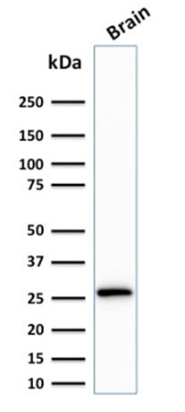
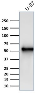 WB (Western Blot)
(Western Blot Analysis of U-87 cell lysate using Vimentin Mouse Monoclonal Antibody (V9).)
WB (Western Blot)
(Western Blot Analysis of U-87 cell lysate using Vimentin Mouse Monoclonal Antibody (V9).)
Vimentin, Monoclonal Antibody (Cat# AAA215254)
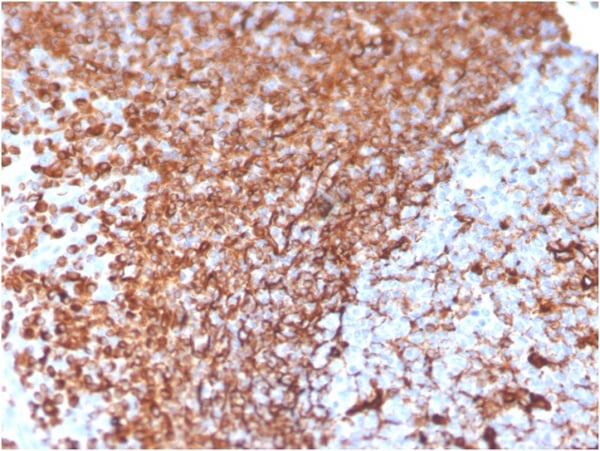
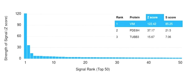 Application Data
(Analysis of Protein Array containing more than 21,000 full-length human proteins using Vimentin Mouse Monoclonal Antibody (VIM/3736) Z- and S- Score: The Z-score represents the strength of a signal that a monoclonal antibody (Monoclonal Antibody) (in combination with a fluorescently-tagged anti-IgG secondary antibody) produces when binding to a particular protein on the HuProtTM array. Z-scores are described in units of standard deviations (SD’s) above the mean value of all signals generated on that array. If targets on HuProtTM are arranged in descending order of the Z-score, the S-score is the difference (also in units of SD’s) between the Z-score. S-score therefore represents the relative target specificity of a Monoclonal Antibody to its intended target. A Monoclonal Antibody is considered to specific to its intended target, if the Monoclonal Antibody has an S-score of at least 2.5. For example, if a Monoclonal Antibody binds to protein X with a Z-score of 43 and to protein Y with a Z-score of 14, then the S-score for the binding of that Monoclonal Antibody to protein X is equal to 29.)
Application Data
(Analysis of Protein Array containing more than 21,000 full-length human proteins using Vimentin Mouse Monoclonal Antibody (VIM/3736) Z- and S- Score: The Z-score represents the strength of a signal that a monoclonal antibody (Monoclonal Antibody) (in combination with a fluorescently-tagged anti-IgG secondary antibody) produces when binding to a particular protein on the HuProtTM array. Z-scores are described in units of standard deviations (SD’s) above the mean value of all signals generated on that array. If targets on HuProtTM are arranged in descending order of the Z-score, the S-score is the difference (also in units of SD’s) between the Z-score. S-score therefore represents the relative target specificity of a Monoclonal Antibody to its intended target. A Monoclonal Antibody is considered to specific to its intended target, if the Monoclonal Antibody has an S-score of at least 2.5. For example, if a Monoclonal Antibody binds to protein X with a Z-score of 43 and to protein Y with a Z-score of 14, then the S-score for the binding of that Monoclonal Antibody to protein X is equal to 29.)
Vimentin, Monoclonal Antibody (Cat# AAA215255)
Does not react with Mouse or Rat.
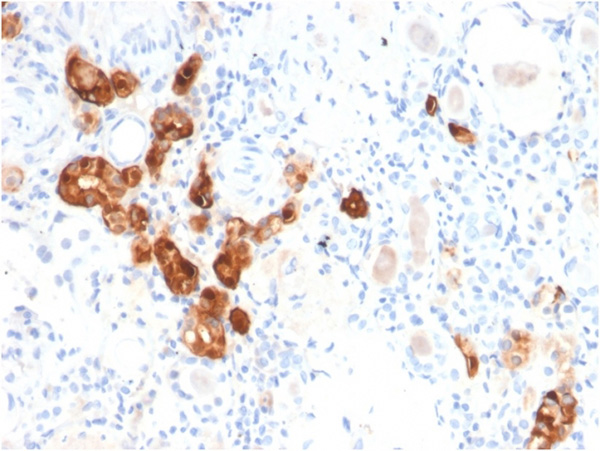
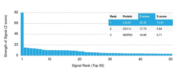 Application Data
(Analysis of Protein Array containing more than 19,000 full-length human proteins using Calbindin Mouse Monoclonal Antibody (CALB1/2782) Z- and S- Score: The Z-score represents the strength of a signal that a monoclonal antibody (Monoclonal Antibody) (in combination with a fluorescently-tagged anti-IgG secondary antibody) produces when binding to a particular protein on the HuProtTM array. Z-scores are described in units of standard deviations (SD's) above the mean value of all signals generated on that array. If targets on HuProtTM are arranged in descending order of the Z-score, the S-score is the difference (also in units of SD's) between the Z-score. S-score therefore represents the relative target specificity of a Monoclonal Antibody to its intended target. A Monoclonal Antibody is considered to specific to its intended target, if the Monoclonal Antibody has an S-score of at least 2.5. For example, if a Monoclonal Antibody binds to protein X with a Z-score of 43 and to protein Y with a Z-score of 14, then the S-score for the binding of that Monoclonal Antibody to protein X is equal to 29.)
Application Data
(Analysis of Protein Array containing more than 19,000 full-length human proteins using Calbindin Mouse Monoclonal Antibody (CALB1/2782) Z- and S- Score: The Z-score represents the strength of a signal that a monoclonal antibody (Monoclonal Antibody) (in combination with a fluorescently-tagged anti-IgG secondary antibody) produces when binding to a particular protein on the HuProtTM array. Z-scores are described in units of standard deviations (SD's) above the mean value of all signals generated on that array. If targets on HuProtTM are arranged in descending order of the Z-score, the S-score is the difference (also in units of SD's) between the Z-score. S-score therefore represents the relative target specificity of a Monoclonal Antibody to its intended target. A Monoclonal Antibody is considered to specific to its intended target, if the Monoclonal Antibody has an S-score of at least 2.5. For example, if a Monoclonal Antibody binds to protein X with a Z-score of 43 and to protein Y with a Z-score of 14, then the S-score for the binding of that Monoclonal Antibody to protein X is equal to 29.)
Calbindin 1 (CALB1), Monoclonal Antibody (Cat# AAA215261)
 SDS-PAGE
(SDS-PAGE Analysis of Purified Calretinin Mouse Monoclonal Antibody (CALB2/2807). Confirmation of Purity and Integrity of Antibody.)
SDS-PAGE
(SDS-PAGE Analysis of Purified Calretinin Mouse Monoclonal Antibody (CALB2/2807). Confirmation of Purity and Integrity of Antibody.)
Calretinin/Calbindin 2, Monoclonal Antibody (Cat# AAA215263)
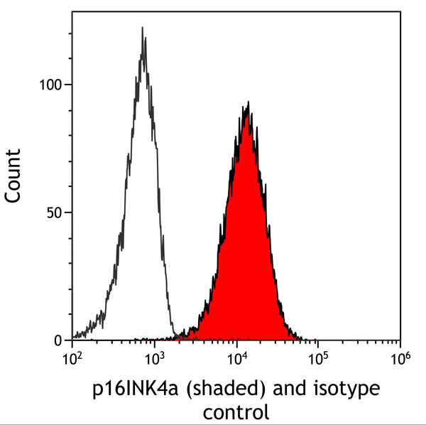
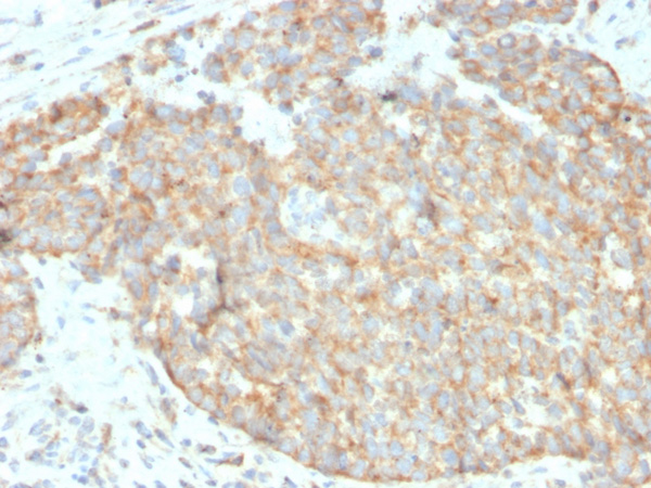
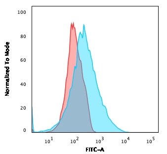 FCM/FACS (Flow Cytometry)
(Flow Cytometric Analysis of Jurkat cells using PD-L2 Mouse Monoclonal Antibody (Z64P2D3*H4) followed by goat anti-Mouse IgG-CF488 (Blue); Isotype Control (Red).)
FCM/FACS (Flow Cytometry)
(Flow Cytometric Analysis of Jurkat cells using PD-L2 Mouse Monoclonal Antibody (Z64P2D3*H4) followed by goat anti-Mouse IgG-CF488 (Blue); Isotype Control (Red).)
PD-L2/PDCD1LG2/CD273, Monoclonal Antibody (Cat# AAA215266)
 SDS-PAGE
(SDS-PAGE Analysis Purified Calpain 1 Mouse Monoclonal Antibody (CAPN1/1530). Confirmation of Purity and Integrity of Antibody.)
SDS-PAGE
(SDS-PAGE Analysis Purified Calpain 1 Mouse Monoclonal Antibody (CAPN1/1530). Confirmation of Purity and Integrity of Antibody.)
Calpain 1, Monoclonal Antibody (Cat# AAA215268)

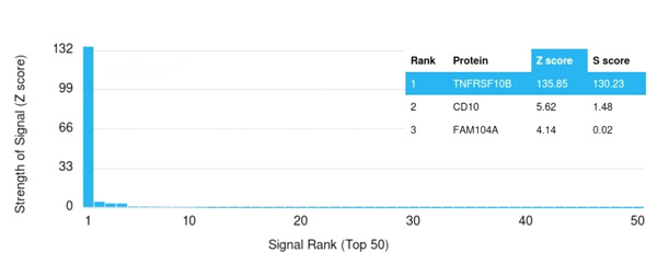 Application Data
(Analysis of Protein Array containing >19,000 full-length human proteins using DR5 Mouse Monoclonal Antibody (DR5/3381) Z- and S- Score: The Z-score represents the strength of a signal that a monoclonal antibody (Monoclonal Antibody) (in combination with a fluorescently-tagged anti-IgG secondary antibody) produces when binding to a particular protein on the HuProtTM array. Z-scores are described in units of standard deviations (SD’s) above the mean value of all signals generated on that array. If targets on HuProtTM are arranged in descending order of the Z-score, the S-score is the difference (also in units of SD’s) between the Z-score. S-score therefore represents the relative target specificity of a Monoclonal Antibody to its intended target. A Monoclonal Antibody is considered to specific to its intended target, if the Monoclonal Antibody has an S-score of at least 2.5. For example, if a Monoclonal Antibody binds to protein X with a Z-score of 43 and to protein Y with a Z-score of 14, then the S-score for the binding of that Monoclonal Antibody to protein X is equal to 29.)
Application Data
(Analysis of Protein Array containing >19,000 full-length human proteins using DR5 Mouse Monoclonal Antibody (DR5/3381) Z- and S- Score: The Z-score represents the strength of a signal that a monoclonal antibody (Monoclonal Antibody) (in combination with a fluorescently-tagged anti-IgG secondary antibody) produces when binding to a particular protein on the HuProtTM array. Z-scores are described in units of standard deviations (SD’s) above the mean value of all signals generated on that array. If targets on HuProtTM are arranged in descending order of the Z-score, the S-score is the difference (also in units of SD’s) between the Z-score. S-score therefore represents the relative target specificity of a Monoclonal Antibody to its intended target. A Monoclonal Antibody is considered to specific to its intended target, if the Monoclonal Antibody has an S-score of at least 2.5. For example, if a Monoclonal Antibody binds to protein X with a Z-score of 43 and to protein Y with a Z-score of 14, then the S-score for the binding of that Monoclonal Antibody to protein X is equal to 29.)
CD262/DR5, Monoclonal Antibody (Cat# AAA215274)

 Application Data
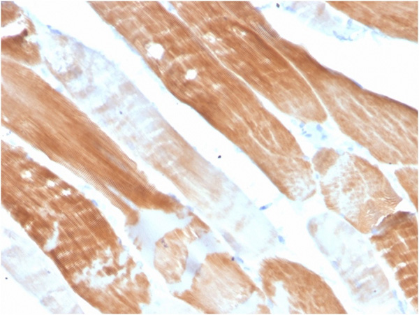
(Analysis of Protein Array containing more than 19,000 full-length human proteins using Sarcomeric Actinin Alpha 2 Mouse Monoclonal Antibody (ACTN2/3291). Z- and S- Score: The Z-score represents the strength of a signal that a monoclonal antibody (MAb) (in combination with a fluorescently-tagged anti-IgG secondary antibody) produces when binding to a particular protein on the HuProtTM array. Z-scores are described in units of standard deviations (SD’s) above the mean value of all signals generated on that array. If targets on HuProtTM are arranged in descending order of the Z-score, the S-score is the difference (also in units of SD’s) between the Z-score. S-score therefore represents the relative target specificity of a MAb to its intended target. A MAb is considered to specific to its intended target, if the MAb has an S-score of at least 2.5. For example, if a MAb binds to protein X with a Z-score of 43 and to protein Y with a Z-score of 14, then the S-score for the binding of that MAb to protein X is equal to 29.)
Application Data
(Analysis of Protein Array containing more than 19,000 full-length human proteins using Sarcomeric Actinin Alpha 2 Mouse Monoclonal Antibody (ACTN2/3291). Z- and S- Score: The Z-score represents the strength of a signal that a monoclonal antibody (MAb) (in combination with a fluorescently-tagged anti-IgG secondary antibody) produces when binding to a particular protein on the HuProtTM array. Z-scores are described in units of standard deviations (SD’s) above the mean value of all signals generated on that array. If targets on HuProtTM are arranged in descending order of the Z-score, the S-score is the difference (also in units of SD’s) between the Z-score. S-score therefore represents the relative target specificity of a MAb to its intended target. A MAb is considered to specific to its intended target, if the MAb has an S-score of at least 2.5. For example, if a MAb binds to protein X with a Z-score of 43 and to protein Y with a Z-score of 14, then the S-score for the binding of that MAb to protein X is equal to 29.)
Sarcomeric Actinin Alpha 2/ACTN2, Monoclonal Antibody (Cat# AAA215275)
 SDS-PAGE
(SDS-PAGE Analysis Purified Aurora B Mouse Monoclonal Antibody (AURKB/1845). Confirmation of Integrity and Purity of Antibody.)
SDS-PAGE
(SDS-PAGE Analysis Purified Aurora B Mouse Monoclonal Antibody (AURKB/1845). Confirmation of Integrity and Purity of Antibody.)
Aurora B, Monoclonal Antibody (Cat# AAA215287)
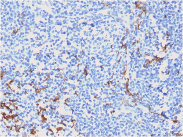
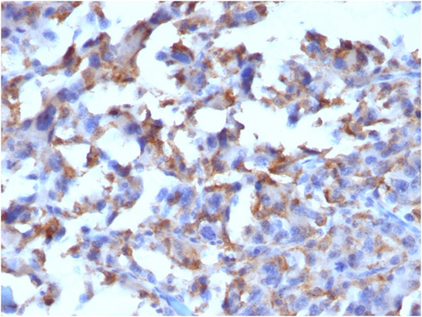
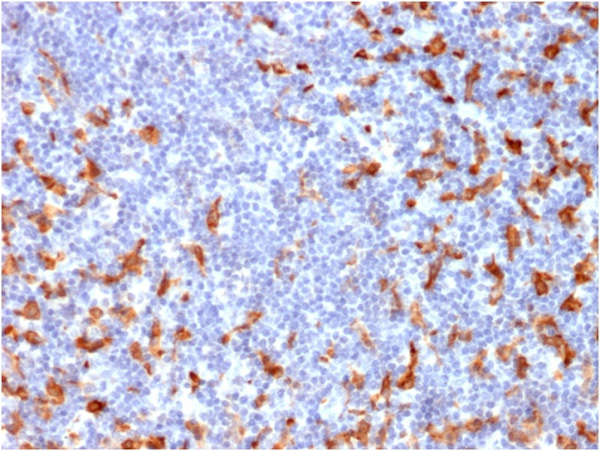
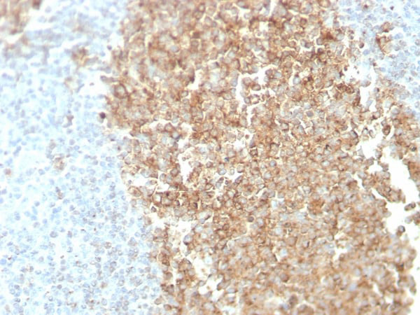
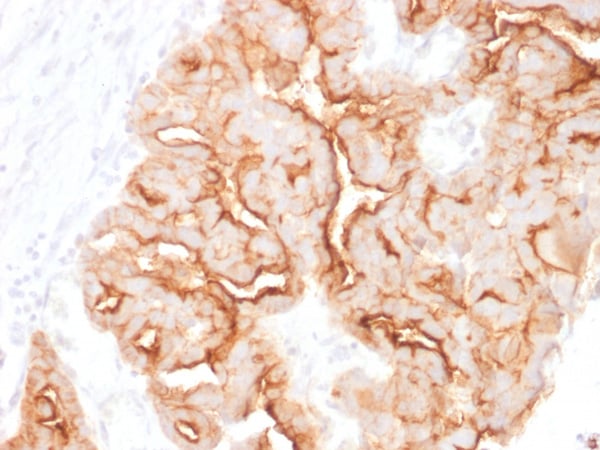
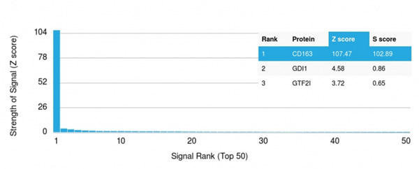 Application Data
(Analysis of Protein Array containing more than 19,000 full-length human proteins using CD163-Monospecific Mouse Monoclonal Antibody (M130/2162). Z- and S- Score: The Z-score represents the strength of a signal that a monoclonal antibody (MAb) (in combination with a fluorescently-tagged anti-IgG secondary antibody) produces when binding to a particular protein on the HuProtTM array. Z-scores are described in units of standard deviations (SD's) above the mean value of all signals generated on that array. If targets on HuProtTM are arranged in descending order of the Z-score, the S-score is the difference (also in units of SD's) between the Z-score. S-score therefore represents the relative target specificity of a MAb to its intended target. A MAb is considered to specific to its intended target, if the MAb has an S-score of at least 2.5. For example, if a MAb binds to protein X with a Z-score of 43 and to protein Y with a Z-score of 14, then the S-score for the binding of that MAb to protein X is equal to 29.)
Application Data
(Analysis of Protein Array containing more than 19,000 full-length human proteins using CD163-Monospecific Mouse Monoclonal Antibody (M130/2162). Z- and S- Score: The Z-score represents the strength of a signal that a monoclonal antibody (MAb) (in combination with a fluorescently-tagged anti-IgG secondary antibody) produces when binding to a particular protein on the HuProtTM array. Z-scores are described in units of standard deviations (SD's) above the mean value of all signals generated on that array. If targets on HuProtTM are arranged in descending order of the Z-score, the S-score is the difference (also in units of SD's) between the Z-score. S-score therefore represents the relative target specificity of a MAb to its intended target. A MAb is considered to specific to its intended target, if the MAb has an S-score of at least 2.5. For example, if a MAb binds to protein X with a Z-score of 43 and to protein Y with a Z-score of 14, then the S-score for the binding of that MAb to protein X is equal to 29.)
CD163, Monoclonal Antibody (Cat# AAA215296)
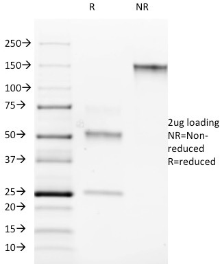
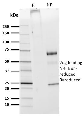
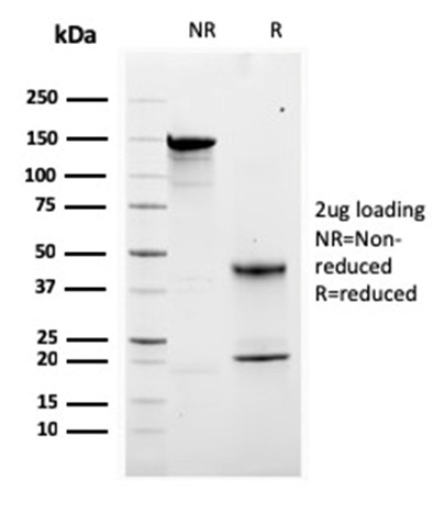
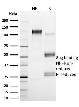
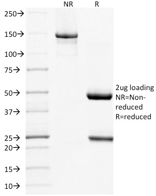 SDS-PAGE
(SDS-PAGE Analysis Purified MUC16 Mouse Monoclonal Antibody (MUC16/1860). Confirmation of Integrity and Purity of Antibody.)
SDS-PAGE
(SDS-PAGE Analysis Purified MUC16 Mouse Monoclonal Antibody (MUC16/1860). Confirmation of Integrity and Purity of Antibody.)
MUC16/CA125, Monoclonal Antibody (Cat# AAA215298)
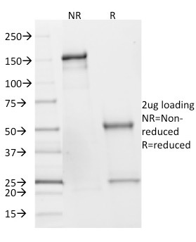
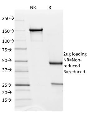
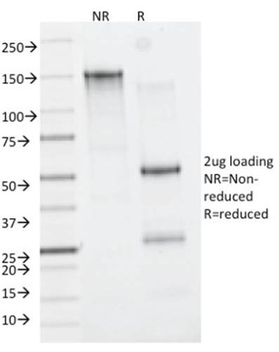
 SDS-PAGE
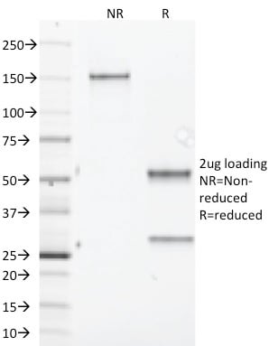
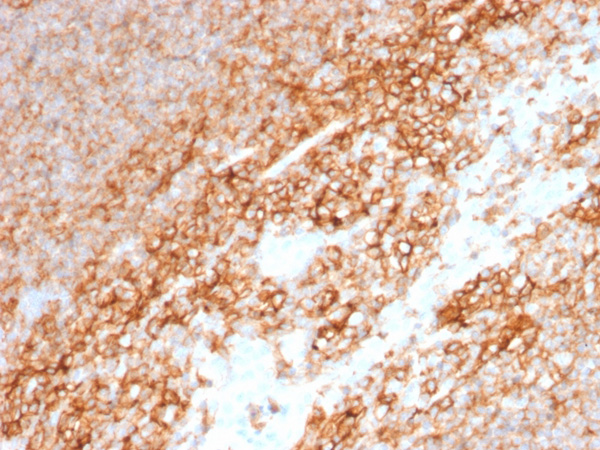
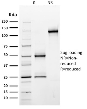
(SDS-PAGE Analysis Purified CD44 Mouse Monoclonal Antibody (BU75). Confirmation of Purity and Integrity of Antibody.)
SDS-PAGE
(SDS-PAGE Analysis Purified CD44 Mouse Monoclonal Antibody (BU75). Confirmation of Purity and Integrity of Antibody.)
CD44/HCAM, Monoclonal Antibody (Cat# AAA215304)
What are Monoclonal Antibodies?
Monoclonal antibodies are specialized laboratory-produced proteins developed for binding to specific biological antigens or other molecular targets. Since they come from a single cell (or clone), they are especially consistent and accurate in the data they are involved in producing.
This type of antibody material has been shown to be a powerful tool in finding and subsequently destroying harmful cells in an organism, such as those found in cancers or various autoimmune diseases. This makes them excellent aids in medical testing and research, which is why they are so widely used.
AAA Biotech offers a comprehensive range of high-quality monoclonal antibodies that perform effectively in various laboratory tests, including (amongst others) ELISA, western blotting, immunohistochemistry, and flow cytometry. All of the products in our catalog are thoroughly quality tested to make sure that they are reliable and will consistently perform well in your research.
What Are The Uses of Monoclonal Antibodies
Monoclonal antibodies are used in many lab tests, including (amongst others) ELISA, western blotting, immunohistochemistry, and flow cytometry.
ELISA is a test that helps detect a specific substance/analyte in a sample. It uses antibodies (often monoclonal) bound to a solid surface (such as the well of a microplate) to “capture” the substance/analyte in the sample and immobilize it so that the detection antibody component can then bind to it and produce a signal, which can then be measured.
Western blotting identifies specific proteins in a sample. The sample is first separated on a gel, and then antibodies are applied that will typically bind to the target, which will all be localized to a single band in a lane.
Immunohistochemistry helps locate specific proteins in cells or tissue samples using antibodies.
Flow cytometry looks at and sorts cells. It uses antibodies that are conjugated to reporter molecules called “fluorophores”, which, under special lights, emit light themselves, which can then be measured by a detector instrument. For a deeper understanding of these techniques, explore our complete guide to monoclonal antibodies and their benefits.
How Monoclonal Antibodies Are Used as Medicine?
Please note that all of the products listed in AAA Biotech’s also known as AAA Bio or AAABio catalog are strictly for research-use only (RUO).
Monoclonal antibodies can also be used as therapeutic/medical treatments, particularly in the context of cancers. They are designed to find and bind to specific cells or proteins, helping the immune system recognize and attack the cancer. These treatments work in different ways, such as:
- Radioimmunotherapy attaches a small amount of radioactive molecule to the antibody, so it delivers the radiation directly to the cancer cells that the antibody is specifically binding to.
- Antibody-directed enzyme prodrug therapy uses antibodies that are specifically bound to special enzymes. These enzymes activate a harmless drug in the body and turn it into a cancer-killing drug only near the cancer cells—this helps avoid harming healthy cells.
- Immunoliposomes are tiny “bubbles” filled with medicine/drug and coated with antibodies. They carry the drug straight to the cancer cells.
Why Buy Monoclonal Antibodies From Us?
At AAA Biotech, we provide high-performance monoclonal antibodies designed to support a wide range of research needs.
1. Validated for Versatile Applications
The antibodies in our catalog are extensively validated and compatible with multiple techniques, including (but not limited to) ELISA, flow cytometry (FC), immunocytochemistry (ICC), immunofluorescence (IF), immunohistochemistry (IHC), immunoprecipitation (IP), and western blotting (WB).
2. Wide Selection & Specialized Options
We offer antibodies for common and rare species, that are available in various conjugated forms, and also in recombinant formats. Essentially, there is almost anything one might need to meet their experimental model’s requirements.
3. High-Quality Proteins
Our proteins meet high purity standards—90% or more as confirmed by SDS-PAGE. Many are available with tags like His, Flag, GST, or MBP, and we also supply native and biologically active proteins for functional studies.
Frequently Asked Questions
1. Are your monoclonal antibodies validated for specific applications?
Yes, our antibodies are tested and validated for use in methods such as ELISA, western blot, IHC, flow cytometry, and more. Refer to specific product pages or datasheets for individual product information.
2. How do I choose the right monoclonal antibody for my application?
Review the product details directly for application validation, species reactivity, and target information. You may also contact our support team at any time for help.
3. How quickly can I receive my order?
Most orders are processed and shipped within 1–3 business days, depending on product availability and your shipping location.