Filters
▼Clonality
▼Type
▼Reactivity
▼Gene Name
▼Isotype
▼Host
▼Application
▼Clone
▼Active Proteins
AAA Biotech also known as AAA Bio or AAABio provides a variety of high-quality recombinant and natural/native proteins that are proven to work in a wide range of experiments. Explore our products to find the active protein that best fits your needs or experimental model.
Viewing 2300-2350 of 2875 product results
 Bioactivity
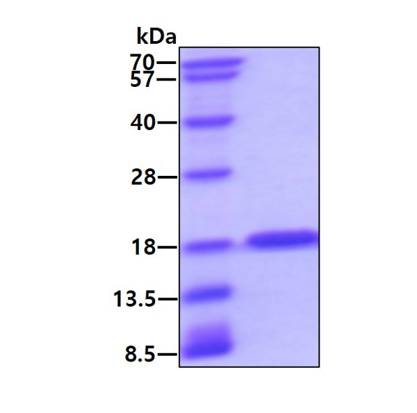
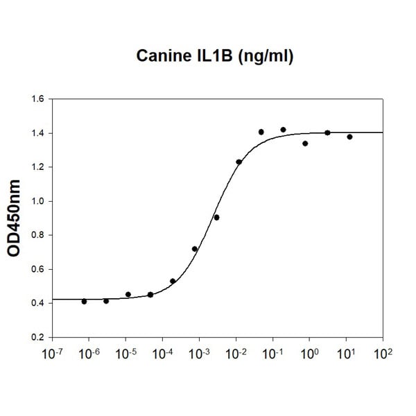
(Canine IL-1B stimulates cell proliferation of the D10.G4.1 mouse helper T cells. The ED50 range range )
Bioactivity
(Canine IL-1B stimulates cell proliferation of the D10.G4.1 mouse helper T cells. The ED50 range range )
IL-1 beta/IL-1F2, Active Protein (Cat# AAA48366)
 Bioactivity
(Small calcium binding protein that S100 Calcium Binding Protein (S100), also known as S100 Alpha (S100A1), is a member of the S100 family of calcium-binding proteins. As with most S100 proteins, S100A1 proteins are localized in the cytoplasm and/or nucleus of a wide range of cells, and it possess a variety of intracellular and extracellular functions. They interact with multiple receptors and signal transducers to regulate pathways that govern inflammation, cell differentiation, proliferation, energy metabolism, apoptosis, calcium homeostasis, cell cytoskeleton and microbial resistance. S100 Calcium Binding Protein A4 (S100A4) is one of targets of S100A1. Thus a functional binding ELISA assay was conducted to detect the interaction of recombinant rat S100A1 and recombinant mouse S100A4. Briefly, S100A1 was diluted serially in PBS with 0.01% BSA (pH 7.4). Duplicate samples of 100 ul were then transferred to S100A4-coated microtiter wells and incubated for 1h at 37 degree C. Wells were washed with PBST and incubated for 1h with anti-S100A1 pAb, then aspirated and washed 3 times. After incubation with HRP labelled secondary antibody for 1h at 37 degree C, wells were aspirated and washed 5 times. With the addition of substrate solution, wells were incubated 15-25 minutes at 37 degree C. Finally, add 50 uL stop solution to the wells and read at 450/630 nm immediately. The binding activity of recombinant rat S100A1 and recombinant mouse S100A4 was shown in Figure 1, the EC50 for this effect is 3.92 ug/mL.)
Bioactivity
(Small calcium binding protein that S100 Calcium Binding Protein (S100), also known as S100 Alpha (S100A1), is a member of the S100 family of calcium-binding proteins. As with most S100 proteins, S100A1 proteins are localized in the cytoplasm and/or nucleus of a wide range of cells, and it possess a variety of intracellular and extracellular functions. They interact with multiple receptors and signal transducers to regulate pathways that govern inflammation, cell differentiation, proliferation, energy metabolism, apoptosis, calcium homeostasis, cell cytoskeleton and microbial resistance. S100 Calcium Binding Protein A4 (S100A4) is one of targets of S100A1. Thus a functional binding ELISA assay was conducted to detect the interaction of recombinant rat S100A1 and recombinant mouse S100A4. Briefly, S100A1 was diluted serially in PBS with 0.01% BSA (pH 7.4). Duplicate samples of 100 ul were then transferred to S100A4-coated microtiter wells and incubated for 1h at 37 degree C. Wells were washed with PBST and incubated for 1h with anti-S100A1 pAb, then aspirated and washed 3 times. After incubation with HRP labelled secondary antibody for 1h at 37 degree C, wells were aspirated and washed 5 times. With the addition of substrate solution, wells were incubated 15-25 minutes at 37 degree C. Finally, add 50 uL stop solution to the wells and read at 450/630 nm immediately. The binding activity of recombinant rat S100A1 and recombinant mouse S100A4 was shown in Figure 1, the EC50 for this effect is 3.92 ug/mL.)
S100 Calcium Binding Protein (S100), Active Protein (Cat# AAA161651)
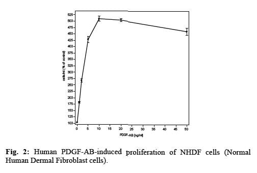 Application Data
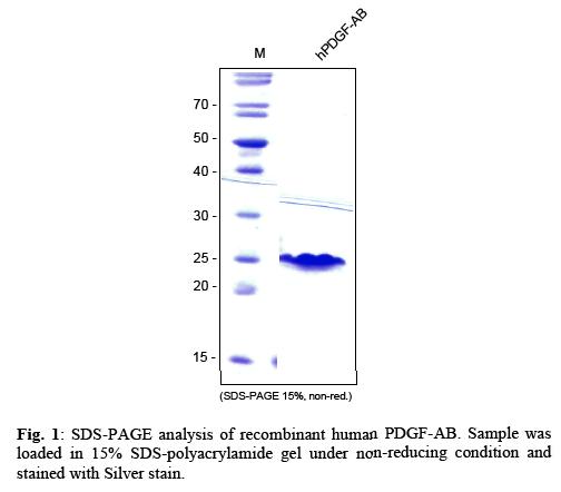
PDGF-AB, Active Protein (Cat# AAA79242)
Artemin, Active Protein (Cat# AAA75568)

 Bioactivity
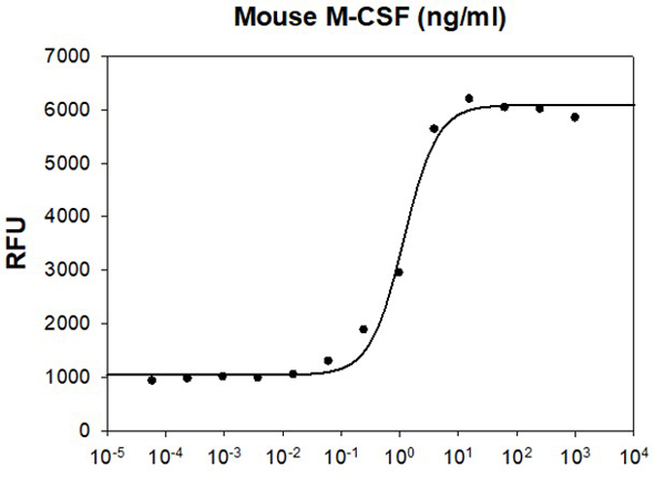
(Mouse M-CSF stimulates cell proliferation of the M-NFS-60 mouse myelogenous leukemia lymphoblast cells. The ED50 range is )
Bioactivity
(Mouse M-CSF stimulates cell proliferation of the M-NFS-60 mouse myelogenous leukemia lymphoblast cells. The ED50 range is )
M-CSF, Active Protein (Cat# AAA48318)
 Bioactivity
(Extracellular matrix metalloproteinase (MMP) inducer (EMMPRIN), also known as basigin and CD147, is a 4466 kDa, variably N and Oglycosylated, type I transmembrane protein that belongs to the immunoglobulin superfamily. EMMPRIN is 269 amino acids (aa) in l)
Bioactivity
(Extracellular matrix metalloproteinase (MMP) inducer (EMMPRIN), also known as basigin and CD147, is a 4466 kDa, variably N and Oglycosylated, type I transmembrane protein that belongs to the immunoglobulin superfamily. EMMPRIN is 269 amino acids (aa) in l)
Cluster Of Differentiation 147 (CD147), Active Protein (Cat# AAA153100)
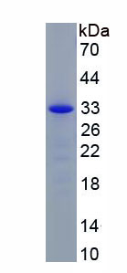
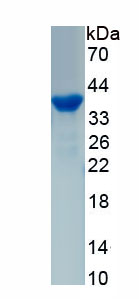
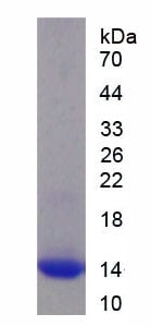
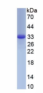
 SDS-PAGE
Delta-like protein 3 (DLL3), Active Protein (Cat# AAA117499)
 Bioactivity
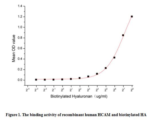
(Homing Associated Cell Adhesion Molecule (HCAM), also known as CD44, is a ubiquitous multistructural and multifunctional cells surface adhesion molecule involved in cell-cell and cell-matrix interactions. CD44 is broadly expressed, including in the membranes of B cells, granulocytes, monocytes, and erythrocytes as well as on many thymocytes and mature T cells, besides it is highly expressed in many cancers and regulates metastasis via recruitment of CD44 to the cell surface. This protein is a receptor for hyaluronic acid (HA) and can also interact with other ligands, such as osteopontin, collagens, and matrix metalloproteinases (MMPs). Thus a functional binding ELISA assay was conducted to detect the interaction of recombinant human HCAM and biotinylated hyaluronan (HA). Briefly, biotin-linked HA was diluted serially in PBS, with 0.01% BSA (pH 7.4). Duplicate samples of 100 ul were then transferred to HCAM-coated microtiter wells and incubated for 2h at 37 degree C. Wells were washed with PBST 3 times and incubation with Streptavidin-HRP for 1 hour, then wells were aspirated and washed 5 times. With the addition of substrate solution, wells were incubated 15-25 minutes at 37 degree C. Finally, add 50 ul stop solution to the wells and read at 450/630 nm immediately. The binding activity of recombinant human HCAM and biotinylated HA was shown in Figure 1, and this effect was in a dose dependent manner.)
Bioactivity
(Homing Associated Cell Adhesion Molecule (HCAM), also known as CD44, is a ubiquitous multistructural and multifunctional cells surface adhesion molecule involved in cell-cell and cell-matrix interactions. CD44 is broadly expressed, including in the membranes of B cells, granulocytes, monocytes, and erythrocytes as well as on many thymocytes and mature T cells, besides it is highly expressed in many cancers and regulates metastasis via recruitment of CD44 to the cell surface. This protein is a receptor for hyaluronic acid (HA) and can also interact with other ligands, such as osteopontin, collagens, and matrix metalloproteinases (MMPs). Thus a functional binding ELISA assay was conducted to detect the interaction of recombinant human HCAM and biotinylated hyaluronan (HA). Briefly, biotin-linked HA was diluted serially in PBS, with 0.01% BSA (pH 7.4). Duplicate samples of 100 ul were then transferred to HCAM-coated microtiter wells and incubated for 2h at 37 degree C. Wells were washed with PBST 3 times and incubation with Streptavidin-HRP for 1 hour, then wells were aspirated and washed 5 times. With the addition of substrate solution, wells were incubated 15-25 minutes at 37 degree C. Finally, add 50 ul stop solution to the wells and read at 450/630 nm immediately. The binding activity of recombinant human HCAM and biotinylated HA was shown in Figure 1, and this effect was in a dose dependent manner.)
Homing Associated Cell Adhesion Molecule (HCAM), Active Protein (Cat# AAA161762)
 Bioactivity
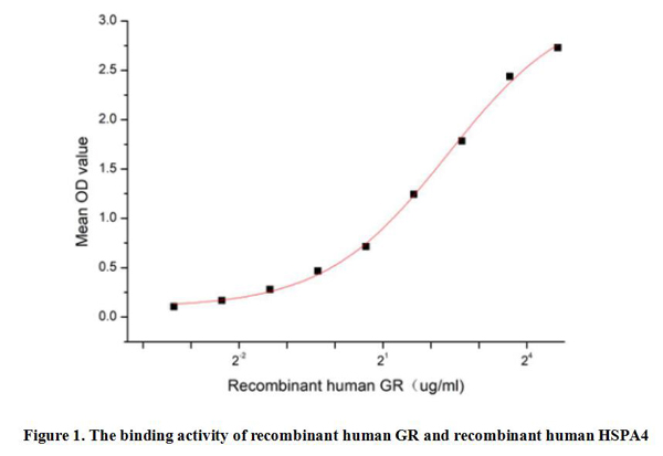
(Glucocorticoid Receptor (GR) is a member of the steroid receptor superfamily, which includes receptors for other steroid hormones such as estrogens, progestogens, androgens and mineralocorticoids. GR is widely distributed in various types of human cells, especially in liver, muscle, adipose tissue, lung, brain and other organs. GR plays a role in signaling within cells. When glucocorticoids enter cells and bind to GR, GR undergoes conformational changes that activate its transcriptional activity. It has been identified that the binding of Heat Shock 70kDa Protein 4 (HSPA4) to GR plays a key role in glucocorticoid signaling, which not only participates in the stabilization and activation of receptors, but also may affect the effect of drugs. Thus a functional binding ELISA assay was conducted to detect the interaction of recombinant human GR and recombinant human HSPA4. Briefly, GR was diluted serially in PBS with 0.01% BSA (pH 7.4). Duplicate samples of 100 ul were then transferred to HSPA4-coated microtiter wells and incubated for 1h at 37 degree C. Wells were washed with PBST and incubated for 1h with anti-GR pAb, then aspirated and washed 3 times. After incubation with HRP labelled secondary antibody for 1h at 37 degree C, wells were aspirated and washed 5 times. With the addition of substrate solution, wells were incubated 15-25 minutes at 37 degree C. Finally, add 50 uL stop solution to the wells and read at 450/630 nm immediately. The binding activity of recombinant human GR and recombinant human HSPA4 was shown in Figure 1, the EC50 for this effect is 5.1 ug/mL.)
Bioactivity
(Glucocorticoid Receptor (GR) is a member of the steroid receptor superfamily, which includes receptors for other steroid hormones such as estrogens, progestogens, androgens and mineralocorticoids. GR is widely distributed in various types of human cells, especially in liver, muscle, adipose tissue, lung, brain and other organs. GR plays a role in signaling within cells. When glucocorticoids enter cells and bind to GR, GR undergoes conformational changes that activate its transcriptional activity. It has been identified that the binding of Heat Shock 70kDa Protein 4 (HSPA4) to GR plays a key role in glucocorticoid signaling, which not only participates in the stabilization and activation of receptors, but also may affect the effect of drugs. Thus a functional binding ELISA assay was conducted to detect the interaction of recombinant human GR and recombinant human HSPA4. Briefly, GR was diluted serially in PBS with 0.01% BSA (pH 7.4). Duplicate samples of 100 ul were then transferred to HSPA4-coated microtiter wells and incubated for 1h at 37 degree C. Wells were washed with PBST and incubated for 1h with anti-GR pAb, then aspirated and washed 3 times. After incubation with HRP labelled secondary antibody for 1h at 37 degree C, wells were aspirated and washed 5 times. With the addition of substrate solution, wells were incubated 15-25 minutes at 37 degree C. Finally, add 50 uL stop solution to the wells and read at 450/630 nm immediately. The binding activity of recombinant human GR and recombinant human HSPA4 was shown in Figure 1, the EC50 for this effect is 5.1 ug/mL.)
Glucocorticoid Receptor (GR), Active Protein (Cat# AAA161853)
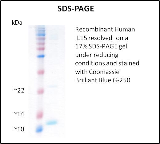
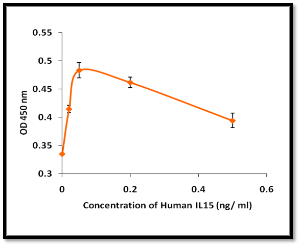 Application Data
IL15, Active Protein (Cat# AAA214232)
 Bioactivity
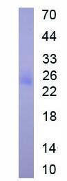
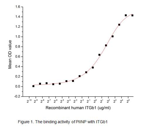
(Procollagen II N-Terminal Propeptide is specific for cartilaginous tissues. It is essential for the normal embryonic development of the skeleton, for linear growth and for the ability of cartilage to resist compressive forces, which was are associated wit)
Bioactivity
(Procollagen II N-Terminal Propeptide is specific for cartilaginous tissues. It is essential for the normal embryonic development of the skeleton, for linear growth and for the ability of cartilage to resist compressive forces, which was are associated wit)
Procollagen II N-Terminal Propeptide (PIINP), Active Protein (Cat# AAA153010)
 Application Data
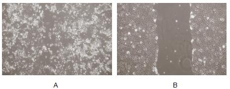
(Interleukin 11 (IL-11) is a multifunctional cytokine. It is a key regulator of multiple events in hematopoiesis, most notably the stimulation of megakaryocyte maturation. IL-11 has been demonstrated to improve platelet recovery after chemotherapy-induced thrombocytopenia, induce acute phase proteins, modulate antigen-antibody responses, participate in the regulation of bone cell proliferation and differentiation IL-11 causes bone-resorption. Besides, IL-11 have been proved can promote migration of A549 cells, 5×10⁴ cells were seeded into 6 well plates. After cell confluent, replace with DMEM without serum overnight. Using a (yellow) pipette tip make a straight scratch, simulating a wound, then washing the wells three times with PBS. Adding 1% serum standard DMEM containing various concentrations of recombinan human IL-11 to each well, incubating the plate for 48 hours at 37, 5% CO2. The results observed by inverted microscope was shown in Figure.(A) A549 cells cultured in DMEM with 100ng/mL IL-11 for 48h; (B) A549 cells cultured in DMEM before addition IL-11.Figure. Wound healing assay of A549 cells after stimulated with IL-11.)
Application Data
(Interleukin 11 (IL-11) is a multifunctional cytokine. It is a key regulator of multiple events in hematopoiesis, most notably the stimulation of megakaryocyte maturation. IL-11 has been demonstrated to improve platelet recovery after chemotherapy-induced thrombocytopenia, induce acute phase proteins, modulate antigen-antibody responses, participate in the regulation of bone cell proliferation and differentiation IL-11 causes bone-resorption. Besides, IL-11 have been proved can promote migration of A549 cells, 5×10⁴ cells were seeded into 6 well plates. After cell confluent, replace with DMEM without serum overnight. Using a (yellow) pipette tip make a straight scratch, simulating a wound, then washing the wells three times with PBS. Adding 1% serum standard DMEM containing various concentrations of recombinan human IL-11 to each well, incubating the plate for 48 hours at 37, 5% CO2. The results observed by inverted microscope was shown in Figure.(A) A549 cells cultured in DMEM with 100ng/mL IL-11 for 48h; (B) A549 cells cultured in DMEM before addition IL-11.Figure. Wound healing assay of A549 cells after stimulated with IL-11.)
Interleukin 11, Active Protein (Cat# AAA150057)
 Bioactivity
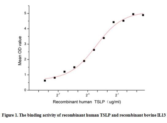
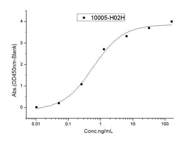
(Thymic stromal lymphopoietin (TSLP) is a member of the IL-2 cytokine family and a distant paralog of IL-7. TSLP is a pleiotropic cytokine that acts on multiple cell lineages, including dendritic cells, T cells, B cells, neutrophils, mast cells, eosinophils and innate lymphoid cells, affecting their maturation, survival and recruitment. It is best known for its role in promoting type 2 immune responses such as in allergic diseases. Interleukin 13 (IL13) is a critical downstream element for TSLP-driven allergic inflammation. Thus a functional binding ELISA assay was conducted to detect the interaction of recombinant human TSLP and recombinant bovine IL13. Briefly, TSLP was diluted serially in PBS with 0.01% BSA (pH 7.4). Duplicate samples of 100 ul were then transferred to IL13-coated microtiter wells and incubated for 1h at 37 degree C. Wells were washed with PBST and incubated for 1h with anti-TSLP pAb, then aspirated and washed 3 times. After incubation with HRP labelled secondary antibody for 1h at 37 degree C, wells were aspirated and washed 5 times. With the addition of substrate solution, wells were incubated 15-25 minutes at 37 degree C. Finally, add 50 uL stop solution to the wells and read at 450/630 nm immediately. The binding activity of recombinant human TSLP and recombinant bovine IL13 was shown in Figure 1, the EC50 for this effect is 0.11 ug/mL.)
Bioactivity
(Thymic stromal lymphopoietin (TSLP) is a member of the IL-2 cytokine family and a distant paralog of IL-7. TSLP is a pleiotropic cytokine that acts on multiple cell lineages, including dendritic cells, T cells, B cells, neutrophils, mast cells, eosinophils and innate lymphoid cells, affecting their maturation, survival and recruitment. It is best known for its role in promoting type 2 immune responses such as in allergic diseases. Interleukin 13 (IL13) is a critical downstream element for TSLP-driven allergic inflammation. Thus a functional binding ELISA assay was conducted to detect the interaction of recombinant human TSLP and recombinant bovine IL13. Briefly, TSLP was diluted serially in PBS with 0.01% BSA (pH 7.4). Duplicate samples of 100 ul were then transferred to IL13-coated microtiter wells and incubated for 1h at 37 degree C. Wells were washed with PBST and incubated for 1h with anti-TSLP pAb, then aspirated and washed 3 times. After incubation with HRP labelled secondary antibody for 1h at 37 degree C, wells were aspirated and washed 5 times. With the addition of substrate solution, wells were incubated 15-25 minutes at 37 degree C. Finally, add 50 uL stop solution to the wells and read at 450/630 nm immediately. The binding activity of recombinant human TSLP and recombinant bovine IL13 was shown in Figure 1, the EC50 for this effect is 0.11 ug/mL.)
Thymic Stromal Lymphopoietin (TSLP), Active Protein (Cat# AAA161842)
 Bioactivity
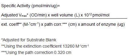
(Complement Factor D (CFD) is a serine protease that catalyzes the initial proteolytic step in the alternative pathway of complement. Expressed in adipose tissue at high levels, factor D is also known as adipsin. It is an exceptionally specific protease and the only known protein substrate is factor B in complex with C3. Factor D protease activity is regulated by reversible conformational changes, which differs from the majority of serine proteases whose regulation involves either activation by processing of the zymogens or inactivation by binding of the inhibitors. Compared to its physiologically important proteolytic activity, factor D has much lower activity toward synthetic peptide substrates. However, thioester substrates have been routinely used for assessing factor D activity. The full-length (amino acid residues 1-263) of rat CFD was expressed which activity was measured by its ability to cleaves a thioester substrate Z-Lys-SBzl•HCl. The reaction was performed in 50 mM Tris, 1 M NaCl, pH 7.5 (Assay Buffer), initiated by addition 50 uL of various concentrations of CFD (diluted by Assay Buffer) to 50 ul substrate mixture of 0.2mM Z-Lys-SBzl•HCl and 0.2 mM DTNB. The final well serves as a negative control with no CFD, replaced with 50 ul assay buffer. Then read in kinetic mode for 5 minutes at an absorbance of 405 nm. The specific activity of recombinant rat CFD is > 10000 pmol/min/ug.)
Bioactivity
(Complement Factor D (CFD) is a serine protease that catalyzes the initial proteolytic step in the alternative pathway of complement. Expressed in adipose tissue at high levels, factor D is also known as adipsin. It is an exceptionally specific protease and the only known protein substrate is factor B in complex with C3. Factor D protease activity is regulated by reversible conformational changes, which differs from the majority of serine proteases whose regulation involves either activation by processing of the zymogens or inactivation by binding of the inhibitors. Compared to its physiologically important proteolytic activity, factor D has much lower activity toward synthetic peptide substrates. However, thioester substrates have been routinely used for assessing factor D activity. The full-length (amino acid residues 1-263) of rat CFD was expressed which activity was measured by its ability to cleaves a thioester substrate Z-Lys-SBzl•HCl. The reaction was performed in 50 mM Tris, 1 M NaCl, pH 7.5 (Assay Buffer), initiated by addition 50 uL of various concentrations of CFD (diluted by Assay Buffer) to 50 ul substrate mixture of 0.2mM Z-Lys-SBzl•HCl and 0.2 mM DTNB. The final well serves as a negative control with no CFD, replaced with 50 ul assay buffer. Then read in kinetic mode for 5 minutes at an absorbance of 405 nm. The specific activity of recombinant rat CFD is > 10000 pmol/min/ug.)
Complement Factor D (CFD), Active Protein (Cat# AAA161865)
 Application Data
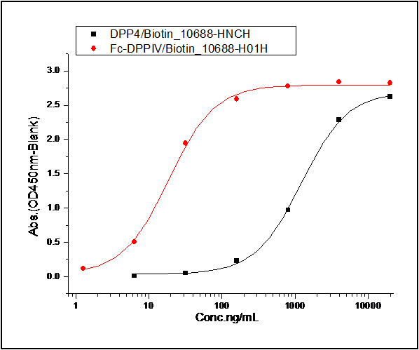
(1. Measured by its binding ability in a functional ELISA. Immobilized Spike Protein S1 (aa 1-725) at 10 ug/ml (100 ul/well) can bind biotinylated human DPP4. The EC50 of of biotinylated DPP4 is 0.6-1.39 ug/ml. 2. Measured by its binding ability in a functional ELISA. Immobilized Spike Protein S1 (aa 1-725) at 10 ug/ml (100 ul/well) can bind biotinylated Fc-DPP4. The EC50 of biotinylated Fc-DPP4 is 0.02-0.05 ug/ml.)
Application Data
(1. Measured by its binding ability in a functional ELISA. Immobilized Spike Protein S1 (aa 1-725) at 10 ug/ml (100 ul/well) can bind biotinylated human DPP4. The EC50 of of biotinylated DPP4 is 0.6-1.39 ug/ml. 2. Measured by its binding ability in a functional ELISA. Immobilized Spike Protein S1 (aa 1-725) at 10 ug/ml (100 ul/well) can bind biotinylated Fc-DPP4. The EC50 of biotinylated Fc-DPP4 is 0.02-0.05 ug/ml.)
MERS-CoV Spike/S1, Active Protein (Cat# AAA258067)
 Application Data
(Measured by its ability to inhibit BMP9 induced alkaline phosphatase production by MC3T3E1 mouse chondrogenic cells. David, L. et al. (2007) Blood 109:1953. The ED50 for this effect is typically 5-15 ng/mL in the presence of 2 ng/mL of recombiant human BMP9.)
Application Data
(Measured by its ability to inhibit BMP9 induced alkaline phosphatase production by MC3T3E1 mouse chondrogenic cells. David, L. et al. (2007) Blood 109:1953. The ED50 for this effect is typically 5-15 ng/mL in the presence of 2 ng/mL of recombiant human BMP9.)
ALK-1, Active Protein (Cat# AAA257825)
 Application Data
(Measured by its binding ability in a functional ELISA. Immobilized Inhibin Human, Mouse, Rat, Cynomolgus, Rhesus Inhibin beta A/Activin A at 2 ug/ml (100 ul/well) can bind Human ACVR2B hFc, the EC50 of Human ACVR2B hFc is 12-60 ng/mL.)
Application Data
(Measured by its binding ability in a functional ELISA. Immobilized Inhibin Human, Mouse, Rat, Cynomolgus, Rhesus Inhibin beta A/Activin A at 2 ug/ml (100 ul/well) can bind Human ACVR2B hFc, the EC50 of Human ACVR2B hFc is 12-60 ng/mL.)
ACVR2B, Active Protein (Cat# AAA257849)


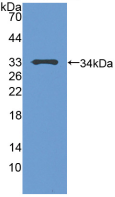
 WB (Western Blot)
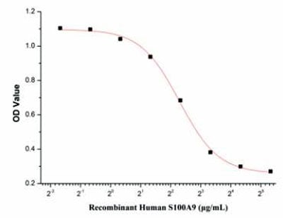
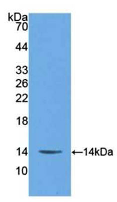
(Sample: Recombinant S100A9, Human;Antibody: Rabbit Anti- Human S100A9 Ab)
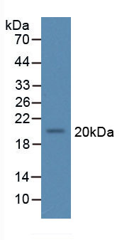
WB (Western Blot)
(Sample: Recombinant S100A9, Human;Antibody: Rabbit Anti- Human S100A9 Ab)
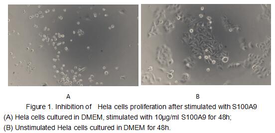
S100 Calcium Binding Protein A9 (S100A9), Active Protein (Cat# AAA153103)
 Application Data
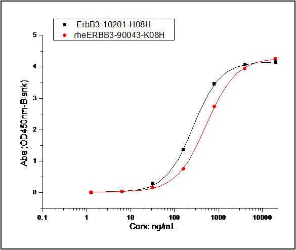
(Measured by its binding ability in a functional ELISA. 1. Immobilized Rhesus ErbB3 at 2 ug/mL (100 ul/well) can bind human NRG1 (isoform Beta1), The EC50 of human NRG1 (isoform Beta1) is 0.58 ug/mL. 2. Immobilized human ErbB3 at 2 ug/mL (100 ul/well) can bind human NRG1 (isoform Beta1), The EC50 of human NRG1 (isoform Beta1) is 0.43 ug/mL.)
Application Data
(Measured by its binding ability in a functional ELISA. 1. Immobilized Rhesus ErbB3 at 2 ug/mL (100 ul/well) can bind human NRG1 (isoform Beta1), The EC50 of human NRG1 (isoform Beta1) is 0.58 ug/mL. 2. Immobilized human ErbB3 at 2 ug/mL (100 ul/well) can bind human NRG1 (isoform Beta1), The EC50 of human NRG1 (isoform Beta1) is 0.43 ug/mL.)
NRG1 Beta 1, Active Protein (Cat# AAA257980)
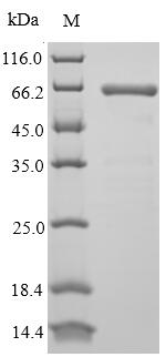
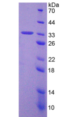
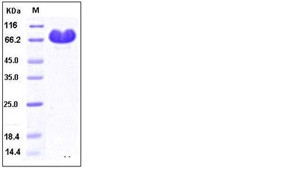
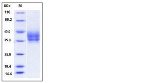
 SDS-PAGE
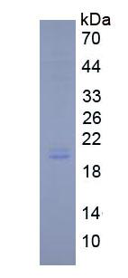
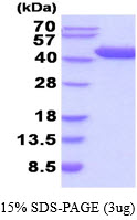
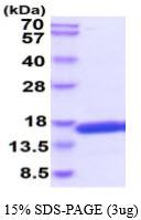
(3ug by SDS-PAGE under reducing condition and visualized by coomassie blue stain)
SDS-PAGE
(3ug by SDS-PAGE under reducing condition and visualized by coomassie blue stain)
GOT1, Active Protein (Cat# AAA48924)
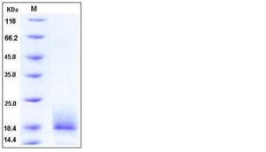
 SDS-PAGE
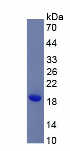
Interleukin-4, Active Protein (Cat# AAA48531)
 Bioactivity
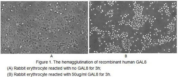
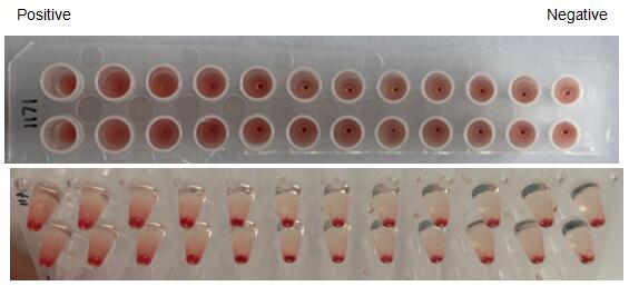
(Figure 2. The hemagglutination assay of GAL8 in V- bottom shaped 96-well microtiter plate.)
Bioactivity
(Figure 2. The hemagglutination assay of GAL8 in V- bottom shaped 96-well microtiter plate.)
Galectin 8 (GAL8), Active Protein (Cat# AAA153022)
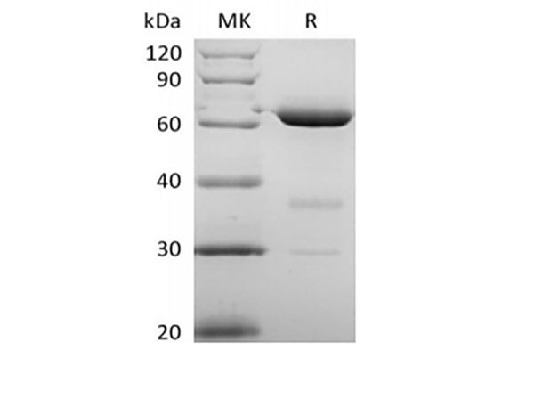
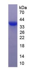
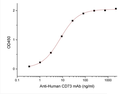 Bioactivity
5'-Nucleotidase/5'-NT/CD73, Active Protein (Cat# AAA177936)
 Application Data
(Tumor protein p53, also known as p53, cellular tumor antigen p53 (UniProt name), phosphoprotein p53, tumor suppressor p53, antigen NY-CO-13, or transformationrelated protein 53 (TRP53), is any isoform of a protein encoded by homologous genes in various organisms, such as TP53 (humans) and Trp53 (mice). TP53 involved in cell cycle regulation as a trans-activator that acts to negatively regulate cell division by controlling a set of genes required for this process. One of the activated genes is an inhibitor of cyclin-dependent kinases. To test the effect of TP53 on cell apoptosis, Jurkat cells were seeded into triplicate wells of 96-well plates at a density of 5,000 cells/well with 1% serum standard 1640 including various concentrations of recombinant human TP53. After incubated for 72h, cells were observed by inverted microscope and cell proliferation was measured by Cell Counting Kit-8 (CCK-8). Briefly, 10uL of CCK-8 solution was added to each well of the plate, then the absorbance at 450nm was measured using a microplate reader after incubating the plate for 1-4 hours at 37?. Proliferation of Jurkat cells after incubation with TP53 for 72h observed by inverted microscope was shown in Figure 1. Cell viability was assessed by CCK-8 (Cell Counting Kit-8) assay after incubation with recombinant TP53 for 72h. The result was shown in Figure 2. It was obvious that TP53 significantly inhibit cell viability of Jurkat cells.)
Application Data
(Tumor protein p53, also known as p53, cellular tumor antigen p53 (UniProt name), phosphoprotein p53, tumor suppressor p53, antigen NY-CO-13, or transformationrelated protein 53 (TRP53), is any isoform of a protein encoded by homologous genes in various organisms, such as TP53 (humans) and Trp53 (mice). TP53 involved in cell cycle regulation as a trans-activator that acts to negatively regulate cell division by controlling a set of genes required for this process. One of the activated genes is an inhibitor of cyclin-dependent kinases. To test the effect of TP53 on cell apoptosis, Jurkat cells were seeded into triplicate wells of 96-well plates at a density of 5,000 cells/well with 1% serum standard 1640 including various concentrations of recombinant human TP53. After incubated for 72h, cells were observed by inverted microscope and cell proliferation was measured by Cell Counting Kit-8 (CCK-8). Briefly, 10uL of CCK-8 solution was added to each well of the plate, then the absorbance at 450nm was measured using a microplate reader after incubating the plate for 1-4 hours at 37?. Proliferation of Jurkat cells after incubation with TP53 for 72h observed by inverted microscope was shown in Figure 1. Cell viability was assessed by CCK-8 (Cell Counting Kit-8) assay after incubation with recombinant TP53 for 72h. The result was shown in Figure 2. It was obvious that TP53 significantly inhibit cell viability of Jurkat cells.)
Tumor Protein p53, Active Protein (Cat# AAA150104)

 Bioactivity
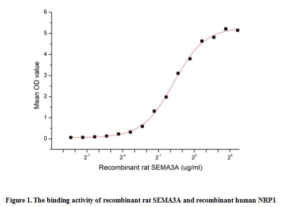
(The Semaphorin 3A(SEMA3A) which belongs to the semaphorin family can function as either a chemorepulsive agent, inhibiting axonal outgrowth, or as a chemoattractive agent, stimulating the growth of apical dendrites. In both cases, the protein is vital for normal neuronal pattern development. Semaphorin 3A is secreted protein containing a Sema domain, an immunoglobulin C2-like domain and a basic domain near the carboxyl tail. It can be secreted by neurons and surrounding tissue to guide migrating cells and axons in the developing nervous system. Besides, Neuropilin 1 (NRP1) has been identified as an interactor of SEMA3A, thus a functional binding ELISA assay was conducted to detect the interaction of recombinant rat SEMA3A and recombinant human NRP1. Briefly, SEMA3A were diluted serially in PBS, with 0.01% BSA (pH 7.4). Duplicate samples of 100 ul were then transferred to NRP1-coated microtiter wells and incubated for 2h at 37 degree C. Wells were washed with PBST and incubated for 1h with anti-SEMA3A pAb, then aspirated and washed 3 times. After incubation with HRP labelled secondary antibody, wells were aspirated and washed 3 times. With the addition of substrate solution, wells were incubated 15-25 minutes at 37 degree C. Finally, add 50 ul stop solution to the wells and read at 450 nm immediately. The binding activity of recombinant rat SEMA3A and recombinant human NRP1 was shown in Figure 1, the EC50 for this effect is 1.22 ug/mL.)
Bioactivity
(The Semaphorin 3A(SEMA3A) which belongs to the semaphorin family can function as either a chemorepulsive agent, inhibiting axonal outgrowth, or as a chemoattractive agent, stimulating the growth of apical dendrites. In both cases, the protein is vital for normal neuronal pattern development. Semaphorin 3A is secreted protein containing a Sema domain, an immunoglobulin C2-like domain and a basic domain near the carboxyl tail. It can be secreted by neurons and surrounding tissue to guide migrating cells and axons in the developing nervous system. Besides, Neuropilin 1 (NRP1) has been identified as an interactor of SEMA3A, thus a functional binding ELISA assay was conducted to detect the interaction of recombinant rat SEMA3A and recombinant human NRP1. Briefly, SEMA3A were diluted serially in PBS, with 0.01% BSA (pH 7.4). Duplicate samples of 100 ul were then transferred to NRP1-coated microtiter wells and incubated for 2h at 37 degree C. Wells were washed with PBST and incubated for 1h with anti-SEMA3A pAb, then aspirated and washed 3 times. After incubation with HRP labelled secondary antibody, wells were aspirated and washed 3 times. With the addition of substrate solution, wells were incubated 15-25 minutes at 37 degree C. Finally, add 50 ul stop solution to the wells and read at 450 nm immediately. The binding activity of recombinant rat SEMA3A and recombinant human NRP1 was shown in Figure 1, the EC50 for this effect is 1.22 ug/mL.)
Semaphorin 3A (SEMA3A), Active Protein (Cat# AAA161937)
 Application Data
(Measured by its ability to neutralize Activin-mediated inhibition on MPC11 cell proliferation. The ED50 for this effect is typically 0.3-2 ug/mL in the presence of 10 ng/mL recombinant Activin A.)
Application Data
(Measured by its ability to neutralize Activin-mediated inhibition on MPC11 cell proliferation. The ED50 for this effect is typically 0.3-2 ug/mL in the presence of 10 ng/mL recombinant Activin A.)
ACVR2B, Active Protein (Cat# AAA257850)
 Bioactivity
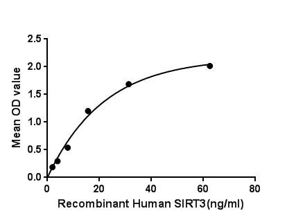
(Figure. The binding activity of SIRT3 with IDH2.)
Bioactivity
(Figure. The binding activity of SIRT3 with IDH2.)
Sirtuin 3, Active Protein (Cat# AAA150138)
VCAM-1, Active Protein (Cat# AAA14954)
 Bioactivity
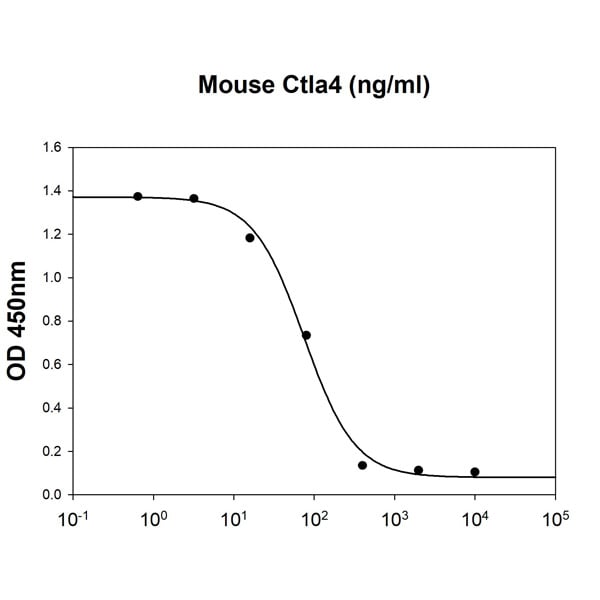
(Mouse Ctla4 inhibits IL-2 secretion of the stimulated Jurkat human acute T cell leukemia cells with Human B7-1/CD80. The ED50 range )
Bioactivity
(Mouse Ctla4 inhibits IL-2 secretion of the stimulated Jurkat human acute T cell leukemia cells with Human B7-1/CD80. The ED50 range )
Ctla-4, Active Protein (Cat# AAA48408)
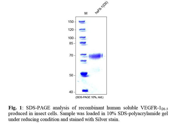
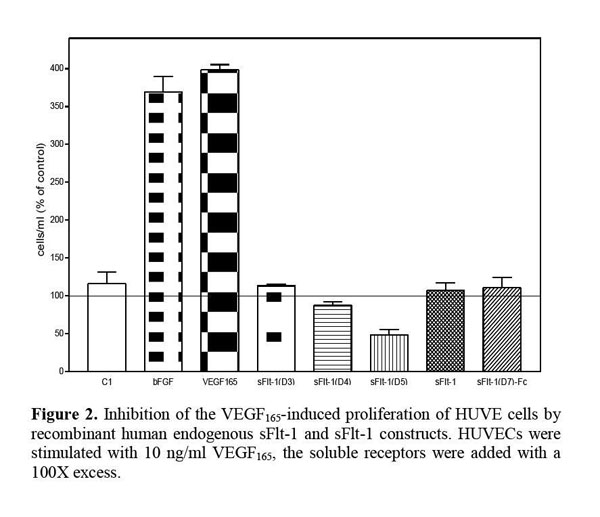 Application Data
VEGFR-1/Flt-1 (D5), soluble, Active Protein (Cat# AAA79171)
Aldo-Keto Reductase Family 1 Member C1, Active Protein (Cat# AAA38788)

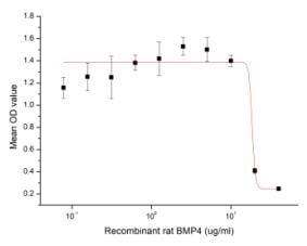 Bioactivity
(Figure 2. Inhibition of HepG2 cells proliferation after stimulated with recombinant rat BMP4)
Bioactivity
(Figure 2. Inhibition of HepG2 cells proliferation after stimulated with recombinant rat BMP4)
Bone Morphogenetic Protein 4 (BMP4), Active Protein (Cat# AAA161655)

 Application Data
(Immobilized PVRIG Protein, Cynomolgus, Rhesus, Recombinant (His Tag) at 2 ug/ml (100 ul/well) can bind Nectin-2 Protein, Human, Recombinant (hFc Tag), the EC50 of Nectin-2 Protein, Human, Recombinant (hFc Tag)is 0.2-1.2 ng/mL.)
Application Data
(Immobilized PVRIG Protein, Cynomolgus, Rhesus, Recombinant (His Tag) at 2 ug/ml (100 ul/well) can bind Nectin-2 Protein, Human, Recombinant (hFc Tag), the EC50 of Nectin-2 Protein, Human, Recombinant (hFc Tag)is 0.2-1.2 ng/mL.)
Nectin-2, Active Protein (Cat# AAA257811)
 Application Data
(Measured by its binding ability in a functional ELISA. Immobilized human ACE2 protein (His tag) at 2ug/mL (100uL/well) can bind SARS-CoV Spike/RBD Protein (RBD, mFc Tag) (40150-V05H), the EC50 of SARS-CoV Spike/RBD Protein (RBD, mFc Tag) is 9-40 ng/mL.)
Application Data
(Measured by its binding ability in a functional ELISA. Immobilized human ACE2 protein (His tag) at 2ug/mL (100uL/well) can bind SARS-CoV Spike/RBD Protein (RBD, mFc Tag) (40150-V05H), the EC50 of SARS-CoV Spike/RBD Protein (RBD, mFc Tag) is 9-40 ng/mL.)
SARS-CoV Spike/RBD, Active Protein (Cat# AAA258069)
 Application Data
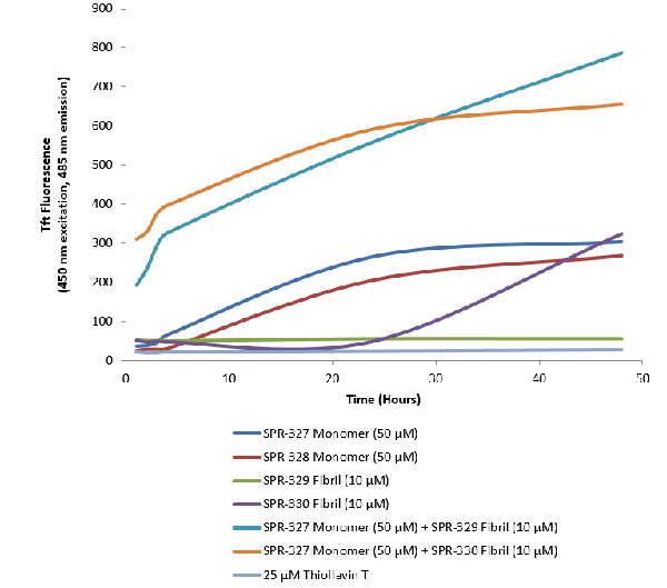
(Thioflavin T is a fluorescent dye that binds to beta sheet-rich structures, such as those in tau fibrils. Upon binding, the emission spectrum of the dye experiences a red-shift and increased fluorescence intensity. Thioflavin T emission curves show increased fluorescence (correlated to tau aggregation) over time in tau monomers. A greater increase in fluorescence is seen when 50 uM monomer is combined with 10 uM PFFs, as the fibrils seed the formation of new fibrils from the pool of monomers. Thioflavin T ex = 450 nm, em = 485 nm.)
Application Data
(Thioflavin T is a fluorescent dye that binds to beta sheet-rich structures, such as those in tau fibrils. Upon binding, the emission spectrum of the dye experiences a red-shift and increased fluorescence intensity. Thioflavin T emission curves show increased fluorescence (correlated to tau aggregation) over time in tau monomers. A greater increase in fluorescence is seen when 50 uM monomer is combined with 10 uM PFFs, as the fibrils seed the formation of new fibrils from the pool of monomers. Thioflavin T ex = 450 nm, em = 485 nm.)
Tau441, Active Protein (Cat# AAA253967)
Ion-Exchange Purified
 SDS_PAGE
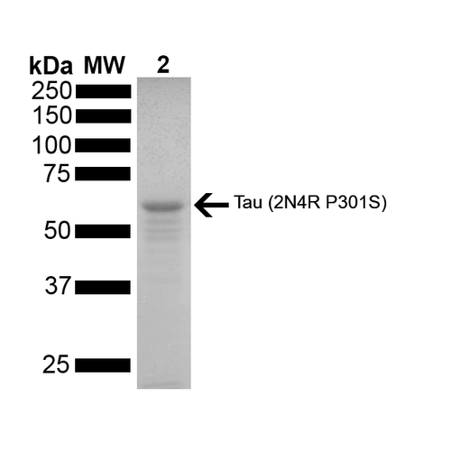
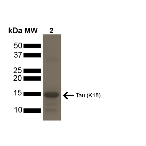
(SDS-PAGE of ~15 kDa Active Human Tau Protein K18 P301L Preformed Fibrils. Lane 1: MW Ladder. Lane 2: Tau Protein Preformed Fibrils.)
SDS_PAGE
(SDS-PAGE of ~15 kDa Active Human Tau Protein K18 P301L Preformed Fibrils. Lane 1: MW Ladder. Lane 2: Tau Protein Preformed Fibrils.)
Tau, Active Protein (Cat# AAA253968)
Ion-Exchange Purified


 Application Data
(Galectin 3 (GAL3) is a member of the lectin family, of which 14 mammalian galectins have been identified. It is also a member of the beta-galactoside-binding protein family that plays an important role in cell-cell adhesion, cell-matrix interactions, macrophage activation, angiogenesis, metastasis, apoptosis. The protein also has been demonstrated to be involved in cancer, inflammation and fibrosis, heart disease, and stroke. GAL3 is expressed in the nucleus, cytoplasm, mitochondrion, cell surface, and extracellular space. It also can agglutinate red blood. In this case, we choose rabbit erythrocyte (RaE) to assay its ability of agglutination. A general procedure for hemagglutination assay (HA) is as follows, two-fold dilute the recombinant rat GAL3 with 0.01M PBS (pH7.4), add 50L a serial dilution of GAL3 to each well of a U or V-bottom shaped 96-well microtiter plate. The final well serves as a negative control with no GAL3, replace with 50L 0.01M PBS. Then add 50L 1% RaE to each well and mixed gently. The plate is incubated for 1-2 hours at room temperature. The results are shown in Figure 1. The minimal effective concentration of GAL3 is 2.5g/mL. (A) 1% RaE treated with 2.5g/mL GAL3 for 2h; (B) Negative control without GAL3.Figure. The hemagglutination activity of recombinant rat GAL3.)
Application Data
(Galectin 3 (GAL3) is a member of the lectin family, of which 14 mammalian galectins have been identified. It is also a member of the beta-galactoside-binding protein family that plays an important role in cell-cell adhesion, cell-matrix interactions, macrophage activation, angiogenesis, metastasis, apoptosis. The protein also has been demonstrated to be involved in cancer, inflammation and fibrosis, heart disease, and stroke. GAL3 is expressed in the nucleus, cytoplasm, mitochondrion, cell surface, and extracellular space. It also can agglutinate red blood. In this case, we choose rabbit erythrocyte (RaE) to assay its ability of agglutination. A general procedure for hemagglutination assay (HA) is as follows, two-fold dilute the recombinant rat GAL3 with 0.01M PBS (pH7.4), add 50L a serial dilution of GAL3 to each well of a U or V-bottom shaped 96-well microtiter plate. The final well serves as a negative control with no GAL3, replace with 50L 0.01M PBS. Then add 50L 1% RaE to each well and mixed gently. The plate is incubated for 1-2 hours at room temperature. The results are shown in Figure 1. The minimal effective concentration of GAL3 is 2.5g/mL. (A) 1% RaE treated with 2.5g/mL GAL3 for 2h; (B) Negative control without GAL3.Figure. The hemagglutination activity of recombinant rat GAL3.)
Galectin 3, Active Protein (Cat# AAA150087)
CLEC9A, Active Protein (Cat# AAA14752)
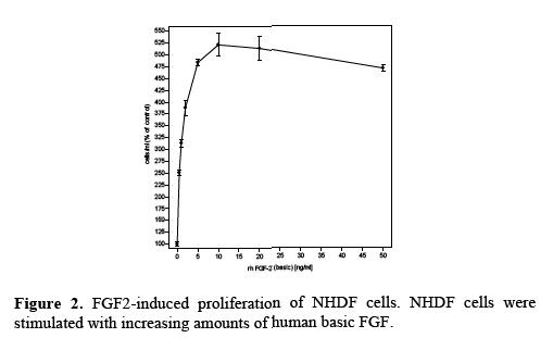
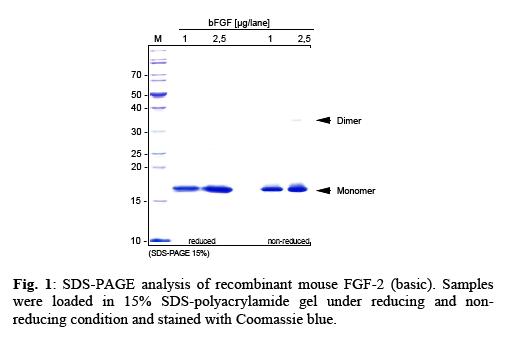
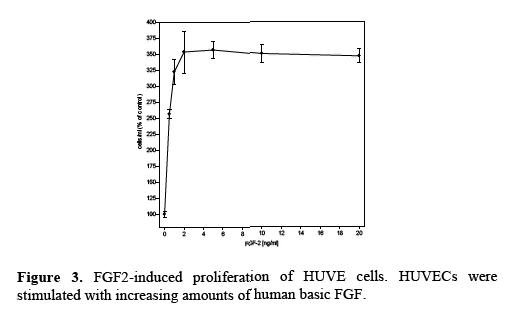 Application Data
FGF-2 (basic), Active Protein (Cat# AAA79146)
Alpha-Amylase, Active Protein (Cat# AAA44778)
 Bioactivity
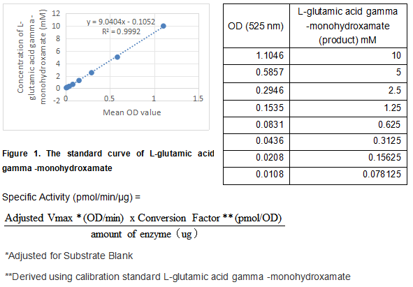
(Transglutaminase 2 (TGM2), encoded by the TGM2 gene, is belongs to the family of transglutaminases that catalyze the posttranslational modification of proteins via calcium dependent cross-linking reactions. In addition to its function in protein cross-linking, TGM2 is also capable of hydrolyzing both GTP and ATP and has intrinsic kinase activity. TGM2 has been implicated in a variety of human diseases including celiac disease, inclusion body myositis, atherosclerosis, and neurodegenerative diseases. The activity of recombinant human TGM2 is measured by its ability to cleave a synthetic peptide Benzyloxycarbonyl-Gln-Gly and NH2OH in the assay buffer 200 mM MES, 10 mM DTT, 10 mM CaCl2, 100 mM Hydroxylamine Hydrochloride, pH 6.0. The rhTGM2 is diluted to 12.5 ug/ml in assay buffer. Loading into a clear well plate 50 uL of 12.5 ug/mL rhTGM2 and start the reaction by adding 50 uL of 100 mM substrate, with a substrate blank containing 50 uL assay buffer, 50 uL substrate, and no rhTGM2. Incubated at 37 degree C for 2 hours and stop the reaction with 400 ul stop solution of 0.37 M FeCl3, 0.67 M HCl, 0.2 M Trichloroacetic Acid. Centrifuge at 2000 rpm for 2 minutes and then load 200 ul of the supernatant into a plate and read at 525 nm (absorbance) in endpoint mode. The specific activity of recombinant human TGM2 is > 800 pmol/min/ug.)
Bioactivity
(Transglutaminase 2 (TGM2), encoded by the TGM2 gene, is belongs to the family of transglutaminases that catalyze the posttranslational modification of proteins via calcium dependent cross-linking reactions. In addition to its function in protein cross-linking, TGM2 is also capable of hydrolyzing both GTP and ATP and has intrinsic kinase activity. TGM2 has been implicated in a variety of human diseases including celiac disease, inclusion body myositis, atherosclerosis, and neurodegenerative diseases. The activity of recombinant human TGM2 is measured by its ability to cleave a synthetic peptide Benzyloxycarbonyl-Gln-Gly and NH2OH in the assay buffer 200 mM MES, 10 mM DTT, 10 mM CaCl2, 100 mM Hydroxylamine Hydrochloride, pH 6.0. The rhTGM2 is diluted to 12.5 ug/ml in assay buffer. Loading into a clear well plate 50 uL of 12.5 ug/mL rhTGM2 and start the reaction by adding 50 uL of 100 mM substrate, with a substrate blank containing 50 uL assay buffer, 50 uL substrate, and no rhTGM2. Incubated at 37 degree C for 2 hours and stop the reaction with 400 ul stop solution of 0.37 M FeCl3, 0.67 M HCl, 0.2 M Trichloroacetic Acid. Centrifuge at 2000 rpm for 2 minutes and then load 200 ul of the supernatant into a plate and read at 525 nm (absorbance) in endpoint mode. The specific activity of recombinant human TGM2 is > 800 pmol/min/ug.)
Transglutaminase 2 (TGM2), Active Protein (Cat# AAA161864)
Resistin, Active Protein (Cat# AAA14423)
 Biological Activity
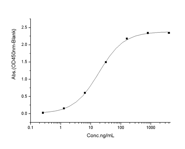
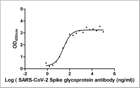
(Measured by its binding ability in a functional ELISA. Immobilized SARS-CoV-2-S1-RBD at 2 ug/ml can bind SARS-CoV-2-S Antibody, the EC50 of SARS-CoV-2-S1-RBD protein is 19.60-39.42 ng/ml.)
Biological Activity
(Measured by its binding ability in a functional ELISA. Immobilized SARS-CoV-2-S1-RBD at 2 ug/ml can bind SARS-CoV-2-S Antibody, the EC50 of SARS-CoV-2-S1-RBD protein is 19.60-39.42 ng/ml.)
COVID 19 Spike Glycoprotein (S) Coronavirus, Active Protein (Cat# AAA27033)
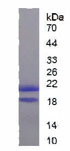
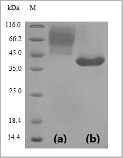
 SDS-PAGE
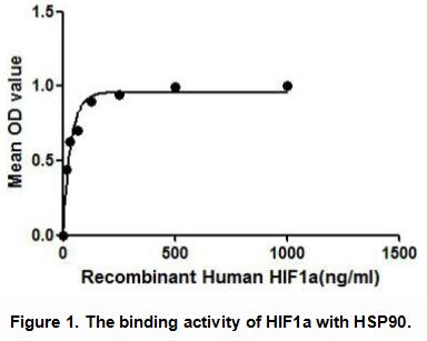
Hypoxia Inducible Factor 1 Alpha (HIF1a), Active Protein (Cat# AAA148241)
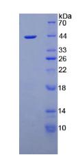
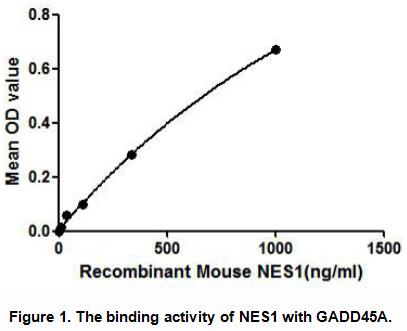
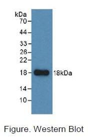
 Application Data
Nesfatin 1 (NES1), Active Protein (Cat# AAA148169)

 Application Data
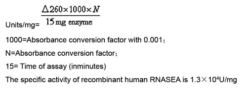
(Activity: Ribonuclease A (RNASEA) is a member of the pancreatic-type of secretory ribonucleases, a subset of the ribonuclease A superfamily. RNASEA cleaves RNA on the 3' side of pyrimidine nucleotides. The protein acts to degrade ds-RNA over ss-RNA. The activity of recombinant human RNASEA measured by cleaving yeast RNA. One unit of the enzyme causes an increase in absorbance of 0.001 at 260 nm in 15 min when yeast RNA is hydrolyzed at 50°C and pH 5.0. Pipette 50ulof respective recombinant human RNASEA dilution into 100ul 0.1M sodium acetate buffer, pH 5.0, then add 150ul of 0.15mg/ml yeast RNA. The blank tube use 50ul ultrapure water instead of enzyme dilution. All the all tubes Incubate at 50°C for 15 minutes. Read 260 verus blankCalculation)
Application Data
(Activity: Ribonuclease A (RNASEA) is a member of the pancreatic-type of secretory ribonucleases, a subset of the ribonuclease A superfamily. RNASEA cleaves RNA on the 3' side of pyrimidine nucleotides. The protein acts to degrade ds-RNA over ss-RNA. The activity of recombinant human RNASEA measured by cleaving yeast RNA. One unit of the enzyme causes an increase in absorbance of 0.001 at 260 nm in 15 min when yeast RNA is hydrolyzed at 50°C and pH 5.0. Pipette 50ulof respective recombinant human RNASEA dilution into 100ul 0.1M sodium acetate buffer, pH 5.0, then add 150ul of 0.15mg/ml yeast RNA. The blank tube use 50ul ultrapure water instead of enzyme dilution. All the all tubes Incubate at 50°C for 15 minutes. Read 260 verus blankCalculation)
Ribonuclease A (RNase A), Active Protein (Cat# AAA150337)
 SDS-PAGE
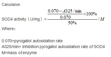
(Sample: Active recombinant SOD4, Mouse)
SDS-PAGE
(Sample: Active recombinant SOD4, Mouse)
Superoxide Dismutase Copper Chaperone, Active Protein (Cat# AAA161750)
 Application Data
(Measured by its binding ability in a functional ELISA. Immobilized mouse PD-L2-his at 10 ug/mL (100 ul/well) can bind mouse PD1-Fc. The EC50 of mouse PD1-Fc is 1.63 ug/mL.)
Application Data
(Measured by its binding ability in a functional ELISA. Immobilized mouse PD-L2-his at 10 ug/mL (100 ul/well) can bind mouse PD1-Fc. The EC50 of mouse PD1-Fc is 1.63 ug/mL.)
PD-L2, Active Protein (Cat# AAA258205)
 Application Data
(Measured by its binding ability in a functional ELISA. Immobilized human TDGF1 at 2 ug/ml (100 ul/well) can bind human ALK-4 with a linear range of 0.032-4 ug/ml.)
Application Data
(Measured by its binding ability in a functional ELISA. Immobilized human TDGF1 at 2 ug/ml (100 ul/well) can bind human ALK-4 with a linear range of 0.032-4 ug/ml.)
ALK4/ACVR1B, Active Protein (Cat# AAA257899)
 Application Data
(Measured by its ability to bind human LIF-Fc in a functional ELISA.)
Application Data
(Measured by its ability to bind human LIF-Fc in a functional ELISA.)
LIFR, Active Protein (Cat# AAA257904)
What Are Active Proteins?
Proteins are large molecules made up of long chains of amino acids. They will typically fold into a very particular 3-dimensional shape/conformation, that is sometimes referred to as their “native” form, which allows them to work properly in the body. For the purposes of product categorization, AAA Biotech will typically refer to proteins purified from their original animal host as being “native” proteins (this is to signify their difference compared to their recombinant proteins or “synthetic” protein counterparts).
If a protein successfully folds into the correct shape, it will typically display high fidelity characteristics to its original protein in its original animal host and be classified as an active protein, as it will be able to function “normally” in most enzymatic or binding capacities. If it loses this shape, due to factors such as heat or strong chemicals (such as detergents), it becomes inactive and is no longer able to perform its basic functions.
All of the proteins in this category are made under strict quality control, and they are active, pure, low in contaminants, and stable. Most are stored as freeze-dried powders and come without extra tags, so they’re very close to the actual natural/native form.
Learn more in our guide “How active proteins work”.
Key Applications of Active Proteins
1. Scientific Research
- Aid in the study of how proteins function in the body
- Aid in understanding various disease processes
2. Drug Development
- Powerful tools to investigate how potential drugs interact with specific proteins
- Ideal for identifying drug targets
3. Cell Culture
- Are routinely utilized to support cell growth and function (e.g., using exogenous growth factors)
- Can be used to promote cellular development into specific types (differentiation)
4. Diagnostics
- Regularly utilized in tests to detect diseases or infections (e.g., COVID-19, cancer)
- Note: All products are strictly for research-use only (RUO).
5. Therapeutics
- Some active proteins are used directly as treatments (e.g., insulin, enzymes)
- Note: All products are strictly for research-use only (RUO).
6. Vaccine Development
- Used to create or test vaccines by mimicking parts of viruses or bacteria
7. Biochemical Assays
- They can facilitate the characterization of enzyme activity, binding strength, or protein interactions in lab tests
Why Buy Active Proteins from AAA Biotech?
- High biological activity – Verified to perform as expected or indicated on datasheet
- Strict quality control – We are confident in our active proteins’ reliability and consistency
- High purity & low endotoxin – Ideal for applications involving sensitive or precious samples/components
- Freeze-dried for stability – Long shelf life and straightforward storage
- Mostly tag-free – Closer to natural/native protein form
FAQ
1. What are active proteins used for in research?
Active proteins are used primarily in the study of how proteins function, in characterizing/discovering drug interactions, supporting cell growth, running biochemical assays, and in development of diagnostics or therapeutics.
2. How are AAA Biotech's active proteins validated?
AAA Biotech’s active proteins are validated through strict quality control and functional assays to ensure they are properly folded and active. “Active”, though, can be an ambiguous term, so if a specific “activity” or “binding” capability of a protein is of crucial interest to you, please inquire with us prior to purchase, and we will provide further details on how the “Active” modifier was determined to be applicable.
3. Are these proteins tested for biological activity?
Yes, all active proteins from AAA Biotech are tested to confirm they have the expected biological activity before being offered for use. Though, said “biological activity” can be either “enzymatic”, “binding”, or both.