Filters
▼Clonality
▼Type
▼Reactivity
▼Gene Name
▼Isotype
▼Host
▼Application
▼Clone
▼Active Proteins
AAA Biotech also known as AAA Bio or AAABio provides a variety of high-quality recombinant and natural/native proteins that are proven to work in a wide range of experiments. Explore our products to find the active protein that best fits your needs or experimental model.
Viewing 2400-2450 of 2875 product results
 Application Data
(Measured by its binding ability in a functional ELISA. Immobilized mouse VEGFR3-His at 10 ug/mL (100 ul/well) can bind mouse Fc-VEGFD, The EC50 of mouse Fc-VEGFD is 44 ng/mL.)
Application Data
(Measured by its binding ability in a functional ELISA. Immobilized mouse VEGFR3-His at 10 ug/mL (100 ul/well) can bind mouse Fc-VEGFD, The EC50 of mouse Fc-VEGFD is 44 ng/mL.)
VEGFR3/FLT4, Active Protein (Cat# AAA258176)
 Application Data
(Immobilized human ACE2 protein (Fc tag)(Cat: 10108-H05H) at 2 ug/mL (100 uL/well) can bind Recombinant SARS-CoV-2 (BA.3) Spike RBD Protein (R319-K537, His Tag)(Cat: AAA258588), the EC50 is 7-21 ng/mL.)
Application Data
(Immobilized human ACE2 protein (Fc tag)(Cat: 10108-H05H) at 2 ug/mL (100 uL/well) can bind Recombinant SARS-CoV-2 (BA.3) Spike RBD Protein (R319-K537, His Tag)(Cat: AAA258588), the EC50 is 7-21 ng/mL.)
COVID 19 (BA.3) Spike RBD Coronavirus, Active Protein (Cat# AAA258588)

 Application Data
(The activity was determined by binding with immobilized Human ACE2 at 2ug/ml, 100ul/well in a functional ELISA assay with a linear range of 0.5-1ug/ml)
Application Data
(The activity was determined by binding with immobilized Human ACE2 at 2ug/ml, 100ul/well in a functional ELISA assay with a linear range of 0.5-1ug/ml)
COVID 19 Spike Protein S Coronavirus, Active Protein (Cat# AAA14435)
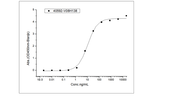
 Application Data
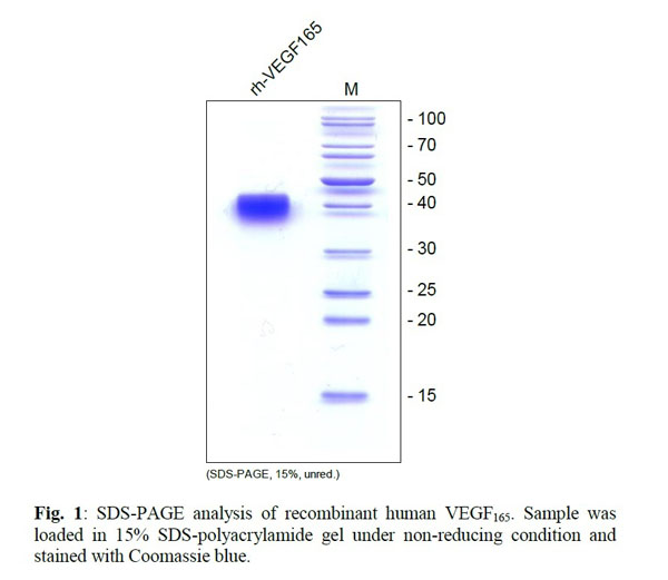
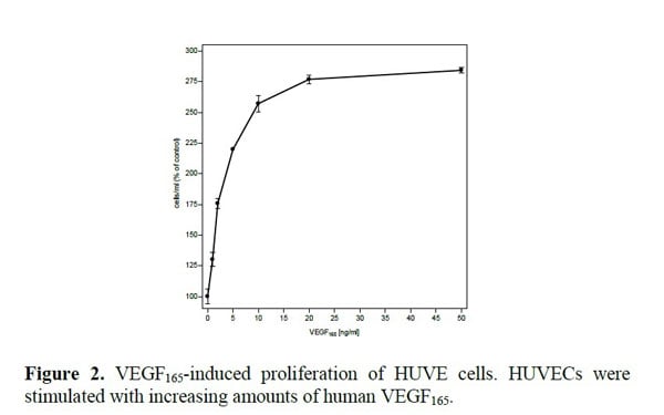
VEGF165, Active Protein (Cat# AAA79220)
 Application Data
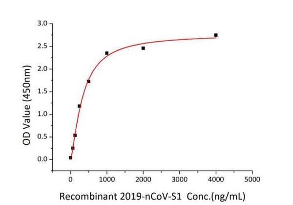
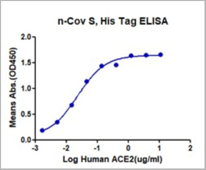
(Fig 2 Immobilized Recombinant Human ACE2 at 2 ugmL (100uLwell) can bind Recombinant nCoV-S1, The EC50 of nCoV-S1 is 0.25-0.45 ugmL.)
Application Data
(Fig 2 Immobilized Recombinant Human ACE2 at 2 ugmL (100uLwell) can bind Recombinant nCoV-S1, The EC50 of nCoV-S1 is 0.25-0.45 ugmL.)
COVID 19 Spike S1 Coronavirus, Active Protein (Cat# AAA78534)

 Application Data
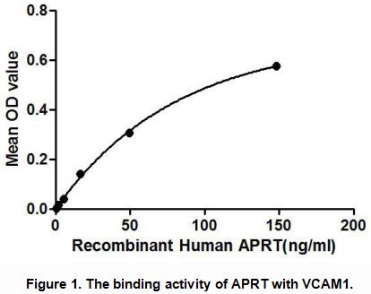
Adenine Phosphoribosyltransferase (APRT), Active Protein (Cat# AAA148201)
 Application Data
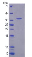
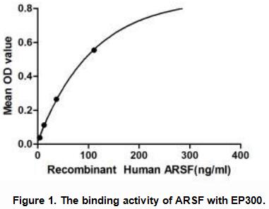
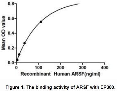
Arylsulfatase F (ARSF), Active Protein (Cat# AAA148215)

 Application Data
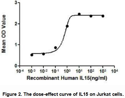
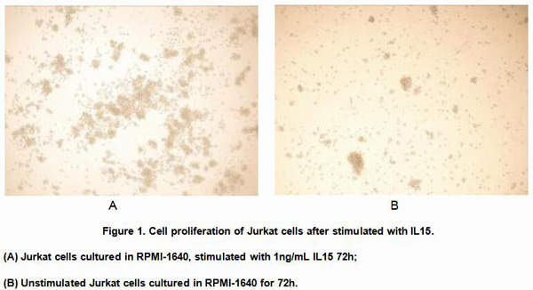
Interleukin 15 (IL15), Active Protein (Cat# AAA148129)
 Bioactivity
(Figure 2. The hemagglutination assay of GAL1 in V- bottom shaped 96-well microtiter plate.)
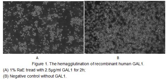
Bioactivity
(Figure 2. The hemagglutination assay of GAL1 in V- bottom shaped 96-well microtiter plate.)
Galectin 1 (GAL1), Active Protein (Cat# AAA153032)
 Bioactivity
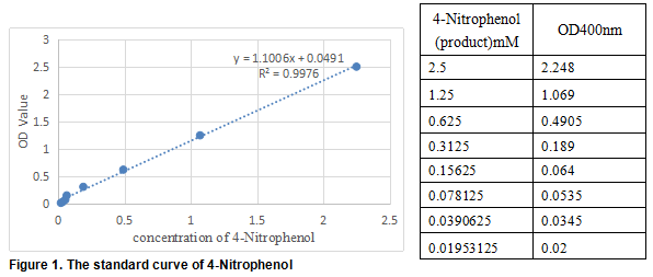
(carboxylesterase 1 (CES1) also known as Liver carboxylesterase 1 is a serine esterase and member of a large multigene carboxylesterase family. The protein Involved in the detoxification of xenobiotics and in the activation of ester and amide prodrugs. Hydrolyzes aromatic and aliphatic esters, but has no catalytic activity toward amides or a fatty acyl-CoA ester. Hydrolyzes the methyl ester group of cocaine to form benzoylecgonine. Thus, the recombinant human CES1 activity was measured by its ability to hydrolyze 4-Nitrophenyl acetate (4-NPA) to 4-Nitrophenol. The reaction was performed in 50 mM Tris, pH 7.5(Assay Buffer), initiated by addition 50 uL of various concentrations of CES1 (dilute by Assay Buffer) to 50 uL of 2 mM Substrate 4-NPA(100 mM stock in Acetone, dilute by deionized water). Incubated at 37 degree C for 10min, then read at a wavelength of 400 nm.)
Bioactivity
(carboxylesterase 1 (CES1) also known as Liver carboxylesterase 1 is a serine esterase and member of a large multigene carboxylesterase family. The protein Involved in the detoxification of xenobiotics and in the activation of ester and amide prodrugs. Hydrolyzes aromatic and aliphatic esters, but has no catalytic activity toward amides or a fatty acyl-CoA ester. Hydrolyzes the methyl ester group of cocaine to form benzoylecgonine. Thus, the recombinant human CES1 activity was measured by its ability to hydrolyze 4-Nitrophenyl acetate (4-NPA) to 4-Nitrophenol. The reaction was performed in 50 mM Tris, pH 7.5(Assay Buffer), initiated by addition 50 uL of various concentrations of CES1 (dilute by Assay Buffer) to 50 uL of 2 mM Substrate 4-NPA(100 mM stock in Acetone, dilute by deionized water). Incubated at 37 degree C for 10min, then read at a wavelength of 400 nm.)
Carboxylesterase 1 (CES1), Active Protein (Cat# AAA152179)
 ELISA
(The activity was determined binding with immobilized Human ACE2 at 2ug/mL, 100ul/well in a functional ELISA assay with a linear range of 0.5-1ug/ml.)
ELISA
(The activity was determined binding with immobilized Human ACE2 at 2ug/mL, 100ul/well in a functional ELISA assay with a linear range of 0.5-1ug/ml.)
COVID 19 B.1.1.529 (Omicron) Spike S Coronavirus, Active Protein (Cat# AAA14438)
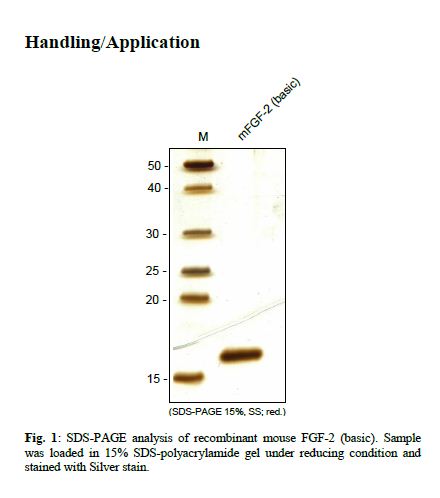
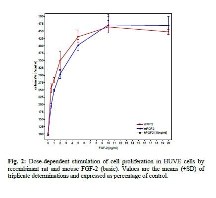 Application Data
FGF-2 (basic), Active Protein (Cat# AAA79158)
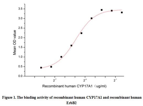 Bioactivity
(Microsomal human cytochrome P450 17A1 (CYP17A1, 17alpha-hydroxylase, 17,20-lyase) belongs to the cytochrome P450 super family, is expressed in the adrenals and gonads, with minor amounts in the brain, placenta, and heart. CYP17A1 enzyme operates at a key juncture of human steroidogenesis, controlling the levels of mineralocorticoids influencing blood pressure, glucocorticoids involved in immune and stress responses, and androgens and estrogens involved in development and homeostasis of reproductive tissues. Receptor Tyrosine Protein Kinase erbB-2 (ErbB2) has been identified as an interactor of CYP17A1, thus a functional binding ELISA assay was conducted to detect the interaction of recombinant human CYP17A1 and recombinant human ErbB2. Briefly, CYP17A1 was diluted serially in PBS with 0.01% BSA (pH 7.4). Duplicate samples of 100 ul were then transferred to ErbB2-coated microtiter wells and incubated for 1h at 37 degree C. Wells were washed with PBST and incubated for 1h with anti-CYP17A1 pAb, then aspirated and washed 3 times. After incubation with HRP labelled secondary antibody for 1h at 37 degree C, wells were aspirated and washed 5 times. With the addition of substrate solution, wells were incubated 15-25 minutes at 37 degree C. Finally, add 50 uL stop solution to the wells and read at 450/630 nm immediately. The binding activity of recombinant human CYP17A1 and recombinant human ErbB2 was shown in Figure 1, the EC50 for this effect is 0.03 ug/mL.)
Bioactivity
(Microsomal human cytochrome P450 17A1 (CYP17A1, 17alpha-hydroxylase, 17,20-lyase) belongs to the cytochrome P450 super family, is expressed in the adrenals and gonads, with minor amounts in the brain, placenta, and heart. CYP17A1 enzyme operates at a key juncture of human steroidogenesis, controlling the levels of mineralocorticoids influencing blood pressure, glucocorticoids involved in immune and stress responses, and androgens and estrogens involved in development and homeostasis of reproductive tissues. Receptor Tyrosine Protein Kinase erbB-2 (ErbB2) has been identified as an interactor of CYP17A1, thus a functional binding ELISA assay was conducted to detect the interaction of recombinant human CYP17A1 and recombinant human ErbB2. Briefly, CYP17A1 was diluted serially in PBS with 0.01% BSA (pH 7.4). Duplicate samples of 100 ul were then transferred to ErbB2-coated microtiter wells and incubated for 1h at 37 degree C. Wells were washed with PBST and incubated for 1h with anti-CYP17A1 pAb, then aspirated and washed 3 times. After incubation with HRP labelled secondary antibody for 1h at 37 degree C, wells were aspirated and washed 5 times. With the addition of substrate solution, wells were incubated 15-25 minutes at 37 degree C. Finally, add 50 uL stop solution to the wells and read at 450/630 nm immediately. The binding activity of recombinant human CYP17A1 and recombinant human ErbB2 was shown in Figure 1, the EC50 for this effect is 0.03 ug/mL.)
Cytochrome P450 17A1 (CYP17A1), Active Protein (Cat# AAA161874)
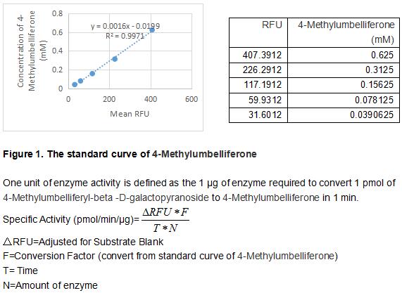 Bioactivity
(GLB1 is a lysosomal beta -galactosidase that hydrolyzes the terminal beta -galactose from ganglioside and keratan sulfate. Defects in this gene are the causes of lysosomal storage diseases for GM1-gangliosidosis and Morquio B syndrome (also known as mucopolysaccharidosis IVB). In GM1 gangliosidosis, GM1 ganglioside accumulates in the neurons of the central nervous system, because of the deficiency of lysosomal beta -galactosidase activity. GM1 gangliosidosis demonstrates varying degrees of clinical severity but is invariably fatal, and children with the most common and severe form of GM1 gangliosidosis usually die within 3 years of birth. Morquio B syndrome patients are neurologically normal, but display severe skeletal dysostosis multiplex because of an accumulation of keratan sulfate. The activity assay of GLB1 was measured by its ability to cleave a peptide substrate, 4-Methylumbelliferyl-beta -D-galactopyranoside. The reaction was performed in 50 mM Sodium Citrate, pH 3.5 (Assay Buffer), ainitiated by addition 50 uL of 1.5 ug/ml uPA (diluted by Assay Buffer) to 50 uL of 1.2 mM Substrate. Read at excitation and emission wavelengths of 365 nm and 445 nm (top read), respectively, in kinetic mode for 5 minutes. The specific activity of recombinant mouse GLB1 is >17000 pmol/min/ug.)
Bioactivity
(GLB1 is a lysosomal beta -galactosidase that hydrolyzes the terminal beta -galactose from ganglioside and keratan sulfate. Defects in this gene are the causes of lysosomal storage diseases for GM1-gangliosidosis and Morquio B syndrome (also known as mucopolysaccharidosis IVB). In GM1 gangliosidosis, GM1 ganglioside accumulates in the neurons of the central nervous system, because of the deficiency of lysosomal beta -galactosidase activity. GM1 gangliosidosis demonstrates varying degrees of clinical severity but is invariably fatal, and children with the most common and severe form of GM1 gangliosidosis usually die within 3 years of birth. Morquio B syndrome patients are neurologically normal, but display severe skeletal dysostosis multiplex because of an accumulation of keratan sulfate. The activity assay of GLB1 was measured by its ability to cleave a peptide substrate, 4-Methylumbelliferyl-beta -D-galactopyranoside. The reaction was performed in 50 mM Sodium Citrate, pH 3.5 (Assay Buffer), ainitiated by addition 50 uL of 1.5 ug/ml uPA (diluted by Assay Buffer) to 50 uL of 1.2 mM Substrate. Read at excitation and emission wavelengths of 365 nm and 445 nm (top read), respectively, in kinetic mode for 5 minutes. The specific activity of recombinant mouse GLB1 is >17000 pmol/min/ug.)
Galactosidase Beta (GLb), Active Protein (Cat# AAA161714)
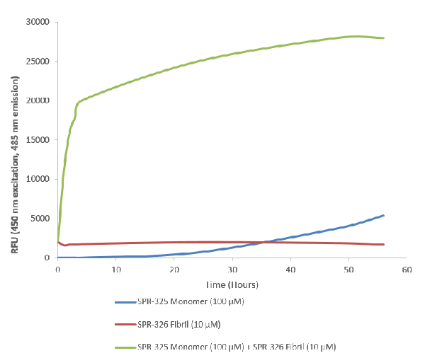 Application Data
(Thioflavin T is a fluorescent dye that binds to beta sheet-rich structures such as those in alpha synuclein fibrils. Upon binding, the emission spectrum of the dye experiences a red-shift and increased fluorescence intensity. Thioflavin T emission curves show a limited increase in fluorescence (correlated to alpha synuclein aggregation) over time in A53T alpha synuclein monomers (SPR-325). A much greater increase in fluorescence is seen when 100 uM monomer (SPR-325) is combined with 10 nM of fibrils (SPR-326) as the fibrils seed the formation of new fibrils from the pool of active monomers. Thioflavin T ex = 450 nm, em = 485 nm.)
Application Data
(Thioflavin T is a fluorescent dye that binds to beta sheet-rich structures such as those in alpha synuclein fibrils. Upon binding, the emission spectrum of the dye experiences a red-shift and increased fluorescence intensity. Thioflavin T emission curves show a limited increase in fluorescence (correlated to alpha synuclein aggregation) over time in A53T alpha synuclein monomers (SPR-325). A much greater increase in fluorescence is seen when 100 uM monomer (SPR-325) is combined with 10 nM of fibrils (SPR-326) as the fibrils seed the formation of new fibrils from the pool of active monomers. Thioflavin T ex = 450 nm, em = 485 nm.)
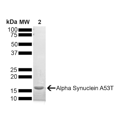
A53T Mutant Alpha Synuclein, Active Protein (Cat# AAA253966)
Ion-Exchange Purified
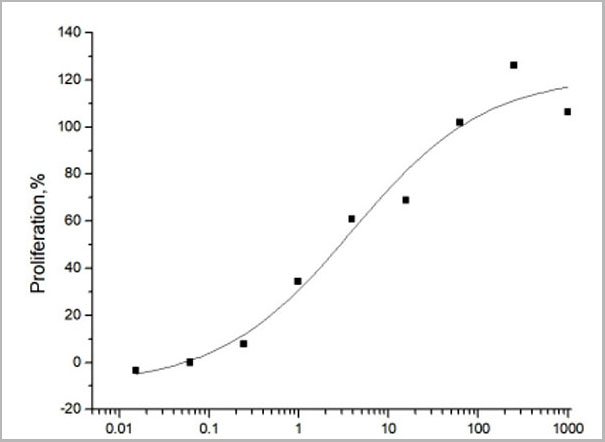 Activity
(The ED(50) was determined by the dose-dependent proliferation of human MCF-7 cells was found to be )
Activity
(The ED(50) was determined by the dose-dependent proliferation of human MCF-7 cells was found to be )
Glial growth factor (GGF2), Active Protein (Cat# AAA14429)

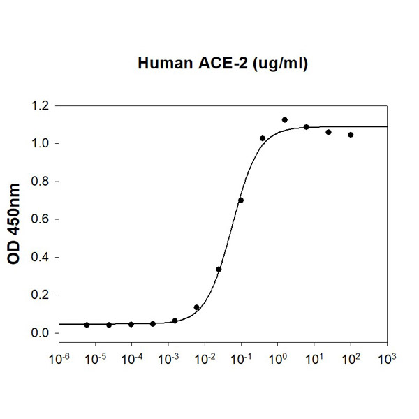 Bioactivity
(Human SARS-CoV-2 Spike RBD is coated at 5ug/ml (100 ul/well) can bind ACE-2 (CAT# ATGP3963) in a Functional ELISA assay)
Bioactivity
(Human SARS-CoV-2 Spike RBD is coated at 5ug/ml (100 ul/well) can bind ACE-2 (CAT# ATGP3963) in a Functional ELISA assay)
COVID 19 Spike RBD Coronavirus, Active Protein (Cat# AAA48384)
 Application Data
(Effect of stem cell factor (SCF) on cardiac stem cell (CSC) migration in vitro.Representative images of migrated CSCs treated with varying concentrations of SCF by Transwell-based migration assays.)
Application Data
(Effect of stem cell factor (SCF) on cardiac stem cell (CSC) migration in vitro.Representative images of migrated CSCs treated with varying concentrations of SCF by Transwell-based migration assays.)
Stem Cell Factor, Active Protein (Cat# AAA76393)
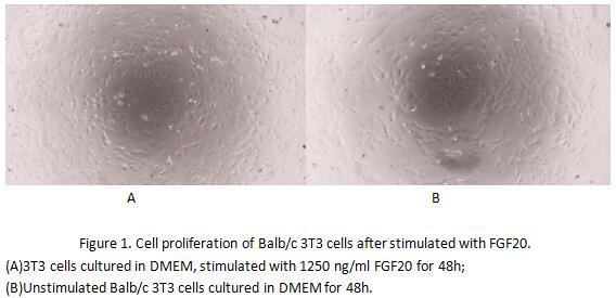 Bioactivity
(Fibroblast growth factor 20 (FGF20) is a member of the fibroblast growth factor family. The fibroblast growth factors possess broad mitogenic and cell survival activities, and are involved in a variety of biological processes including embryonic developme)
Bioactivity
(Fibroblast growth factor 20 (FGF20) is a member of the fibroblast growth factor family. The fibroblast growth factors possess broad mitogenic and cell survival activities, and are involved in a variety of biological processes including embryonic developme)
Fibroblast Growth Factor 20 (FGF20), Active Protein (Cat# AAA153120)
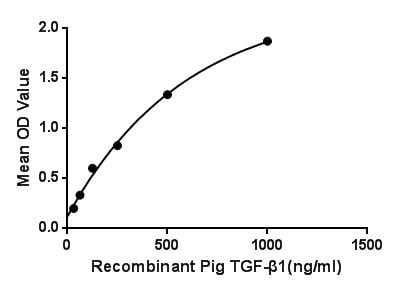 Bioactivity
(Figure. The binding activity of TGF-1 with LTBP1.Transforming growth factor beta 1 or TGF-1 is a polypeptide member of the transforming growth factor beta superfamily of cytokines. It is a secreted protein that performs many cellular functions, including the control of cell growth, cell proliferation, cell differentiation and apoptosis. TGF-1 plays an important role in controlling the immune system, and shows different activities on different types of cell, or cells at different developmental stages. Besides, Latent Transforming Growth Factor Beta Binding Protein 1 (LTBP1) has been identified as an interactor of TGF-1, thus a binding ELISA assay was conducted to detect the interaction of recombinant pig TGF-1 and recombinant pig LTBP1. Briefly, TGF-1 were diluted serially in PBS, with 0.01% BSA (pH 7.4). Duplicate samples of 100L were then transferred to LTBP1-coated microtiter wells and incubated for 2h at 37. Wells were washed with PBST and incubated for 1h with anti-TGF-1 pAb, then aspirated and washed 3 times. After incubation with HRP labelled secondary antibody, wells were aspirated and washed 3 times. With the addition of substrate solution, wells were incubated 15-25 minutes at 37. Finally, add 50uL stop solution to the wells and read at 450nm immediately. The binding activity of TGF-1 and LTBP1 was shown in Figure 1, and this effect was in a dose dependent manner.)
Bioactivity
(Figure. The binding activity of TGF-1 with LTBP1.Transforming growth factor beta 1 or TGF-1 is a polypeptide member of the transforming growth factor beta superfamily of cytokines. It is a secreted protein that performs many cellular functions, including the control of cell growth, cell proliferation, cell differentiation and apoptosis. TGF-1 plays an important role in controlling the immune system, and shows different activities on different types of cell, or cells at different developmental stages. Besides, Latent Transforming Growth Factor Beta Binding Protein 1 (LTBP1) has been identified as an interactor of TGF-1, thus a binding ELISA assay was conducted to detect the interaction of recombinant pig TGF-1 and recombinant pig LTBP1. Briefly, TGF-1 were diluted serially in PBS, with 0.01% BSA (pH 7.4). Duplicate samples of 100L were then transferred to LTBP1-coated microtiter wells and incubated for 2h at 37. Wells were washed with PBST and incubated for 1h with anti-TGF-1 pAb, then aspirated and washed 3 times. After incubation with HRP labelled secondary antibody, wells were aspirated and washed 3 times. With the addition of substrate solution, wells were incubated 15-25 minutes at 37. Finally, add 50uL stop solution to the wells and read at 450nm immediately. The binding activity of TGF-1 and LTBP1 was shown in Figure 1, and this effect was in a dose dependent manner.)
Transforming Growth Factor Beta 1, Active Protein (Cat# AAA150075)
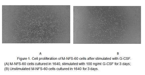
 Bioactivity
(Figure 2. Cell proliferation of M-NFS-60 cells after stimulated with G-CSF.)
Bioactivity
(Figure 2. Cell proliferation of M-NFS-60 cells after stimulated with G-CSF.)
Colony Stimulating Factor 3, Granulocyte (GCSF), Active Protein (Cat# AAA161663)
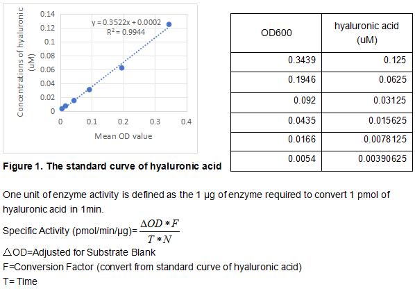 Bioactivity
(Hyaluronidase (HAase) is a general term for enzymes that can hydrolyze hyaluronic acid. It is an enzyme that can reduce the activity of hyaluronic acid in the body, thereby improving the fluid permeability in tissues. When used in the human body, it can temporarily reduce the viscosity of the intercellular matrix, promote subcutaneous infusion, locally accumulated exudate or blood to accelerate diffusion and facilitate absorption, and is an important drug dispersant. Clinically used as a drug penetrating agent to promote drug absorption, promote local edema or hematoma dissipation after surgery and trauma. Human HAase consists of 435 amino acids which contains a signal peptide of 1-21 amino acids and it shares 74% and 75% amino acid sequence homology with mouse and rat respectively. The activity assay of recombinant human HAase was measured by its ability to hydrolyze the substrate hyaluronic acid. The rhHAase was diluted to 0.5 ug/ml in 8.46 mM NaH2PO4, 11.54 mM Na2HPO4, 77 mM NaCl, 0.1 mg/ml BSA, pH 7. 50 ul 0.5 ug/ml rhHAase was added into the microplate and start the reaction by adding 50 uL of 0.3 mg/ml substrate which was diluted in 300mM NaH2PO4, pH 5.35. Incubated at 37 degree C for 5min and add 50 ul reaction mixture to 250 ul 1 mg/ml BSA in 24 mM sodium acetic, 79 mM acetic acid, pH 3.75. Incubated at room temperature for 10min and read at a wavelength of 600 nm. The specific activity of recombinant human HAase is >30 pmol/min/ug.)
Bioactivity
(Hyaluronidase (HAase) is a general term for enzymes that can hydrolyze hyaluronic acid. It is an enzyme that can reduce the activity of hyaluronic acid in the body, thereby improving the fluid permeability in tissues. When used in the human body, it can temporarily reduce the viscosity of the intercellular matrix, promote subcutaneous infusion, locally accumulated exudate or blood to accelerate diffusion and facilitate absorption, and is an important drug dispersant. Clinically used as a drug penetrating agent to promote drug absorption, promote local edema or hematoma dissipation after surgery and trauma. Human HAase consists of 435 amino acids which contains a signal peptide of 1-21 amino acids and it shares 74% and 75% amino acid sequence homology with mouse and rat respectively. The activity assay of recombinant human HAase was measured by its ability to hydrolyze the substrate hyaluronic acid. The rhHAase was diluted to 0.5 ug/ml in 8.46 mM NaH2PO4, 11.54 mM Na2HPO4, 77 mM NaCl, 0.1 mg/ml BSA, pH 7. 50 ul 0.5 ug/ml rhHAase was added into the microplate and start the reaction by adding 50 uL of 0.3 mg/ml substrate which was diluted in 300mM NaH2PO4, pH 5.35. Incubated at 37 degree C for 5min and add 50 ul reaction mixture to 250 ul 1 mg/ml BSA in 24 mM sodium acetic, 79 mM acetic acid, pH 3.75. Incubated at room temperature for 10min and read at a wavelength of 600 nm. The specific activity of recombinant human HAase is >30 pmol/min/ug.)
Hyaluronidase (HAase), Active Protein (Cat# AAA161835)
 Bioactivity
Nectin-4, Active Protein (Cat# AAA177966)
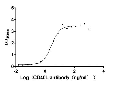 Bioactivity
CD40 ligand (CD40LG), Active Protein (Cat# AAA244028)
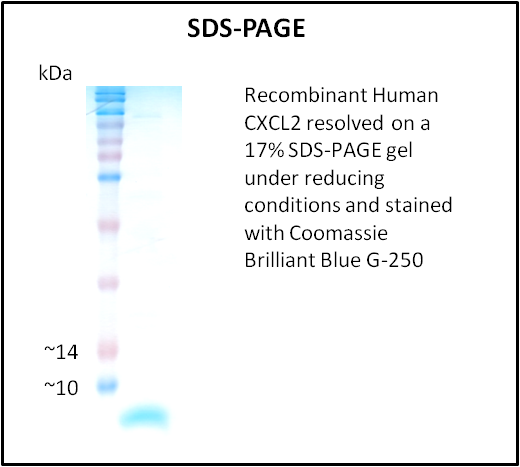
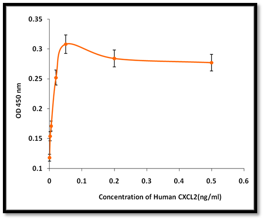 Application Data
CXCL2, Active Protein (Cat# AAA214224)
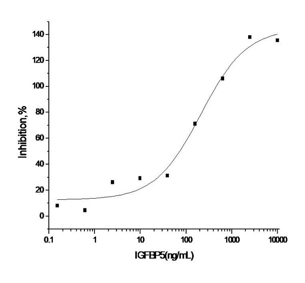 Application Data
(Measured by its ability to inhibit the biological activity of IGF-I or IGF-II on MCF-7 human breast cancer cells. The ED50 for this effect is 0.1-0.5 ug/mL in the presence of 14 ng/mL rhIGF-II.)
Application Data
(Measured by its ability to inhibit the biological activity of IGF-I or IGF-II on MCF-7 human breast cancer cells. The ED50 for this effect is 0.1-0.5 ug/mL in the presence of 14 ng/mL rhIGF-II.)
IGFBP5, Active Protein (Cat# AAA257845)
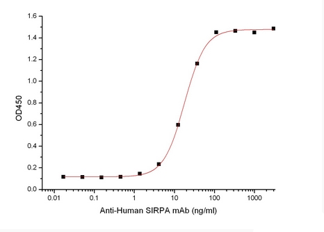 Bioactivity
Signal-Regulatory Protein alpha-1/SIRPA/CD172a, Active Protein (Cat# AAA177975)
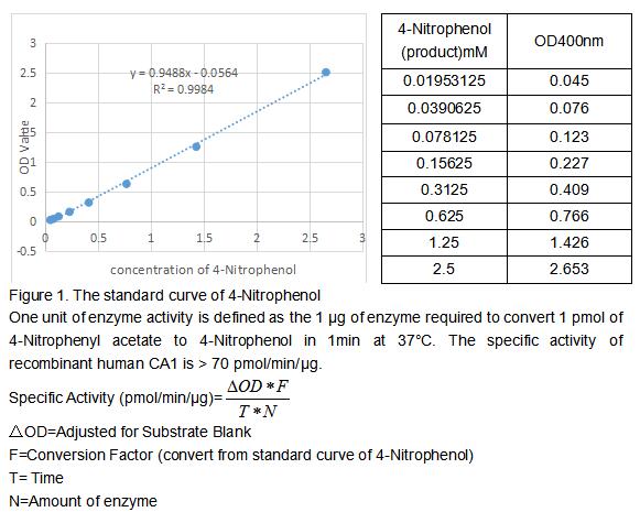 Bioactivity
(Carbonic Anhydrase (CA) catalyzes the reversible reaction of CO2 H2O = HCO3- H, which is fundamental to many processes such as respiration, renal tubular acidification and bone resorption. CA1 is a cytosolic enzyme with the highest levels in erythrocytes and is a very early marker for erythroid differentiation. The activity of recombinant human CA1 was measured by its ability to hydrolyze 4-Nitrophenyl acetate (4-NPA) to 4-Nitrophenol. The reaction was performed in 12.5 mM Tris, 75 mM NaCl, pH 7.5 (assay buffer), initiated by addition 50 uL of various concentrations of CA1 (diluted by assay buffer) to 50 uL of 2 mM substrate 4-NPA (100 mM stock in Acetone, diluted by assay buffer). Incubated at 37 degree C for 5min, then read at a wavelength of 400 nm.)
Bioactivity
(Carbonic Anhydrase (CA) catalyzes the reversible reaction of CO2 H2O = HCO3- H, which is fundamental to many processes such as respiration, renal tubular acidification and bone resorption. CA1 is a cytosolic enzyme with the highest levels in erythrocytes and is a very early marker for erythroid differentiation. The activity of recombinant human CA1 was measured by its ability to hydrolyze 4-Nitrophenyl acetate (4-NPA) to 4-Nitrophenol. The reaction was performed in 12.5 mM Tris, 75 mM NaCl, pH 7.5 (assay buffer), initiated by addition 50 uL of various concentrations of CA1 (diluted by assay buffer) to 50 uL of 2 mM substrate 4-NPA (100 mM stock in Acetone, diluted by assay buffer). Incubated at 37 degree C for 5min, then read at a wavelength of 400 nm.)
Carbonic Anhydrase I (CA1), Active Protein (Cat# AAA161796)
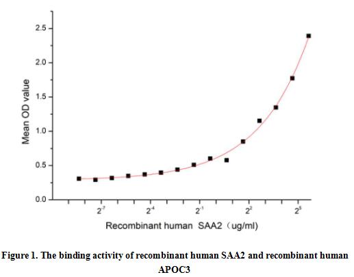 Bioactivity
(Serum Amyloid A2 (SAA2) is a multifunctional apolipoprotein that belongs to SAA family. SAA2 is produced by hepatocytes in response to pro-inflammatory cytokines and is the most prominent members of the acute phase response (APR) during which their serum levels rise dramatically after trauma, infection and other stimuli. Besides, Apolipoprotein C3 (APOC3) has been identified as an interactor of SAA2, thus a functional binding ELISA assay was conducted to detect the interaction of recombinant human SAA2 and recombinant human APOC3. Briefly, SAA2 were diluted serially in PBS, with 0.01% BSA (pH 7.4). Duplicate samples of 100 ul were then transferred to APOC3-coated microtiter wells and incubated for 2h at 37 degree C. Wells were washed with PBST and incubated for 1h with anti-SAA2 pAb, then aspirated and washed 3 times. After incubation with HRP labelled secondary antibody, wells were aspirated and washed 5 times. With the addition of substrate solution, wells were incubated 15-25 minutes at 37 degree C. Finally, add 50 uL stop solution to the wells and read at 450 nm immediately. The binding activity of recombinant human SAA2 and recombinant human APOC3 was shown in Figure 1, and this effect was in a dose dependent manner.)
Bioactivity
(Serum Amyloid A2 (SAA2) is a multifunctional apolipoprotein that belongs to SAA family. SAA2 is produced by hepatocytes in response to pro-inflammatory cytokines and is the most prominent members of the acute phase response (APR) during which their serum levels rise dramatically after trauma, infection and other stimuli. Besides, Apolipoprotein C3 (APOC3) has been identified as an interactor of SAA2, thus a functional binding ELISA assay was conducted to detect the interaction of recombinant human SAA2 and recombinant human APOC3. Briefly, SAA2 were diluted serially in PBS, with 0.01% BSA (pH 7.4). Duplicate samples of 100 ul were then transferred to APOC3-coated microtiter wells and incubated for 2h at 37 degree C. Wells were washed with PBST and incubated for 1h with anti-SAA2 pAb, then aspirated and washed 3 times. After incubation with HRP labelled secondary antibody, wells were aspirated and washed 5 times. With the addition of substrate solution, wells were incubated 15-25 minutes at 37 degree C. Finally, add 50 uL stop solution to the wells and read at 450 nm immediately. The binding activity of recombinant human SAA2 and recombinant human APOC3 was shown in Figure 1, and this effect was in a dose dependent manner.)
Serum Amyloid A2 (SAA2), Active Protein (Cat# AAA161860)


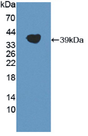
 WB (Western Blot)
(Figure 4. Western BlotSample: Recombinant TUBb, Human;Antibody: Rabbit Anti-Human TUBb Ab)
WB (Western Blot)
(Figure 4. Western BlotSample: Recombinant TUBb, Human;Antibody: Rabbit Anti-Human TUBb Ab)
Tubulin Beta (TUBb), Active Protein (Cat# AAA148197)
 Application Data
(Measured by its ability to neutralize Activin-mediated inhibition on MPC11 cell proliferation. The ED50 for this effect is typically 10-50 ng/mL in the presence of 10 ng/mL recombinant Activin A.)
Application Data
(Measured by its ability to neutralize Activin-mediated inhibition on MPC11 cell proliferation. The ED50 for this effect is typically 10-50 ng/mL in the presence of 10 ng/mL recombinant Activin A.)
ACVR2B, Active Protein (Cat# AAA258136)
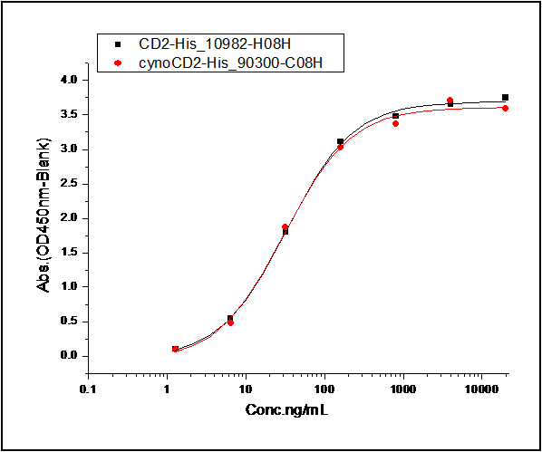 Application Data
(1. Measured by its binding ability in a functional ELISA. Immobilized human CD2-His at 10 ug/ml (100 ul/well) can bind human CD58-Fc, The EC50 of human CD58-Fc is 0.04-0.1 ug/ml. 2. Measured by its binding ability in a functional ELISA. Immobilized Cynomolgus CD2-His at 10 ug/ml (100 ul/well) can bind human CD58-Fc, The EC50 of human CD58-Fc is 0.04-0.10 ug/ml.)
Application Data
(1. Measured by its binding ability in a functional ELISA. Immobilized human CD2-His at 10 ug/ml (100 ul/well) can bind human CD58-Fc, The EC50 of human CD58-Fc is 0.04-0.1 ug/ml. 2. Measured by its binding ability in a functional ELISA. Immobilized Cynomolgus CD2-His at 10 ug/ml (100 ul/well) can bind human CD58-Fc, The EC50 of human CD58-Fc is 0.04-0.10 ug/ml.)
CD58, Active Protein (Cat# AAA258011)
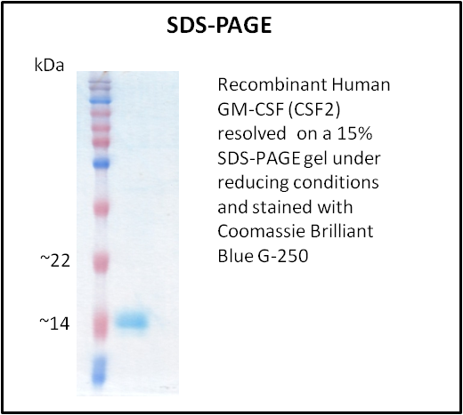
 Application Data
GM-CSF (CSF2), Active Protein (Cat# AAA214222)
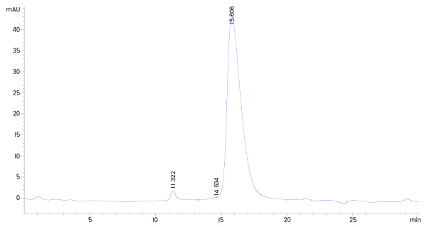
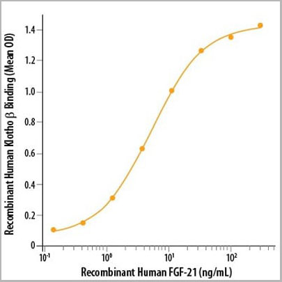 ELISA
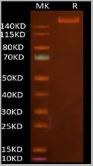
(Western Blot (reducing (R) conditions) analysis using AAA14751 (1ug/lane) and visualized by silver staining, showing a single band at 126kD.)
ELISA
(Western Blot (reducing (R) conditions) analysis using AAA14751 (1ug/lane) and visualized by silver staining, showing a single band at 126kD.)
Klotho beta, Active Protein (Cat# AAA14751)
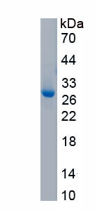
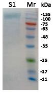
>95% by SDS-PAGE, reducing conditions and visualized with silver stain.
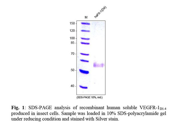
 Application Data
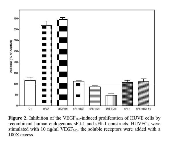
VEGFR-1/Flt-1 (D4), soluble, Active Protein (Cat# AAA79219)
 Bioactivity
(Glutathione S-Transferases (GSTs) are members of the phase II detoxification enzyme family that conjugate glutathione to various electrophilic compounds, including metabolites generated by oxidative processes in the body, environmental toxins or carcinogens, and anti-cancer drugs. GSTM1 is a cytosolic protein that belongs to the mu class of the GST superfamily. GSTM1 catalyze the endogenous glutathione conjugation 1-Chloro-2,4-dinitrobenzene (CDNB), which can increase in the absorbance at 340 nm. The reaction was performed in adding 10 ul 200 mM glutathione (reduced) and 10 ul 100 mM CDNB in 980 ul 100 mM NaH2PO4 (pH7.0), rapidly mixed. Then add 50 ul mixed substrates to 50 ul different concentrations of recombinant chicken GSTM1, mix gently. Incubated at 37 degree C for 5min, then read at a wavelength of 340 nm. The specific activity of recombinant chicken GSTM1 is >2500 pmol/min/ug.)
Bioactivity
(Glutathione S-Transferases (GSTs) are members of the phase II detoxification enzyme family that conjugate glutathione to various electrophilic compounds, including metabolites generated by oxidative processes in the body, environmental toxins or carcinogens, and anti-cancer drugs. GSTM1 is a cytosolic protein that belongs to the mu class of the GST superfamily. GSTM1 catalyze the endogenous glutathione conjugation 1-Chloro-2,4-dinitrobenzene (CDNB), which can increase in the absorbance at 340 nm. The reaction was performed in adding 10 ul 200 mM glutathione (reduced) and 10 ul 100 mM CDNB in 980 ul 100 mM NaH2PO4 (pH7.0), rapidly mixed. Then add 50 ul mixed substrates to 50 ul different concentrations of recombinant chicken GSTM1, mix gently. Incubated at 37 degree C for 5min, then read at a wavelength of 340 nm. The specific activity of recombinant chicken GSTM1 is >2500 pmol/min/ug.)
Glutathione S Transferase Mu 1 (GSTM1), Active Protein (Cat# AAA161759)

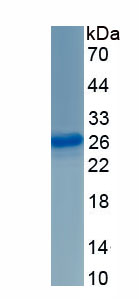
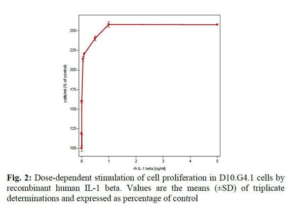 Application Data
IL-1 beta, Active Protein (Cat# AAA79259)
 Bioactivity
(Mouse OX40 Ligand/TNFSF4 (CAT# ATGP4022) is coated at 1 ug/ml (100 ul/well) can bind Human TNFRSF4. The ED50 range )
Bioactivity
(Mouse OX40 Ligand/TNFSF4 (CAT# ATGP4022) is coated at 1 ug/ml (100 ul/well) can bind Human TNFRSF4. The ED50 range )
OX40/TNFRSF4, Active Protein (Cat# AAA48407)
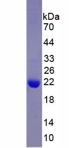
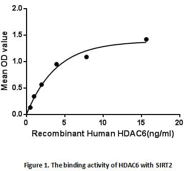 Bioactivity
(Histone Deacetylase 6 (HDAC6) belongs to class II of the histone deacetylase/acuc/apha family. It contains an internal duplication of two catalytic domains that appear to function independently of each other. This protein possesses histone deacetylase act)
Bioactivity
(Histone Deacetylase 6 (HDAC6) belongs to class II of the histone deacetylase/acuc/apha family. It contains an internal duplication of two catalytic domains that appear to function independently of each other. This protein possesses histone deacetylase act)
Histone Deacetylase 6 (HDAC6), Active Protein (Cat# AAA153129)
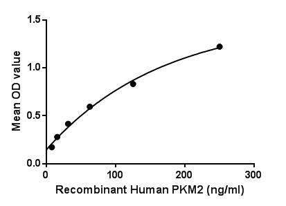 Bioactivity
(Figure. The binding activity of PKM2 with PIN1.Pyruvate Kinase, Muscle (PKM2) is one of four isozymes of pyruvate kinase. In vertebrates there are four isozymes of pyruvate kinase: L (liver), R (erythrocytes), M1 (muscles, hearts and brain) and M2 (only form detectable in early fetal tissue and present in most adult tissues). Pyruvate kinase is the enzyme that catalyzes the final step of glycolysis. It catalyzes the transfer of a phosphate group from phosphoenolpyruvate (PEP) to adenosine diphosphate (ADP), yielding one molecule of pyruvate and one molecule of ATP. Besides, Peptidyl Prolyl Cis/Trans Isomerase NIMA Interacting Protein 1 (PIN1) has been identified as an interactor of PKM2, thus a binding ELISA assay was conducted to detect the interaction of recombinant human PKM2 and recombinant human PIN1. Briefly, PKM2 were diluted serially in PBS, with 0.01% BSA (pH 7.4). Duplicate samples of 100uL were then transferred to PIN1-coated microtiter wells and incubated for 2h at 37. Wells were washed with PBST and incubated for 1h with anti-PKM2 pAb, then aspirated and washed 3 times. After incubation with HRP labelled secondary antibody, wells were aspirated and washed 3 times. With the addition of substrate solution, wells were incubated 15-25 minutes at 37. Finally, add 50uL stop solution to the wells and read at 450nm immediately. The binding activity of PKM2 and PIN1 was shown in Figure 1, and this effect was in a dose dependent manner.)
Bioactivity
(Figure. The binding activity of PKM2 with PIN1.Pyruvate Kinase, Muscle (PKM2) is one of four isozymes of pyruvate kinase. In vertebrates there are four isozymes of pyruvate kinase: L (liver), R (erythrocytes), M1 (muscles, hearts and brain) and M2 (only form detectable in early fetal tissue and present in most adult tissues). Pyruvate kinase is the enzyme that catalyzes the final step of glycolysis. It catalyzes the transfer of a phosphate group from phosphoenolpyruvate (PEP) to adenosine diphosphate (ADP), yielding one molecule of pyruvate and one molecule of ATP. Besides, Peptidyl Prolyl Cis/Trans Isomerase NIMA Interacting Protein 1 (PIN1) has been identified as an interactor of PKM2, thus a binding ELISA assay was conducted to detect the interaction of recombinant human PKM2 and recombinant human PIN1. Briefly, PKM2 were diluted serially in PBS, with 0.01% BSA (pH 7.4). Duplicate samples of 100uL were then transferred to PIN1-coated microtiter wells and incubated for 2h at 37. Wells were washed with PBST and incubated for 1h with anti-PKM2 pAb, then aspirated and washed 3 times. After incubation with HRP labelled secondary antibody, wells were aspirated and washed 3 times. With the addition of substrate solution, wells were incubated 15-25 minutes at 37. Finally, add 50uL stop solution to the wells and read at 450nm immediately. The binding activity of PKM2 and PIN1 was shown in Figure 1, and this effect was in a dose dependent manner.)
Pyruvate kinase isozymes M2, Active Protein (Cat# AAA150096)
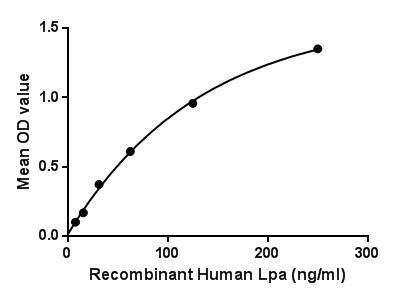
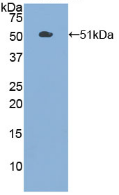
 WB (Western Blot)
(Sample: Recombinant Lpa, Human;Antibody: Rabbit Anti-Human Lpa Ab)
WB (Western Blot)
(Sample: Recombinant Lpa, Human;Antibody: Rabbit Anti-Human Lpa Ab)
Lipoprotein, a, Active Protein (Cat# AAA150102)
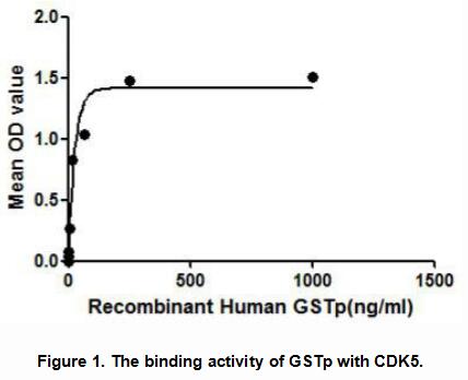
 WB (Western Blot)
(Western Blot; Sample: Recombinant GSTp, HumanAntibody: Rabbit Anti-Human GSTp Ab )
WB (Western Blot)
(Western Blot; Sample: Recombinant GSTp, HumanAntibody: Rabbit Anti-Human GSTp Ab )
Glutathione S Transferase Pi (GSTp), Active Protein (Cat# AAA148150)
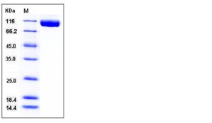
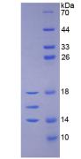
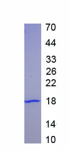
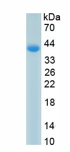
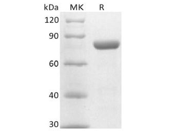
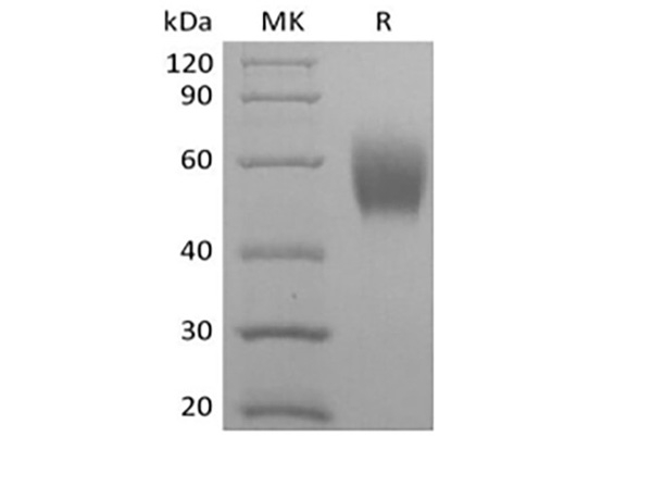
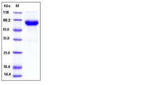
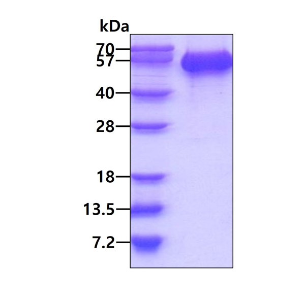
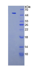
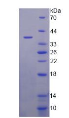
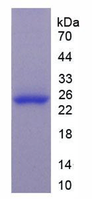
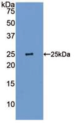
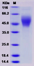
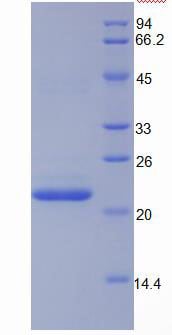 SDS-PAGE
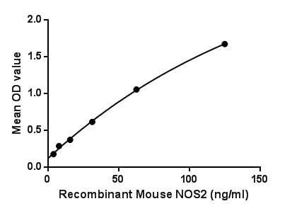
Nitric Oxide Synthase 2, Inducible (NOS2), Active Protein (Cat# AAA149243)

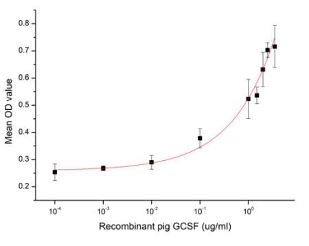 Bioactivity
(Figure 2. Cell proliferation of M-NFS-60 cells after stimulated with G-CSF.)
Bioactivity
(Figure 2. Cell proliferation of M-NFS-60 cells after stimulated with G-CSF.)
Colony Stimulating Factor 3, Granulocyte (GCSF), Active Protein (Cat# AAA161664)
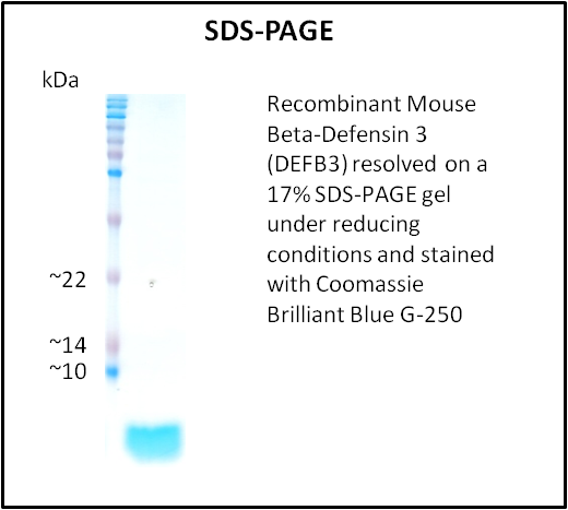
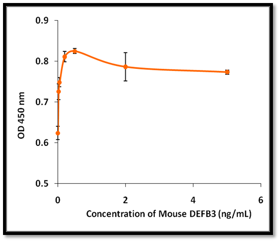 Application Data
Beta-Defensin 3 (DEFB3), Active Protein (Cat# AAA214325)
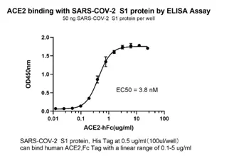 Bioactivity
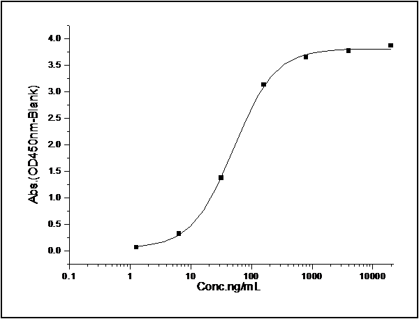
(The activity was determined by immobilized Spike Protein S1 binding with human ACE2 in a functional ELISA assay, the ED50 was determined to be 3.8nM.2019-ncov S1-his tagged (coating at 0.5ug-well) binding with Human ACE2-Fc (cat. The linear range was found to be 0.1-5 ug/ml)
Bioactivity
(The activity was determined by immobilized Spike Protein S1 binding with human ACE2 in a functional ELISA assay, the ED50 was determined to be 3.8nM.2019-ncov S1-his tagged (coating at 0.5ug-well) binding with Human ACE2-Fc (cat. The linear range was found to be 0.1-5 ug/ml)
COVID 19 Spike S1 Coronavirus, Active Protein (Cat# AAA76120)
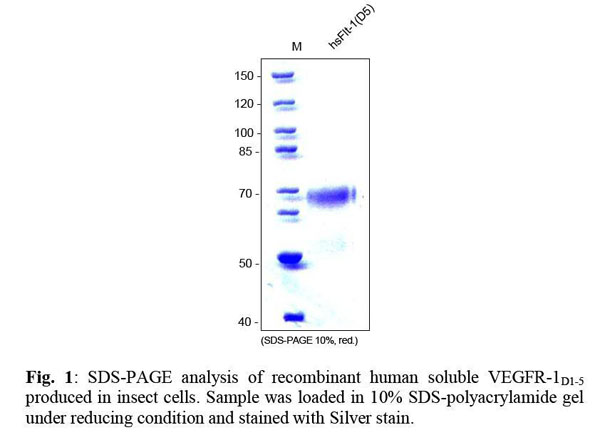
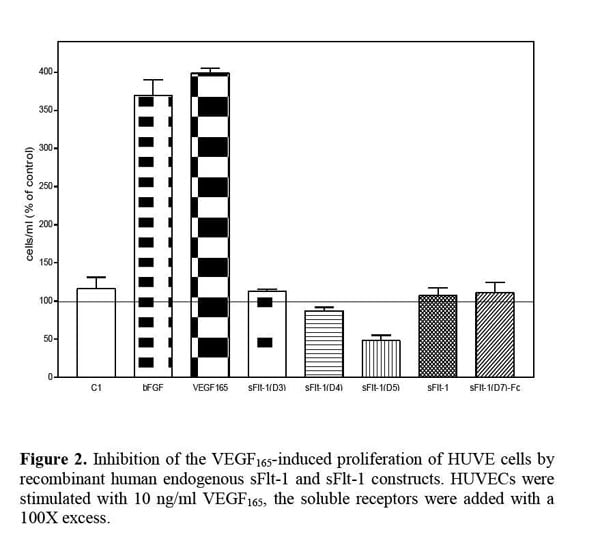
 Application Data
VEGFR-1/Flt-1 (D5), soluble, Active Protein (Cat# AAA79174)
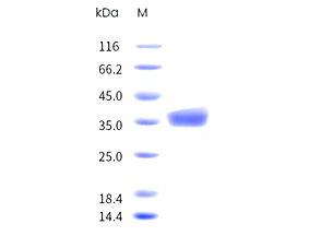
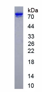
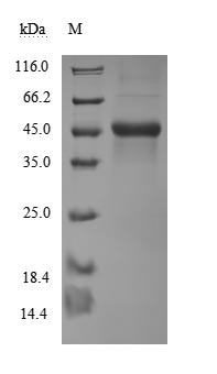
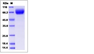
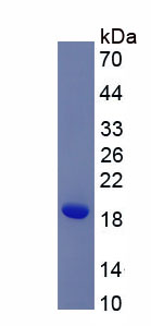
 SDS-PAGE
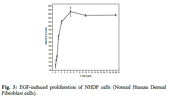
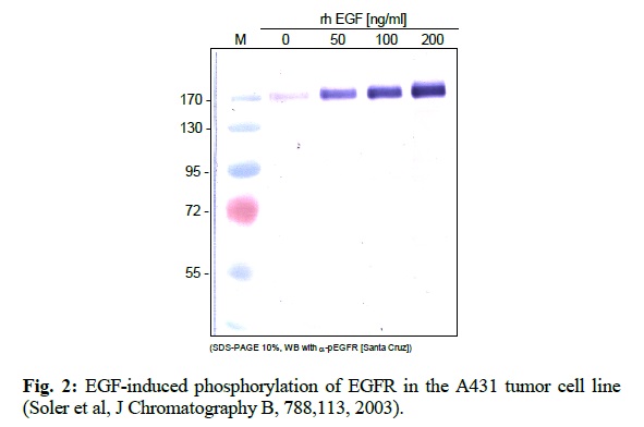
EGF, Active Protein (Cat# AAA79221)
 SDS-PAGE
B7-H6 / B7H6, Active Protein (Cat# AAA173515)

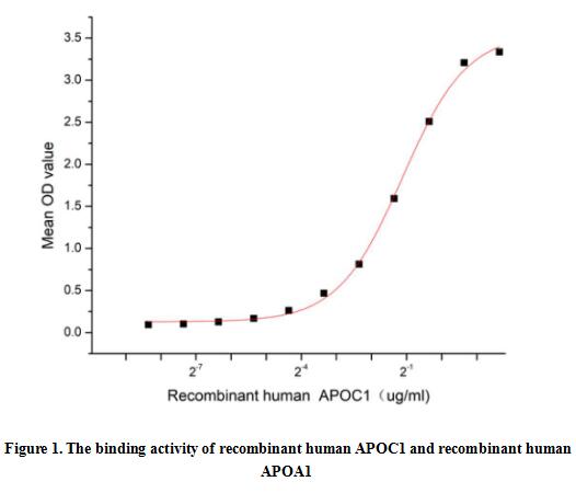 Bioactivity
(Apolipoprotein C1 (APOC1), also known as Apo-CI; ApoC-I; apo-CIB; apoC-IB, is the smallest size apolipoprotein of all apolipoprotein C family (Mr = 6.6 kDa) and located at position 19q13.32. APOC1 is primarily expressed in the liver and activated when monocytes differentiate into macrophages. It plays important roles in the innate immune response as effector of glucocorticoid-mediated responses and regulator of the inflammatory process. It has anti-inflammatory activity and also can promote the differentiation of T-cells into Th1 cells and negatively regulates differentiation into Th2 cells. Besides, Apolipoprotein A1 (APOA1) has been identified as an interactor of APOC1, thus a functional binding ELISA assay was conducted to detect the interaction of recombinant human APOC1 and recombinant human APOA1. Briefly, APOC1 was diluted serially in PBS with 0.01% BSA (pH 7.4). Duplicate samples of 100 ul were then transferred to APOA1-coated microtiter wells and incubated for 1h at 37 degree C. Wells were washed with PBST and incubated for 1h with anti-APOC1 pAb, then aspirated and washed 3 times. After incubation with HRP labelled secondary antibody for 1h at 37 degree C, wells were aspirated and washed 5 times. With the addition of substrate solution, wells were incubated 15-25 minutes at 37 degree C. Finally, add 50 uL stop solution to the wells and read at 450/630 nm immediately. The binding activity of recombinant human APOC1 and recombinant human APOA1 was shown in Figure 1, the EC50 for this effect is 0.46 ug/mL.)
Bioactivity
(Apolipoprotein C1 (APOC1), also known as Apo-CI; ApoC-I; apo-CIB; apoC-IB, is the smallest size apolipoprotein of all apolipoprotein C family (Mr = 6.6 kDa) and located at position 19q13.32. APOC1 is primarily expressed in the liver and activated when monocytes differentiate into macrophages. It plays important roles in the innate immune response as effector of glucocorticoid-mediated responses and regulator of the inflammatory process. It has anti-inflammatory activity and also can promote the differentiation of T-cells into Th1 cells and negatively regulates differentiation into Th2 cells. Besides, Apolipoprotein A1 (APOA1) has been identified as an interactor of APOC1, thus a functional binding ELISA assay was conducted to detect the interaction of recombinant human APOC1 and recombinant human APOA1. Briefly, APOC1 was diluted serially in PBS with 0.01% BSA (pH 7.4). Duplicate samples of 100 ul were then transferred to APOA1-coated microtiter wells and incubated for 1h at 37 degree C. Wells were washed with PBST and incubated for 1h with anti-APOC1 pAb, then aspirated and washed 3 times. After incubation with HRP labelled secondary antibody for 1h at 37 degree C, wells were aspirated and washed 5 times. With the addition of substrate solution, wells were incubated 15-25 minutes at 37 degree C. Finally, add 50 uL stop solution to the wells and read at 450/630 nm immediately. The binding activity of recombinant human APOC1 and recombinant human APOA1 was shown in Figure 1, the EC50 for this effect is 0.46 ug/mL.)
Apolipoprotein C1 (APOC1), Active Protein (Cat# AAA161720)
What Are Active Proteins?
Proteins are large molecules made up of long chains of amino acids. They will typically fold into a very particular 3-dimensional shape/conformation, that is sometimes referred to as their “native” form, which allows them to work properly in the body. For the purposes of product categorization, AAA Biotech will typically refer to proteins purified from their original animal host as being “native” proteins (this is to signify their difference compared to their recombinant proteins or “synthetic” protein counterparts).
If a protein successfully folds into the correct shape, it will typically display high fidelity characteristics to its original protein in its original animal host and be classified as an active protein, as it will be able to function “normally” in most enzymatic or binding capacities. If it loses this shape, due to factors such as heat or strong chemicals (such as detergents), it becomes inactive and is no longer able to perform its basic functions.
All of the proteins in this category are made under strict quality control, and they are active, pure, low in contaminants, and stable. Most are stored as freeze-dried powders and come without extra tags, so they’re very close to the actual natural/native form.
Learn more in our guide “How active proteins work”.
Key Applications of Active Proteins
1. Scientific Research
- Aid in the study of how proteins function in the body
- Aid in understanding various disease processes
2. Drug Development
- Powerful tools to investigate how potential drugs interact with specific proteins
- Ideal for identifying drug targets
3. Cell Culture
- Are routinely utilized to support cell growth and function (e.g., using exogenous growth factors)
- Can be used to promote cellular development into specific types (differentiation)
4. Diagnostics
- Regularly utilized in tests to detect diseases or infections (e.g., COVID-19, cancer)
- Note: All products are strictly for research-use only (RUO).
5. Therapeutics
- Some active proteins are used directly as treatments (e.g., insulin, enzymes)
- Note: All products are strictly for research-use only (RUO).
6. Vaccine Development
- Used to create or test vaccines by mimicking parts of viruses or bacteria
7. Biochemical Assays
- They can facilitate the characterization of enzyme activity, binding strength, or protein interactions in lab tests
Why Buy Active Proteins from AAA Biotech?
- High biological activity – Verified to perform as expected or indicated on datasheet
- Strict quality control – We are confident in our active proteins’ reliability and consistency
- High purity & low endotoxin – Ideal for applications involving sensitive or precious samples/components
- Freeze-dried for stability – Long shelf life and straightforward storage
- Mostly tag-free – Closer to natural/native protein form
FAQ
1. What are active proteins used for in research?
Active proteins are used primarily in the study of how proteins function, in characterizing/discovering drug interactions, supporting cell growth, running biochemical assays, and in development of diagnostics or therapeutics.
2. How are AAA Biotech's active proteins validated?
AAA Biotech’s active proteins are validated through strict quality control and functional assays to ensure they are properly folded and active. “Active”, though, can be an ambiguous term, so if a specific “activity” or “binding” capability of a protein is of crucial interest to you, please inquire with us prior to purchase, and we will provide further details on how the “Active” modifier was determined to be applicable.
3. Are these proteins tested for biological activity?
Yes, all active proteins from AAA Biotech are tested to confirm they have the expected biological activity before being offered for use. Though, said “biological activity” can be either “enzymatic”, “binding”, or both.