Filters
▼Clonality
▼Type
▼Reactivity
▼Gene Name
▼Isotype
▼Host
▼Application
▼Clone
▼Active Proteins
AAA Biotech also known as AAA Bio or AAABio provides a variety of high-quality recombinant and natural/native proteins that are proven to work in a wide range of experiments. Explore our products to find the active protein that best fits your needs or experimental model.
Viewing 2000-2050 of 2875 product results
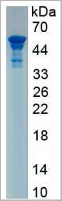 SDS-PAGE
(Sample: Active recombinant LOX, Human)
SDS-PAGE
(Sample: Active recombinant LOX, Human)
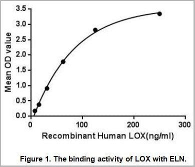
Lysyl Oxidase (LOX), Active Protein (Cat# AAA21109)
 SDS-PAGE
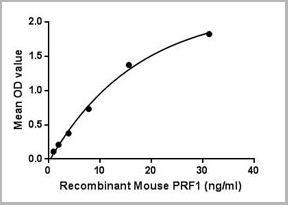
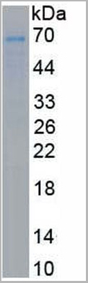
(Sample: Active recombinant PRF1, Mouse)
SDS-PAGE
(Sample: Active recombinant PRF1, Mouse)
Perforin 1 (PRF1), Active Protein (Cat# AAA21137)
 Bioactivity
(Pregnancy-associated plasma protein A(PAPPA), also known as pappalysin-1, is a secreted protease whose main substrate is insulin-like growth factor binding proteins. PAPPA's proteolytic function is activated upon collagen binding. It is thought to be involved in local proliferative processes such as wound healing and bone remodeling. Low plasma level of this protein has been suggested as a biochemical marker for pregnancies with aneuploid fetuses (fetuses with an abnormal number of chromosomes). Besides, Plasminogen (Plg) has been identified as an interactor of PAPPA, thus a binding ELISA assay was conducted to detect the interaction of recombinant human PAPPA and recombinant human Plg. Briefly, biotinylated PAPPA were diluted serially in PBS with 0.01% BSA (pH 7.4). Duplicate samples of 100ul were then transferred to Plg-coated microtiter wells and incubated for 1 h at 37 degree C. Wells were washed 3 times with PBST and incubated for 0.5 h at 37 degree C with SA labelled HRP. After incubation with SA labelled HRP, wells were aspirated and washed 5 times. With the addition of substrate solution, wells were incubated 15-25 minutes at 37 degree C. Finally, add 50uL stop solution to the wells and read at 450/630 nm immediately. The binding activity of PAPPA and Plg was shown in Figure 1, and the ED50 for this effect is 0.04936 ug/ml.)
Bioactivity
(Pregnancy-associated plasma protein A(PAPPA), also known as pappalysin-1, is a secreted protease whose main substrate is insulin-like growth factor binding proteins. PAPPA's proteolytic function is activated upon collagen binding. It is thought to be involved in local proliferative processes such as wound healing and bone remodeling. Low plasma level of this protein has been suggested as a biochemical marker for pregnancies with aneuploid fetuses (fetuses with an abnormal number of chromosomes). Besides, Plasminogen (Plg) has been identified as an interactor of PAPPA, thus a binding ELISA assay was conducted to detect the interaction of recombinant human PAPPA and recombinant human Plg. Briefly, biotinylated PAPPA were diluted serially in PBS with 0.01% BSA (pH 7.4). Duplicate samples of 100ul were then transferred to Plg-coated microtiter wells and incubated for 1 h at 37 degree C. Wells were washed 3 times with PBST and incubated for 0.5 h at 37 degree C with SA labelled HRP. After incubation with SA labelled HRP, wells were aspirated and washed 5 times. With the addition of substrate solution, wells were incubated 15-25 minutes at 37 degree C. Finally, add 50uL stop solution to the wells and read at 450/630 nm immediately. The binding activity of PAPPA and Plg was shown in Figure 1, and the ED50 for this effect is 0.04936 ug/ml.)
Pregnancy Associated Plasma Protein A (PAPPA), Active Protein (Cat# AAA152234)
 Bioactivity
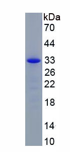
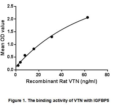
(Vitronectin (VTN ) is a glycoprotein of the hemopexin family which is abundantly found in serum, the extracellular matrix and bone. It is a secreted protein and exists in either a single chain form or a clipped, two chain form held together by a disulfide)
Bioactivity
(Vitronectin (VTN ) is a glycoprotein of the hemopexin family which is abundantly found in serum, the extracellular matrix and bone. It is a secreted protein and exists in either a single chain form or a clipped, two chain form held together by a disulfide)
Vitronectin (VTN), Active Protein (Cat# AAA153067)
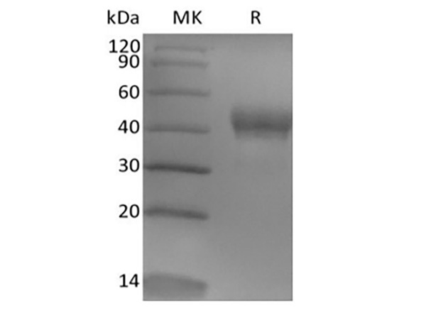
 SDS-PAGE
CD32a / FCGR2A, Active Protein (Cat# AAA173506)
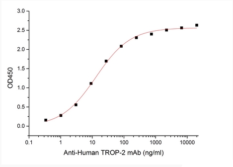 Bioactivity
Tumor-associated Calcium Signal Transducer 2/TROP-2, Active Protein (Cat# AAA177960)
 Application Data
(Measured by its binding ability in a functional ELISA. Immobilized recombinant Mouse/Human BDNF Protein (Native) at 10 ug/ml (100 ul/well) can bind biotinylated mouse TrkB-His with a linear range of 10-80 ng/ml.)
Application Data
(Measured by its binding ability in a functional ELISA. Immobilized recombinant Mouse/Human BDNF Protein (Native) at 10 ug/ml (100 ul/well) can bind biotinylated mouse TrkB-His with a linear range of 10-80 ng/ml.)
TrkB, Active Protein (Cat# AAA258131)
 Application Data
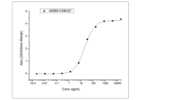
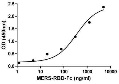
(Immobilized human ACE2 protein (Fc tag)(Cat: 10108-H05H) at 2 ug/mL (100 uL/well) can bind Recombinant SARS-CoV-2 (BA.4.6) Spike S1+S2 trimer Protein (ECD, His Tag)(Cat: AAA258586), the EC50 is 8-24 ng/mL.)
Application Data
(Immobilized human ACE2 protein (Fc tag)(Cat: 10108-H05H) at 2 ug/mL (100 uL/well) can bind Recombinant SARS-CoV-2 (BA.4.6) Spike S1+S2 trimer Protein (ECD, His Tag)(Cat: AAA258586), the EC50 is 8-24 ng/mL.)
COVID 19 (BA.4.6) Spike S1+S2 trimer Coronavirus, Active Protein (Cat# AAA258586)
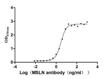
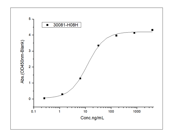 Application Data
(Immobilized Recombinant Human MSLN/Mesothelin Protein(Cat:13128-HNCH) at 2 ug/ml (100 ul/well) can bind Recombinant Human CA125 Protein (ECD, His Tag)(Cat:AAA258594), the EC50 is 6-18 ng/mL.)
Application Data
(Immobilized Recombinant Human MSLN/Mesothelin Protein(Cat:13128-HNCH) at 2 ug/ml (100 ul/well) can bind Recombinant Human CA125 Protein (ECD, His Tag)(Cat:AAA258594), the EC50 is 6-18 ng/mL.)
CA125, Active Protein (Cat# AAA258594)
ApoE2, Active Protein (Cat# AAA79218)
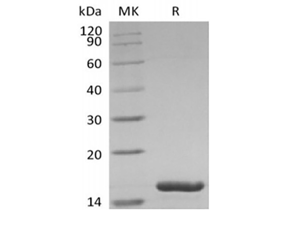
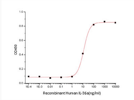 Bioactivity
Interleukin-36 alpha/IL-36 alpha, Active Protein (Cat# AAA177939)
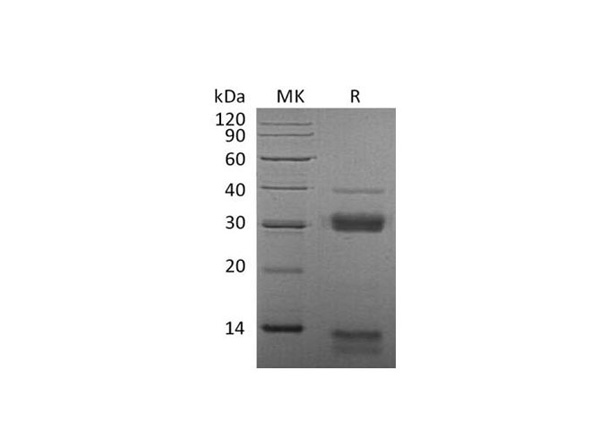 SDS-PAGE
Cathepsin S, Active Protein (Cat# AAA174601)
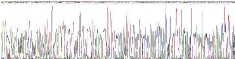
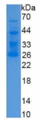
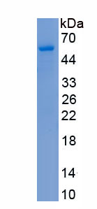
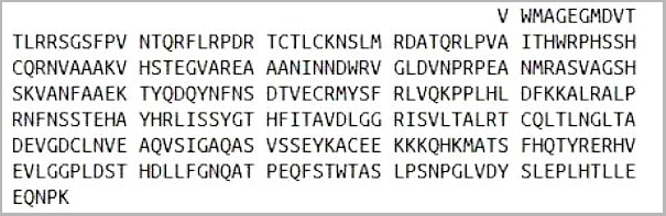
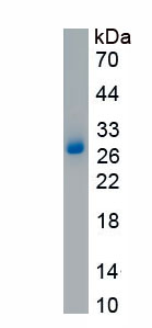
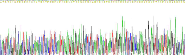
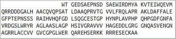
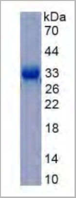
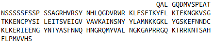
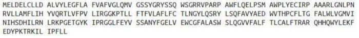 Sequence
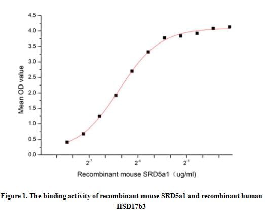
Steroid 5 Alpha Reductase 1 (SRD5a1), Active Protein (Cat# AAA161940)
 Bioactivity
(Ephrin-A1 (EFNA1), also known as EPLG1, LERK1, TNFAIP4, Immediate early response protein B61, is a member of the A-type ephrin family of cell surface proteins that function as ligands for the A-type Eph receptor tyrosine kinase family. EFNA1 plays an important role in angiogenesis and tumor neovascularization. EFNA1 widely affects tumor growth through enhancing tumor angiogenesis, malignant cell events and invasiveness. Ephrin Type A Receptor 1 (EPHA1) is one of high-affinity ligands for EFNA1, thus a functional binding ELISA assay was conducted to detect the interaction of recombinant human EFNA1 and recombinant mouse EPHA1. Briefly, biotin-linked EFNA1 were diluted serially in PBS, with 0.01% BSA (pH 7.4). Duplicate samples of 100 ul were then transferred to EPHA1-coated microtiter wells and incubated for 1h at 37 degree C. Wells were washed with PBST 3 times and incubation with Streptavidin-HRP for 30min, then wells were aspirated and washed 5 times. With the addition of substrate solution, wells were incubated 15-25 minutes at 37 degree C. Finally, add 50 ul stop solution to the wells and read at 450 nm immediately. The binding activity of recombinant human EFNA1 and recombinant mouse EPHA1 was shown in Figure 1, the EC50 for this effect is 0.24 ug/mL.)
Bioactivity
(Ephrin-A1 (EFNA1), also known as EPLG1, LERK1, TNFAIP4, Immediate early response protein B61, is a member of the A-type ephrin family of cell surface proteins that function as ligands for the A-type Eph receptor tyrosine kinase family. EFNA1 plays an important role in angiogenesis and tumor neovascularization. EFNA1 widely affects tumor growth through enhancing tumor angiogenesis, malignant cell events and invasiveness. Ephrin Type A Receptor 1 (EPHA1) is one of high-affinity ligands for EFNA1, thus a functional binding ELISA assay was conducted to detect the interaction of recombinant human EFNA1 and recombinant mouse EPHA1. Briefly, biotin-linked EFNA1 were diluted serially in PBS, with 0.01% BSA (pH 7.4). Duplicate samples of 100 ul were then transferred to EPHA1-coated microtiter wells and incubated for 1h at 37 degree C. Wells were washed with PBST 3 times and incubation with Streptavidin-HRP for 30min, then wells were aspirated and washed 5 times. With the addition of substrate solution, wells were incubated 15-25 minutes at 37 degree C. Finally, add 50 ul stop solution to the wells and read at 450 nm immediately. The binding activity of recombinant human EFNA1 and recombinant mouse EPHA1 was shown in Figure 1, the EC50 for this effect is 0.24 ug/mL.)
Ephrin A1 (EFNA1), Active Protein (Cat# AAA161914)
 Application Data
(Figure.The chemotactic effect of IL8 on Jurkat cells.)
Application Data
(Figure.The chemotactic effect of IL8 on Jurkat cells.)
Interleukin 8, Active Protein (Cat# AAA150064)

 Application Data
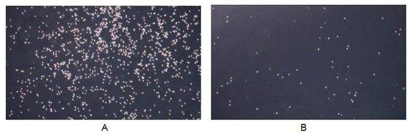
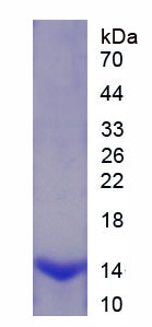
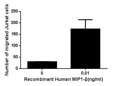
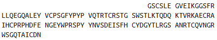
(Figure. The chemotactic effect of recombinant human MIP1-on Jurkat cells.)
Application Data
(Figure. The chemotactic effect of recombinant human MIP1-on Jurkat cells.)
Macrophage Inflammatory Protein 1 Beta, Active Protein (Cat# AAA150067)
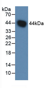
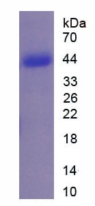
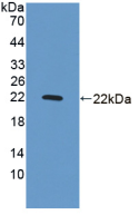
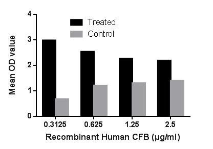 Application Data
(Figure. The hemolysis activity of recombinant human CFB)
Application Data
(Figure. The hemolysis activity of recombinant human CFB)
Complement Factor B, Active Protein (Cat# AAA150124)
 Bioactivity
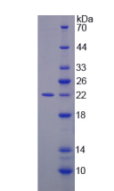
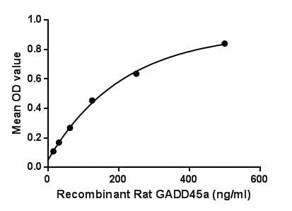
(Growth arrest and DNA-damage-inducible protein GADD45 alpha (GADD45a) is a member of the GADD45 family. It may affect PCNA interaction with some CDK (cell division protein kinase) complexes; stimulates DNA excision repair in vitro and inhibits entry of cells into S phase. In T-cells, functions as a regulator of p38 MAPKs by inhibiting p88 phosphorylation and activity. Besides, Proliferating Cell Nuclear Antigen (PCNA) has been identified as an interactor of GADD45a, thus a binding ELISA assay was conducted to detect the interaction of recombinant rat GADD45a and recombinant rat PCNA. Briefly, GADD45a were diluted serially in PBS, with 0.01% BSA (pH 7.4). Duplicate samples of 100L were then transferred to PCNA-coated microtiter wells and incubated for 2h at 37. Wells were washed with PBST and incubated for 1h with anti-GADD45a pAb, then aspirated and washed 3 times. After incubation with HRP labelled secondary antibody, wells were aspirated and washed 3 times. With the addition of substrate solution, wells were incubated 15-25 minutes at 37. Finally, add 50uL stop solution to the wells and read at 450nm immediately. The binding activity of GADD45a and PCNA was shown in Figure 1, and this effect was in a dose dependent manner.Figure. The binding activity of GADD45a with PCNA.)
Bioactivity
(Growth arrest and DNA-damage-inducible protein GADD45 alpha (GADD45a) is a member of the GADD45 family. It may affect PCNA interaction with some CDK (cell division protein kinase) complexes; stimulates DNA excision repair in vitro and inhibits entry of cells into S phase. In T-cells, functions as a regulator of p38 MAPKs by inhibiting p88 phosphorylation and activity. Besides, Proliferating Cell Nuclear Antigen (PCNA) has been identified as an interactor of GADD45a, thus a binding ELISA assay was conducted to detect the interaction of recombinant rat GADD45a and recombinant rat PCNA. Briefly, GADD45a were diluted serially in PBS, with 0.01% BSA (pH 7.4). Duplicate samples of 100L were then transferred to PCNA-coated microtiter wells and incubated for 2h at 37. Wells were washed with PBST and incubated for 1h with anti-GADD45a pAb, then aspirated and washed 3 times. After incubation with HRP labelled secondary antibody, wells were aspirated and washed 3 times. With the addition of substrate solution, wells were incubated 15-25 minutes at 37. Finally, add 50uL stop solution to the wells and read at 450nm immediately. The binding activity of GADD45a and PCNA was shown in Figure 1, and this effect was in a dose dependent manner.Figure. The binding activity of GADD45a with PCNA.)
Growth Arrest And DNA Damage Inducible Protein Alpha, Active Protein (Cat# AAA150146)
 Bioactivity
(Caspase 3 is a member of the cysteine-aspartic acid protease (caspase) family. Sequential activation of caspases plays a central role in the execution-phase of cell apoptosis. Caspases exist as inactive proenzymes that undergo proteolytic processing at co)
Bioactivity
(Caspase 3 is a member of the cysteine-aspartic acid protease (caspase) family. Sequential activation of caspases plays a central role in the execution-phase of cell apoptosis. Caspases exist as inactive proenzymes that undergo proteolytic processing at co)
Caspase 3 (CASP3), Active Protein (Cat# AAA153051)
 Application Data
(Measured by its ability to neutralize Activin-mediated inhibition on MPC11 cell proliferation. The ED50 for this effect is typically 40-200 ng/mL in the presence of 10 ng/mL rhActivin A.)
Application Data
(Measured by its ability to neutralize Activin-mediated inhibition on MPC11 cell proliferation. The ED50 for this effect is typically 40-200 ng/mL in the presence of 10 ng/mL rhActivin A.)
Follistatin, Active Protein (Cat# AAA258120)
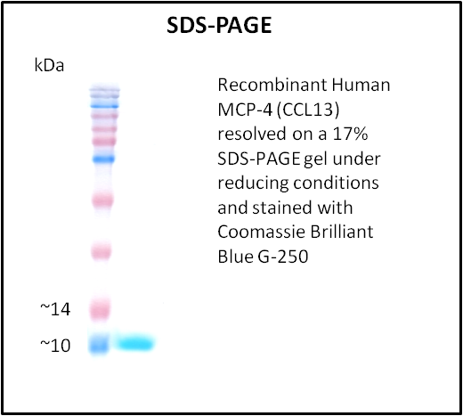
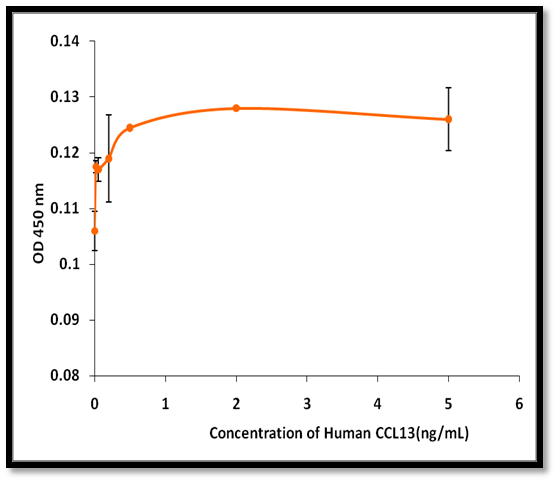 Application Data
MCP-4 (CCL13), Active Protein (Cat# AAA214244)
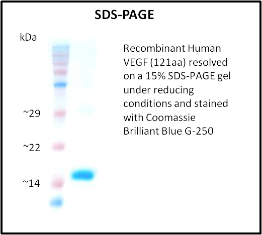
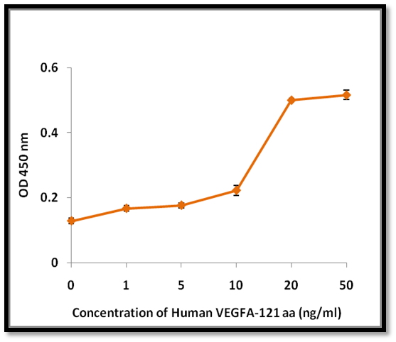 Application Data
VEGF, Active Protein (Cat# AAA214288)
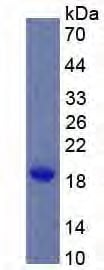
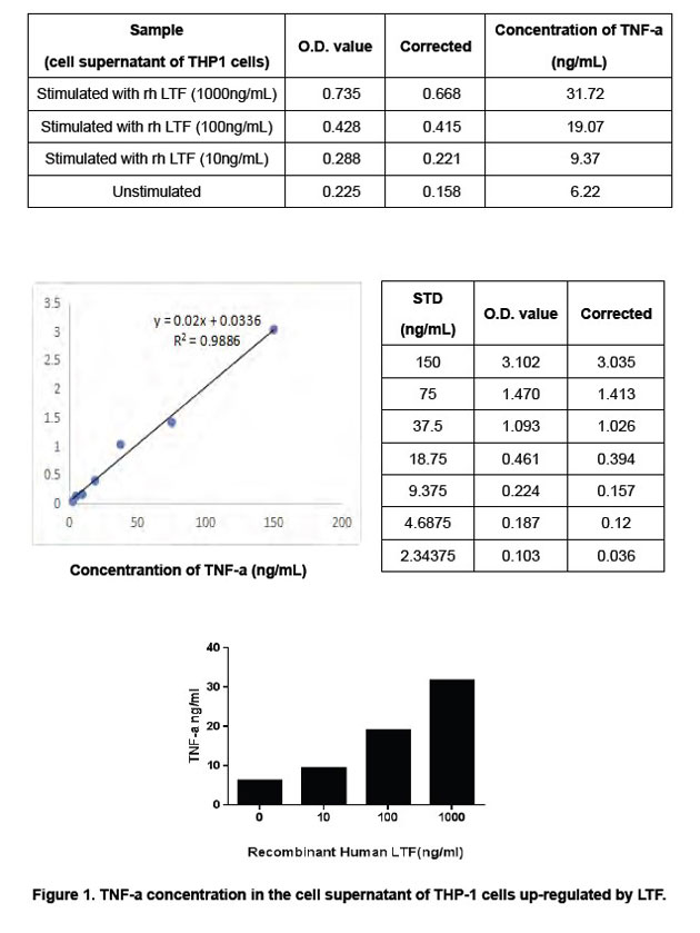
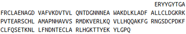
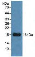 WB (Western Blot)
(Sample: Recombinant LTF, Human; Antibody: Rabbit Anti-Human LTF Ab)
WB (Western Blot)
(Sample: Recombinant LTF, Human; Antibody: Rabbit Anti-Human LTF Ab)
Lactoferrin (LTF), Active Protein (Cat# AAA149239)
Antistreptolysin O, Active Protein (Cat# AAA44827)
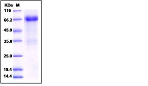
 SDS-PAGE
(3ug by SDS-PAGE under reducing condition and visualized by coomassie blue stain.)
SDS-PAGE
(3ug by SDS-PAGE under reducing condition and visualized by coomassie blue stain.)
Carbonic anhydrase 2, Active Protein (Cat# AAA48538)
 SDS-PAGE
GFRA1 / GFR alpha-1, Active Protein (Cat# AAA173594)
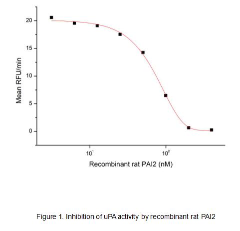 Bioactivity
(Serpin B2, also known as PAI-2, is an approximately 60 kDa serine protease inhibitor. It is primarily secreted by macrophages and monocytes and can form disulfide-linked multimers. Serpin B2 inhibits both the urokinase-type and tissue-type plasminogen activators (uPA and tPA). Serpin B2 also promotes the clearance of uPA by enhancing its binding and uptake by LRP. It limits fibril formation by Huntington protein (HTT) and beta-Amyloid peptides. It promotes Th2 biased immune responses and is important for intestinal CCL2 production, monocyte recruitment, and nematode clearance. A non-glycosylated form of Serpin B2 is retained intracellularly where it interferes with TNF-a induced apoptosis by protecting the Retinoblastoma protein (RB1) from calpain digestion. It also inhibits proteasome activity in activated endothelial cells. The activity of recombinant rat PAI-2 was measured by its ability to inhibit uPA cleavage of a peptide substrate, N-carbobenzyloxy-Gly-Gly-Arg-7-amido-4-methylcoumarin (Z-GGR-AMC) in the assay buffer 50 mM Tris, 0.01% Tween 20, pH 8.5. The 50 ul different concentrations of rrPAI-2 (MW: 78.3 KD) was incubated with 50ul 2ug/ml rhuPA (EPA140Mu61) at room temperature for 15 minutes. Loading 50 uL of the incubated mixtures into empty wells of a plate, and start the reaction by adding 50 uL of 200 uM substrate (Z-GGR-AMC). Include a substrate blank containing 50 uL of assay buffer and 50 uL of 200 uM substrate. Then read at excitiation and emission wavelengths of 380 nm and 460 nm, respectively, in kinetic mode for 5 minutes. The result was shown in Figure 1 and it was obvious that recombinant rat PAI2 significantly decreased uPA activity. The inhibition IC50 was )
Bioactivity
(Serpin B2, also known as PAI-2, is an approximately 60 kDa serine protease inhibitor. It is primarily secreted by macrophages and monocytes and can form disulfide-linked multimers. Serpin B2 inhibits both the urokinase-type and tissue-type plasminogen activators (uPA and tPA). Serpin B2 also promotes the clearance of uPA by enhancing its binding and uptake by LRP. It limits fibril formation by Huntington protein (HTT) and beta-Amyloid peptides. It promotes Th2 biased immune responses and is important for intestinal CCL2 production, monocyte recruitment, and nematode clearance. A non-glycosylated form of Serpin B2 is retained intracellularly where it interferes with TNF-a induced apoptosis by protecting the Retinoblastoma protein (RB1) from calpain digestion. It also inhibits proteasome activity in activated endothelial cells. The activity of recombinant rat PAI-2 was measured by its ability to inhibit uPA cleavage of a peptide substrate, N-carbobenzyloxy-Gly-Gly-Arg-7-amido-4-methylcoumarin (Z-GGR-AMC) in the assay buffer 50 mM Tris, 0.01% Tween 20, pH 8.5. The 50 ul different concentrations of rrPAI-2 (MW: 78.3 KD) was incubated with 50ul 2ug/ml rhuPA (EPA140Mu61) at room temperature for 15 minutes. Loading 50 uL of the incubated mixtures into empty wells of a plate, and start the reaction by adding 50 uL of 200 uM substrate (Z-GGR-AMC). Include a substrate blank containing 50 uL of assay buffer and 50 uL of 200 uM substrate. Then read at excitiation and emission wavelengths of 380 nm and 460 nm, respectively, in kinetic mode for 5 minutes. The result was shown in Figure 1 and it was obvious that recombinant rat PAI2 significantly decreased uPA activity. The inhibition IC50 was )
Plasminogen Activator Inhibitor 2 (PAI2), Active Protein (Cat# AAA161741)
 Bioactivity
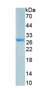
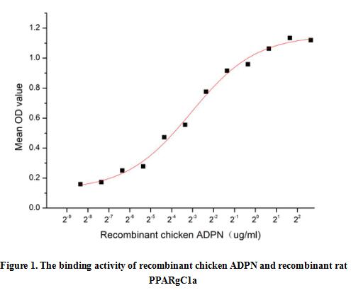
(Human Adiponectin (ADPN) is a 53 kDa member of the PNPLA family of phospholipase A2 enzymes. It is a plasma protein secreted by adipose tissue showing pleiotropic effects with anti-diabetic, anti-atherogenic and anti-inflammatory properties. Adiponectin and AdipoR1 regulate PPARgC1a and mitochondria by Ca(2) and AMPK/SIRT1. Thus a functional binding ELISA assay was conducted to detect the interaction of recombinant chicken ADPN and recombinant rat PPARgC1a. Briefly, ADPN was diluted serially in PBS with 0.01% BSA (pH 7.4). Duplicate samples of 100 ul were then transferred to PPARgC1a-coated microtiter wells and incubated for 1h at 37 degree C. Wells were washed with PBST and incubated for 1h with anti-ADPN pAb, then aspirated and washed 3 times. After incubation with HRP labelled secondary antibody for 1h at 37 degree C, wells were aspirated and washed 5 times. With the addition of substrate solution, wells were incubated 15-25 minutes at 37 degree C. Finally, add 50 uL stop solution to the wells and read at 450/630 nm immediately. The binding activity of recombinant chicken ADPN and recombinant rat PPARgC1a was shown in Figure 1, the EC50 for this effect is 0.12 ug/mL.)
Bioactivity
(Human Adiponectin (ADPN) is a 53 kDa member of the PNPLA family of phospholipase A2 enzymes. It is a plasma protein secreted by adipose tissue showing pleiotropic effects with anti-diabetic, anti-atherogenic and anti-inflammatory properties. Adiponectin and AdipoR1 regulate PPARgC1a and mitochondria by Ca(2) and AMPK/SIRT1. Thus a functional binding ELISA assay was conducted to detect the interaction of recombinant chicken ADPN and recombinant rat PPARgC1a. Briefly, ADPN was diluted serially in PBS with 0.01% BSA (pH 7.4). Duplicate samples of 100 ul were then transferred to PPARgC1a-coated microtiter wells and incubated for 1h at 37 degree C. Wells were washed with PBST and incubated for 1h with anti-ADPN pAb, then aspirated and washed 3 times. After incubation with HRP labelled secondary antibody for 1h at 37 degree C, wells were aspirated and washed 5 times. With the addition of substrate solution, wells were incubated 15-25 minutes at 37 degree C. Finally, add 50 uL stop solution to the wells and read at 450/630 nm immediately. The binding activity of recombinant chicken ADPN and recombinant rat PPARgC1a was shown in Figure 1, the EC50 for this effect is 0.12 ug/mL.)
Adiponectin (ADPN), Active Protein (Cat# AAA161753)
 Bioactivity
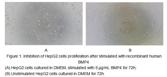
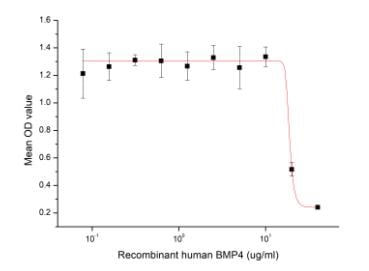
(Figure 2. Inhibition of HepG2 cells proliferation after stimulated with recombinant human BMP4)
Bioactivity
(Figure 2. Inhibition of HepG2 cells proliferation after stimulated with recombinant human BMP4)
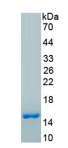
Bone Morphogenetic Protein 4 (BMP4), Active Protein (Cat# AAA161654)
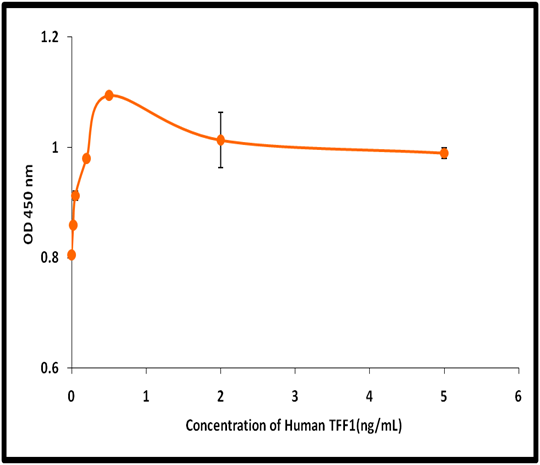 Application Data
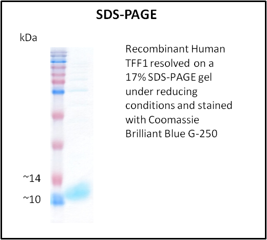
TFF1, Active Protein (Cat# AAA214281)
 Application Data
(Measured in a cell proliferation assay using NIH3T3 cells. The ED50 for this effect is typically 2-12 ng/mL.)
Application Data
(Measured in a cell proliferation assay using NIH3T3 cells. The ED50 for this effect is typically 2-12 ng/mL.)
Oncostatin M/OSM, Active Protein (Cat# AAA258128)
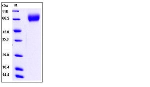
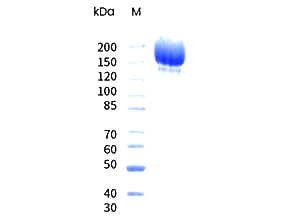
 Purity Data
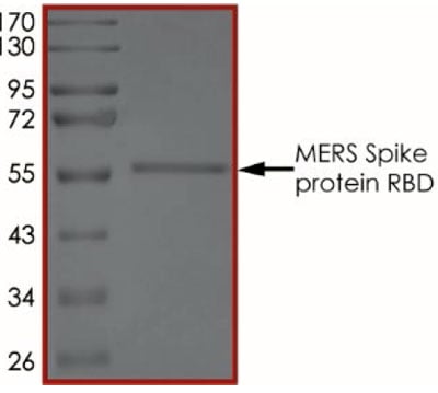
(The purity of MERS Spike Protein RBD was determined to be >90% by densitometry, approx. MW 58 kDa.)
Purity Data
(The purity of MERS Spike Protein RBD was determined to be >90% by densitometry, approx. MW 58 kDa.)
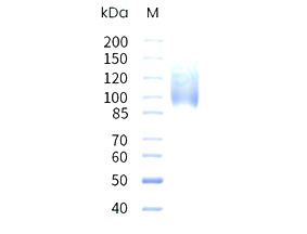
 SDS-PAGE
(3ug by SDS-PAGE under reducing condition and visualized by coomassie blue stain)
SDS-PAGE
(3ug by SDS-PAGE under reducing condition and visualized by coomassie blue stain)
BACE-1, Active Protein (Cat# AAA48325)
 SDS-PAGE
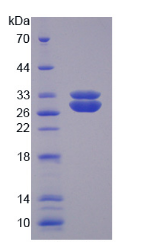
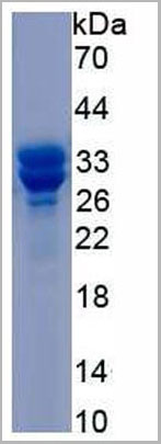
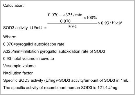
(Sample: Active recombinant SOD3, Human)
SDS-PAGE
(Sample: Active recombinant SOD3, Human)
Superoxide Dismutase 3, Extracellular, Active Protein (Cat# AAA21162)
 Application Data
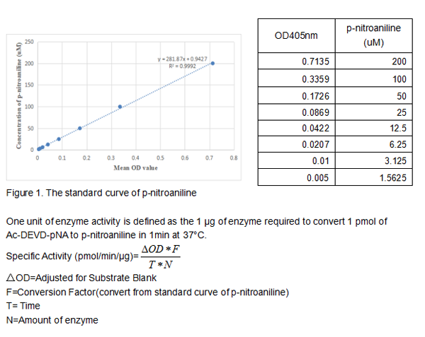
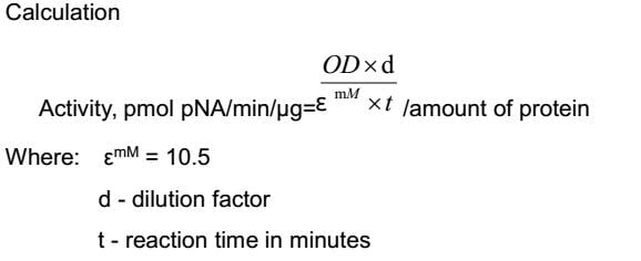
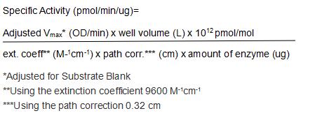
(Caspase 3 is a member of the cysteine-aspartic acid protease (caspase) family. Sequential activation of caspases plays a central role in the execution-phase of cell apoptosis. Caspases exist as inactive proenzymes that undergo proteolytic processing at conserved aspartic residues to produce two subunits, large and small, that dimerize to form the active enzyme. This protein cleaves and activates caspases 6 and 7; and the protein itself is processed and activated by caspases 8, 9, and 10. Caspase 3 can hydrolyze the peptide substrate acetyl-Asp-Glu-Val-Asp-p-nitroanilide (Ac-DEVD-pNA) resulting in the release of the p-nitroaniline (pNA) moiety. p-Nitroaniline has a high absorbance at 405nm. Thus the activity of recombinant human caspase 3 can be measured by calculate the concentration of the pNA released from the substrate. The reaction was performed in adding 50L 2×buffer (50mM HEPES, 100mM NaCl, 10mM DTT, 2mM EDTA, 10% glycerol) to 96 well plates, then add 50L various concentration of caspe 3 (diluted by 1×buffer, 25mM HEPES, 50mM NaCl, 5mM DTT, 1mM EDTA, 5% glycerol) to each well, finally, add 5L 4mmol Ac-DEVD-pNA to each well. Cover the 96 well plates and incubate at 37 for 2h. p-Nitroaniline (pNA) standard curve prepare by double dilute 200M pNA with 1×buffer and record the OD value at 405nm. Calculate the caspase 3 activity in pmol of pNA released per min per g recombinat human caspase 3.The specific activity of recombinant human caspase 3 is 2196pmol/min/g.)
Application Data
(Caspase 3 is a member of the cysteine-aspartic acid protease (caspase) family. Sequential activation of caspases plays a central role in the execution-phase of cell apoptosis. Caspases exist as inactive proenzymes that undergo proteolytic processing at conserved aspartic residues to produce two subunits, large and small, that dimerize to form the active enzyme. This protein cleaves and activates caspases 6 and 7; and the protein itself is processed and activated by caspases 8, 9, and 10. Caspase 3 can hydrolyze the peptide substrate acetyl-Asp-Glu-Val-Asp-p-nitroanilide (Ac-DEVD-pNA) resulting in the release of the p-nitroaniline (pNA) moiety. p-Nitroaniline has a high absorbance at 405nm. Thus the activity of recombinant human caspase 3 can be measured by calculate the concentration of the pNA released from the substrate. The reaction was performed in adding 50L 2×buffer (50mM HEPES, 100mM NaCl, 10mM DTT, 2mM EDTA, 10% glycerol) to 96 well plates, then add 50L various concentration of caspe 3 (diluted by 1×buffer, 25mM HEPES, 50mM NaCl, 5mM DTT, 1mM EDTA, 5% glycerol) to each well, finally, add 5L 4mmol Ac-DEVD-pNA to each well. Cover the 96 well plates and incubate at 37 for 2h. p-Nitroaniline (pNA) standard curve prepare by double dilute 200M pNA with 1×buffer and record the OD value at 405nm. Calculate the caspase 3 activity in pmol of pNA released per min per g recombinat human caspase 3.The specific activity of recombinant human caspase 3 is 2196pmol/min/g.)
Caspase 3, Active Protein (Cat# AAA150098)

 Bioactivity
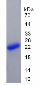
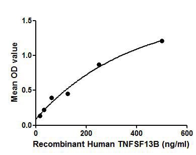
(Figure. The binding activity of TNFSF13B with ITGb1.Tumor necrosis factor ligand superfamily member 13B protein (TNFSF13B) also known as B-cell activating factor (BAFF) is a cytokine that belongs to the tumor necrosis factor (TNF) ligand family. This cytokine is a ligand for receptors TNFRSF13B/TACI, TNFRSF17/BCMA, and TNFRSF13C/BAFF-R. This cytokine is expressed in B cell lineage cells, and acts as a potent B cell activator. It has been also shown to play an important role in the proliferation and differentiation of B cells. Besides, Integrin Beta 1 (ITGb1) has been identified as an interactor of TNFSF13B, thus a binding ELISA assay was conducted to detect the interaction of recombinant human TNFSF13B and recombinant human ITGb1. Briefly, TNFSF13B were diluted serially in PBS, with 0.01% BSA (pH 7.4). Duplicate samples of 100uL were then transferred to ITGb1-coated microtiter wells and incubated for 2h at 37. Wells were washed with PBST and incubated for 1h with anti-TNFSF13B pAb, then aspirated and washed 3 times. After incubation with HRP labelled secondary antibody, wells were aspirated and washed 3 times. With the addition of substrate solution, wells were incubated 15-25 minutes at 37. Finally, add 50uL stop solution to the wells and read at 450nm immediately. The binding activity of TNFSF13B and ITGb1 was shown in Figure 1, and this effect was in a dose dependent manner.)
Bioactivity
(Figure. The binding activity of TNFSF13B with ITGb1.Tumor necrosis factor ligand superfamily member 13B protein (TNFSF13B) also known as B-cell activating factor (BAFF) is a cytokine that belongs to the tumor necrosis factor (TNF) ligand family. This cytokine is a ligand for receptors TNFRSF13B/TACI, TNFRSF17/BCMA, and TNFRSF13C/BAFF-R. This cytokine is expressed in B cell lineage cells, and acts as a potent B cell activator. It has been also shown to play an important role in the proliferation and differentiation of B cells. Besides, Integrin Beta 1 (ITGb1) has been identified as an interactor of TNFSF13B, thus a binding ELISA assay was conducted to detect the interaction of recombinant human TNFSF13B and recombinant human ITGb1. Briefly, TNFSF13B were diluted serially in PBS, with 0.01% BSA (pH 7.4). Duplicate samples of 100uL were then transferred to ITGb1-coated microtiter wells and incubated for 2h at 37. Wells were washed with PBST and incubated for 1h with anti-TNFSF13B pAb, then aspirated and washed 3 times. After incubation with HRP labelled secondary antibody, wells were aspirated and washed 3 times. With the addition of substrate solution, wells were incubated 15-25 minutes at 37. Finally, add 50uL stop solution to the wells and read at 450nm immediately. The binding activity of TNFSF13B and ITGb1 was shown in Figure 1, and this effect was in a dose dependent manner.)
B-Cell Activating Factor, Active Protein (Cat# AAA150113)
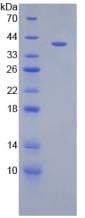
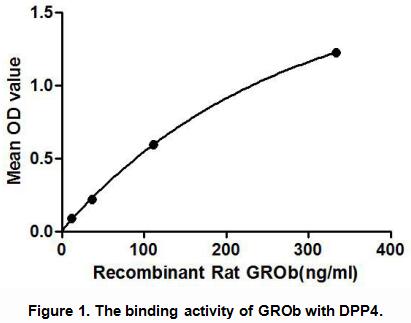
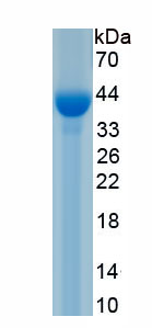
 Application Data
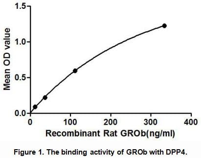
Growth Regulated Oncogene Beta (GROb), Active Protein (Cat# AAA148193)
 Bioactivity
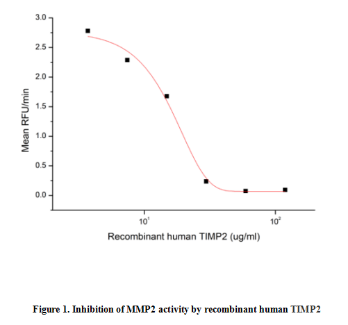
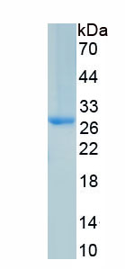
(Tissue inhibitors of metalloproteinases or TIMPs are a family of proteins that regulate the activation and proteolytic activity of the zinc enzymes known as matrix metalloproteinases (MMPs). There are four members of the family, TIMP-1, TIMP-2, TIMP-3, and TIMP-4. TIMP-2 is a non N-glycosylated protein with a molecular mass of 22 kDa produced by a wide range of cell types, which inhibits MMPs non-covalently by the formation of binary complexes. TIMP-2 also has erythroidpotentiating and cell growth promoting activities. The activity of recombinant human TIMP2 was measured by its ability to inhibit rhMMP2 cleavage of a fluorogenic peptide substrate MCA-Pro-Leu-Gly-Leu-DPA-Ala-Arg-NH2 in the assay buffer 50 mM Tris, 10 mM CaCl2, 150 mM NaCl, 0.05% (w/v) Brij-35, pH 7.5. rhMMP2 was diluted to 100 ug/ml and activated with 1 mM APMA at 37 degree C for 1 hour and rhTIMP2 (MW: 51.75 KD) was diluted to different concentrations with the assay buffer. Mix 8 ul of rhTIMP2 curve dilutions, 12.8 ul of activated rhMMP-2, and 59.2 ul of assay buffer, including a control containing assay buffer and the diluted rhMMP-2 and incubate the reactions for 2 hours at 37 degree C. Loading 50 ul of the incubated mixtures which were diluted five-fold in assay buffer into empty wells of a plate, and start the reaction by adding 50 ul of 20 uM substrate. Include a substrate blank containing 50 ul of assay buffer and 50 ul of 20 uM substrate. Then read at excitiation and emission wavelengths of 320 nm and 405 nm, respectively, in kinetic mode for 5 minutes. The result was shown in Figure 1 and it was obvious that recombinant human TIMP2 significantly decreased rhMMP2 activity. The inhibition IC50 was )
Bioactivity
(Tissue inhibitors of metalloproteinases or TIMPs are a family of proteins that regulate the activation and proteolytic activity of the zinc enzymes known as matrix metalloproteinases (MMPs). There are four members of the family, TIMP-1, TIMP-2, TIMP-3, and TIMP-4. TIMP-2 is a non N-glycosylated protein with a molecular mass of 22 kDa produced by a wide range of cell types, which inhibits MMPs non-covalently by the formation of binary complexes. TIMP-2 also has erythroidpotentiating and cell growth promoting activities. The activity of recombinant human TIMP2 was measured by its ability to inhibit rhMMP2 cleavage of a fluorogenic peptide substrate MCA-Pro-Leu-Gly-Leu-DPA-Ala-Arg-NH2 in the assay buffer 50 mM Tris, 10 mM CaCl2, 150 mM NaCl, 0.05% (w/v) Brij-35, pH 7.5. rhMMP2 was diluted to 100 ug/ml and activated with 1 mM APMA at 37 degree C for 1 hour and rhTIMP2 (MW: 51.75 KD) was diluted to different concentrations with the assay buffer. Mix 8 ul of rhTIMP2 curve dilutions, 12.8 ul of activated rhMMP-2, and 59.2 ul of assay buffer, including a control containing assay buffer and the diluted rhMMP-2 and incubate the reactions for 2 hours at 37 degree C. Loading 50 ul of the incubated mixtures which were diluted five-fold in assay buffer into empty wells of a plate, and start the reaction by adding 50 ul of 20 uM substrate. Include a substrate blank containing 50 ul of assay buffer and 50 ul of 20 uM substrate. Then read at excitiation and emission wavelengths of 320 nm and 405 nm, respectively, in kinetic mode for 5 minutes. The result was shown in Figure 1 and it was obvious that recombinant human TIMP2 significantly decreased rhMMP2 activity. The inhibition IC50 was )
Tissue Inhibitors Of Metalloproteinase 2 (TIMP2), Active Protein (Cat# AAA161698)
 Bioactivity
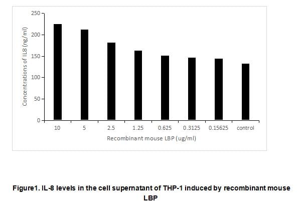
(Lipopolysaccharide Binding Protein (LBP) is a soluble acute-phase protein that binds to bacterial lipopolysaccharide (or LPS) to elicit immune responses by presenting the LPS to important cell surface pattern recognition receptors called CD14 and TLR4. This protein is part of a family of structurally and functionally related proteins, including BPI, plasma cholesteryl ester transfer protein (CETP), and phospholipid transfer protein (PLTP). It has been reported that LBP can enhance LPS-stimulated IL-8 secretion by THP-1 cells. To test the bioactivity of recombinant mouse LBP, THP-1 cells were seeded into 24-well plate at a density of 1x10⁶ cells/mL including 1 ug/mL LPS, and treated with certain concentrations (0.15625 ug/mL-10 ug/mL) of rmLBP for 24h and IL-8 levels in the cell supernatant were determined by ELISA. IL-8 levels in the cell supernatant of THP-1 cells increased significantly after stimulated with recombinant mouse LBP have shown in Figure 1.)
Bioactivity
(Lipopolysaccharide Binding Protein (LBP) is a soluble acute-phase protein that binds to bacterial lipopolysaccharide (or LPS) to elicit immune responses by presenting the LPS to important cell surface pattern recognition receptors called CD14 and TLR4. This protein is part of a family of structurally and functionally related proteins, including BPI, plasma cholesteryl ester transfer protein (CETP), and phospholipid transfer protein (PLTP). It has been reported that LBP can enhance LPS-stimulated IL-8 secretion by THP-1 cells. To test the bioactivity of recombinant mouse LBP, THP-1 cells were seeded into 24-well plate at a density of 1x10⁶ cells/mL including 1 ug/mL LPS, and treated with certain concentrations (0.15625 ug/mL-10 ug/mL) of rmLBP for 24h and IL-8 levels in the cell supernatant were determined by ELISA. IL-8 levels in the cell supernatant of THP-1 cells increased significantly after stimulated with recombinant mouse LBP have shown in Figure 1.)
Lipopolysaccharide Binding Protein (LBP), Active Protein (Cat# AAA161847)
 SDS-PAGE
Leukemia inhibitory factor protein (LIF), Active Protein (Cat# AAA114239)
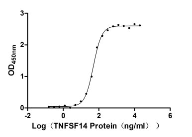 Bioactivity
Tumor necrosis factor ligand superfamily member 14 (TNFSF14), Active Protein (Cat# AAA244031)

 Bioactivity
Mesothelin (MSLN), Active Protein (Cat# AAA243732)
 Application Data
(Measured by its ability to inhibit TNF-alpha mediated cytotoxicity in L-929 mouse fibrosarcoma cells in the presence of the metabolic inhibitor actinomycin D. The ED50 for this effect is typically 5-40 ng/mL in the presence of 1 ng/mL recombinant human TNF-alpha.)
Application Data
(Measured by its ability to inhibit TNF-alpha mediated cytotoxicity in L-929 mouse fibrosarcoma cells in the presence of the metabolic inhibitor actinomycin D. The ED50 for this effect is typically 5-40 ng/mL in the presence of 1 ng/mL recombinant human TNF-alpha.)
TNFR2/TNFRSF1B, Active Protein (Cat# AAA257881)
 Application Data
(Measured by its binding ability in a functional ELISA. Immobilized human CD7-His at 10 ug/ml (100 ul /well) can bind biotinylated SECTM1-His, The EC50 of biotinylated SECTM1-His is 11-26ng/ml.)
Application Data
(Measured by its binding ability in a functional ELISA. Immobilized human CD7-His at 10 ug/ml (100 ul /well) can bind biotinylated SECTM1-His, The EC50 of biotinylated SECTM1-His is 11-26ng/ml.)
CD7, Active Protein (Cat# AAA257959)
Activin-A, Active Protein (Cat# AAA38992)
Aeromonas Aminopeptidase, Active Protein (Cat# AAA38247)
ALKALINE PHOSPHATASE, Active Protein (Cat# AAA50488)
 Bioactivity
(GSTa3 (Glutathione S-transferase a3) is an enzyme that plays an important role in detoxification by catalyzing the conjugation of many hydrophobic and electrophilic compounds with reduced glutathione. This subfamily of enzymes has a particular role in protecting cells from Reactive Oxygen Species and the products of peroxidation. Polymorphisms in this gene influence the ability of individuals to metabolize different drugs. GSTa3 catalyze the endogenous glutathione conjugation 1-Chloro-2,4-dinitrobenzene (CDNB), which can increase in the absorbance at 340 nm. The reaction was performed in adding 10 ul 200 mM glutathione (reduced) and 10 ul 100 mM CDNB in 980 ul 100 mM NaH2PO4 (pH7.0), rapidly mixed. Then add 50 ul mixed substrates to 50 ul different concentrations of recombinant rat GSTa3, mix gently. Incubated at 37 degree C for 5min, then read at a wavelength of 340 nm. The specific activity of recombinant rat GSTa3 is >37000 pmol/min/ug.)
Bioactivity
(GSTa3 (Glutathione S-transferase a3) is an enzyme that plays an important role in detoxification by catalyzing the conjugation of many hydrophobic and electrophilic compounds with reduced glutathione. This subfamily of enzymes has a particular role in protecting cells from Reactive Oxygen Species and the products of peroxidation. Polymorphisms in this gene influence the ability of individuals to metabolize different drugs. GSTa3 catalyze the endogenous glutathione conjugation 1-Chloro-2,4-dinitrobenzene (CDNB), which can increase in the absorbance at 340 nm. The reaction was performed in adding 10 ul 200 mM glutathione (reduced) and 10 ul 100 mM CDNB in 980 ul 100 mM NaH2PO4 (pH7.0), rapidly mixed. Then add 50 ul mixed substrates to 50 ul different concentrations of recombinant rat GSTa3, mix gently. Incubated at 37 degree C for 5min, then read at a wavelength of 340 nm. The specific activity of recombinant rat GSTa3 is >37000 pmol/min/ug.)
Glutathione S Transferase Alpha 3 (GSTa3), Active Protein (Cat# AAA161768)
 Bioactivity
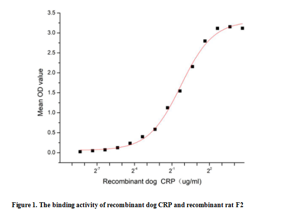
(C reactive protein (CRP) is an annular (ring-shaped), pentameric protein found in blood plasma, whose levels rise in response to inflammation. It is an acute-phase protein of hepatic origin that increases following interleukin-6 secretion by macrophages and T cells. Its physiological role is to bind to lysophosphatidylcholine expressed on the surface of dead or dying cells (and some types of bacteria) in order to activate the complement system via C1q. Besides, Coagulation Factor II (F2) has been identified as an interactor of CRP, thus a functional binding ELISA assay was conducted to detect the interaction of recombinant dog CRP and recombinant rat F2. Briefly, CRP was diluted serially in PBS with 0.01% BSA (pH 7.4). Duplicate samples of 100 ul were then transferred to F2-coated microtiter wells and incubated for 1h at 37 degree C. Wells were washed with PBST and incubated for 1h with anti-CRP pAb, then aspirated and washed 3 times. After incubation with HRP labelled secondary antibody for 1h at 37 degree C, wells were aspirated and washed 5 times. With the addition of substrate solution, wells were incubated 15-25 minutes at 37 degree C. Finally, add 50 uL stop solution to the wells and read at 450/630 nm immediately. The binding activity of recombinant dog CRP and recombinant rat F2 was shown in Figure 1, the EC50 for this effect is 0.82 ug/mL.)
Bioactivity
(C reactive protein (CRP) is an annular (ring-shaped), pentameric protein found in blood plasma, whose levels rise in response to inflammation. It is an acute-phase protein of hepatic origin that increases following interleukin-6 secretion by macrophages and T cells. Its physiological role is to bind to lysophosphatidylcholine expressed on the surface of dead or dying cells (and some types of bacteria) in order to activate the complement system via C1q. Besides, Coagulation Factor II (F2) has been identified as an interactor of CRP, thus a functional binding ELISA assay was conducted to detect the interaction of recombinant dog CRP and recombinant rat F2. Briefly, CRP was diluted serially in PBS with 0.01% BSA (pH 7.4). Duplicate samples of 100 ul were then transferred to F2-coated microtiter wells and incubated for 1h at 37 degree C. Wells were washed with PBST and incubated for 1h with anti-CRP pAb, then aspirated and washed 3 times. After incubation with HRP labelled secondary antibody for 1h at 37 degree C, wells were aspirated and washed 5 times. With the addition of substrate solution, wells were incubated 15-25 minutes at 37 degree C. Finally, add 50 uL stop solution to the wells and read at 450/630 nm immediately. The binding activity of recombinant dog CRP and recombinant rat F2 was shown in Figure 1, the EC50 for this effect is 0.82 ug/mL.)
C ReProtein (CRP), Active Protein (Cat# AAA161778)

 SDS-PAGE
(Figure. SDS-PAGE)
SDS-PAGE
(Figure. SDS-PAGE)
Fibroblast Growth Factor 10 (FGF10), Active Protein (Cat# AAA150560)
What Are Active Proteins?
Proteins are large molecules made up of long chains of amino acids. They will typically fold into a very particular 3-dimensional shape/conformation, that is sometimes referred to as their “native” form, which allows them to work properly in the body. For the purposes of product categorization, AAA Biotech will typically refer to proteins purified from their original animal host as being “native” proteins (this is to signify their difference compared to their recombinant proteins or “synthetic” protein counterparts).
If a protein successfully folds into the correct shape, it will typically display high fidelity characteristics to its original protein in its original animal host and be classified as an active protein, as it will be able to function “normally” in most enzymatic or binding capacities. If it loses this shape, due to factors such as heat or strong chemicals (such as detergents), it becomes inactive and is no longer able to perform its basic functions.
All of the proteins in this category are made under strict quality control, and they are active, pure, low in contaminants, and stable. Most are stored as freeze-dried powders and come without extra tags, so they’re very close to the actual natural/native form.
Learn more in our guide “How active proteins work”.
Key Applications of Active Proteins
1. Scientific Research
- Aid in the study of how proteins function in the body
- Aid in understanding various disease processes
2. Drug Development
- Powerful tools to investigate how potential drugs interact with specific proteins
- Ideal for identifying drug targets
3. Cell Culture
- Are routinely utilized to support cell growth and function (e.g., using exogenous growth factors)
- Can be used to promote cellular development into specific types (differentiation)
4. Diagnostics
- Regularly utilized in tests to detect diseases or infections (e.g., COVID-19, cancer)
- Note: All products are strictly for research-use only (RUO).
5. Therapeutics
- Some active proteins are used directly as treatments (e.g., insulin, enzymes)
- Note: All products are strictly for research-use only (RUO).
6. Vaccine Development
- Used to create or test vaccines by mimicking parts of viruses or bacteria
7. Biochemical Assays
- They can facilitate the characterization of enzyme activity, binding strength, or protein interactions in lab tests
Why Buy Active Proteins from AAA Biotech?
- High biological activity – Verified to perform as expected or indicated on datasheet
- Strict quality control – We are confident in our active proteins’ reliability and consistency
- High purity & low endotoxin – Ideal for applications involving sensitive or precious samples/components
- Freeze-dried for stability – Long shelf life and straightforward storage
- Mostly tag-free – Closer to natural/native protein form
FAQ
1. What are active proteins used for in research?
Active proteins are used primarily in the study of how proteins function, in characterizing/discovering drug interactions, supporting cell growth, running biochemical assays, and in development of diagnostics or therapeutics.
2. How are AAA Biotech's active proteins validated?
AAA Biotech’s active proteins are validated through strict quality control and functional assays to ensure they are properly folded and active. “Active”, though, can be an ambiguous term, so if a specific “activity” or “binding” capability of a protein is of crucial interest to you, please inquire with us prior to purchase, and we will provide further details on how the “Active” modifier was determined to be applicable.
3. Are these proteins tested for biological activity?
Yes, all active proteins from AAA Biotech are tested to confirm they have the expected biological activity before being offered for use. Though, said “biological activity” can be either “enzymatic”, “binding”, or both.